-
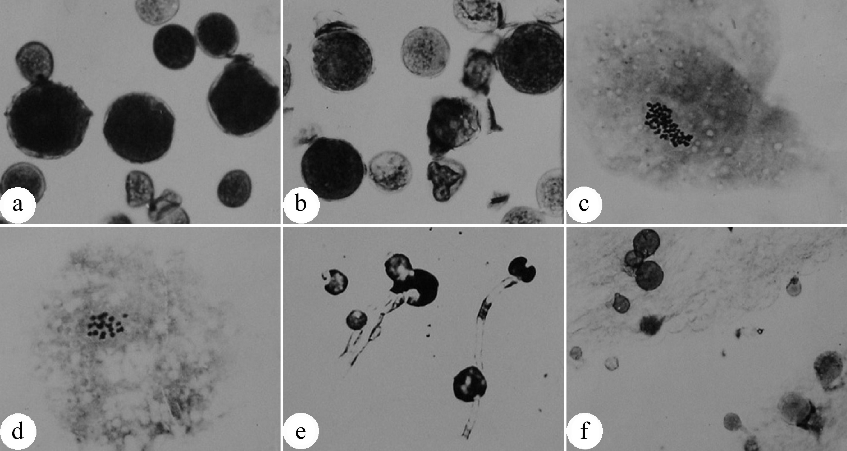
Figure 1.
Natural occurrence of unreduced 2n pollens in Populus tomentosa. (a) Enlarged pollen grains produced by male clone B105 of P. tomentosa; (b) Enlarged pollen grains produced by male clone B111 P. tomentosa; (c) Chromosome number (n = 2x = 38) of an enlarged pollen; (d) Chromosome number (n = x = 19) of a normal pollen; (e) Germinated 2n pollens on medium; (f) Germinated 2n pollens on the stigma. (Figure source: References [9], [11])
-
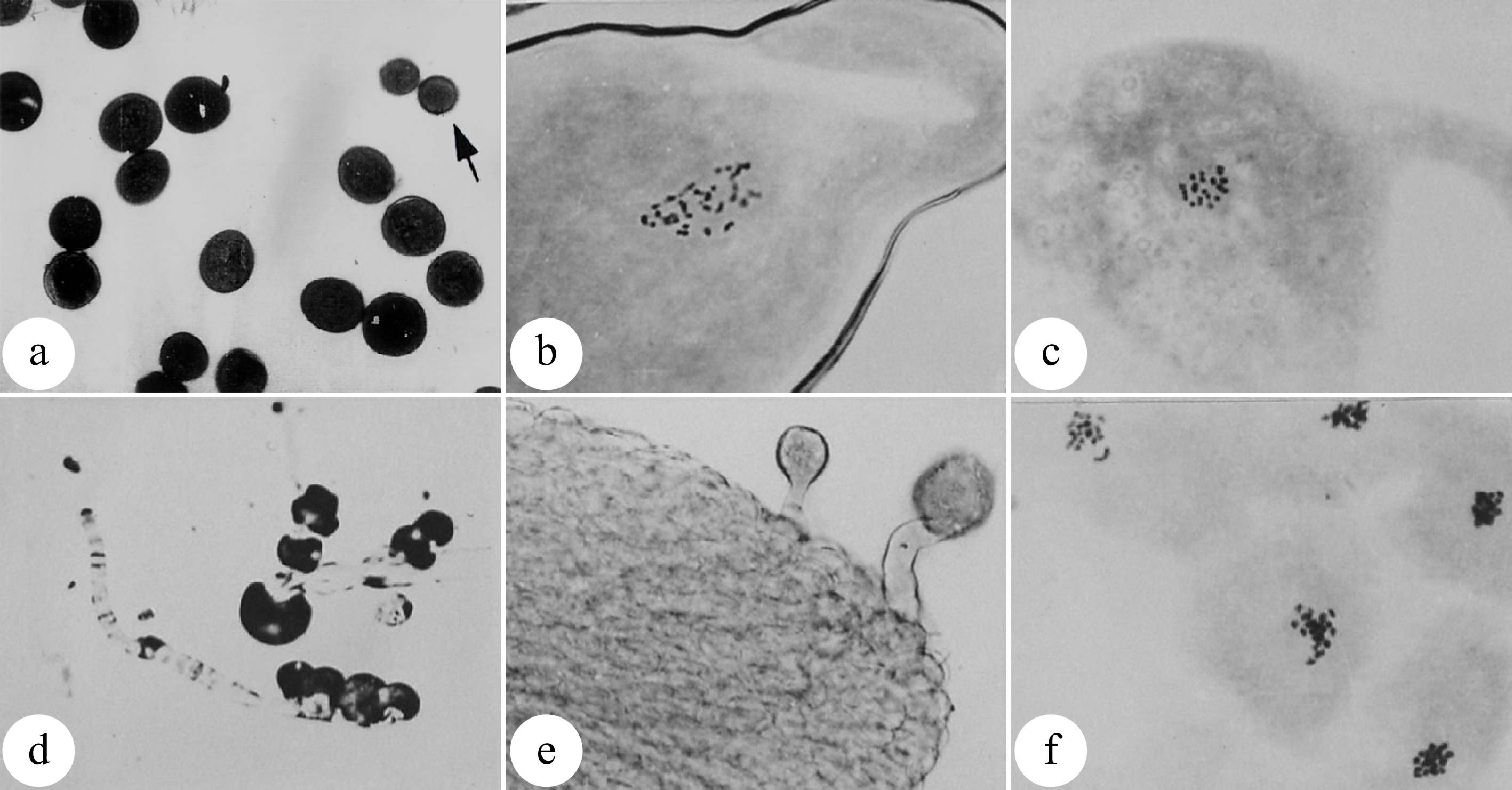
Figure 2.
Pollen chromosome doubling of P. tomentosa × P. bolleana induced by colchicine. (a) 2n pollens induced by colchicine. The arrow indicates a normal pollen. (b) Chromosome number (n = 2x = 38) of an induced 2n pollen. (c) Chromosome number (n = x = 19) of a normal pollen. (d) Germination of induced 2n pollen on medium. (e) Germination of induced 2n pollens on the pistil stigma. (f) Chromosomes are aggregated into clusters after colchicine treatment. (Figure source: References [9], [23])
-
-
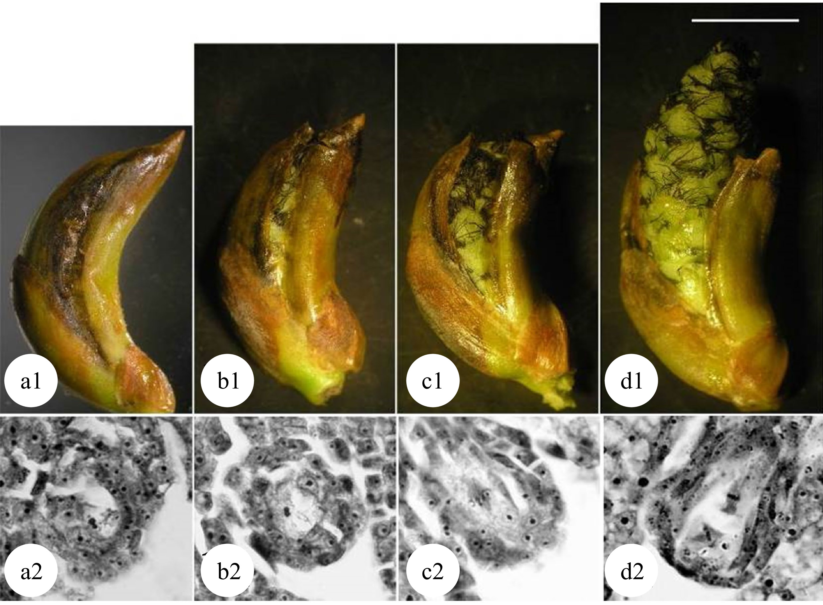
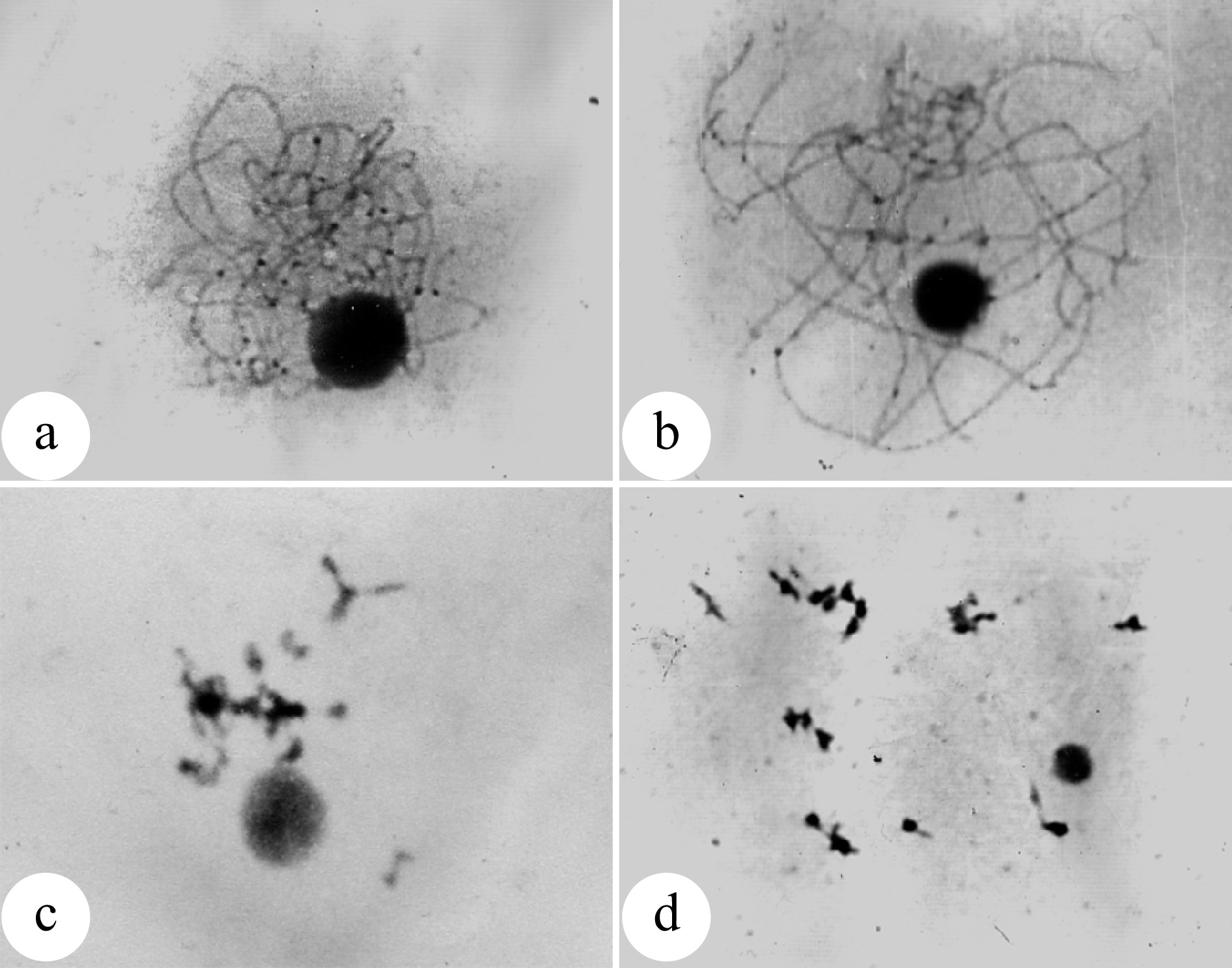
Figure 4.
Meiosis of megaspore mother cells in 'Zheyin #3' and its relationship to female flower bud development. (a) In the pachytene stage, the flower buds swell, and the bud scales crack but are not yet open. (b) In the diplotene stage, the bud scales are slightly open, but the inflorescence is not exposed. (c) In metaphase I, the bud scales are open, and the inflorescence is slightly exposed. (d) In metaphase II, the bud scales are fully open one third of the inflorescence has emerged from the bud scales. Scale = 10 μm. (Figure source: Reference [38])
-
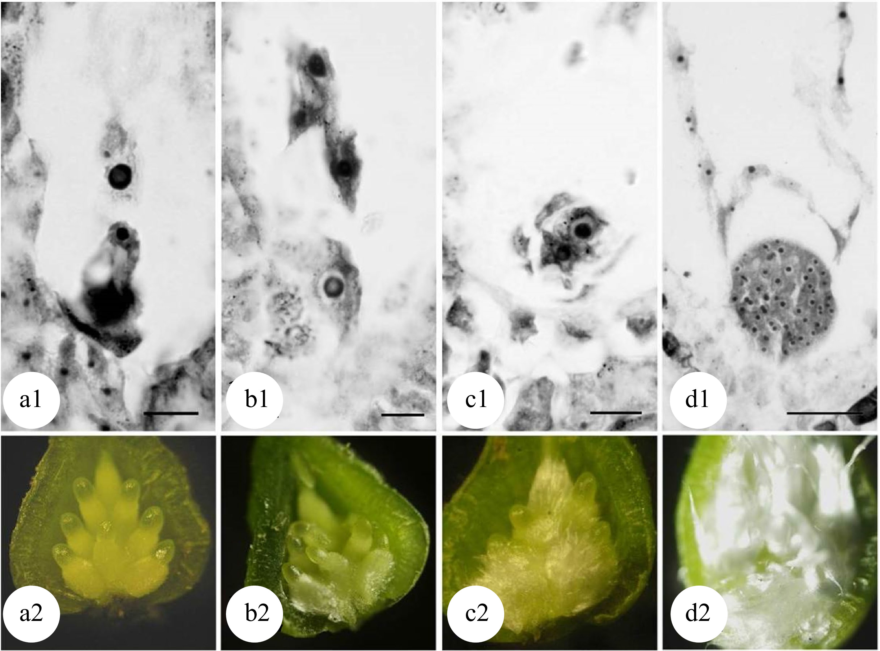
Figure 5.
Developmental stages of zygote and flocculent fibers in the ovary of 'Zheyin #3' × P. beijingensis. (a) During double fertilization, flocculent fibers occur around the funicle in the ovary, but they are not visible to the naked eye. (b) The dormant zygote forms after fertilization, and the flocculent fibers cover the surface of the funicle and are visible to the naked eye. (c) In the two-celled embryo stage, the flocculent fibers begin to wrap the ovule but do not fill the ovary. (d) In the globular embryo stage, flocculent fibers completely wrap the ovule and fill the ovary. Scale = 10 μm. (Figure source: Referencecs [49])
-
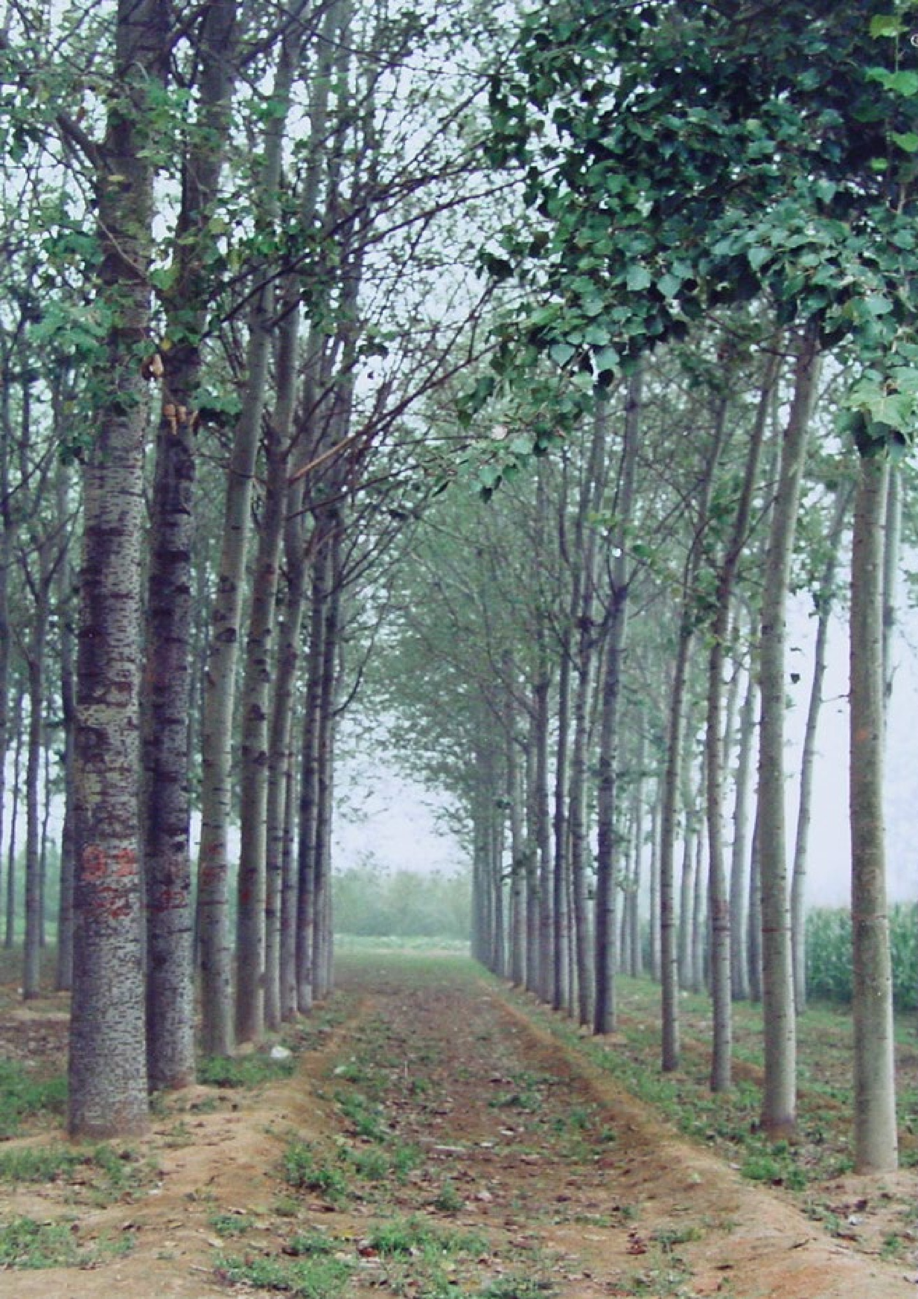
Figure 6.
Eight-year-old triploid P. tomentosa trees show significant advantages in growth over diploid clones (not the same parents). Triploids are on the left, and diploids are on the right.
-
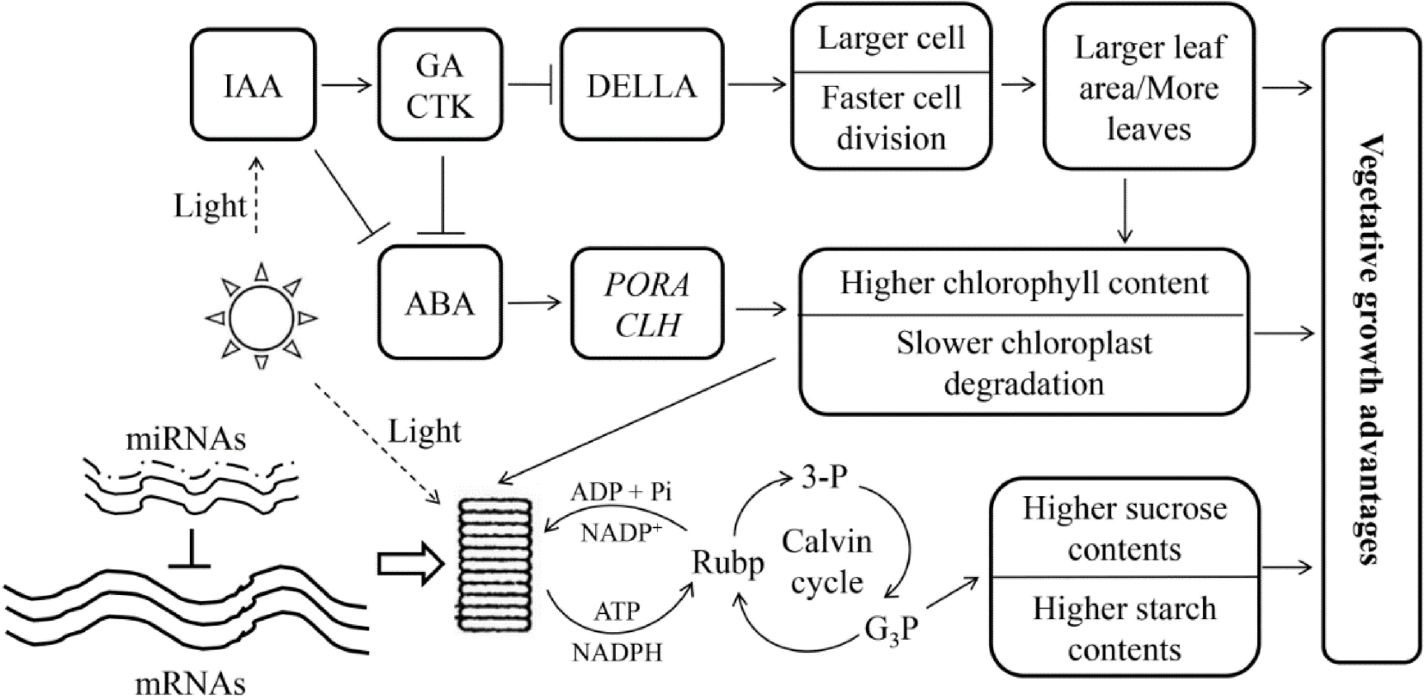
Figure 7.
Genetic regulation model of allotriploid P. cathayana with enhanced vegetative growth. Arrows indicate promotion, and short lines indicate inhibition. The three solid lines next to 'mRNAs' indicate the dose effect in the triploid, and the dotted lines next to 'miRNAs' indicate the lack of a dose effect. (Figure source: Reference [82])
-
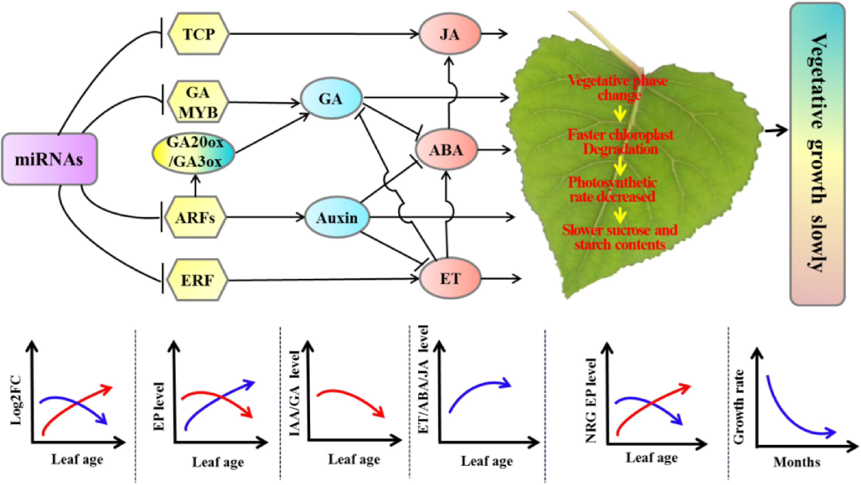
Figure 8.
Molecular regulatory model of tetraploid poplars with slower vegetative growth than triploid poplars. Arrows indicate promotion, and short lines indicate inhibition. The red curves represent the expression of differentially expressed miRNAs positively associated with tetraploid vegetative growth or DEGs positively associated with vegetative growth. The blue curves represent the expression of differentially expressed miRNAs or DEGs negatively associated with tetraploid vegetative growth. ET, ethylene; EP: expressed protein; NRG, negative related growth; ABA: Abscisic acid; JA: asmonic acid; GA: gibberellic acid; FC: fold change. (Figure source: Reference [66])
Figures
(8)
Tables
(0)