-
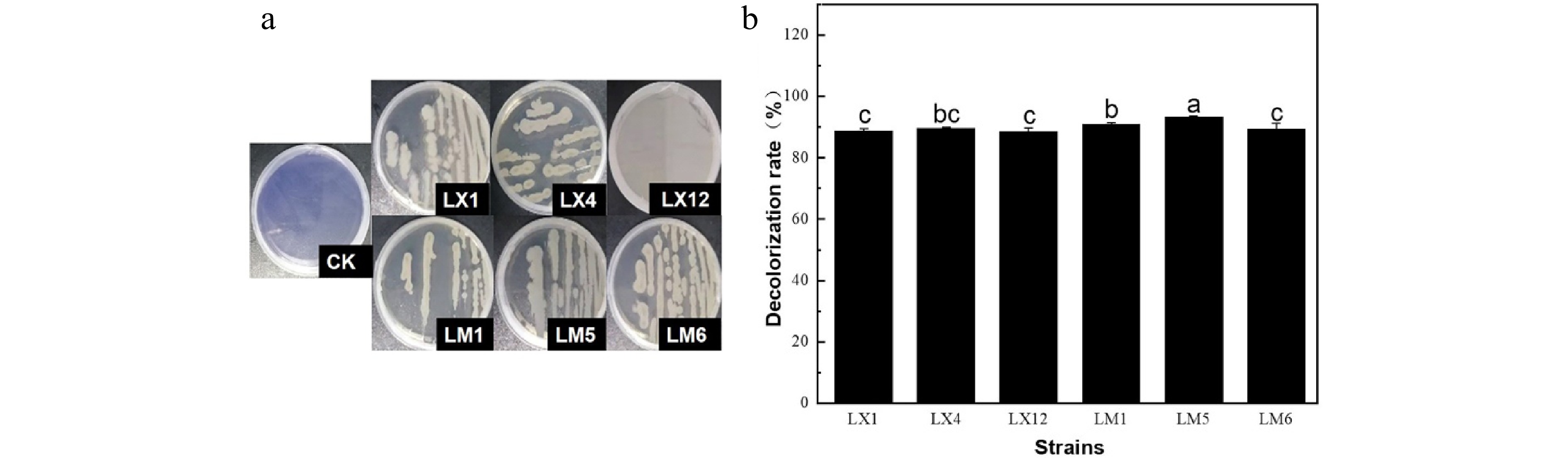
Figure 1.
(a) Aniline blue fading results and (b) decolorization rate of different strains.
-
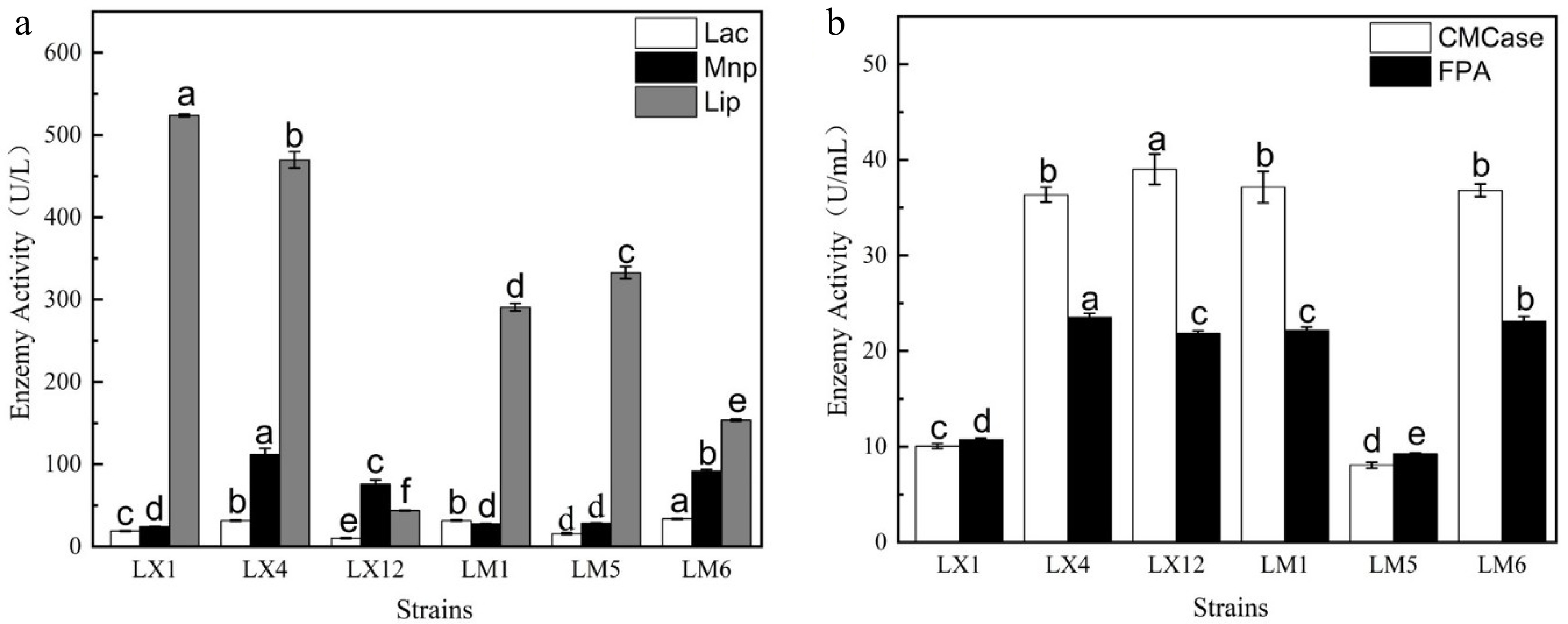
Figure 2.
(a) Ligninase activity and (b) cellulase activity of different strains.
-
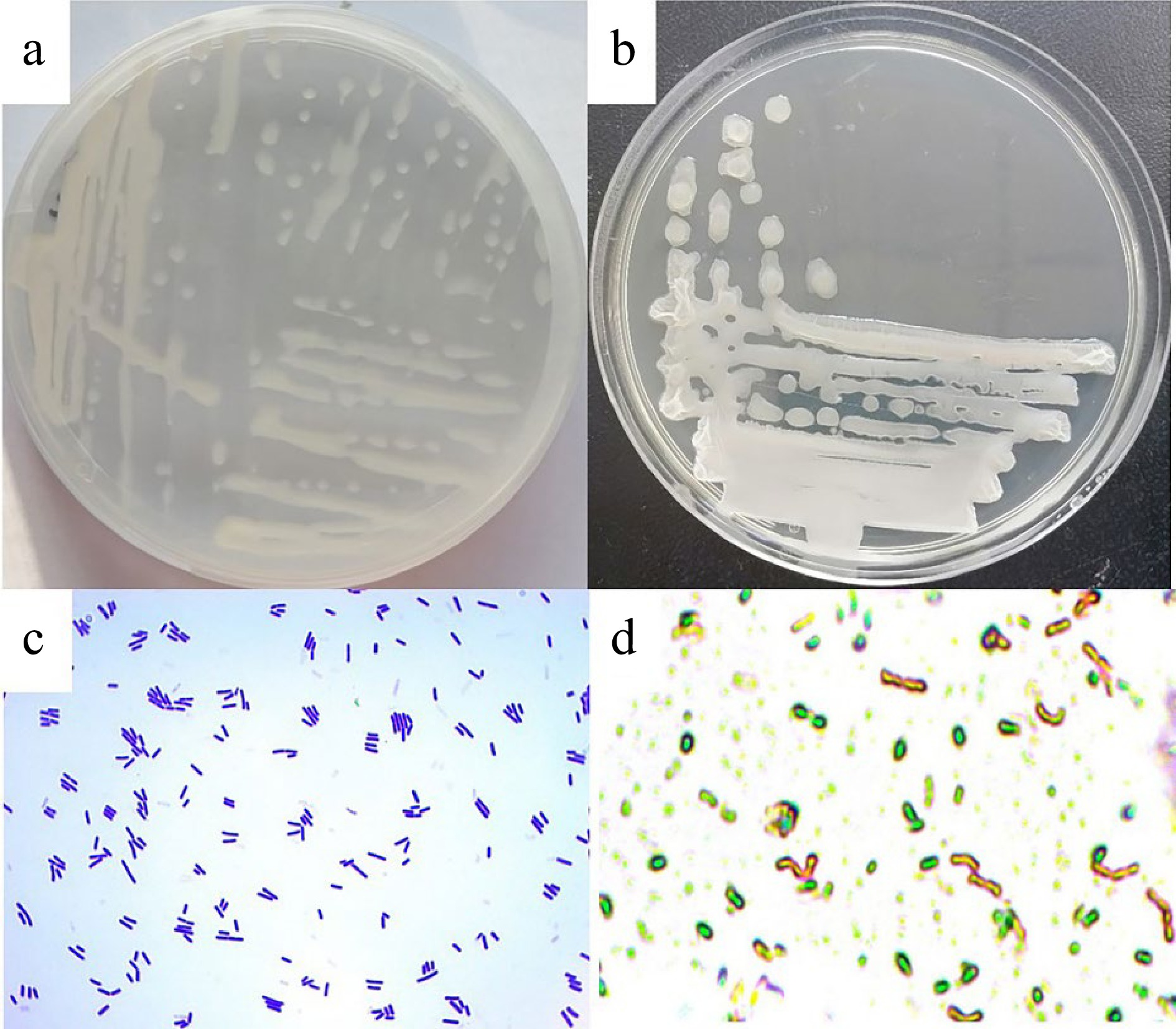
Figure 3.
Colony, cell morphologies and spore of the isolated ligninase and cellulase-producing strain LM1. (a) Colony morphology 12 h, (b) colony morphology 24 h, (c) cell morphology, (d) spore.
-
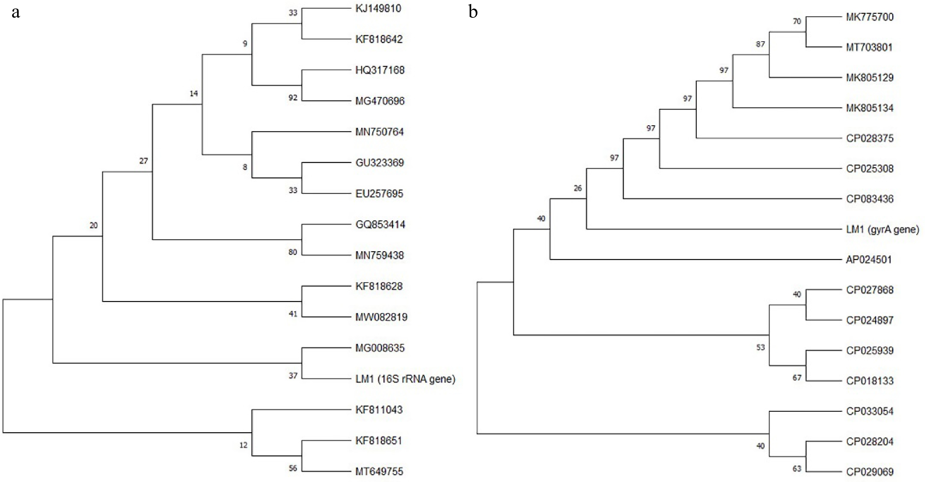
Figure 4.
Phylogenetic trees based on (a) 16S rRNA gene sequence and (b) gyrA gene sequence of strain LM1.
-
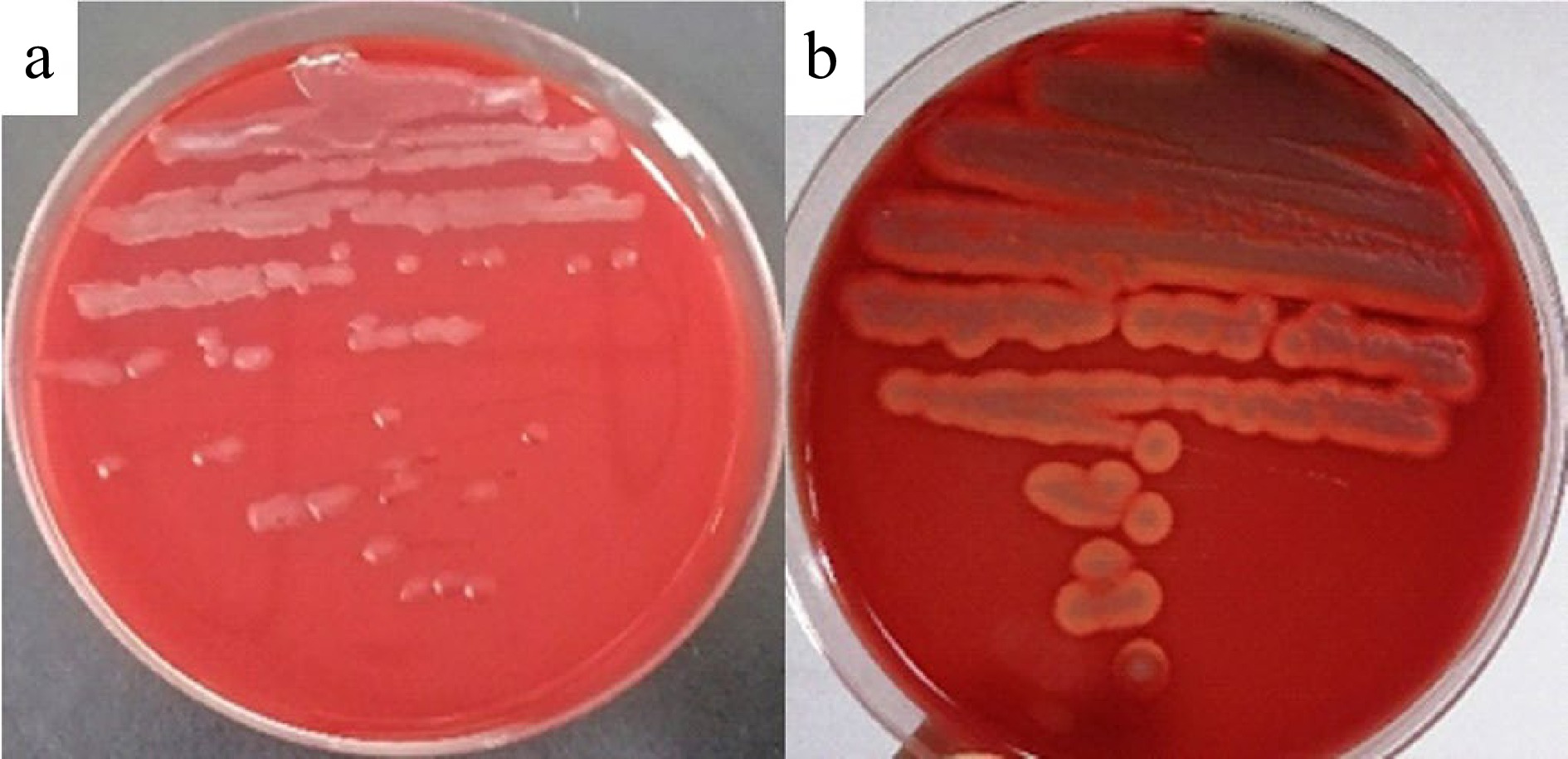
Figure 5.
Hemolysis test of (a) B. velezensis LM1 and (b) B. cereus ATCC14579.
-
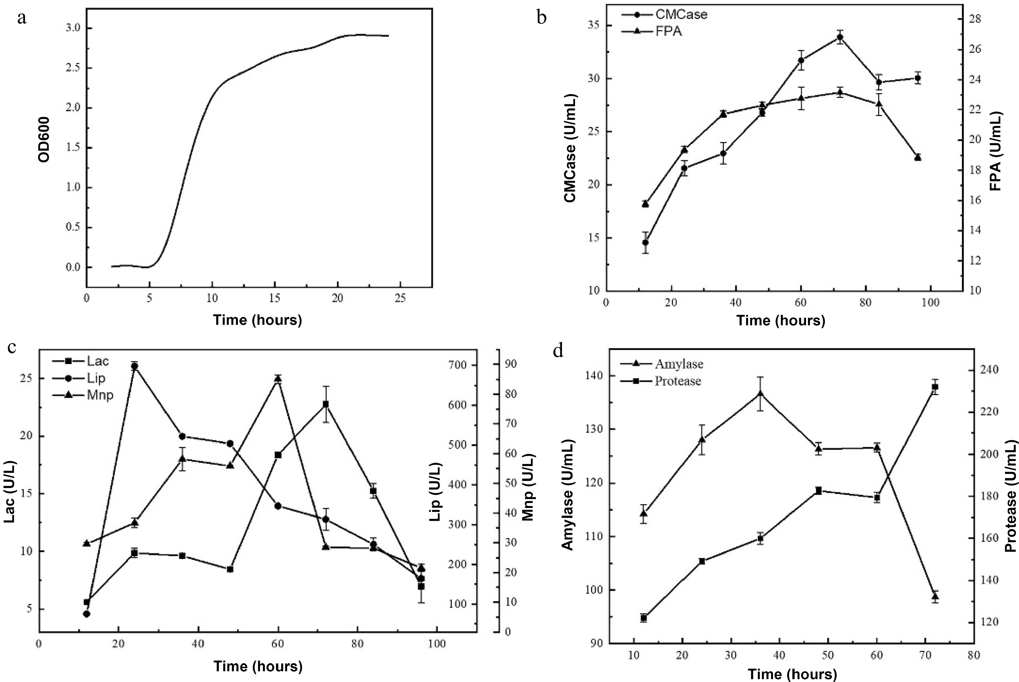
Figure 6.
(a) Growth curve and the enzyme activities of (b) CMCase and FPA, (c) Lac, Lip and Mnp, (d) amylase and protease of B. velezensis LM1.
-
Strains Diameter of colony (D/mm) Diameter of transparent circle (d/mm) d/D Degree of disintegration LX1 4.23 ± 0.01b 14.62 ± 0.21d 3.46 ++ LX4 4.61 ± 0.18a 23.79 ± 0.64b 5.16 ++++ LX12 3.55 ± 0.05c 18.07 ± 0.09c 5.10 +++ LM1 4.77 ± 0.01a 27.02 ± 0.02a 5.66 +++++ LM5 4.29 ± 0.04b 9.25 ± 0.52e 2.16 + LM6 4.84 ± 0.18a 27.83 ± 1.39a 5.76 ++++ Lowercase letters represent significant differences in the same column. '+' represents the degree of damage to the filter paper, and the more '+', the stronger the degree of damage. Table 1.
Congo red transparent circle and filter paper strip disintegration effects of different strains.
-
Sample Yield (%) Purity (%) RSDF 6.07 ± 0.01B 87.00 ± 0.30A FSDF 12.57 ± 0.22A 87.13 ± 0.99A RIDF 22.45 ± 0.02a 91.70 ± 0.82a FIDF 18.68 ± 0.21b 77.27 ± 0.50b A, B reflect the significance of SDF, and a, b reflect the significance of IDF.
P < 0.05.Table 2.
The yield and purity of dietary fiber.
Figures
(6)
Tables
(2)