-
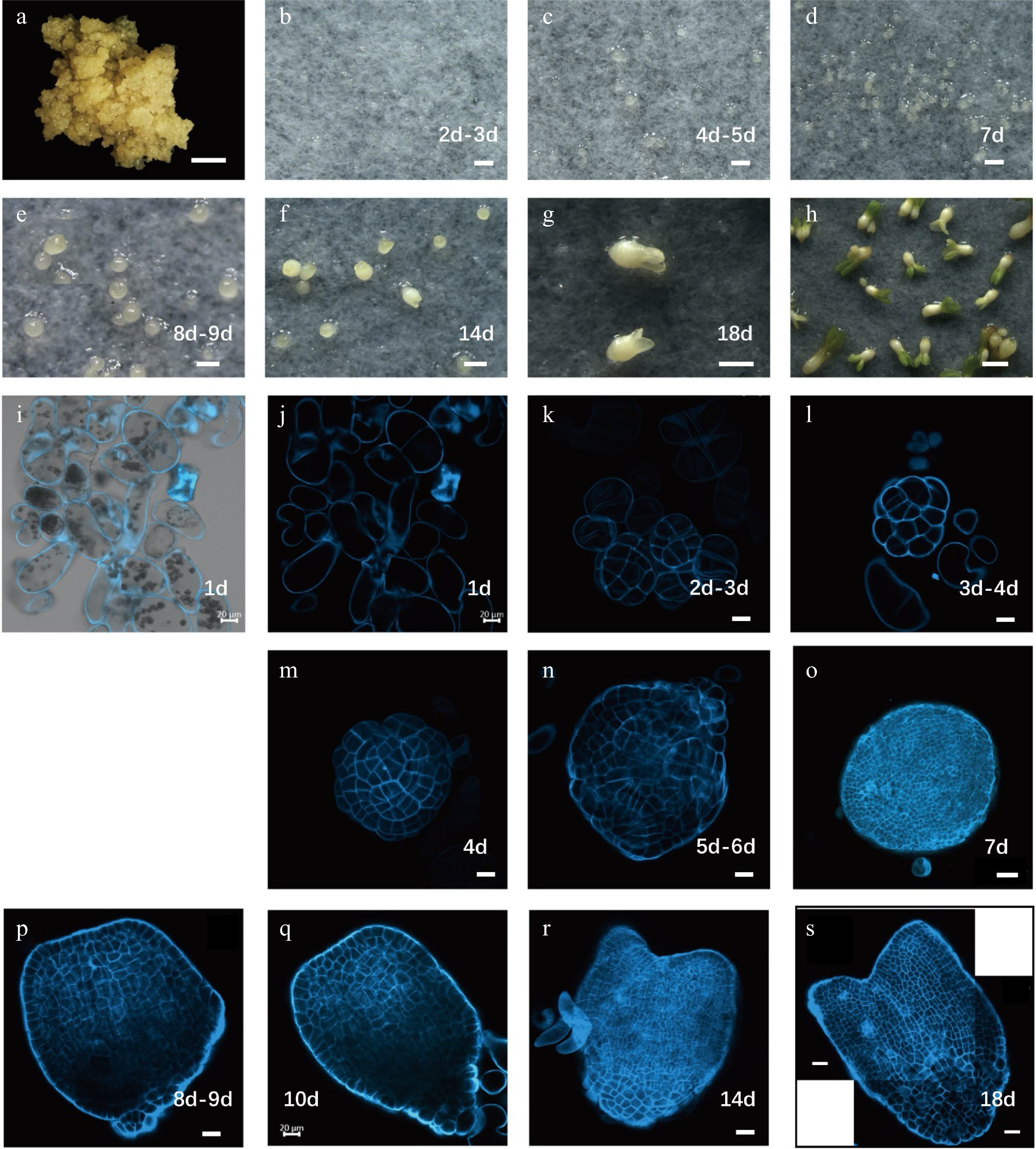
Figure 1.
Developmental stages of Liriodendron hybrids somatic embryos. (a) Embryonic callus; (b) embryos after 2–3 d on embryo induction medium (EIM); (c) pre-globular embryo after 4–5 d on EIM; (d) globular embryo after 7 d on EIM; (e) transition stage embryo after 8–9 d on EIM; (f) heart-shaped embryo after 14 d on EIM; (g) cotyledon embryos after 18 d on EIM; (h) plantlets after cultivation in the light; (i–s) different stages of somatic embryos observed by confocal microscopy with SCRI 2200 staining. Scale bars: (a)–(d) 200 μm; (e)–(h) 100 μm; (i)–(n) 20 μm; (o) 50 μm; (p)–(s) 20 μm.
-
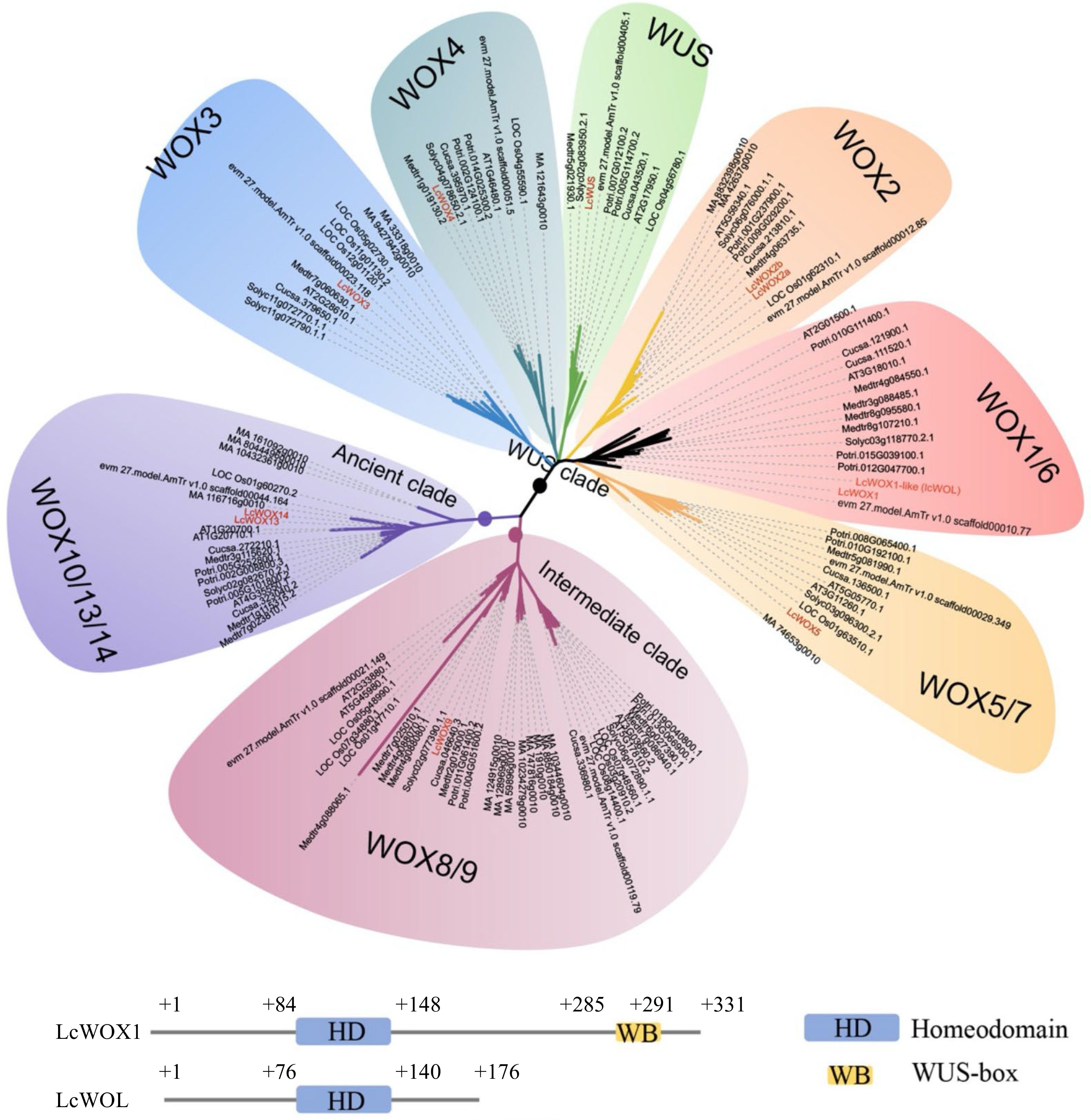
Figure 2.
LcWOX genes were grouped into three distinct, well-supported clades. WOX orthologs come from fully sequenced genomes of five eudicot species, Arabidopsis thaliana(AT), Medicago truncatula (Medtr), Cucumis sativus (Cucsa), Solanum lycopersicum(Solyc) and Populus trichocarpa (Potri), as well as a monocot species: Oryza sativa(LOC Os). WOX gene sequences from the gymnosperm Picea abies(MA) and basal Magnoliophyte, Amborella trichopoda(evm27.model.AmTr.V1.0) also have been included.
-
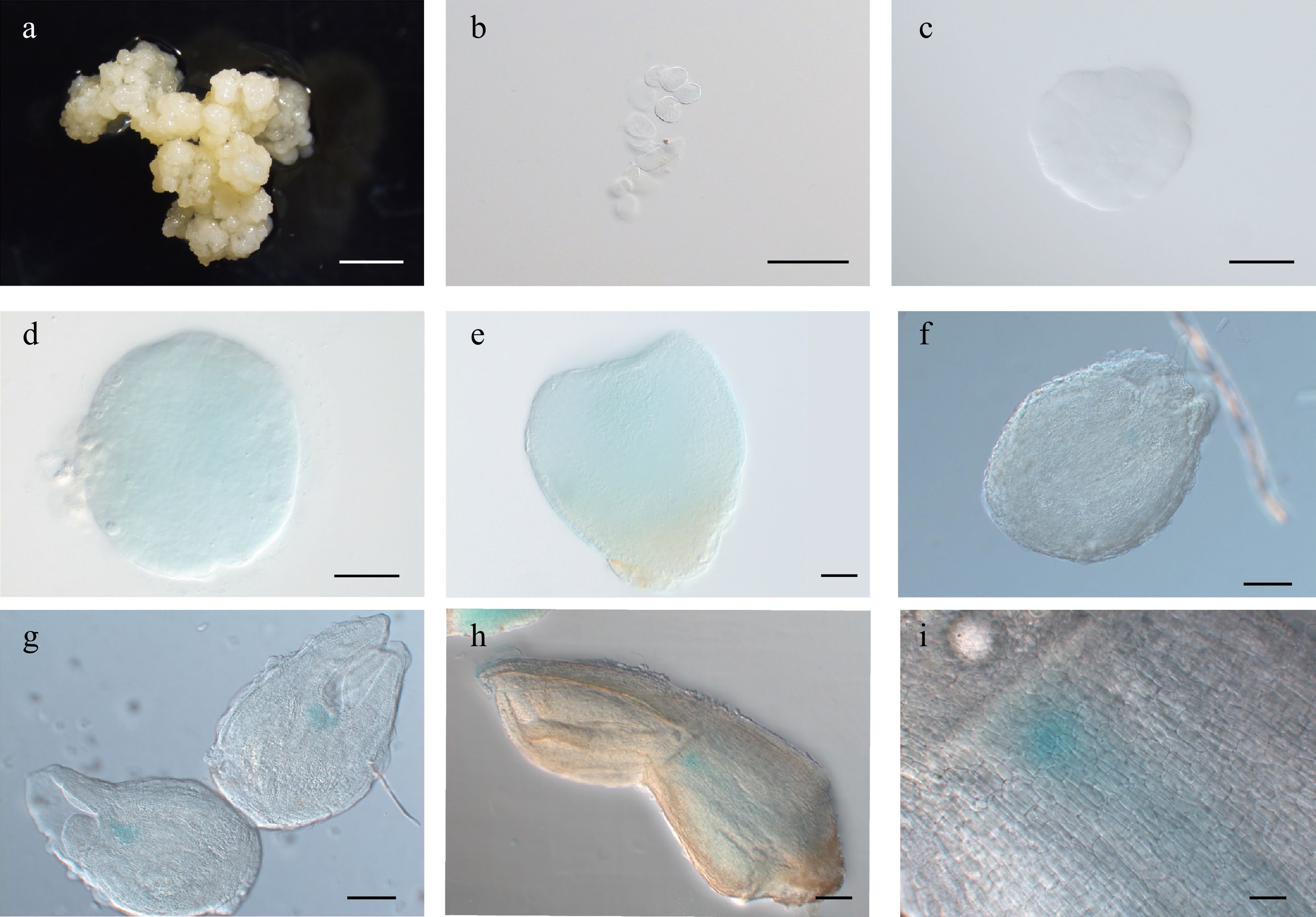
Figure 3.
Expression pattern of LcWUSpro:GUS during somatic embryogenesis in Lirodendron. (a) – (c) LcWUSpro:GUS was not expressed in (a) embryonic callus, (b) single cells after 1 d on induction medium (IM), or (c) the pre-globular embryo after 4 ds on IM. (d), (e) LcWUSpro:GUS was weakly expressed in the (d) globular embryo and (e) heart-shaped embryo. (f) – (i) LcWUSpro:GUS was expressed in a tissue-specific manner in the (f) late heart-shaped embryo, (g) torpedo embryo, and (h) mature cotyledon embryo. (i) Magnification of the OC area in (h). Scale bars: (a) 2000 μm; (b) 100 μm; (c) – (e) 50 μm; (f), (g) 100 μm; (h) 200 μm; (i) 500 μm.
-
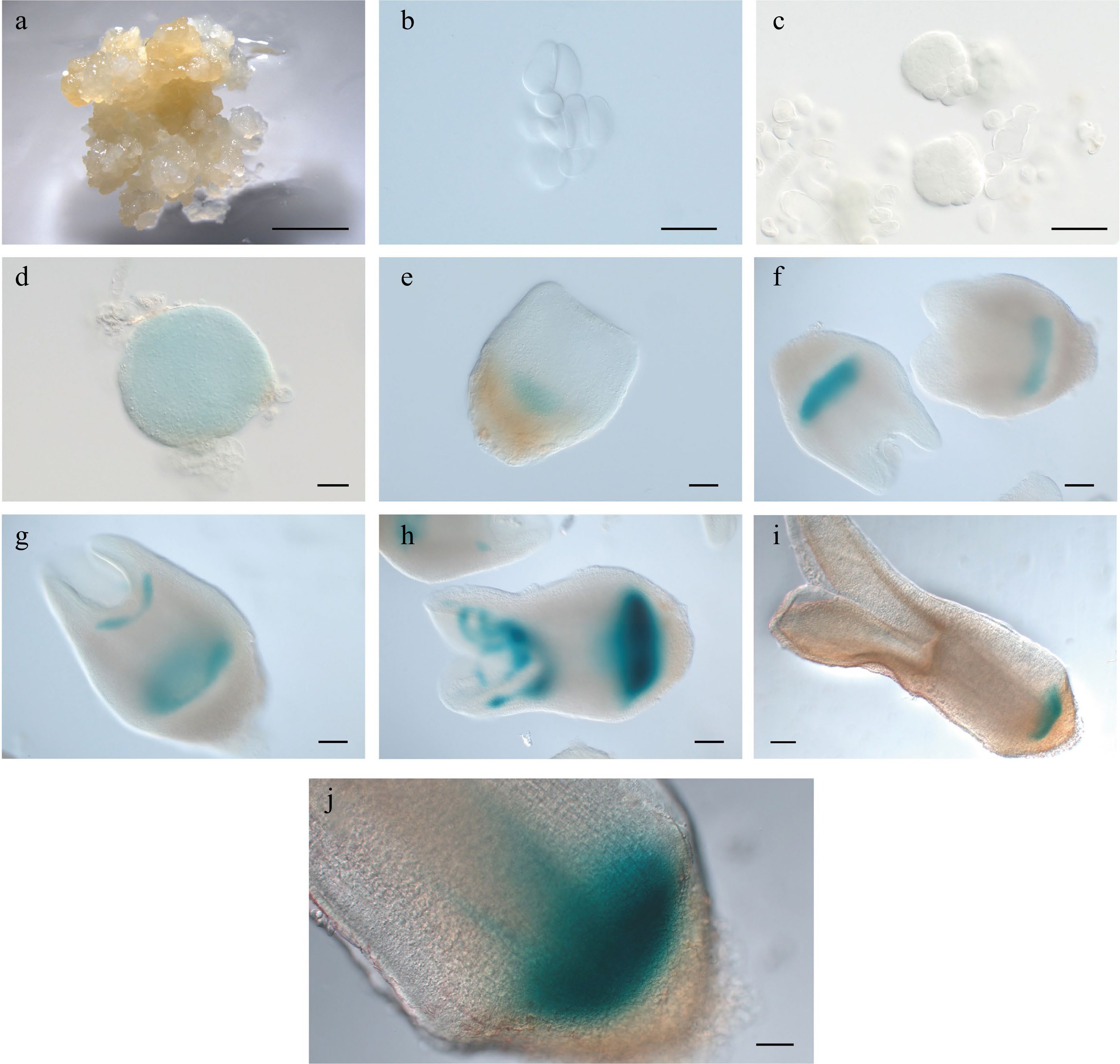
Figure 4.
Expression pattern of LcWOX5pro:GUS during somatic embryogenesis in Liriodendron. (a)−(c) LcWOX5pro:GUS was not expressed in (a) embryonic callus, single cells after one day on (b) induction medium (IM), and (c) pre-globular embryos after four days on IM. (d) LcWOX5pro:GUS was expressed in the globular embryo. (e)−(j) LcWOX5pro:GUS was expressed in a tissue-specific manner in the (e) transition-stage embryo, (f), (g) late heart-shaped embryo, (h) torpedo embryo, (i) mature cotyledon embryo, and (j) plantlet root apical meristem of. Scale bar: (a) 2,000 μm; (b)−(e) 50 μm; (f)−(h) 100 μm; (i) 200 μm; (j) 100 μm.
-
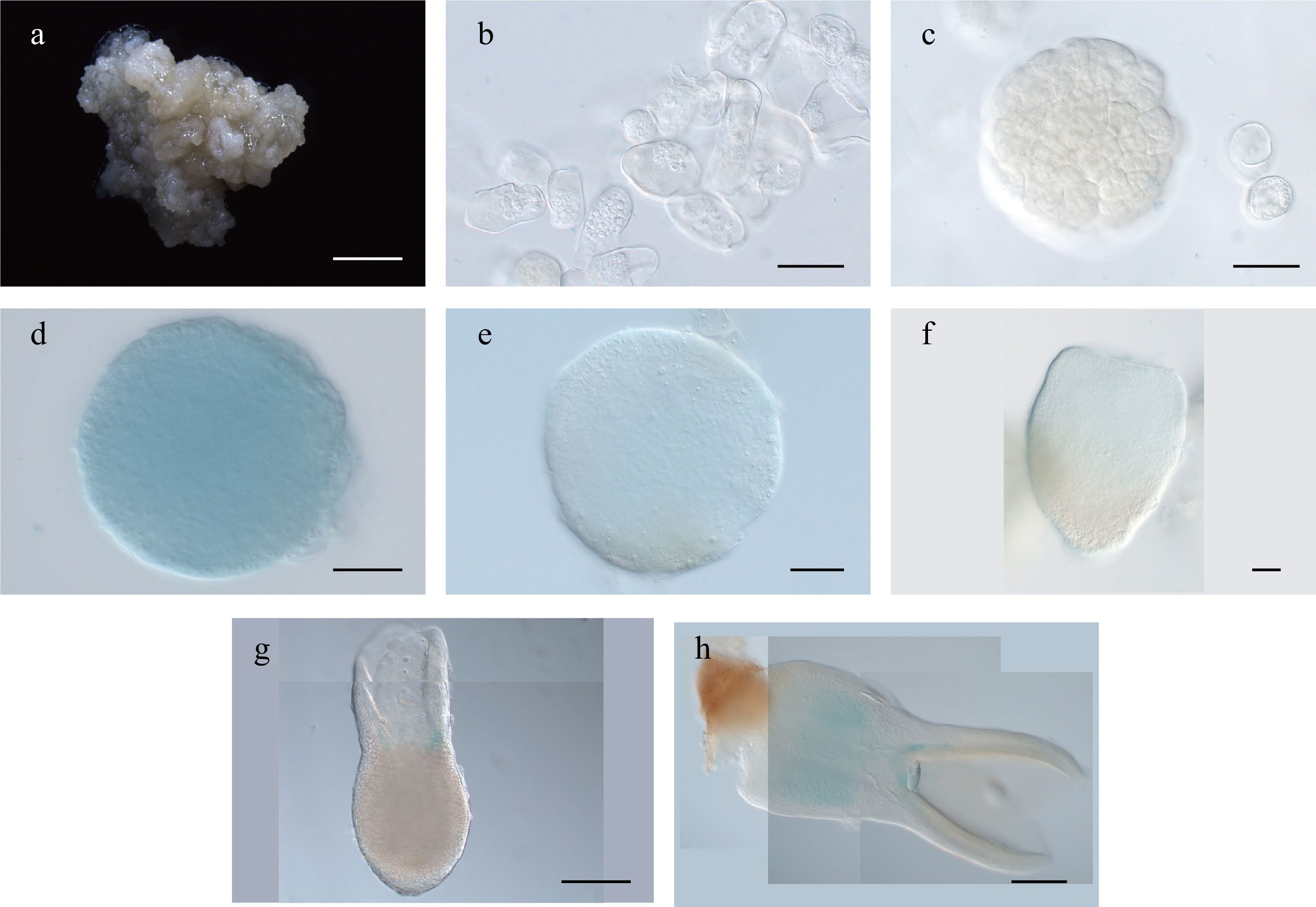
Figure 5.
Expression pattern of LcWOX1pro:GUS during somatic embryogenesis in Liriodendron. (a)–(c) LcWOX1pro:GUS was not expressed in (a) embryonic callus, (b) single cells after 1 d on induction medium (IM), or the (c) pre-globular embryo. (d)–(f) LcWOX1pro:GUS was weakly expressed in the (d), (e) globular embryo and (f) transition-stage embryo. (g) LcWOX1pro:GUS was expressed at the base of the cotyledon in the torpedo embryo. (h) LcWOX1pro:GUS was expressed in the cotyledon and hypocotyl of the mature cotyledon embryo. Scale bars: (a) 2,000 μm; (b)–(e) 50 μm; (f)–(h) 200 μm.
-
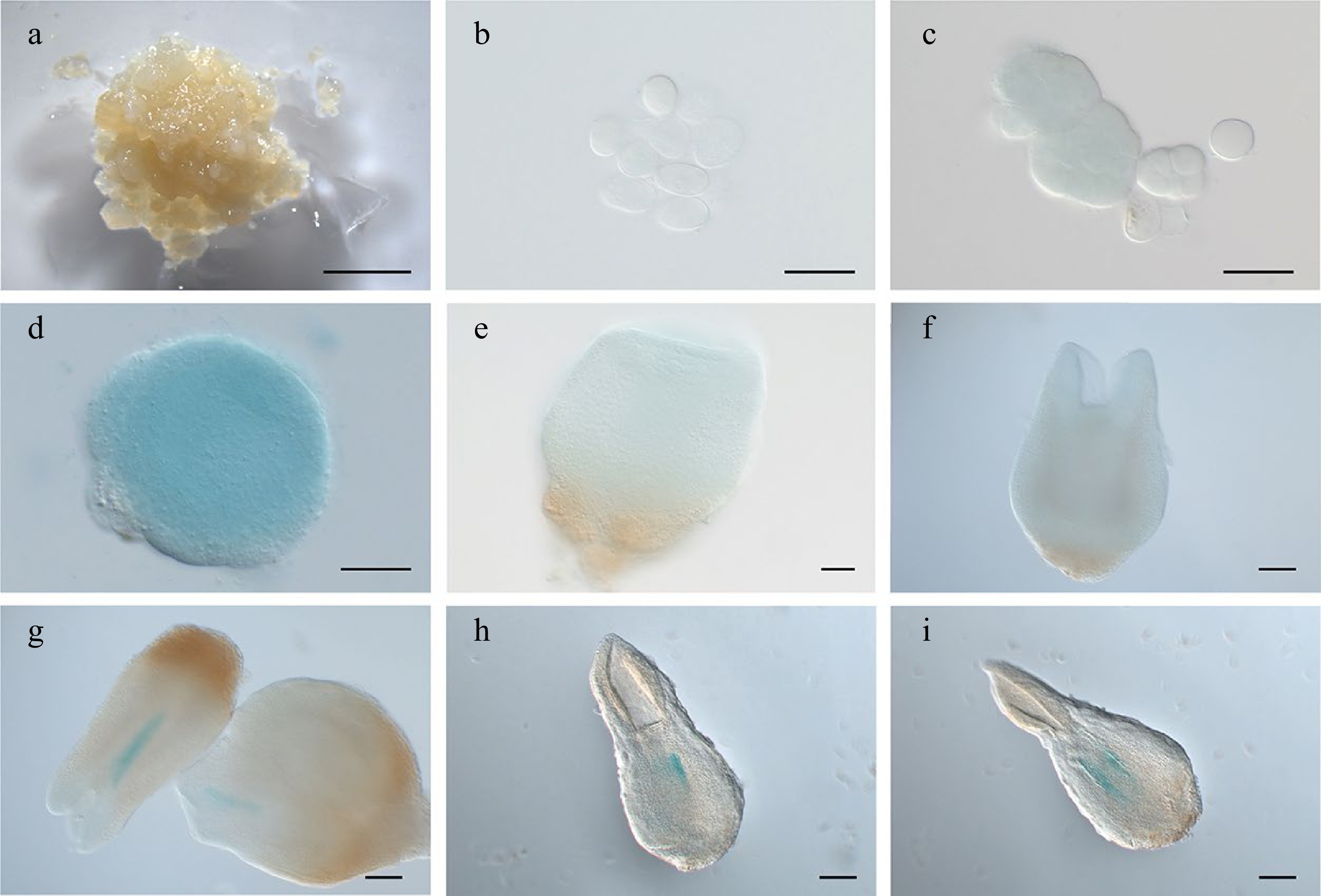
Figure 6.
Expression pattern of LcWOX4pro:GUS during somatic embryogenesis in Liriodendron. (a), (b) LcWOX4pro:GUS was not expressed in (a) embryonic callus or (b) single cells after 1 d on induction medium (IM). (c) LcWOX4pro:GUS was weakly expressed in the (d) pre-globular embryo but highly expressed in the globular embryo. (e), (f) LcWOX4pro:GUS was weakly expressed in the (e) transition-stage embryo and(f) late heart-shaped embryo. (g)–(i) LcWOX4pro:GUS was expressed in a tissue-specific manner in the (g) torpedo embryo and (h), (i) cotyledon embryo. Scale bars: (a) 2,000 μm; (b)–(e) 50 μm; (f)–(h) 200 μm.
-
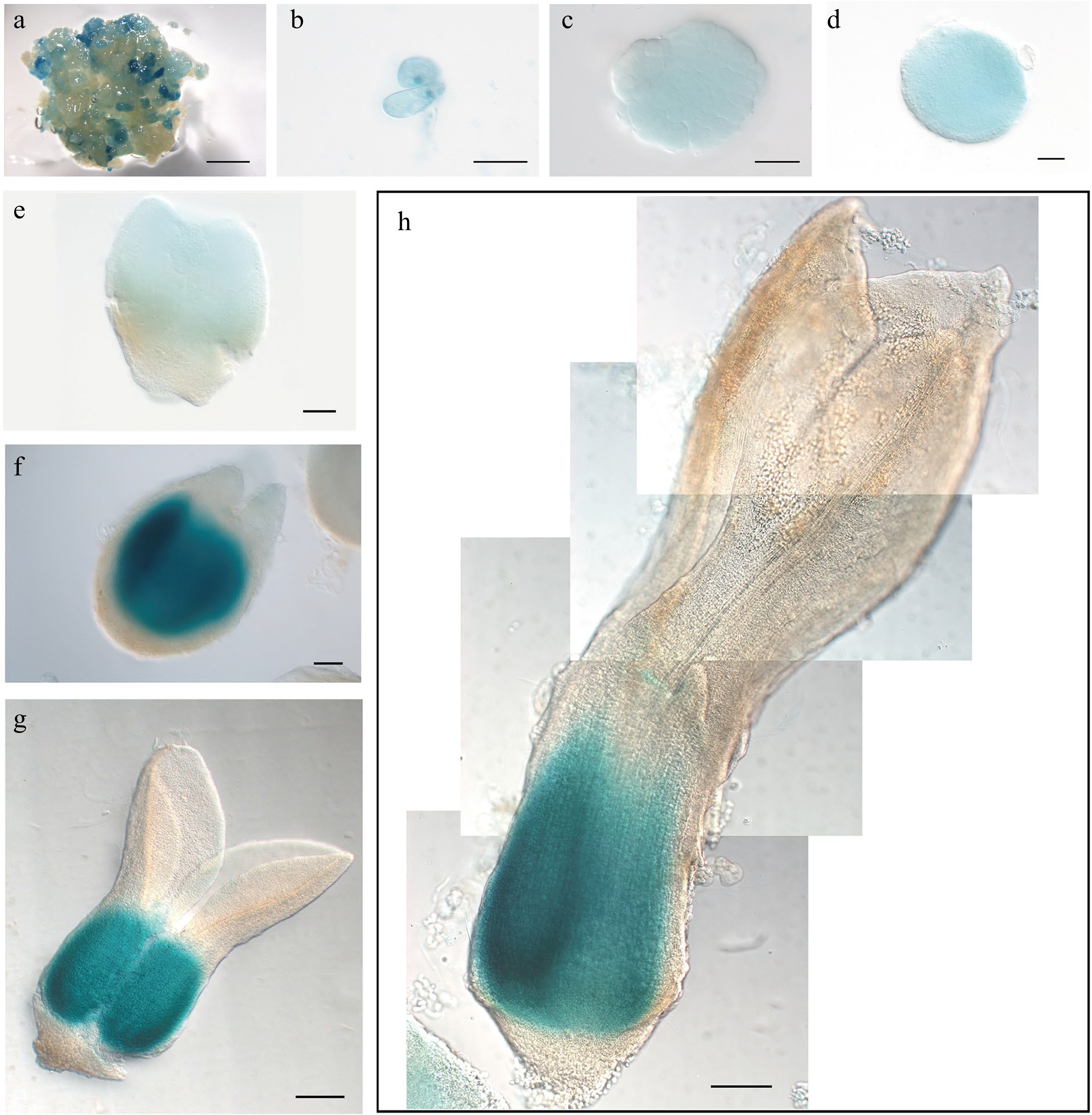
Figure 7.
Expression pattern of LcWOX9pro:GUS during somatic embryogenesis in Liriodendron. (a) LcWOX9pro:GUS was very highly expressed in embryonic callus. (b) LcWOX9pro:GUS was expressed in single cells after 1 d on induction medium (IM). (c)–(e) LcWOX9pro:GUS was expressed in the (c) pre-globular embryo, the (d) globular embryo, and the (e) early heart-shaped embryo. (f) LcWOX9pro:GUS was expressed in a tissue-specific manner in the late heart-shaped embryo. (g) LcWOX9pro:GUS was highly expressed in the cotyledon embryo. (h) LcWOX9pro:GUS was expressed in a tissue-specific manner in the mature cotyledon embryo. Scale bars: (a) 2,000 μm; (b)–(e) 50 μm; (f) 100 μm; (g), (h) 200 μm.
-
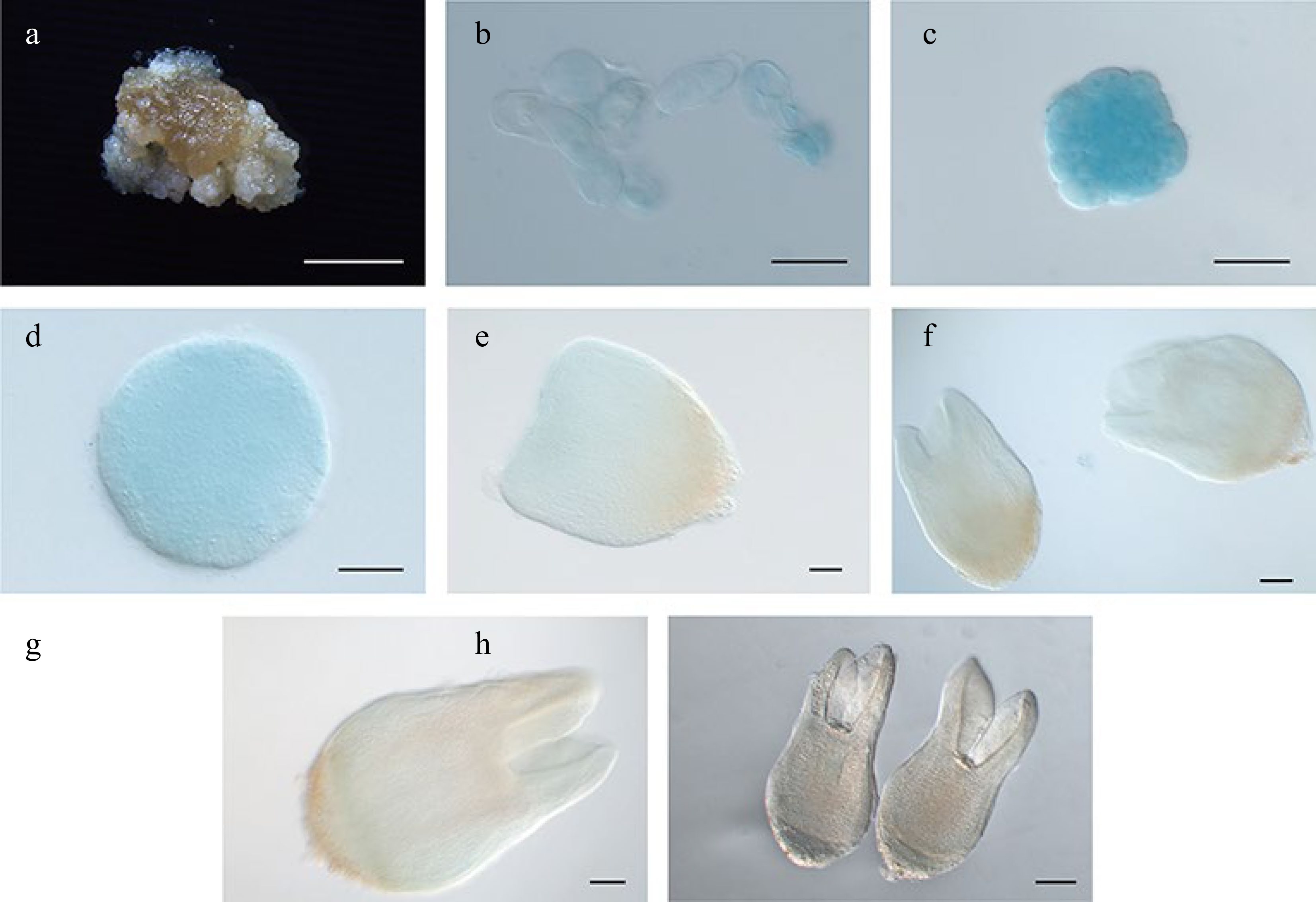
Figure 8.
Expression pattern of LcWOX14pro:GUS during somatic embryogenesis in Liriodendron. (a) LcWOX14pro:GUS was not expressed in embryonic callus. (b), (c) LcWOX14pro:GUS was highly expressed in single cells after 1 d on (b) induction medium (IM) and in the (c) pre-globular embryo. (d)–(g) Expression of LcWOX4pro:GUS gradually declined in the (d) globular embryo, the (e) transition-stage embryo, the (f) late heart-shaped embryo, and (g) the torpedo embryo. (h) LcWOX14pro:GUS was not expressed in the cotyledon embryo. Scale bars: (a) 2,000 μm; (b)–(e) 50 μm; (f), (g) 100 μm; (h) 200 μm.
-
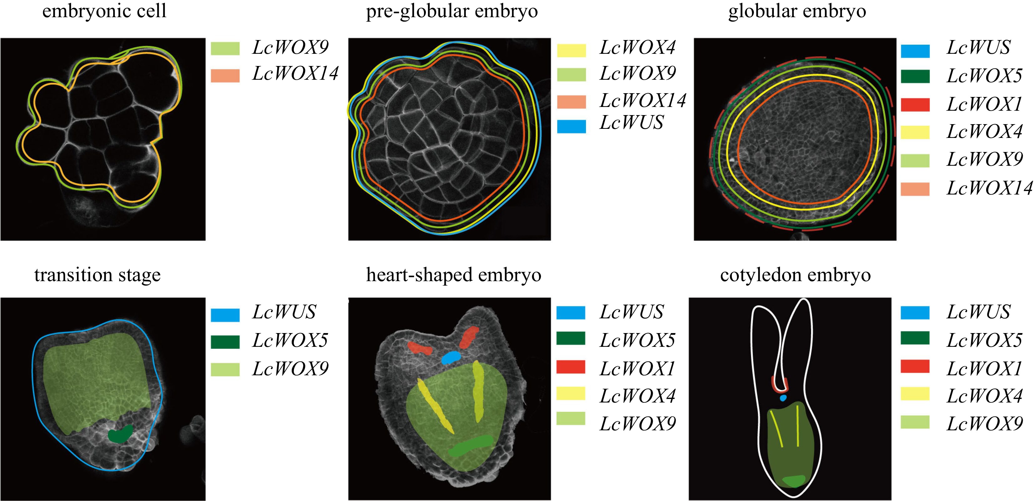
Figure 9.
Diagrams illustrating the dynamic expression of LcWOX genes during somatic embryogenesis in Liriodendron.
Figures
(9)
Tables
(0)