-
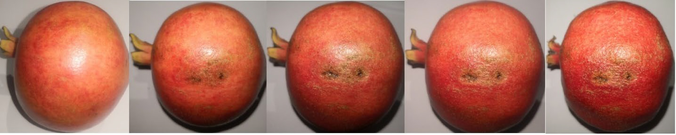
Figure 1.
Evolution of bruising damage over 8 d in pomegranate fruit (cv. Wonderful) that was dropped from 60 cm vertically (impact energy of about 1.8 J).
-
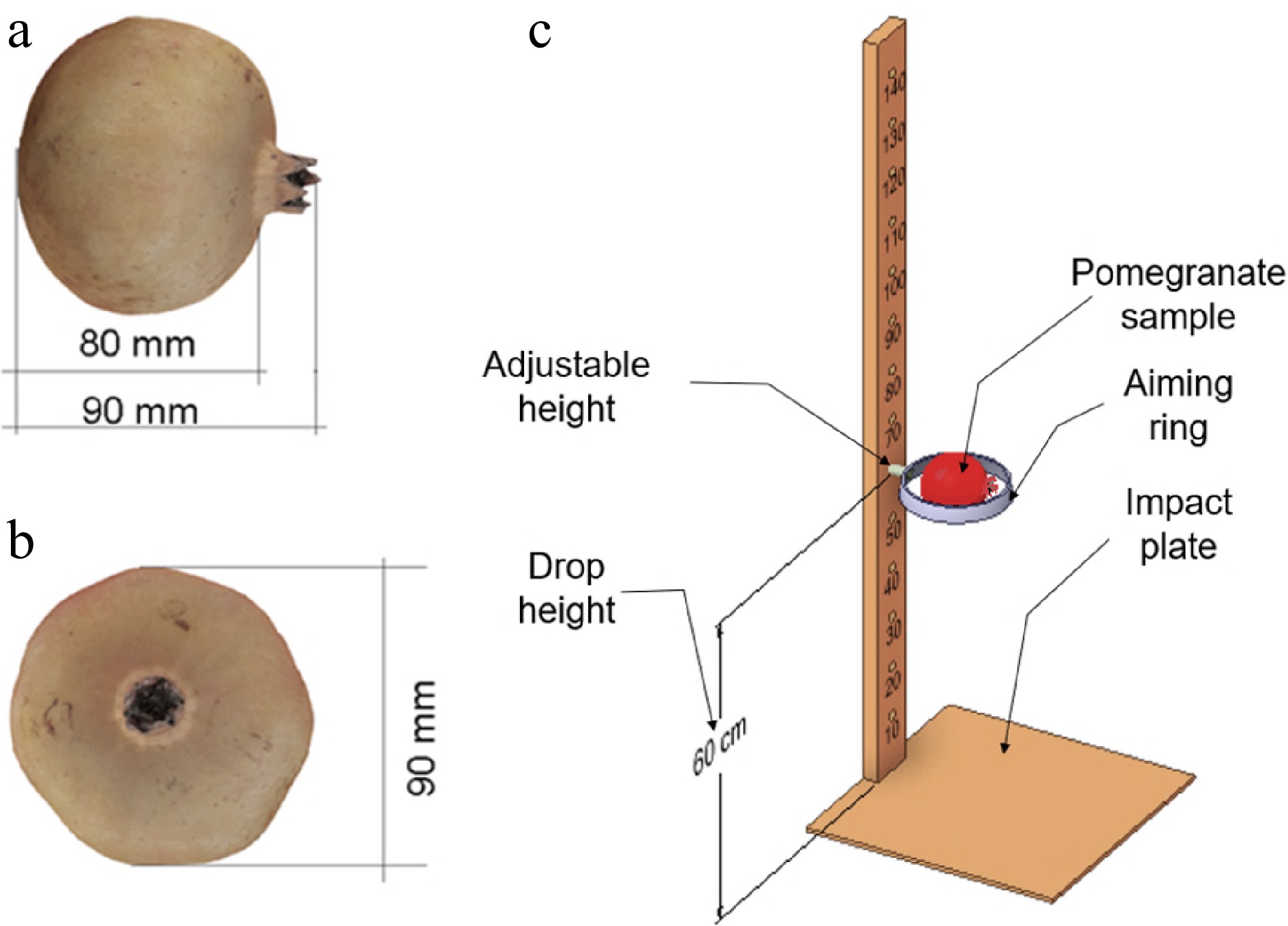
Figure 2.
Schematic showing dimensions of 'Class 1 pomegranate' (cv Wonderful) fruit used in the test ((a) front view and (b) side view) and (c) illustration of the drop simulation setup.
-
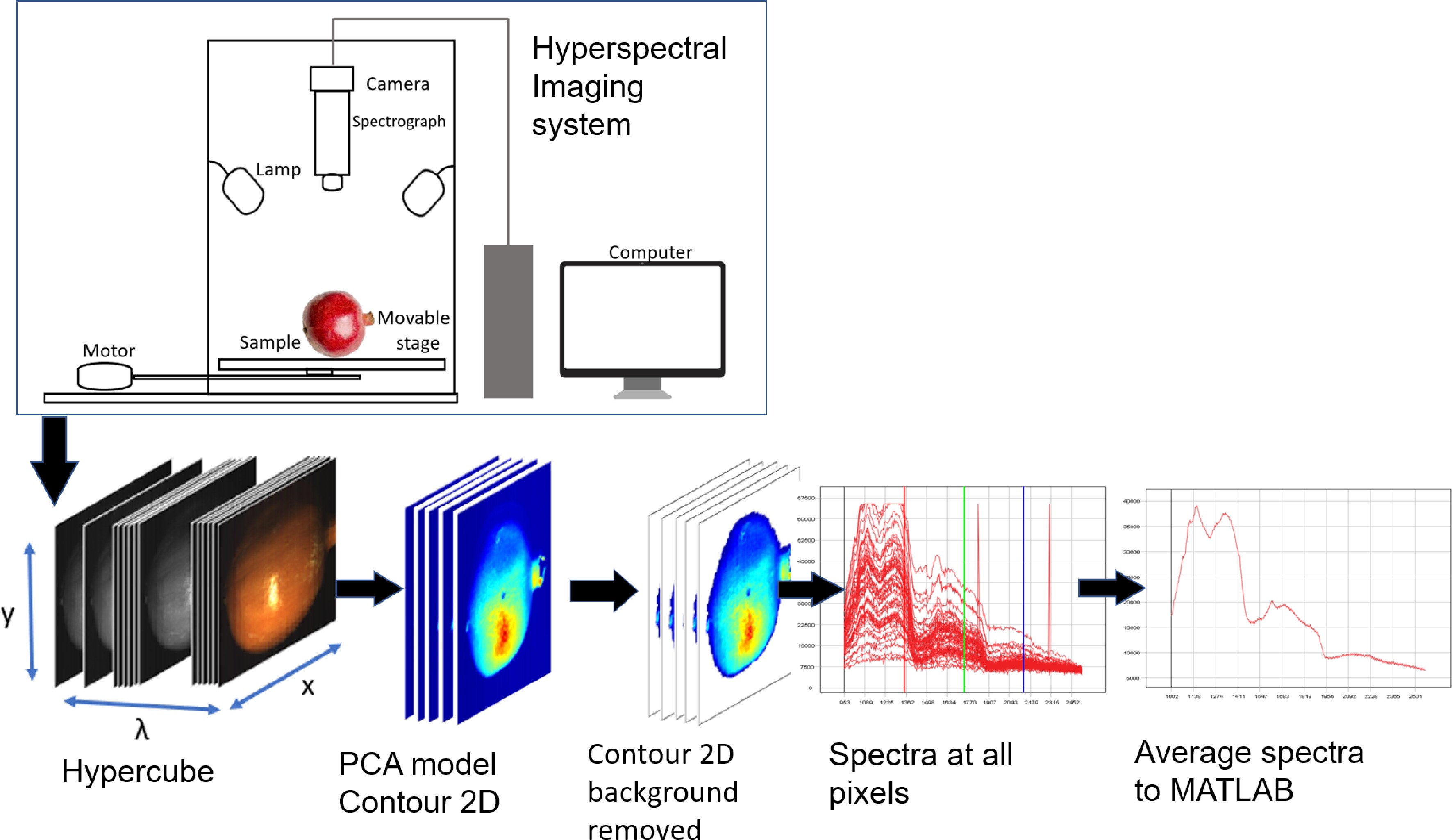
Figure 3.
Schematic showing the hyperspectral image system and image acquisition and preprocessing, process.
-
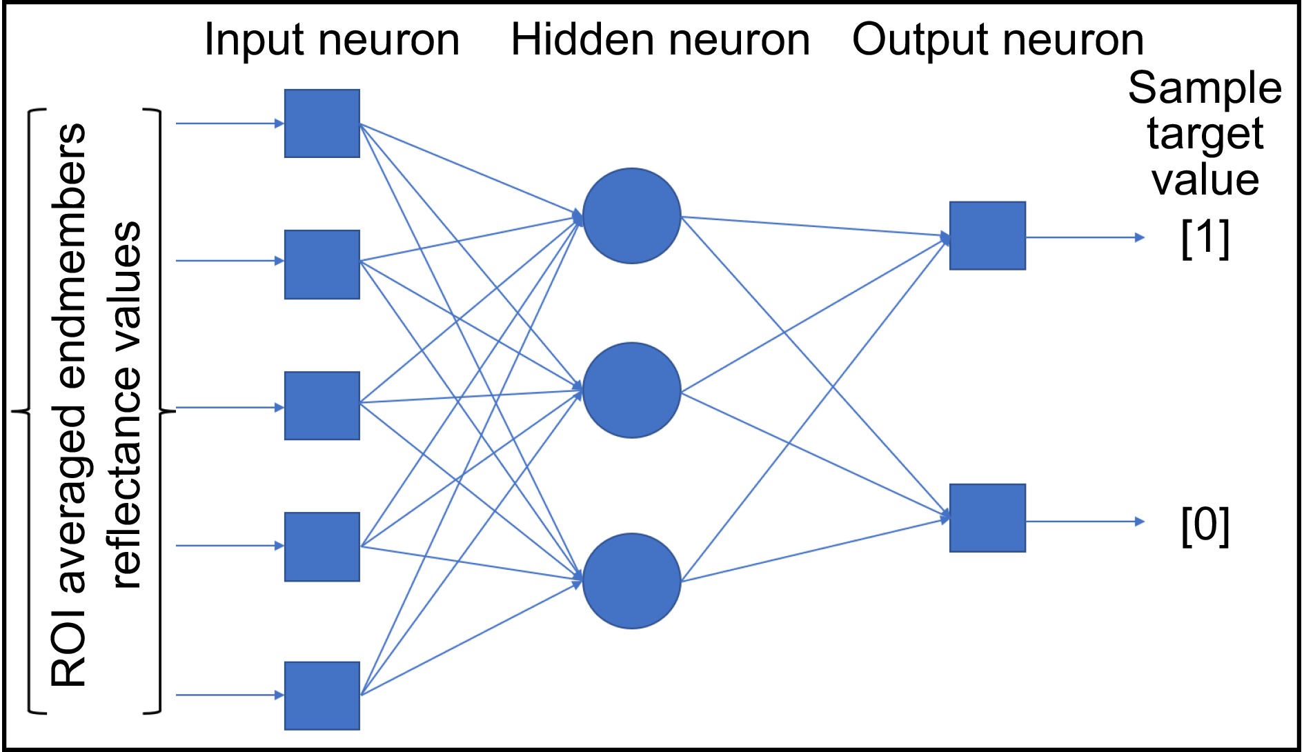
Figure 4.
Artificial neural network architecture for bruise classification.
-
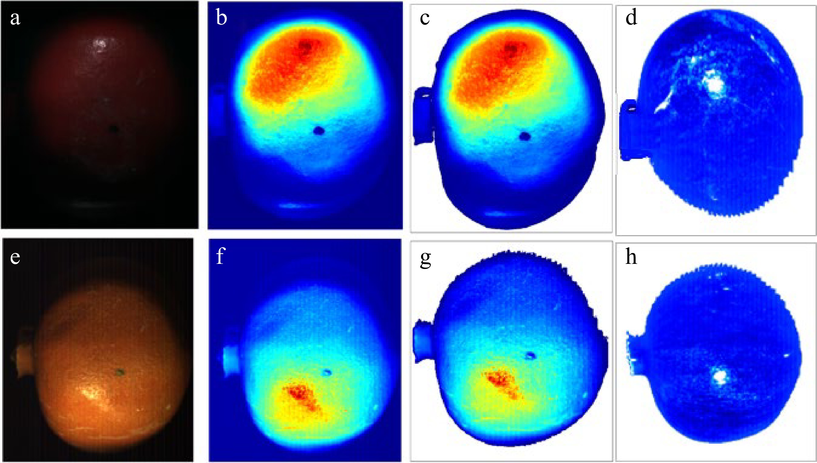
Figure 5.
Segmentation and pre-processing of images from the VNIR (upper raw) and SWIR (bottom raw) cameras. Raw (RGB) images (a) and (e), false color image (b) and (f), segmented images (c) and (g), with corrected spatial intensity variations (d) and (h).
-
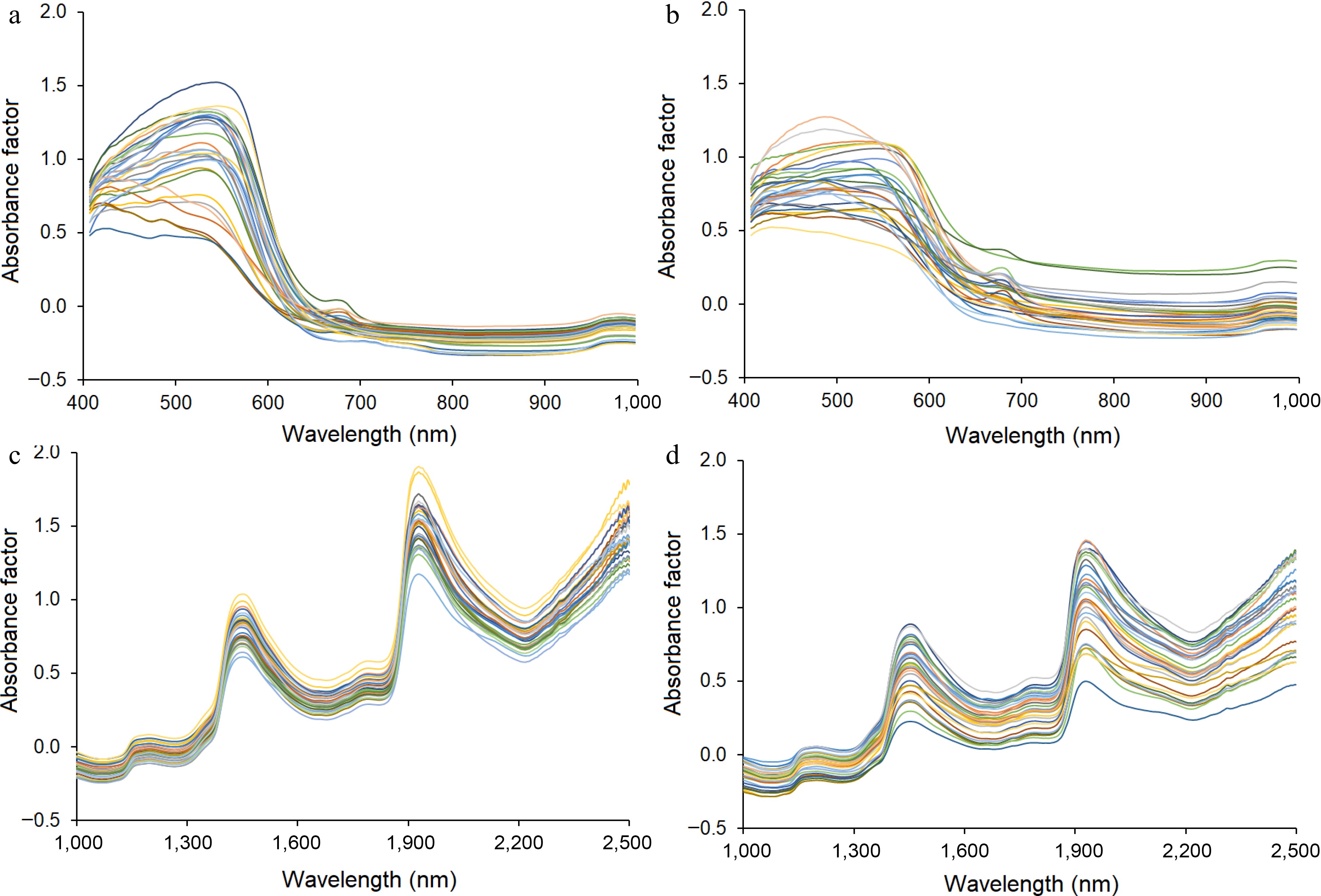
Figure 6.
Average absorbance spectra of (a) unbruised and (b) bruised pomegranates under the VNIR camera and (c) unbruised and (d) bruised under the SWIR camera. Data corresponds to imaging immediately after bruising.
-
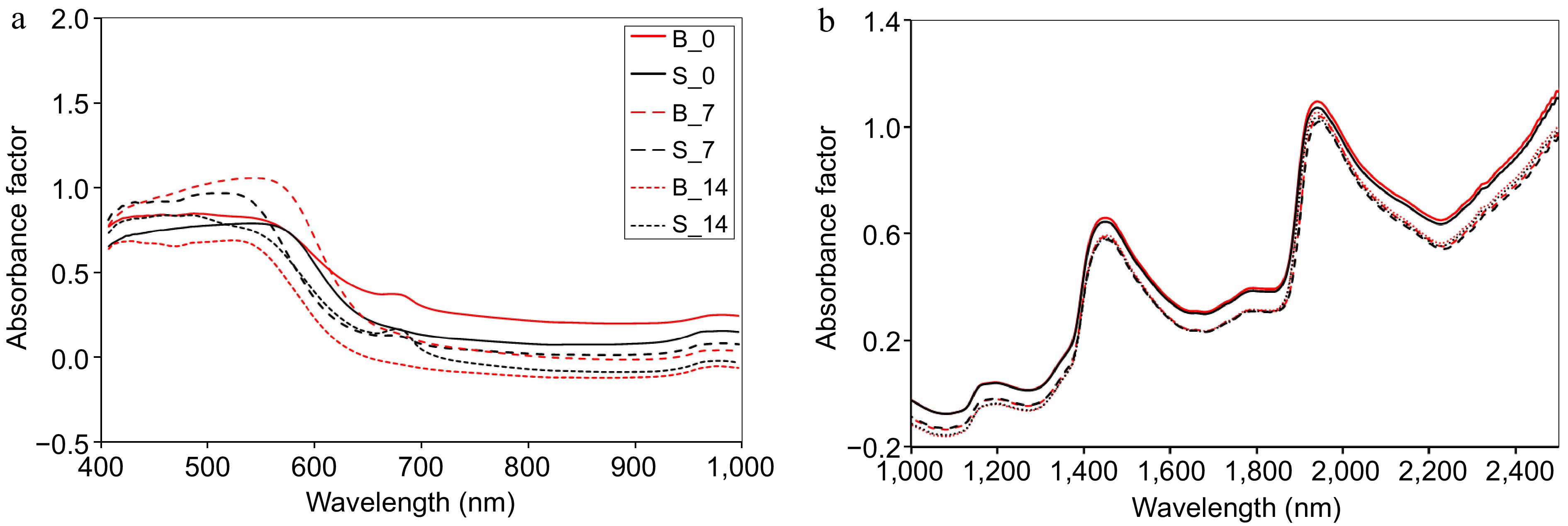
Figure 7.
The average spectra absorbance of unbruised (black curves) and bruised (red curves) of pomegranates under (a) VNIR and (b) SWIR cameras. Where the spectra of not bruised pomegranate in day 0 is S_0, sound fruit in day 7 is S_7, sound in day 14 is S_14, bruised in day 0 is B_0, bruised in day 7 is B_7 and bruised scanned in day 14 is B_14.
-
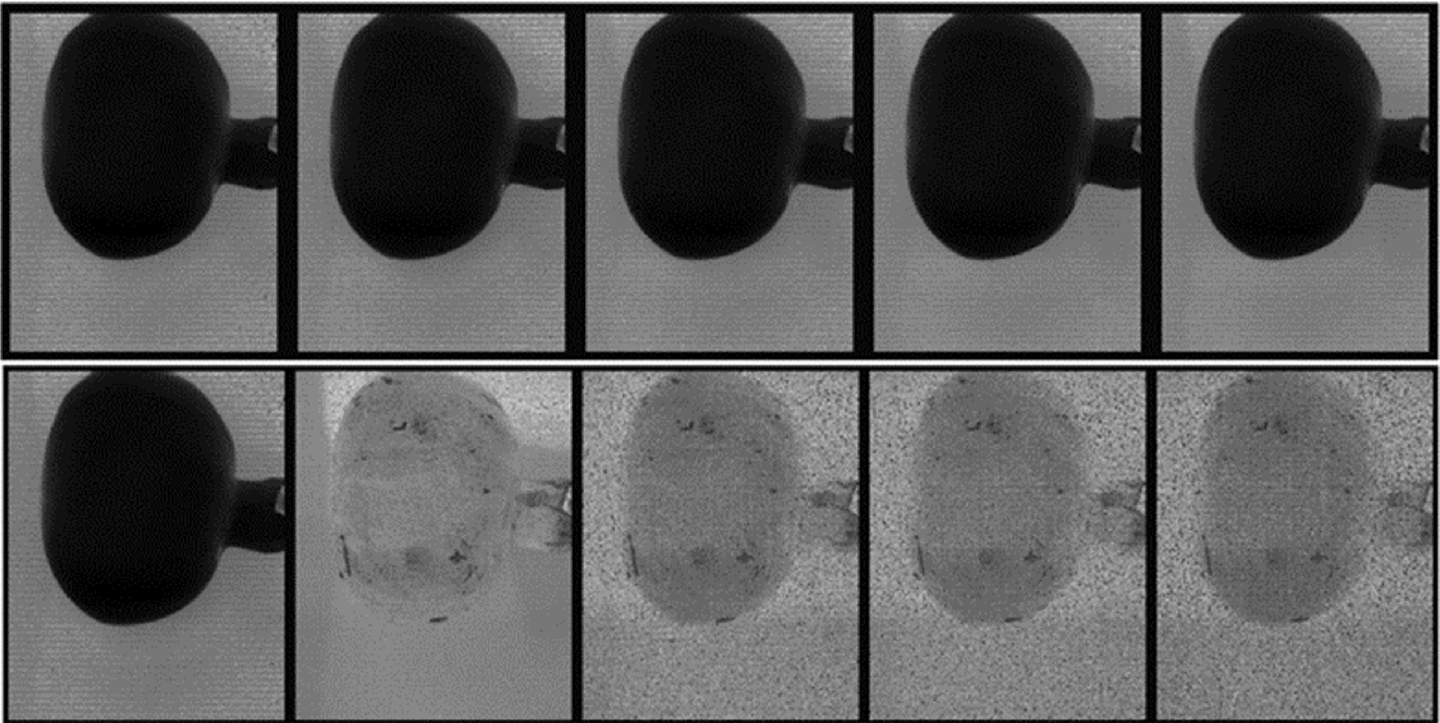
Figure 8.
Display of the first five spectral bands in the input data cube (top row) and the five most informative bands (bottom row) of a typical pomegranate fruit without bruising.
-

Figure 9.
Display of the first five spectral bands in the input data cube (top row) and the five most informative bands (bottom row) of a typical unbruised pomegranate fruit.
-
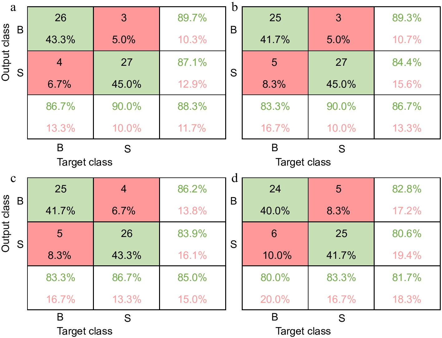
Figure 10.
Confusion matrix for the independent test set on the VNIR (upper raw) and SWIR (bottom raw) spectral data immediately after bruise damage. Model based on full wavelengths (left, (a) and (c)) and model based on selected wavelengths (right, (b) and (d)).
-
Main specifications SWIR VNIR Spectral range 930–2,500 nm 400–1,000 nm Spatial pixels 384 1,800 Spectral channels 288 186 Spectral sampling 5.45 nm 3.26 nm FOV 16° 17° Pixel FOV across/along 0.73/0.73 mrad 0.16/0.32 mrad Bit resolution 16 bit 16 bit Dynamic range 7,500 20,000 Peak SNR (at full resolution) > 1,100 >255 Max speed (at full resolution) 400 fps 260 fps Power consumption 30 W 30 W Dimensions (l-w-h) 38–12–17.5 cm 39–9.9–15 cm Weight 5.7 kg 5.0 kg Camera interface CameraLink CameraLink FOV is field of view. Table 1.
Specifications of the HySpex SWIR and VNIR cameras used in the study.
-
SWIR VNIR Sample number Correct CA (%) OA (%) Sample number Correct CA (%) OA (%) Day 0 AW Sound 30 26 86.7 85.0 30 27 90.0 88.3 Bruised 30 25 83.3 30 26 86.7 SW Sound 30 25 83.3 81.7 30 27 90.0 86.7 Bruised 30 24 80.0 30 25 83.3 Day 7 AW Sound 30 30 100.0 96.7 30 30 100.0 98.3 Bruised 30 28 93.3 30 29 96.7 SW Sound 30 28 93.3 93.3 30 29 96.7 93.3 Bruised 30 28 93.3 30 27 90.0 Day 14 AW Sound 30 30 100.0 98.3 30 30 100.0 100.0 Bruised 30 29 96.7 30 30 100.0 SW Sound 30 30 100.0 96.7 30 30 100.0 100.0 Bruised 30 28 93.3 30 30 100.0 AW is a classification prediction model based on all wavelengths; SW is model based on selected wavelengths; CA is component accuracy; OA overall accuracy. Table 2.
Classification results of test data set of ANN model for distinguishing sound and bruised tissues based on VNIR and SWIR reflected for the extracted (ROI) and the WFS of pomegranate fruit.
Figures
(10)
Tables
(2)