-
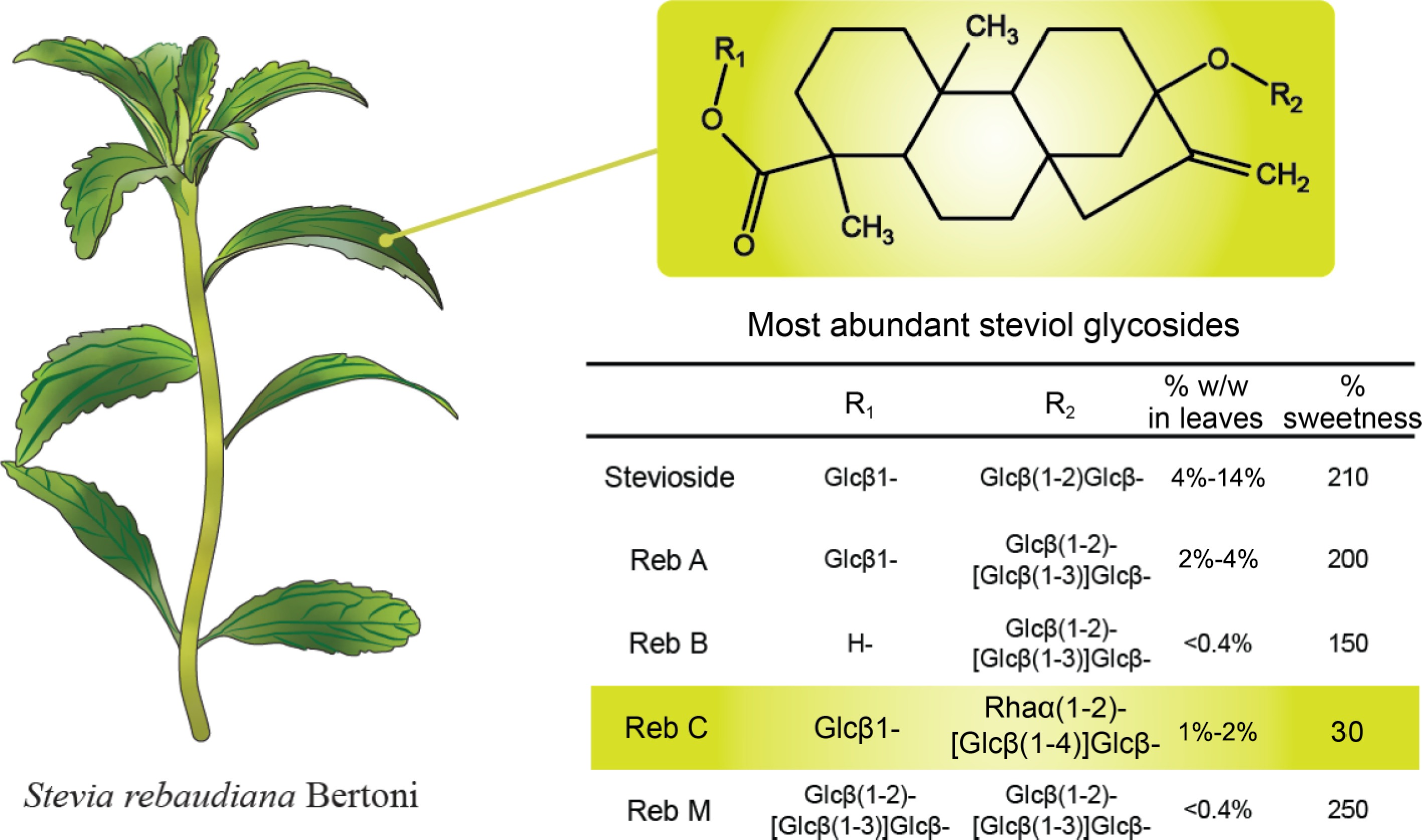
Figure 1.
Stevia rebaudiana Bertoni plant and the structure of relevant steviol glycoside (Reb) variants.
-
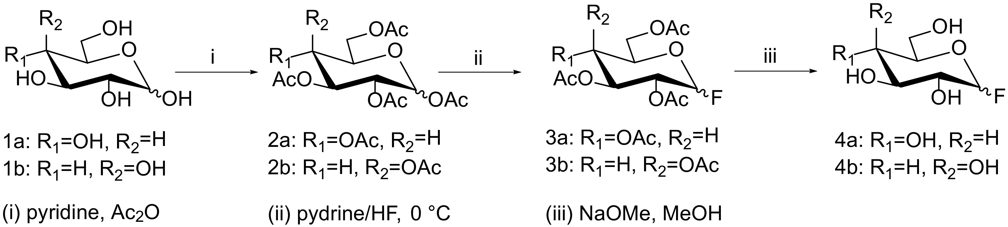
Figure 2.
Synthesis scheme for 1-F-glucose (4a) and 1-F-galactose (4b) from glucose (1a) and galactose (1b). Reagents and conditions: (i) pyridine, acetic anhydride; (ii) 70% hydrogen fluoride-pyridine, 0 °C; (iii) sodium methoxide, methanol.
-
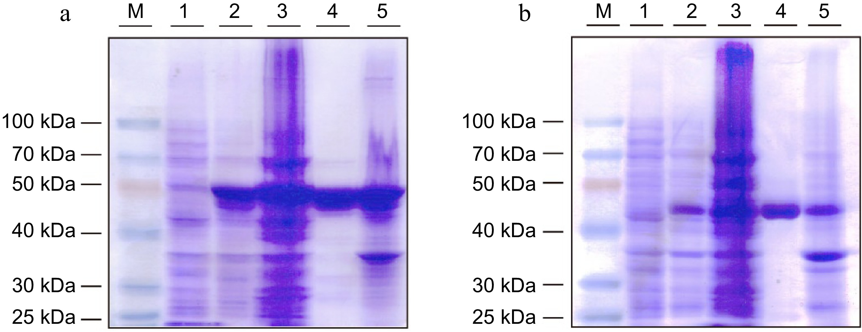
Figure 3.
SDS-PAGE analysis of recombinant (a) StspBGlcE383A and (b) AgtuBGlcE358S protein expressed in E. coli BL21 (DE3) cells. M: Protein marker, Lane 1: cell suspension before induction, Lane 2: cell suspension after induction, Lane 3: supernatant of cell lysis, Lane 4: purified recombinant protein, Lane 5: precipitate of cell lysis.
-
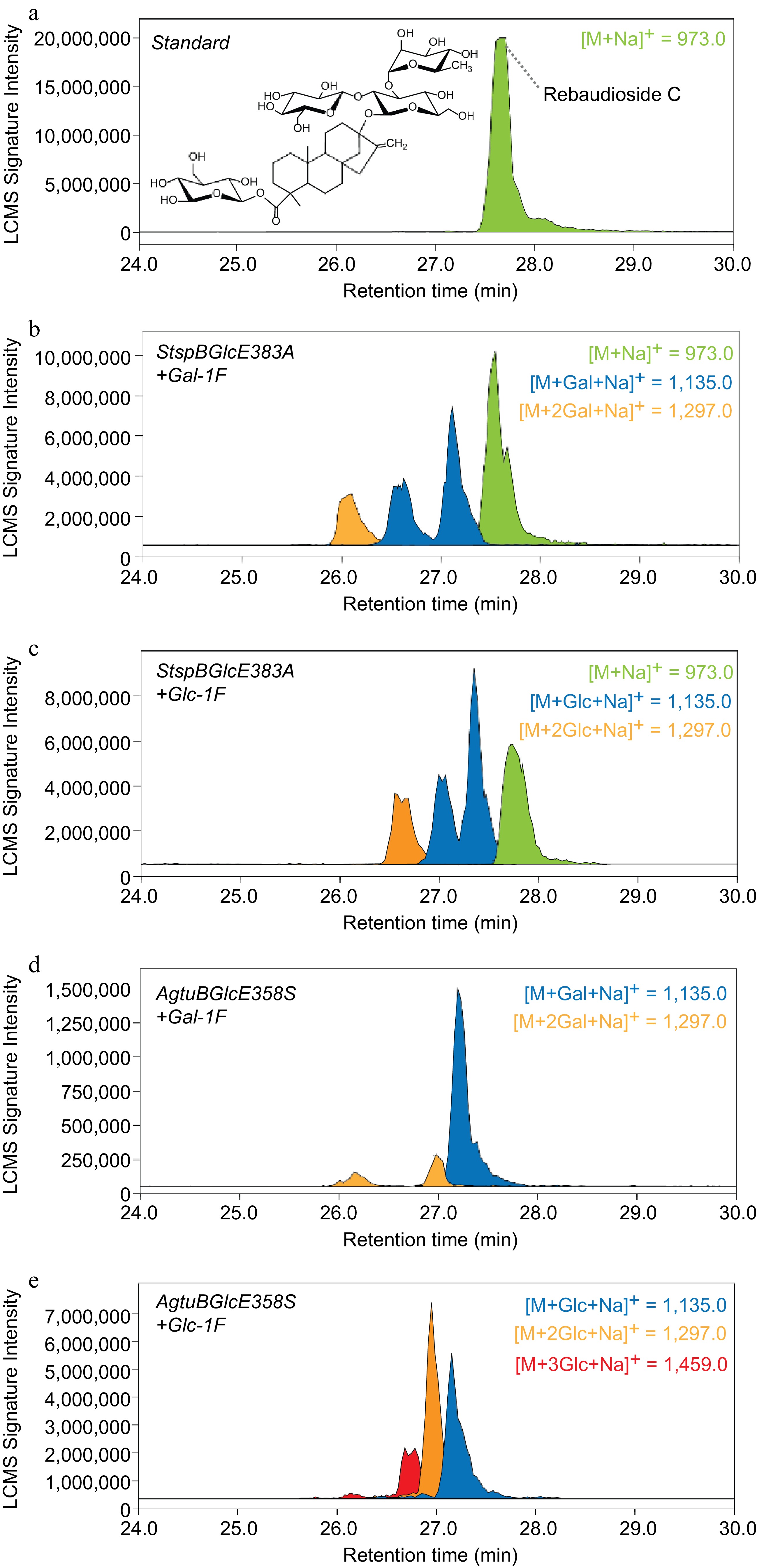
Figure 4.
Extracted ion masses of Reb C and its enzymatically glycosylated reaction products. The analytes were separated using a reversed-phase C18 column and detected using a m/z range between 500−2,000.
Figures
(4)
Tables
(0)