-
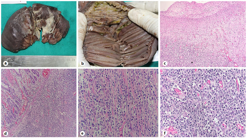
Figure 1.
a Resected ileum with exudate covered serosa. b Ileum showing perforation of the mucosal surface. c Granulation tissues and serosal exudate at the perforation site. (H&E, ×200). d Tumor cells infiltrating lamina propria and the submucosa (H&E, ×100). e Tumor cells infiltrating into the muscularis propria (H&E, ×400). f Atypical lymphoid cells (H&E, ×400).
-
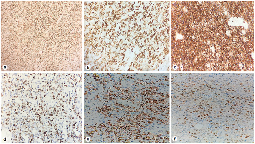
Figure 2.
a LCA immunoreactivity (IHC, ×100). b CD79a immunoreactivity (IHC, ×200). c CD10 immunoreactivity (IHC, ×200). d Ki-67 > 85% (IHC, ×100). e Bcl2 immunoreactivity (IHC, ×100). f Bcl6 immunoreactivity (IHC, ×100).
Figures
(2)
Tables
(0)