-
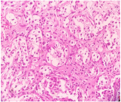
Figure 1.
Photomicrograph showing tumor cells organized in nests with clear cytoplasm, well-defined cytoplasmic borders, little cytological atypia, and hyperchromatic nuclei (H&E, ×400).
-
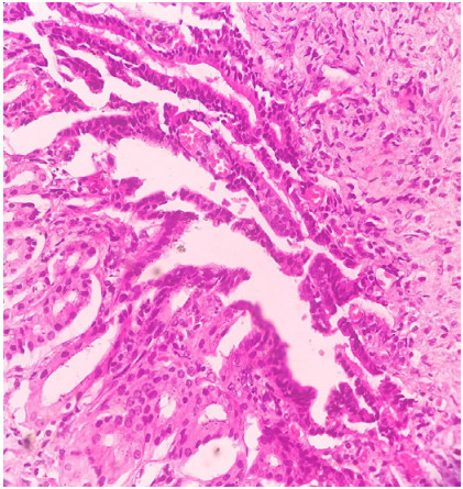
Figure 2.
Photomicrograph showing foci of conventional adenocarcinoma (H&E, ×400).
-
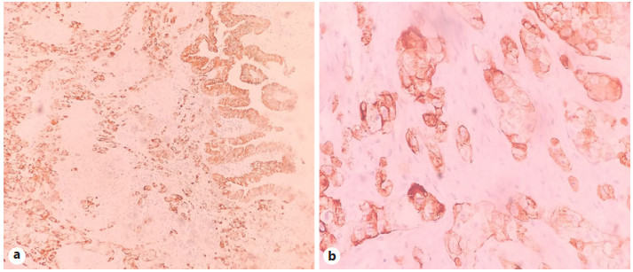
Figure 3.
Photomicrograph showing tumor cells diffuse positivity for CK-7 by IHC at low power (×100) (a) and at high power (×400) (b). IHC, immunohistochemistry; CK-7, cytokeratin-7.
-
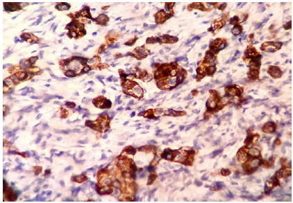
Figure 4.
Photomicrograph showing tumor cells positivity for AFP by IHC (×400). AFP, alpha-fetoprotein; IHC, immunohistochemistry.
-
Author Number of reported cases Age (years)/sex Cholelithiasis Histology Lymph node metastasis Outcome Maharaj et al. [5] 1 56/F yes CCG no AWD, on CT Zhang et al. [14] 1 80/M no CCG with hepatoid differentiation + FOCA yes 19 months disease-free Eken et al. [8] 1 56/F no CCG nd nd Sentani et al. [12] 1 78/F no AFP-producing CCG with neuroendocrine differentiation no 8 months disease-free Vaillo et al. [24] 1 72/F no CCG nd AWD, on CT Piana et al. [25] 1 66/F yes Combined CCG + small cell carcinoma + FOCA no 3 years DOD Bittinger et al. [15] 3 53–77/F yes (in 2 cases) CCG yes (in all cases) nd Vardaman Albores-Saavedra et al. [6] 7 56–68/F yes (in all cases) AFP-producing CCG and hepatoid differentiation in 1 case
Others-CCG + FOCA + areas of mucinous carcinoma (in 2 cases)yes (in 2 cases) 5 cases-DOD
2 cases-AWDPresent case 1 60/F yes AFP-producing CCG + FOCA no AWD, on CT CCG, clear-cell carcinoma of the gallbladder; FOCA, foci of conventional adenocarcinoma; ND, not determined; CT, chemotherapy; AWD, alive with disease; DOD, dead of disease; AFP, alpha-fetoprotein. -
Other clear cell tumors Age, years/sex Microscopy IHC Metastatic RCC [16–18] 39–84/M Solid, alveolar, and acinar pattern of cells with abundant clear cytoplasm and central or eccentric hyperchromatic nuclei. The characteristic network of small, thin-walled vasculature +ve for PAX-8
PAX-2, CD-10, vimentin, and CA IX
−ve for CK-7, CK-20, and CEAClear-cell carcinoma of the ovary [15, 19] 50–70/F Solid, papillary, and tubulocystic pattern lined by clear or hobnail cells. Papillae are frequently hyalinized +ve for EMA, CK and CA-125
−ve for CK-7, CK-20, and CEAEndometrial clearcell carcinoma [15, 20] 62–67/F Solid, papillary, tubular, and cystic architecture composed of polygonal cells with abundant clear or eosinophilic cytoplasm, and hobnail cells. Marked nuclear atypia and high mitotic activity +ve for 34βE12
Leu-M1, vimentin, BCL-2, CEA, P-53, and CA-125
ER, HER-2, and CK-7 are +/−. -ve for CK-20 and PRClear-cell hepatocellular carcinoma [15, 21] Elderly/F Trabecular, acinar, or compact pattern of polygonal cells with clear, vacuolated or foamy cytoplasm. Central or eccentric nucleus, frequently dispersed among conventional neoplastic hepatocytes +ve for Hep par 1, arginase-1, and AFP
−ve for CK-7, CK-19, EMA, CK-20, and CEACCG, clear-cell carcinoma of the gallbladder; IHC, immunohistochemistry; RCC, clear-cell renal carcinoma; CEA, carcinoembryonic antigen; CK-7, cytokeratin-7; PAX-8, paired box gene 8.
Figures
(4)
Tables
(2)