-
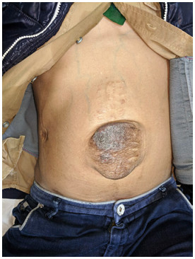
Figure 1.
Abdomen showing an incisional hernia over the infraumbilical region of the midline scar with overlying, hyperpigmented, and thinned out skin along with dilated veins over the abdomen.
-
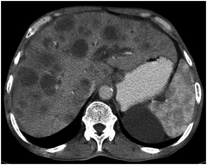
Figure 2.
CECT abdomen showing hepatomegaly with architectural distortion of the liver with multiple discrete and confluent lesions in the liver parenchyma showing peripheral enhancement in the arterial phase with central hypo-attenuating non-enhancing core. CECT, contrast-enhanced computed tomography.
-
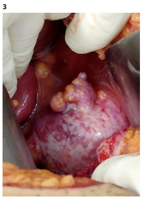
Figure 3.
Picture showing shrunken liver with finely nodular surface along with a deeply embedded porcelain gall bladder.
-
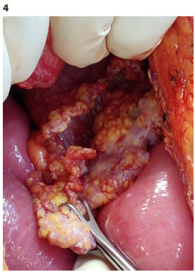
Figure 4.
Picture showing multiple hard and smooth nodules within the omentum.
-
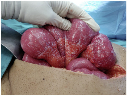
Figure 5.
Picture showing multiple nodules within the bowel mesentery.
-
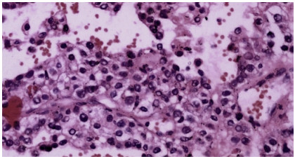
Figure 6.
Photomicrograph of an H&E-stained section s/o borderline vascular malignancy, that is, epithelioid hemangioendothelioma.
Figures
(6)
Tables
(0)