-
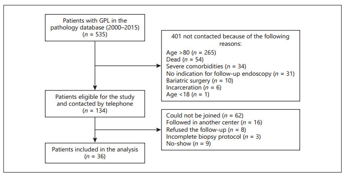
Figure 1.
Flow chart of the patients with gastric precancerous lesions (GPL) identified within the pathology database. From 535 patients with GPL initially identified, 134 were eligible for the study and were contacted by telephone. From these, 36 were finally included in the analysis.
-
Males, n (%) 22 (61) Age, years, median (range) 65 (38–79) Helicobacter pylori positive by histology, n (%) 9 (25) Delay between initial and final endoscopy, months, median (range) 57 (11–121) Proton pump inhibitors intake, n (%) 4 (11) No symptoms, n (%) 22 (61) Epigastralgia, dyspepsia, n (%) 8 (22) GERD, n (%) 6 (16) Initial endoscopy, the last endoscopy before recall; Final endoscopy, endoscopy after recall; GERD, gastro-esophageal reflux disease. Table 1.
Characteristics of all the patients included in the analysis (n = 36)
-
Initial endoscopy Final endoscopy (after recall) normal AG IM antrum IM corpus IM antrum + corpus LGD cancer AG, n = 2 1a – – 1c – – – IM (antrum only), n = 16 5a – 8b – 3c – – IM (corpus only), n = 8 1a – 2b 4b 1c – – IM (antrum + corpus), n = 4 – – – 1a 1b 1c 1c LGD, n = 6 – – 2a 1a – 3b – Patients marked by a superscript letter a showed a regression of GPL, those with a superscript letter b showed a progression of GPL, and those with a superscript letter c showed a stability of GPL during the follow-up. GPL, gastric precancerous lesions; AG, atrophic gastritis; IM, intestinal metaplasia; LGD, low-grade dysplasia. Table 2.
Evolution of GPL between the initial and final endoscopy
Figures
(1)
Tables
(2)