-
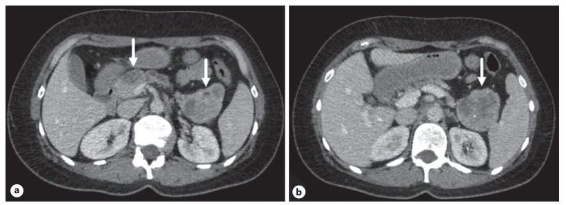
Figure 1.
Preoperative data. a CT scan, axial view, venous phase: tumors of the head and tail of the pancreas, and dilated pancreatic duct. b CT scan, axial view: tumor of the tail of the pancreas.
-
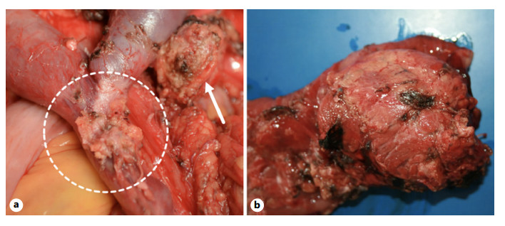
Figure 2.
Intra- and postoperative data. a Intraoperative view after removal of the pancreatoduodenal complex. Tumor growth can be seen on the mesentericoportal trunk; the arrow indicates the remaining part of the pancreatic parenchyma. b Specimen: intraductal papillary mucinous neoplasm associated with ductal adenocarcinoma.
-
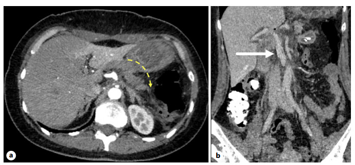
Figure 3.
Postoperative data. a CT scan, native phase, axial view. The dotted line marks the remaining pancreatic parenchyma after surgery. b CT scan, venous phase, frontal view. The arrow indicates a functioning venous prosthesis.
Figures
(3)
Tables
(0)