-
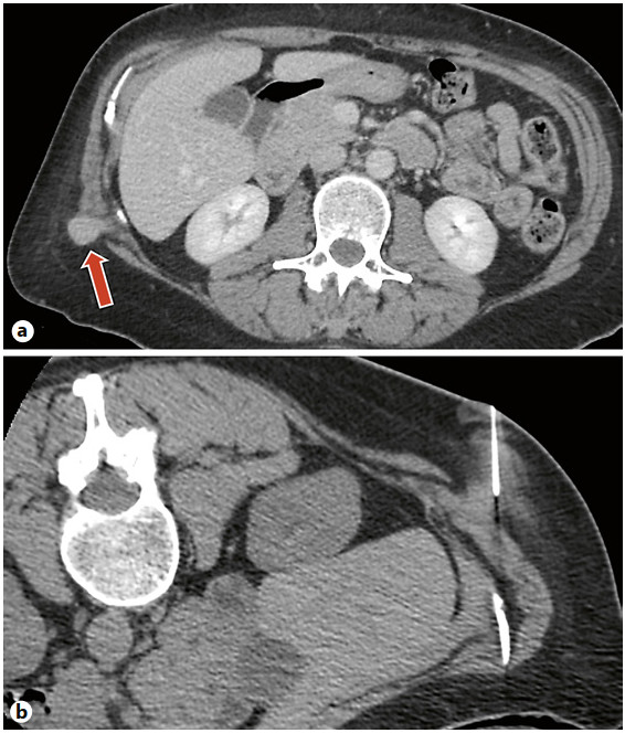
Figure 1.
a Axial contrast-enhanced CT scan at the level of the kidneys, in a 58-year-old female who presented with flank pain, demonstrating an incidental round, enhanced soft-tissue mass in the subcutaneous tissue of the right flank measuring 1.8 × 1.7 × 1.7 cm. b Axial unenhanced intraoperative CT image, with the patient prone, demonstrating biopsy of the soft-tissue mass with an 18-gauge BioPince full-core biopsy instrument (Argon Medical Devices).
-
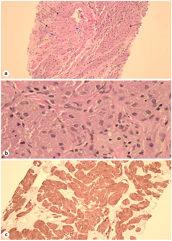
Figure 2.
GCT pathology microscopy of 58-year-old female who presented with flank pain. a Histopathology image demonstrating large tumor cells (black arrows) percolating between skeletal muscle fibers (blue arrows) HE. b Histopathology image demonstrating tumor cells with ill-defined cytoplasmic borders and abundant granular material (black arrows). Larger eosinophilic droplets are also present (yellow arrows). HE. ×40. c Immunohistochemistry image is diffusely positive and highlights the lysosomes (granular material) in the tumor cells. S-100. ×10.
-
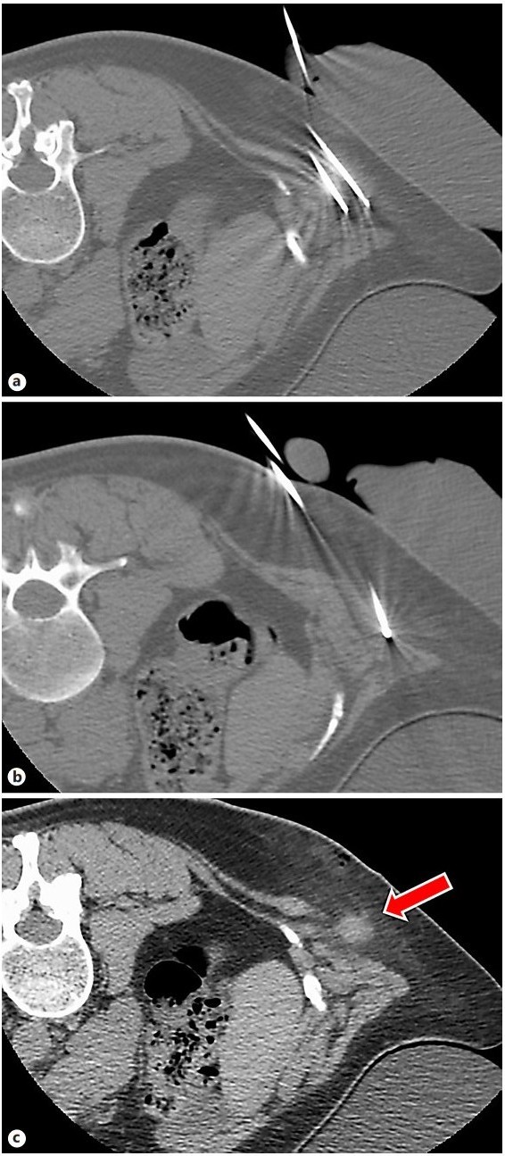
Figure 3.
Axial intraoperative CT image at the level of the kidneys, in a 58-year-old female who presented with flank pain, demonstrating two 17-gauge 1.5 IceRodCX cryoablation probes in the medial and lateral portion of the mass (a) and an additional 17-gauge probe in the inferior portion of the mass (b). The mass has "disappeared" as the cryoablation ice ball is closer in attenuation to the surrounding fat tissue than the index mass. c Immediate postprocedure axial CT shows increased conspicuity of the mass versus the intraprocedural images. There is faint hazy hyperdensity surrounding the mass and hypodensity at the interface of the mass and the adjacent musculature representing residual ice. This atypical appearance is related to surrounding fat density and is in contradistinction to the typical appearance of a cryoablation ice ball in solid organ tissue.
-
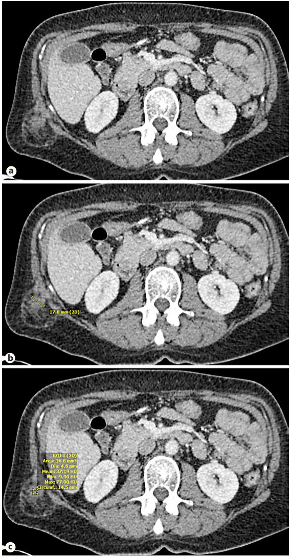
Figure 4.
A 6-week postoperative axial CT scan at the level of the kidneys, in a 58-year-old female who presented with flank pain, demonstrating a soft-tissue mass in the subcutaneous tissue of the right posterior flank (a), now measuring 1.7 × 1.4 cm (previously measured 1.7 × 1.9 cm) after cryoablation (b). There are adjacent inflammatory changes of the surrounding subcutaneous fat and no internal gas. c There is no internal enhancement (37 HU on precontrast exam and 34 HU on postcontrast exam). This is evidence of no viability.
Figures
(4)
Tables
(0)