-
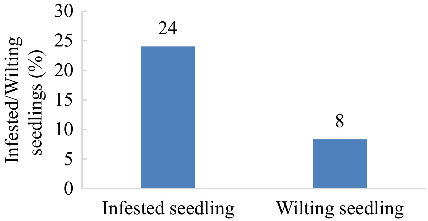
Figure 1.
Percentage of infested and wilting cacao seedlings under X. compactus infestation in East Luwu, South Sulawesi, Indonesia.
-
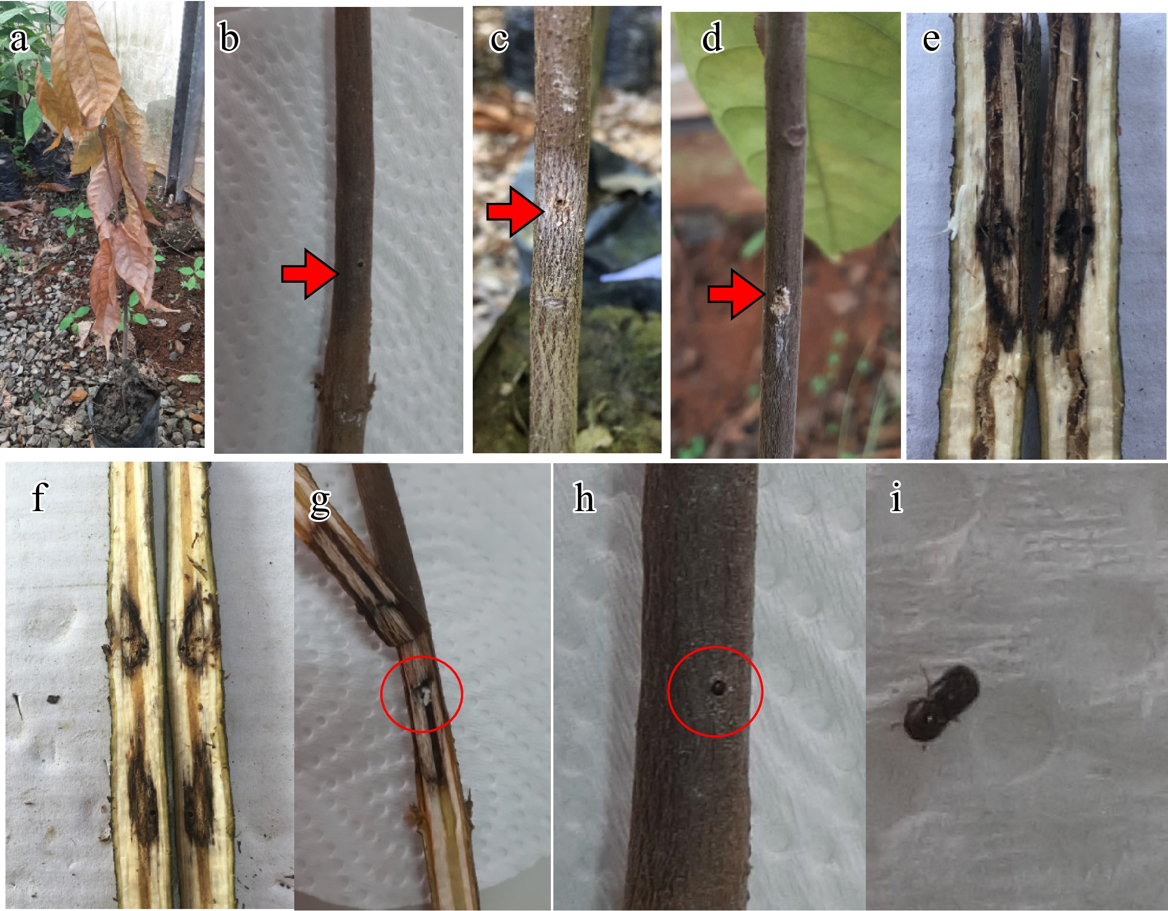
Figure 2.
External and internal symptoms of Xylosandrus colonisation on cacao seedlings in East Luwu, South Sulawesi, Indonesia. (a) Decline/wilting seedling. (b) Borehole with dark brown to black lesion around the hole (arrowhead). (c), (d) Borehole with powdery frass (arrowhead). (e), (f) Internal galleries with dark brown to black lesions around the inside holes. (g) Dark brown to black necrotic on wood tissue around the bore hole and internal gallery with a group of larvae inside (red circle). (h) Beetle in entry/exit hole. (i) Beetle.
-
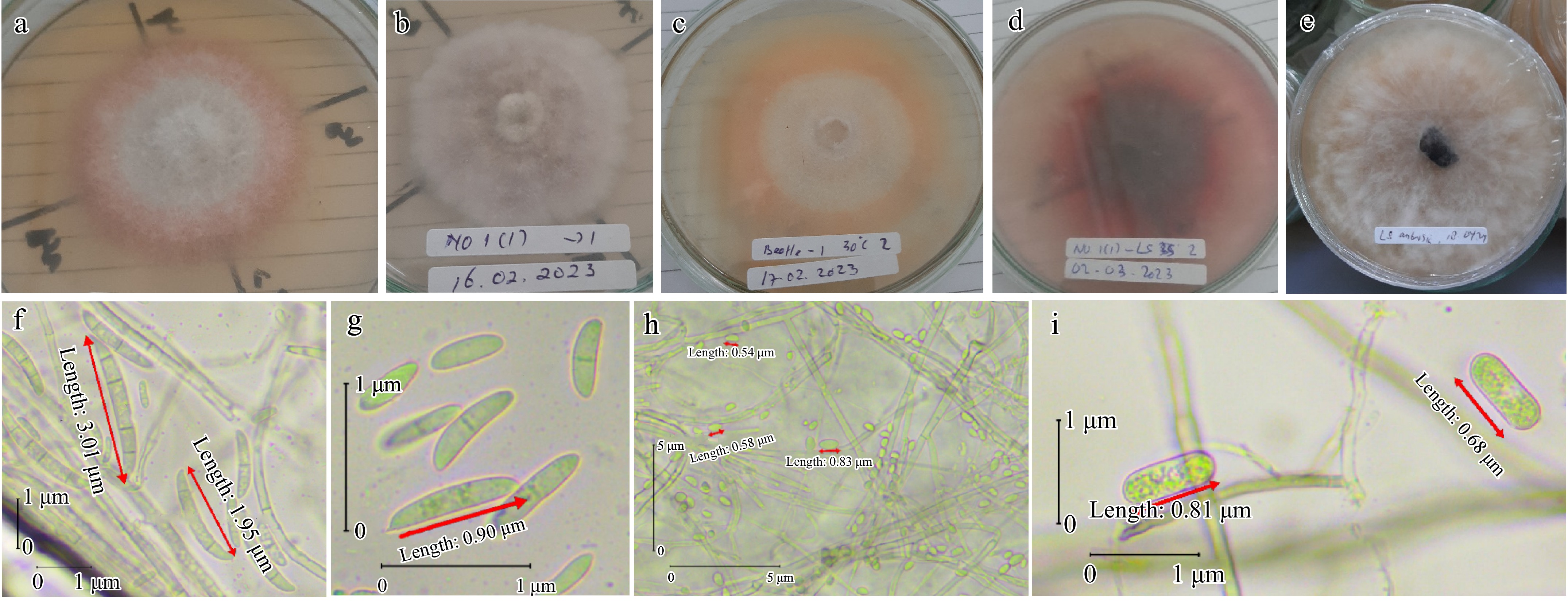
Figure 3.
Morphological and conidia features of fungi associated with wilting seedling of cacao. (a) Fusarium sp. colony features after 10 d at 30 °C. (b) Colletotrichum sp. colony features after 12 d at 26–29 °C. (c) Fusarium sp. colony features after 12 d at 30 °C. (d) Lasiodiplodia sp. colony features after 6 d at 35 °C. (e) Lasiodiplodia sp. colony features after 6 d at 26–29 °C. (f) Microconidia (two-celled oval) and macroconidia (typical Fusarium macroconidium; apical cell; blunt; basal cell; foot-shape; four-six cell). (g) Microconidia (oval and two-celled oval) of Fusarium sp. (h) Aseptate, hyaline, subovoid, ovoid, and globose conidia of Colletotrichum sp. (i) Ellipsoid young conidia, hyaline, aseptate, and granular contents of Lasiodiplodia sp.
-
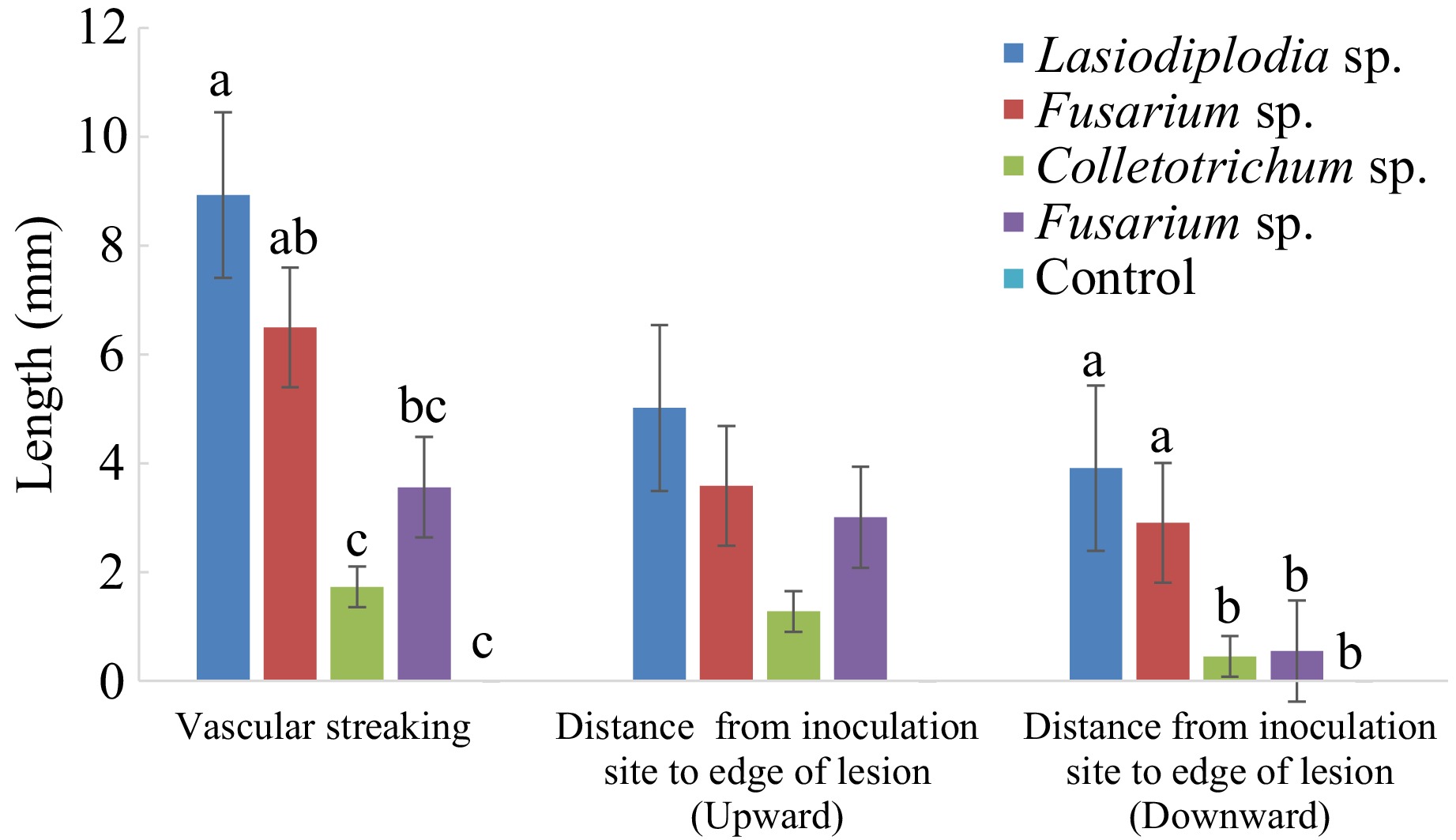
Figure 4.
Vascular streaking length (mm) in the stem of cacao seedlings inoculated with associated fungi with Xylosandrus and its infected stem with PDA as a control inoculum for 95 d. Bars with the same letter do not differ significantly according to the Least Significant Difference (LSD) test at p ≤ 0.05.
-
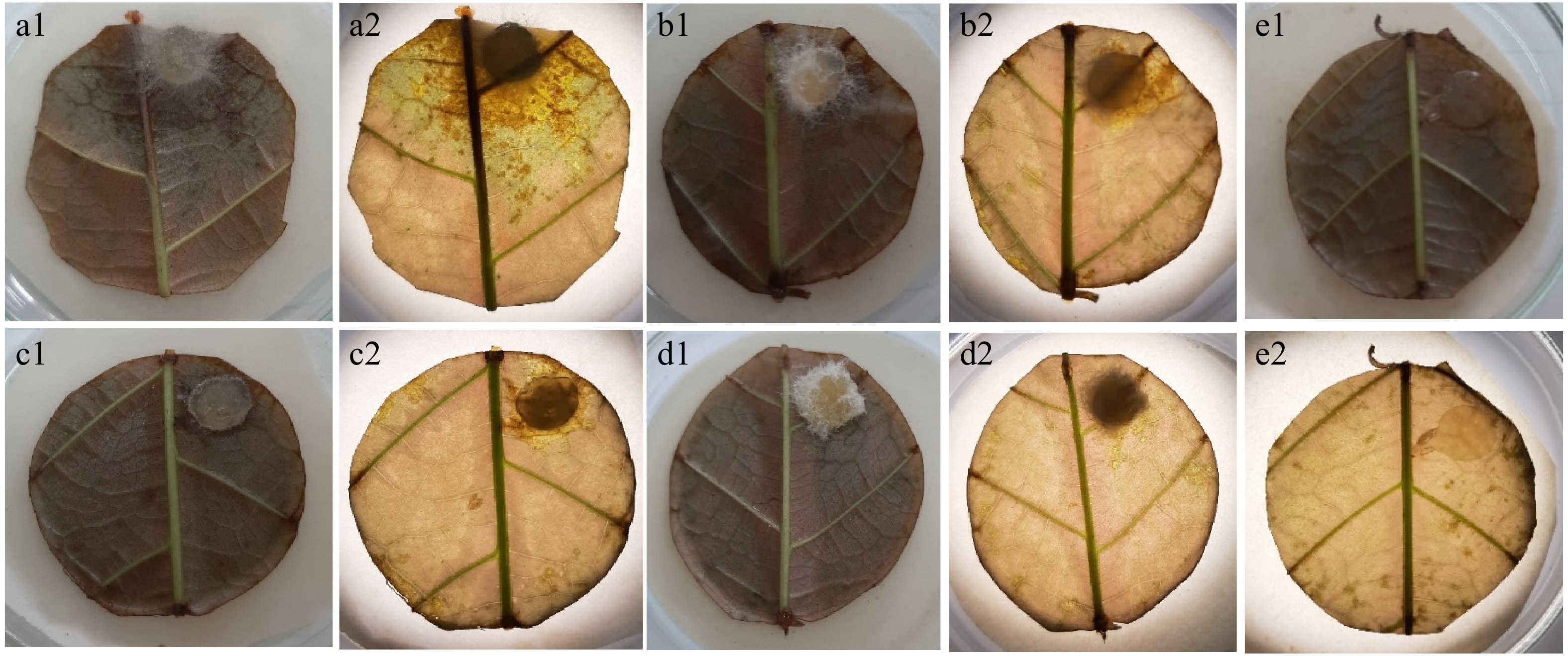
Figure 5.
Necrotic lesions on stage 3 leaf disks 48 h after inoculation in the leaf disk bioassay. (a1) Inoculation with Lasiodiplodia isolate; (a2) observed under a microscope. (b1) Inoculation with Fusarium isolate; (b2) observed under a microscope. (c1) Inoculation with Fusarium isolate; (c2) observed under a microscope. (d1) Inoculation with Colletotrichum isolate; (d2) observed under a microscope. (e1) No necrotic lesions obtained on controls treated with sterile PDA agar plugs; (e2) observed under a microscope.
-
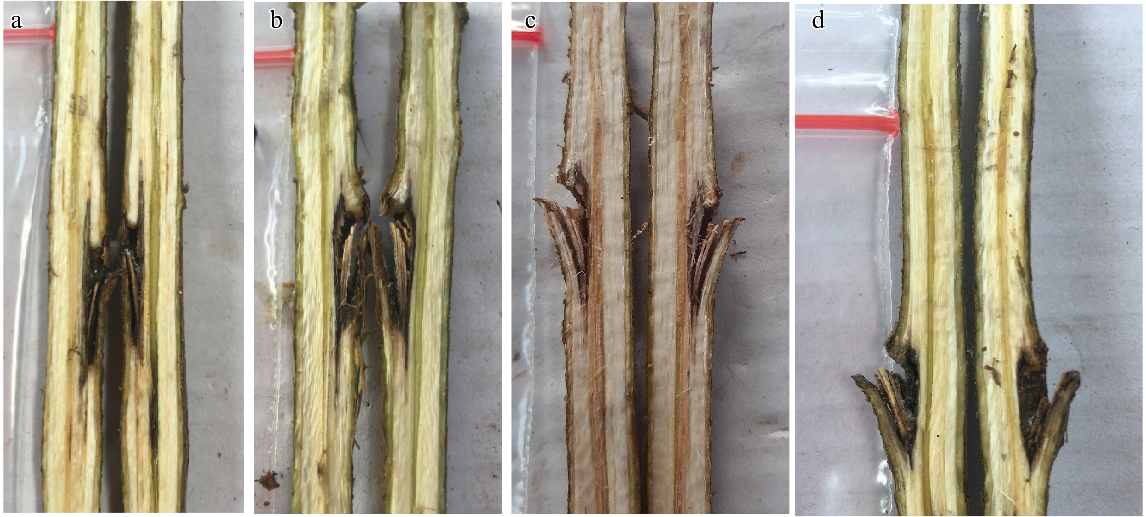
Figure 6.
The vertical necrotic section of the fungal inoculated stem showed various sizes of dark brown to black vascular streaking 95 d after inoculation. (a) Lasiodiplodia isolate. (b) Fusarium isolate. (c) Colletotrichum isolate. (d) The vertical section of control showed symptomless/no vascular streaking.
-
Material
originFusarium sp. Lasiodiplodia sp. Colletotrichum sp. Total Wood sections of infected beetle galleries Surface sterilization 2 1 1 4 Without surface sterilization 3 1 1 5 Larvae 3 0 2 5 Total 8 2 4 14 Table 1.
Associated fungi with infested plant parts under X. compactus infestation and larvae of X. compactus on cacao seedlings in East Luwu, South Sulawesi, Indonesia.
-
Fungal isolates Leaf blade Main vein 24-h 48-h 72-h AUDPC 24-h 48-h AUDPC Lasiodiplodia sp. 14.4a 55.0a 81.3a 102.8a 22.5a 67.5a 45.0a Fusarium sp. 0.8b 2.5b 8.1b 6.9b 0.0b 0.0b 0.0b Fusarium sp. 0.0b 1.8b 5.0b 4.3b 0.0b 0.0b 0.0b Colletotrichum sp. 0.0b 0.8b 4.5b 3.0b 0.0b 0.0b 0.0b Control 0.0b 0.0b 0.0b 0.0b 0.0b 0.0b 0.0b Numbers in the same column followed by the same letter are not significantly different by LSD's test analysis (p < 0.05). Table 2.
Lesion development and AUDPC values (%) of lesions 24, 48, and 72 h after inoculation on the leaf blade and 24 and 48 h after inoculation on the main vein. Leaf blade necrosis was analyzed separately from the main vein necrosis.
-
Fungi 30 °C 35 °C 1 d 2 d 4 d 6 d 8 d 1 d 2 d 4 d 6 d 8 d Lasiodiplodia sp. 22.0 40.0 -* -* -* 8.1 16.9 35.8 36.7 -* Fusarium sp. 0.0 0.0 12.3 17.4 21.3 0.0 0.0 0.0 0.0 0.0 Fusarium sp. 0.0 0.0 18.5 26.4 33.1 0.0 0.0 6.6 11.3 13.3 Colletotrichum sp. 0.0 0.0 7.8 11.9 17.4 0.0 0.0 0.0 0.0 0.0 *The growth has reached the end of the medium. Table 3.
Growth rates (mm/d) of the fungi associated with cacao decline seedlings under X. compactus infestation at 30 and 35 °C. Isolates were grown on PDA at the temperatures shown in the figures.
Figures
(6)
Tables
(3)