-
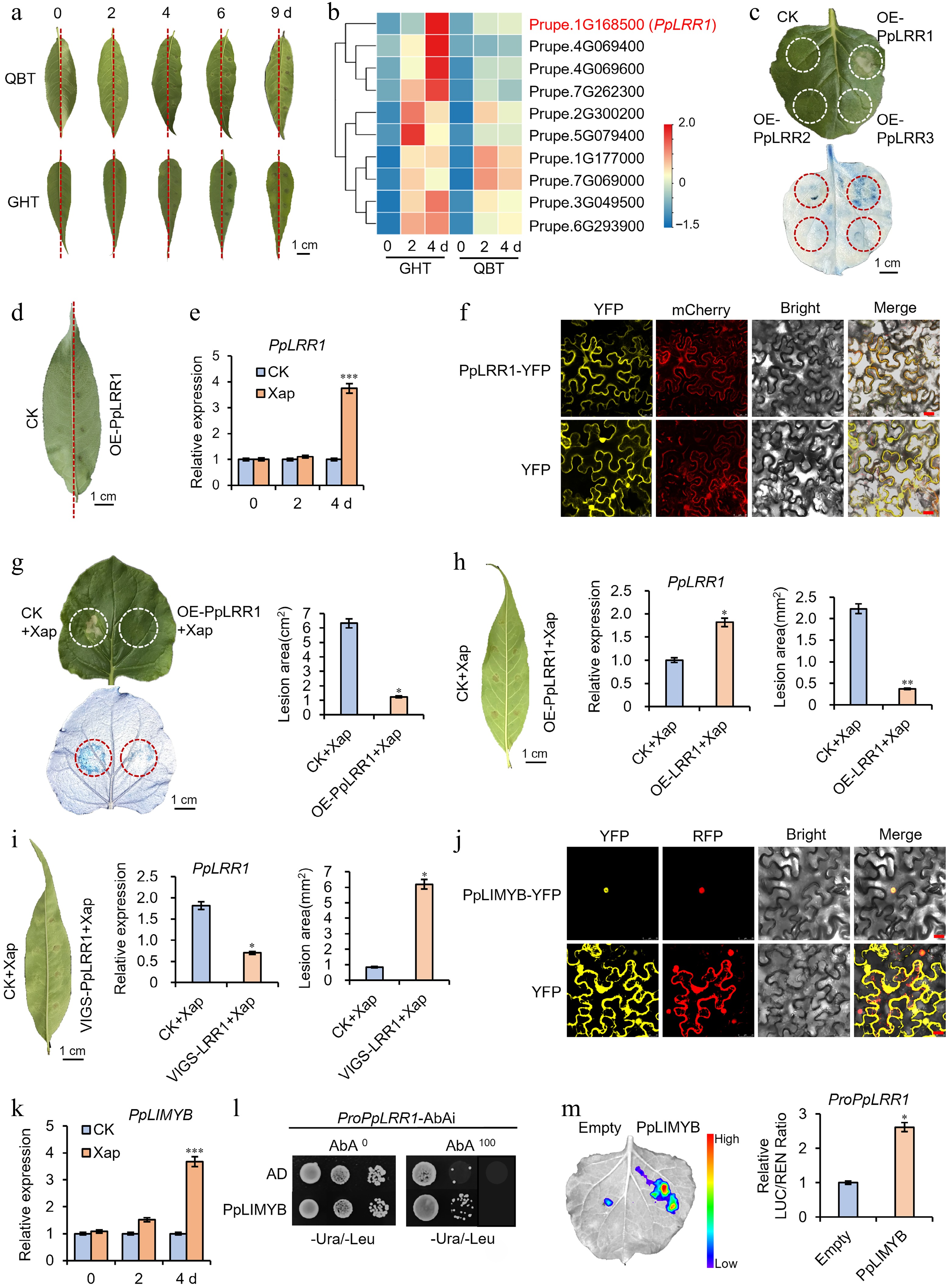
Figure 1.
Functional validation of PpLRR1 in resistance to peach bacterial spot disease. (a) Phenotypic comparison of peach leaves from 'Qiubaitao (QBT, Prunus persica)' and 'Guanghetao (GHT, Prunus mira)' cultivars inoculated with Xanthomonas arboricola pv. pruni (Xap) vs control at 4 dpi. Transcriptome sequencing was conducted on leaf samples at 0, 2, and 4 dpi. Scale bar = 1 cm. (b) Heatmap of differentially expressed genes (|log2FC| > 1, p < 0.05) identified from RNA-seq data. PpLRR1 is highlighted in red. (c) Trypan blue-stained tobacco leaves transiently overexpressing PpLRR1, PpLRR2, or PpLRR3 compared to the empty vector control at 3 dpi. (d) Hypersensitive response phenotype in peach leaves transiently overexpressing PpLRR1 at 4 dpi. (e) Relative expression of PpLRR1 in 'Mantianhong (MTH)' peach leaves at 0, 2, and 4 dpi, measured by qRT-PCR. Expression was normalized to the sterile water-injected control, set to 1. (f) Subcellular localization of PpLRR1-YFP fusion protein in Nicotiana benthamiana epidermal cells. Yellow fluorescence (YFP) co-localizes with the red fluorescent plasma membrane marker (mCherry). OD600 = 1.0. Cultivate for 48 h after injection. Scale bar = 25 μm. (g) Quantification of lesion area in tobacco leaves co-inoculated with Xap and overexpressing PpLRR1 at 3 dpi (trypan blue staining). (h) Lesion area and relative PpLRR1 expression in MTH peach leaves co-inoculated with Xap and overexpressing PpLRR1 at 4 dpi. (i) Lesion area and PpLRR1 expression in MTH peach leaves co-inoculated with Xap following PpLRR1 silencing. TRV, TRV1 + TRV2; PpLRR1-TRV, TRV1 + PpLRR1-TRV2. (j) Subcellular localization of PpLIMYB-YFP fusion protein in transgenic N. benthamiana epidermal cells. Yellow fluorescent signal (YFP) co-localizes with RFP (red). TM–NR: tonoplast marker and red fluorescent nuclear marker (Nucleus–RFP). OD600 = 1.0. Cultivate for 48 h after injection. Scale bar = 25 μm. (k) Relative expression levels of PpLIMYB in peach leaves at 0, 2, and 4 dpi assessed by qRT-PCR. Expression was normalized to the sterile water-injected control, set to 1. (l) Yeast one-hybrid assay showing direct binding of PpLIMYB to the PpLRR1 promoter. AbA0: medium without AbA; AbA100: medium with 100 ng/mL AbA. (m) Dual-luciferase reporter assay quantifying PpLIMYB-mediated activation of the PpLRR1 promoter. Data represent mean ± SD of three biological replicates (*p < 0.05). Each treatment was performed in triplicate, and each replicate contained three to five peach leaves. Error bars indicate standard deviation. Statistical significance was assessed by one-way ANOVA and t-test: * p < 0.05; ** p < 0.01; *** p < 0.001.
Figures
(1)
Tables
(0)