-

Figure 1.
Maximum Likelihood tree illustrating the phylogeny of Thelephora based on combined ITS + LSU sequence data. Branches are labeled with maximum-likelihood bootstrap proportions higher than 50% and Bayesian posterior probabilities greater than 0.90, respectively. * Indicate the type species. The new species are in bold.
-
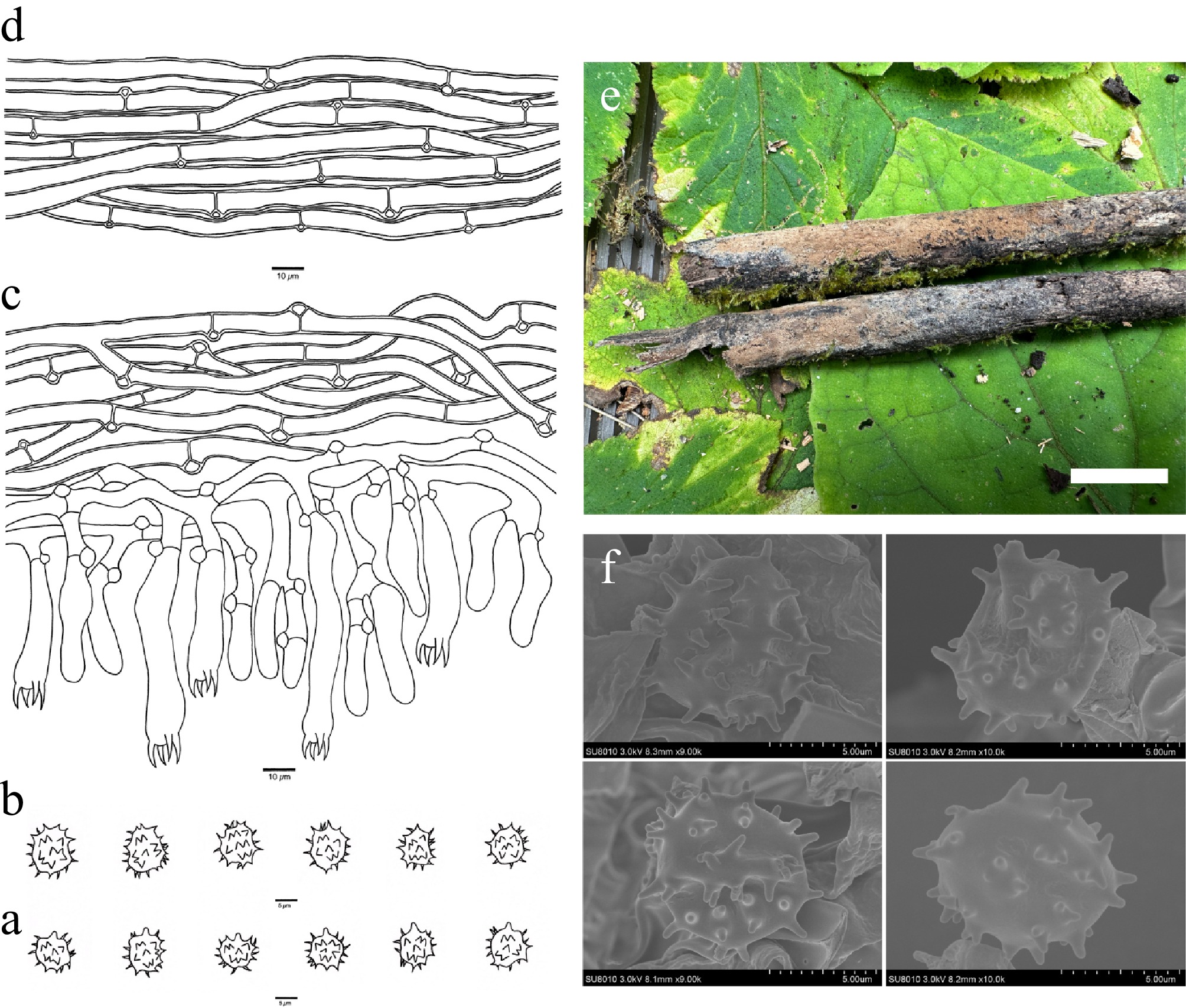
Figure 2.
Basidiomata and microscopic structures of Thelephora bambusicola (holotype, Dai 31713). (a) Basidiospores in lateral view. (b) Basidiospores in frontal view. (c) A section through a basidioma. (d) Hyphae from a rhizomorph. (e) Basidiomata on the host substrates. (f) Scanning electron microscope (SEM) of basidiospores. Scale bars: (a), (b) = 5 µm; (c), (d) = 10 µm; (e) = 1 cm.
-
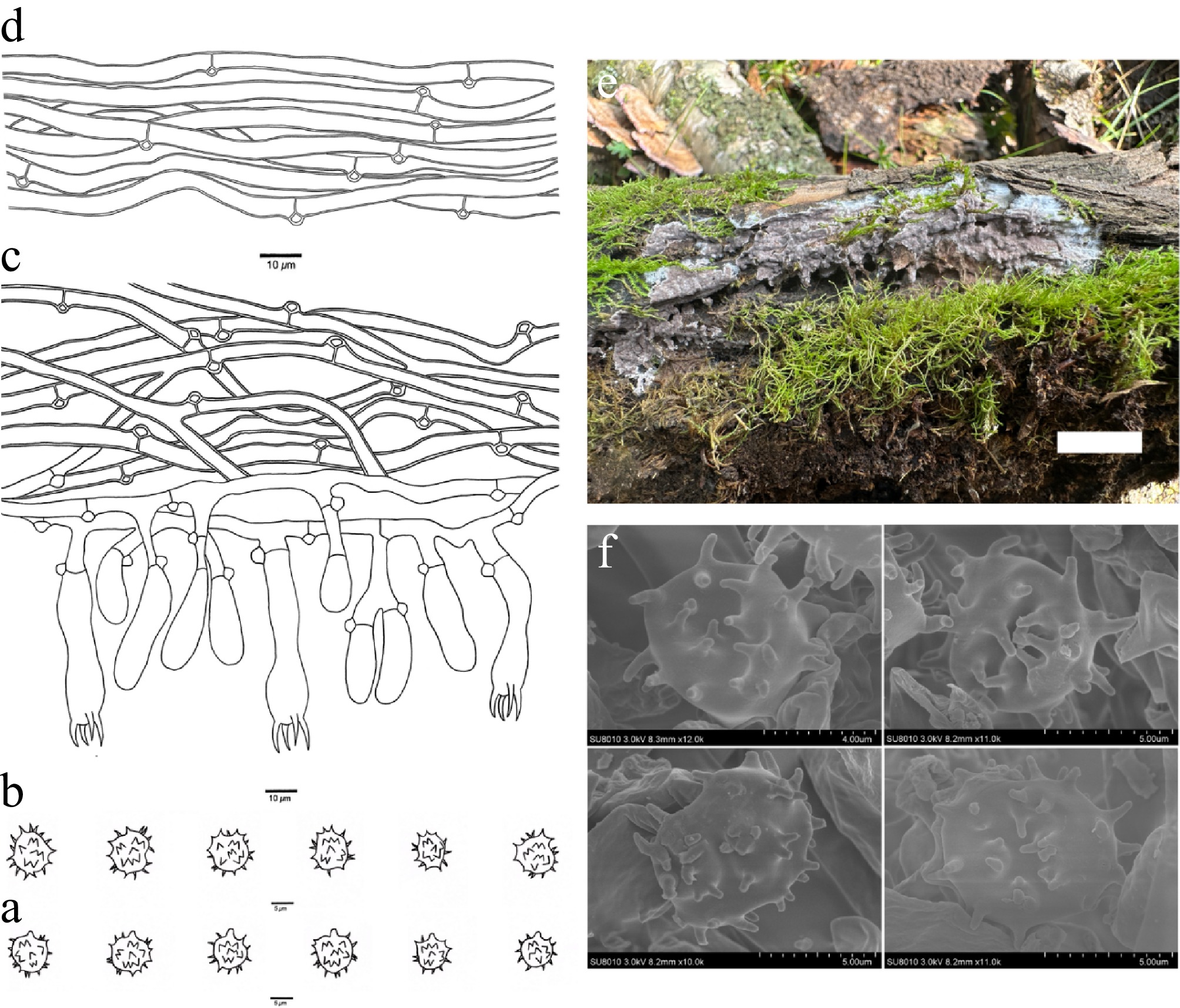
Figure 3.
Basidiomata and microscopic structures of Thelephora caeruleoalba (holotype, Dai 29452). (a) Basidiospores in lateral view. (b) Basidiospores in frontal view. (c) A section through a basidioma. (d) Hyphae from a rhizomorph. (e) Basidiomata on the host substrates. (f) Scanning electron microscope (SEM) of basidiospores. Scale bars: (a), (b) = 5 µm; (c), (d) = 10 µm; (e) = 1 cm.
-
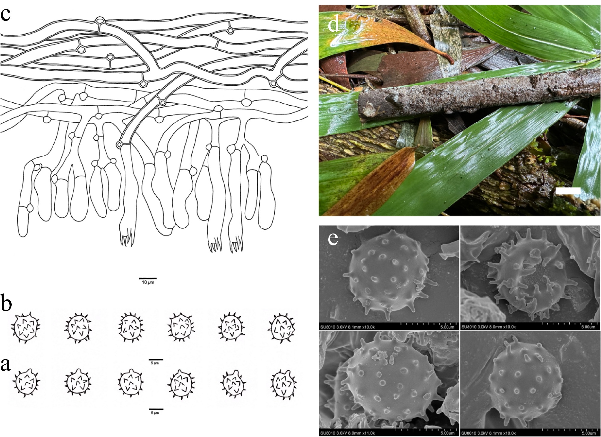
Figure 4.
Basidiomata and microscopic structures of Thelephora ceracea (holotype, Dai 30115). (a) Basidiospores in lateral view. (b) Basidiospores in frontal view. (c) A section through a basidioma. (d) Basidiomata on the host substrates. (e) Scanning electron microscope (SEM) of basidiospores. Scale bars: (a), (b) = 5 µm; (c) = 10 µm; (d) = 1 cm.
-
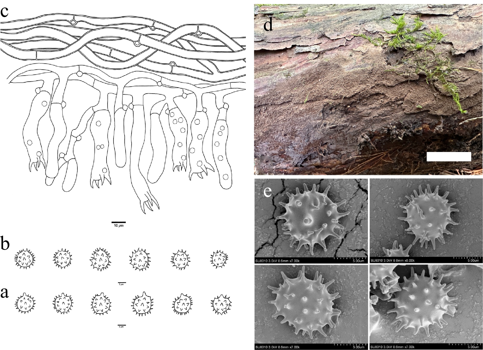
Figure 5.
Basidiomata and microscopic structures of Thelephora griseosepiacea (holotype, Dai 37654). (a) Basidiospores in lateral view. (b) Basidiospores in frontal view. (c) A section through a basidioma. (d) Basidiomata on the host substrates. (e) Scanning electron microscope (SEM) of basidiospores. Scale bars: (a), (b) = 5 µm; (c) = 10 µm; (d) = 1 cm.
-
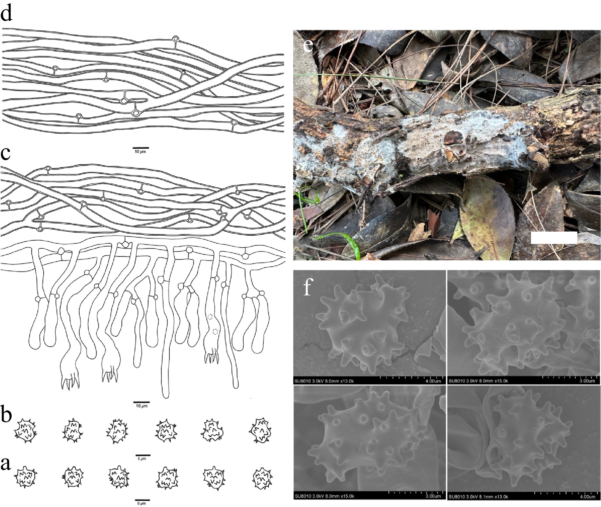
Figure 6.
Basidiomata and microscopic structures of Thelephora keteleeriae (holotype, Dai 28478). (a) Basidiospores in lateral view. (b) Basidiospores in frontal view. (c) A section through a basidioma. (d) Hyphae from a rhizomorph. (e) Basidiomata on the host substrates. (f) Scanning electron microscope (SEM) of basidiospores. Scale bars: (a), (b) = 5 µm; (c), (d) = 10 µm; (e) = 1 cm.
-
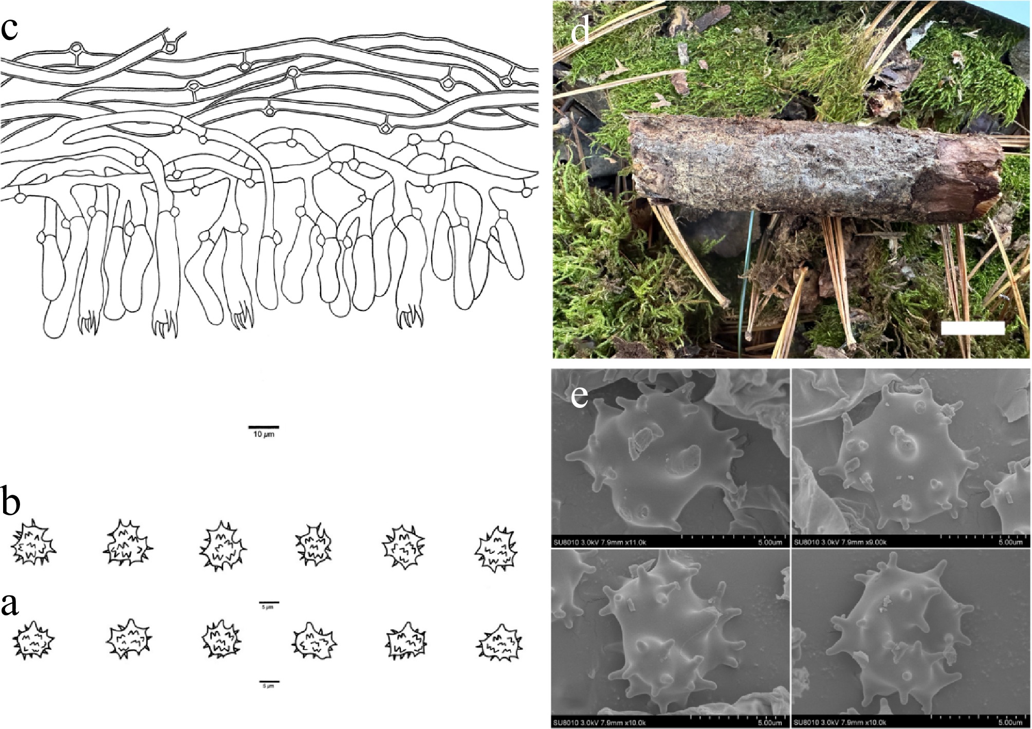
Figure 7.
Basidiomata and microscopic structures of Thelephora koraiensis (holotype, Dai 38336). (a) Basidiospores in lateral view. (b) Basidiospores in frontal view. (c) A section through a basidioma. (d) Basidiomata on the host substrates. (e) Scanning electron microscope (SEM) of basidiospores. Scale bars: (a), (b) = 5 µm; (c) = 10 µm; (d) = 1 cm.
-
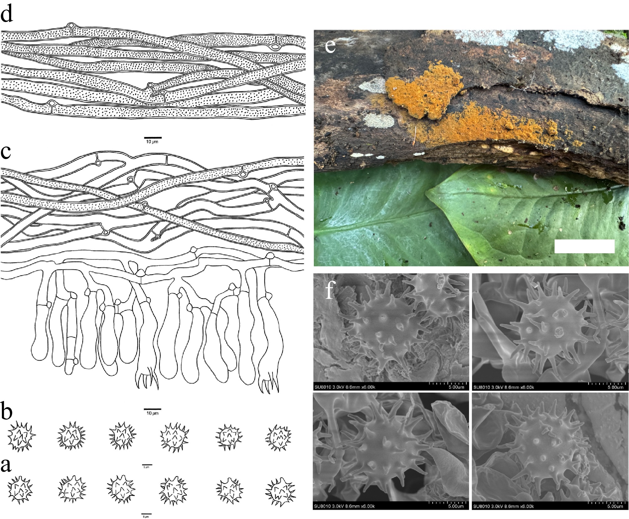
Figure 8.
Basidiomata and microscopic structures of Thelephora longissima (holotype, Dai 39708). (a) Basidiospores in lateral view. (b) Basidiospores in frontal view. (c) A section through a basidioma. (d) Hyphae from a rhizomorph. (e) Basidiomata on the host substrates. (f) Scanning electron microscope (SEM) of basidiospores. Scale bars: (a), (b) = 5 µm; (c), (d) = 10 µm; (e) = 1 cm.
-
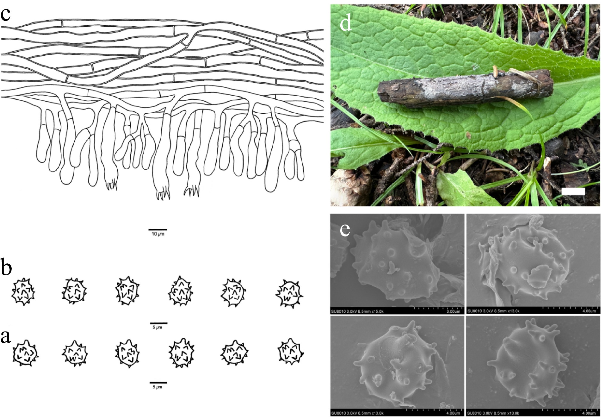
Figure 9.
Basidiomata and microscopic structures of Thelephora mongolica (holotype, Dai 37245). (a) Basidiospores in lateral view. (b) Basidiospores in frontal view. (c) A section through a basidioma. (d) Basidiomata on the host substrates. (e) Scanning electron microscope (SEM) of basidiospores. Scale bars: (a), (b) = 5 µm; (c) = 10 µm; (d) = 1 cm.
-
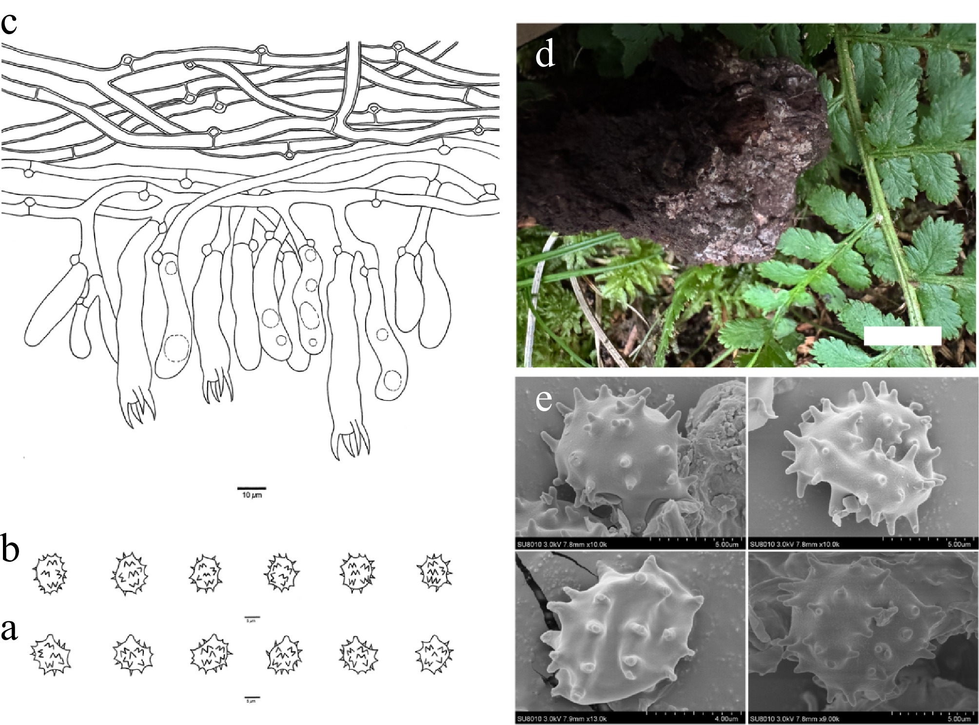
Figure 10.
Basidiomata and microscopic structures of Thelephora murina (holotype, Dai 35286). (a) Basidiospores in lateral view. (b) Basidiospores in frontal view. (c) A section through a basidioma. (d) Basidiomata on the host substrates. (e) Scanning electron microscope (SEM) of basidiospores. Scale bars: (a), (b) = 5 µm; (c) = 10 µm; (d) = 1 cm.
-
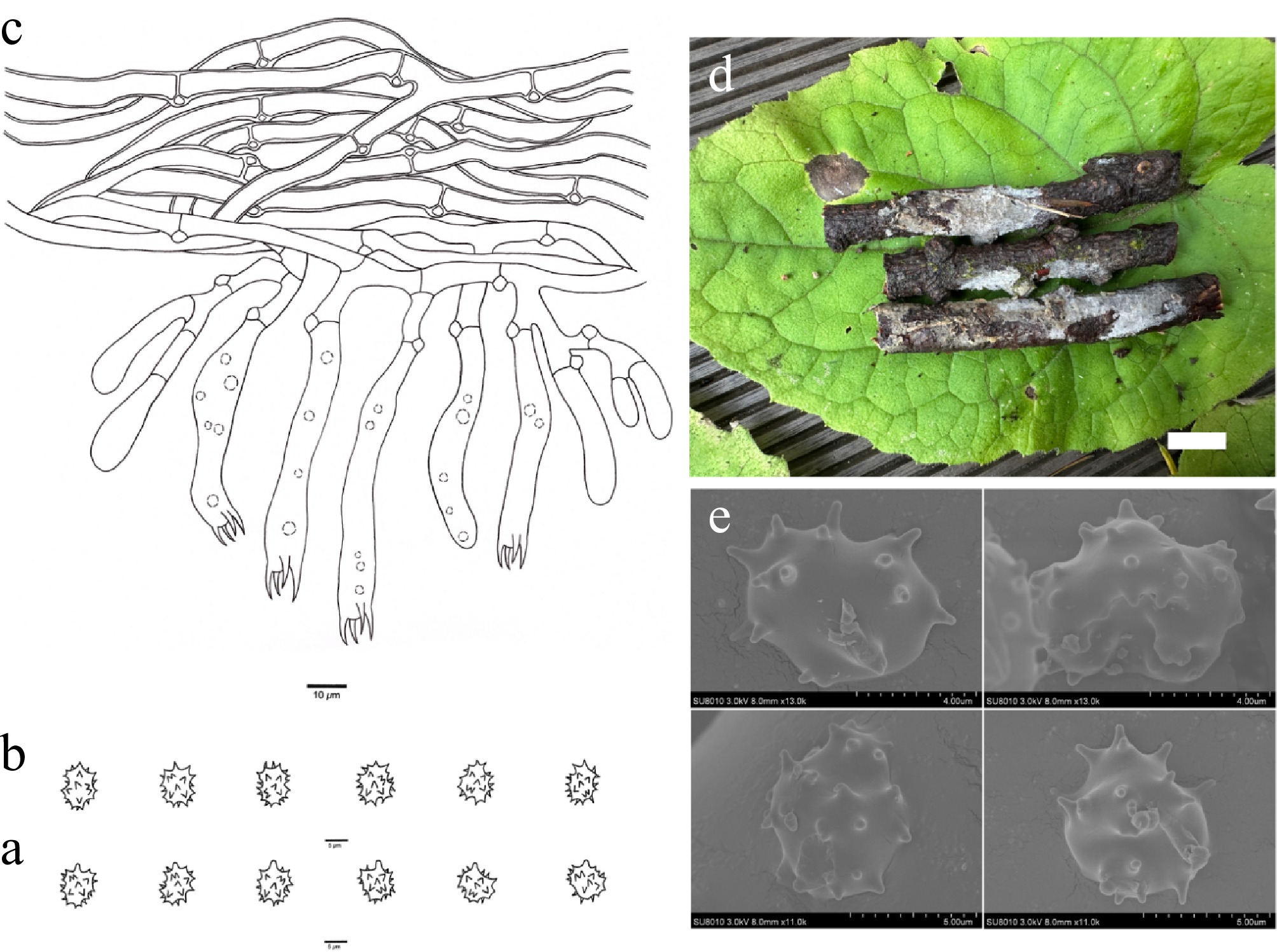
Figure 11.
Basidiomata and microscopic structures of Thelephora niveomarginata (holotype, Dai 31735). (a) Basidiospores in lateral view. (b) Basidiospores in frontal view. (c) A section through a basidioma. (d) Basidiomata on the host substrates. (e) Scanning electron microscope (SEM) of basidiospores. Scale bars: (a), (b) = 5 µm; (c) = 10 µm; (d) = 1 cm.
-
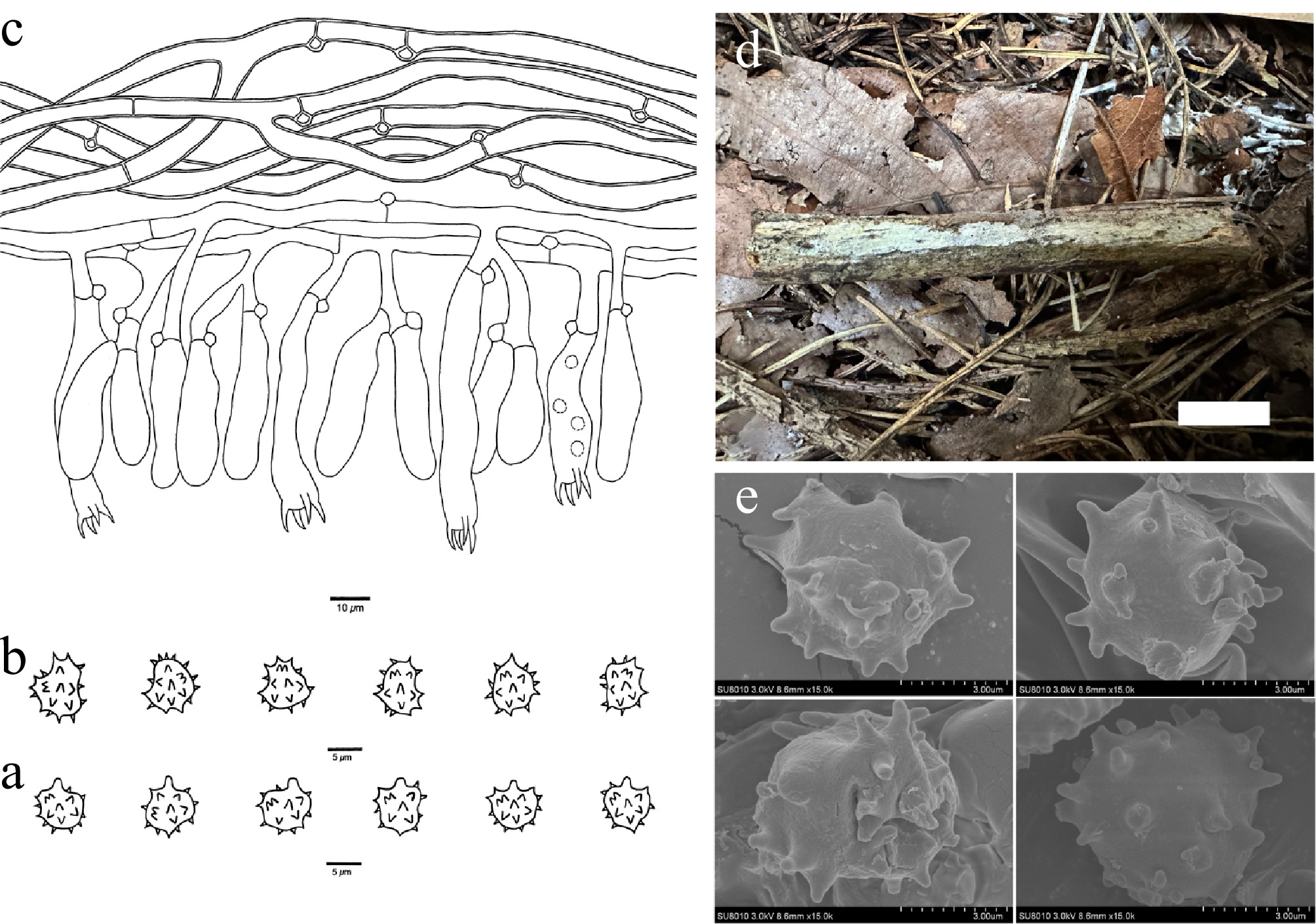
Figure 12.
Basidiomata and microscopic structures of Thelephora nivea (holotype, Dai 37773). (a) Basidiospores in lateral view. (b) Basidiospores in frontal view. (c) A section through a basidioma. (d) Basidiomata on the host substrates. (e) Scanning electron microscope (SEM) of basidiospores. Scale bars: (a), (b) = 5 µm; (c) = 10 µm; (d) = 1 cm.
-
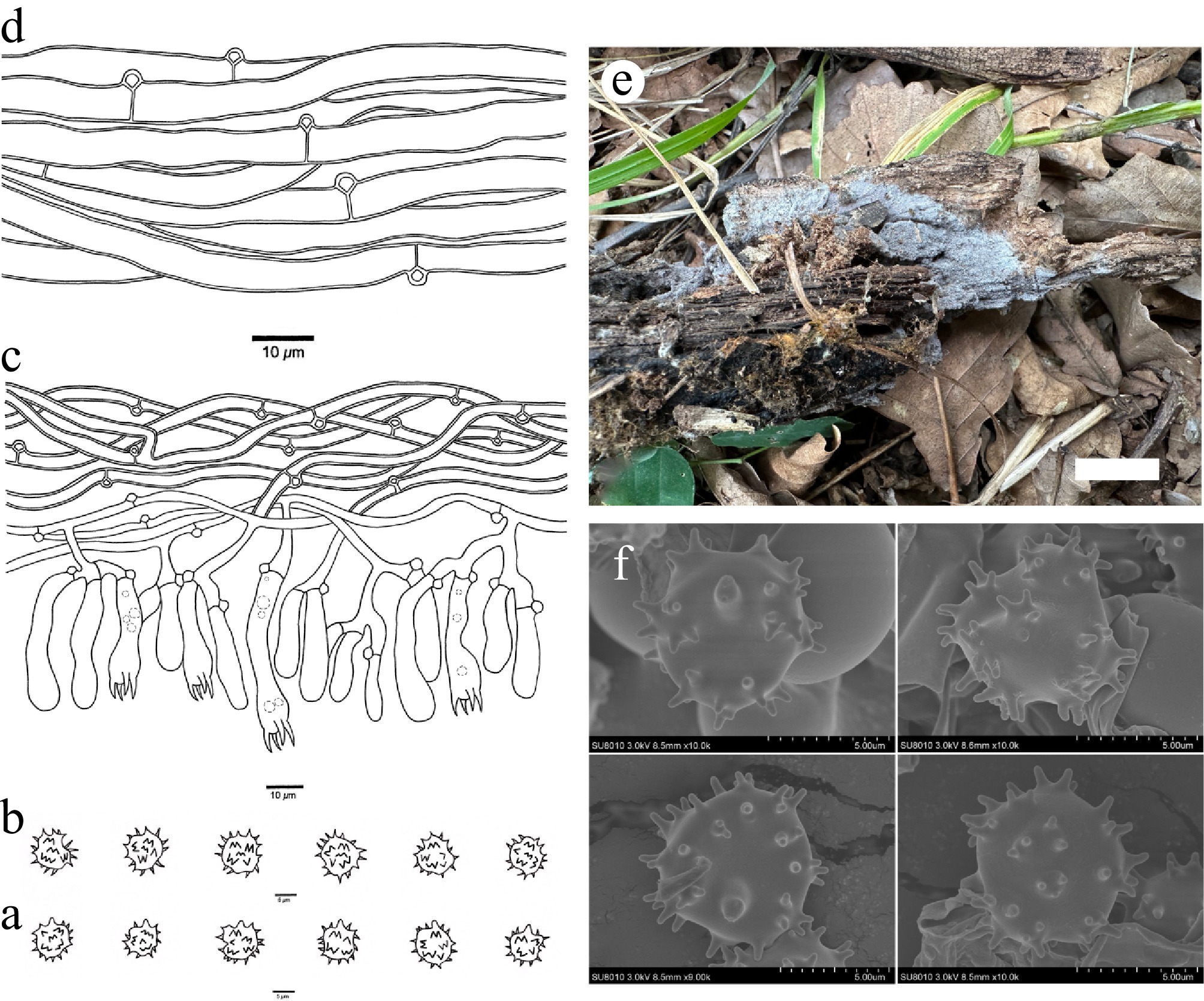
Figure 13.
Basidiomata and microscopic structures of Thelephora quercicola (holotype, Dai 37262). (a) Basidiospores in lateral view. (b) Basidiospores in frontal view. (c) A section through a basidioma. (d) Hyphae from a rhizomorph. (e) Basidiomata on the host substrates. (f) Scanning electron microscope (SEM) of basidiospores. Scale bars: (a), (b) = 5 µm; (c), (d) = 10 µm; (e) = 1 cm.
-
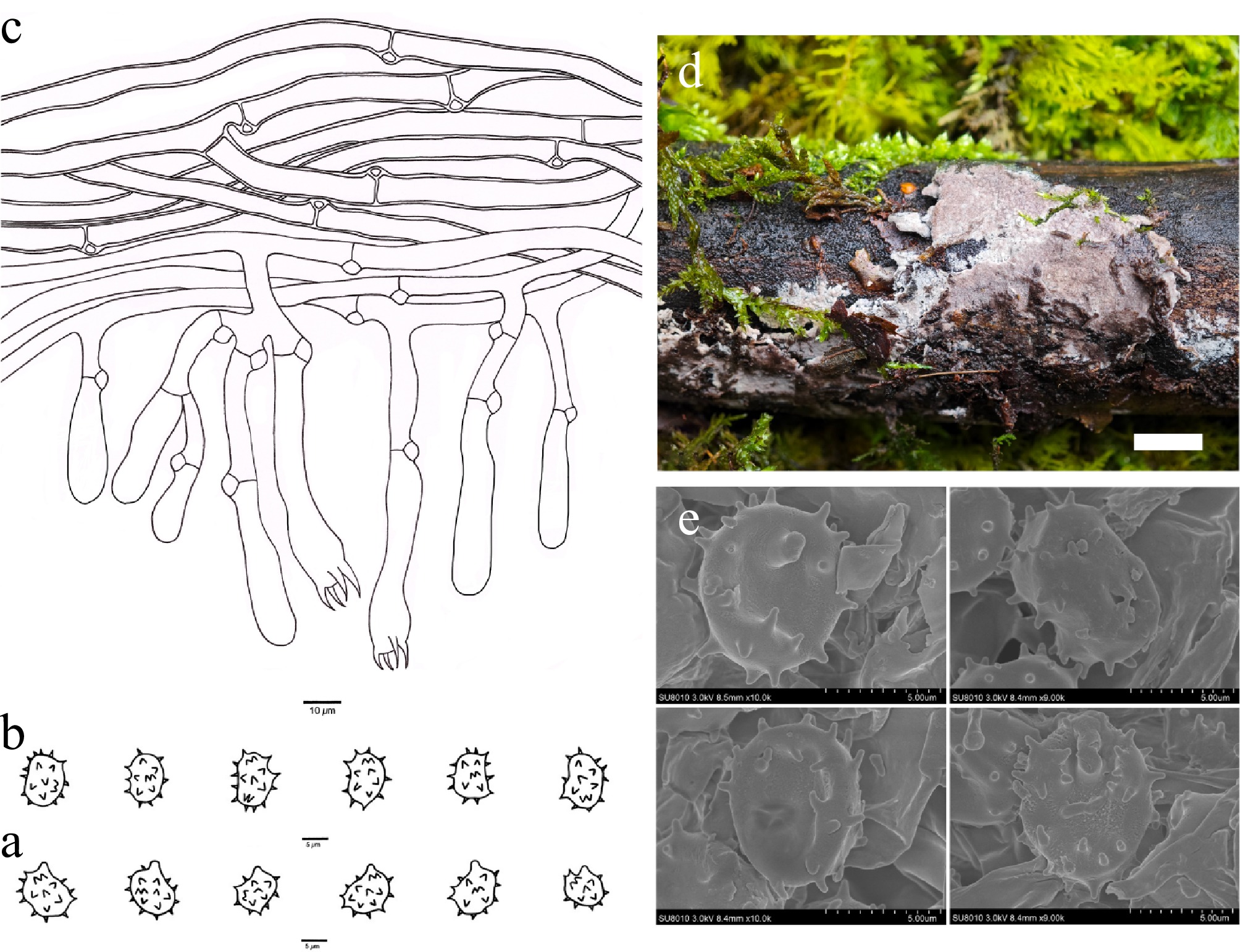
Figure 14.
Basidiomata and microscopic structures of Thelephora reniformis (holotype, Dai 31229). (a) Basidiospores in lateral view. (b) Basidiospores in frontal view. (c) A section through a basidioma. (d) Basidiomata on the host substrates. (e) Scanning electron microscope (SEM) of basidiospores. Scale bars: (a), (b) = 5 µm; (c) = 10 µm; (d) = 1 cm.
-
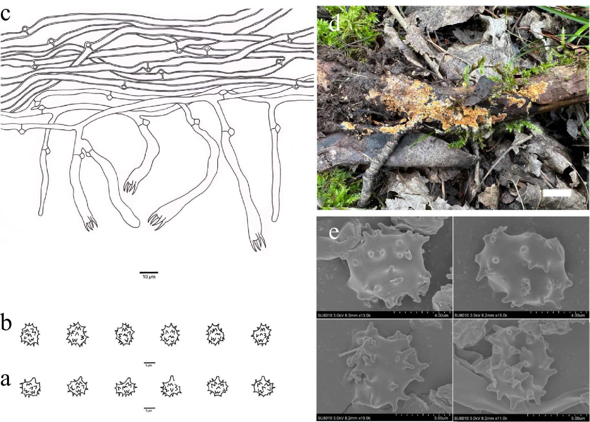
Figure 15.
Basidiomata and microscopic structures of Thelephora rhododendri (holotype, Dai 29574). (a) Basidiospores in lateral view. (b) Basidiospores in frontal view. (c) A section through a basidioma. (d) Basidiomata on the host substrates. (e) Scanning electron microscope (SEM) of basidiospores. Scale bars: (a), (b) = 5 µm; (c) = 10 µm; (d) = 1 cm.
-
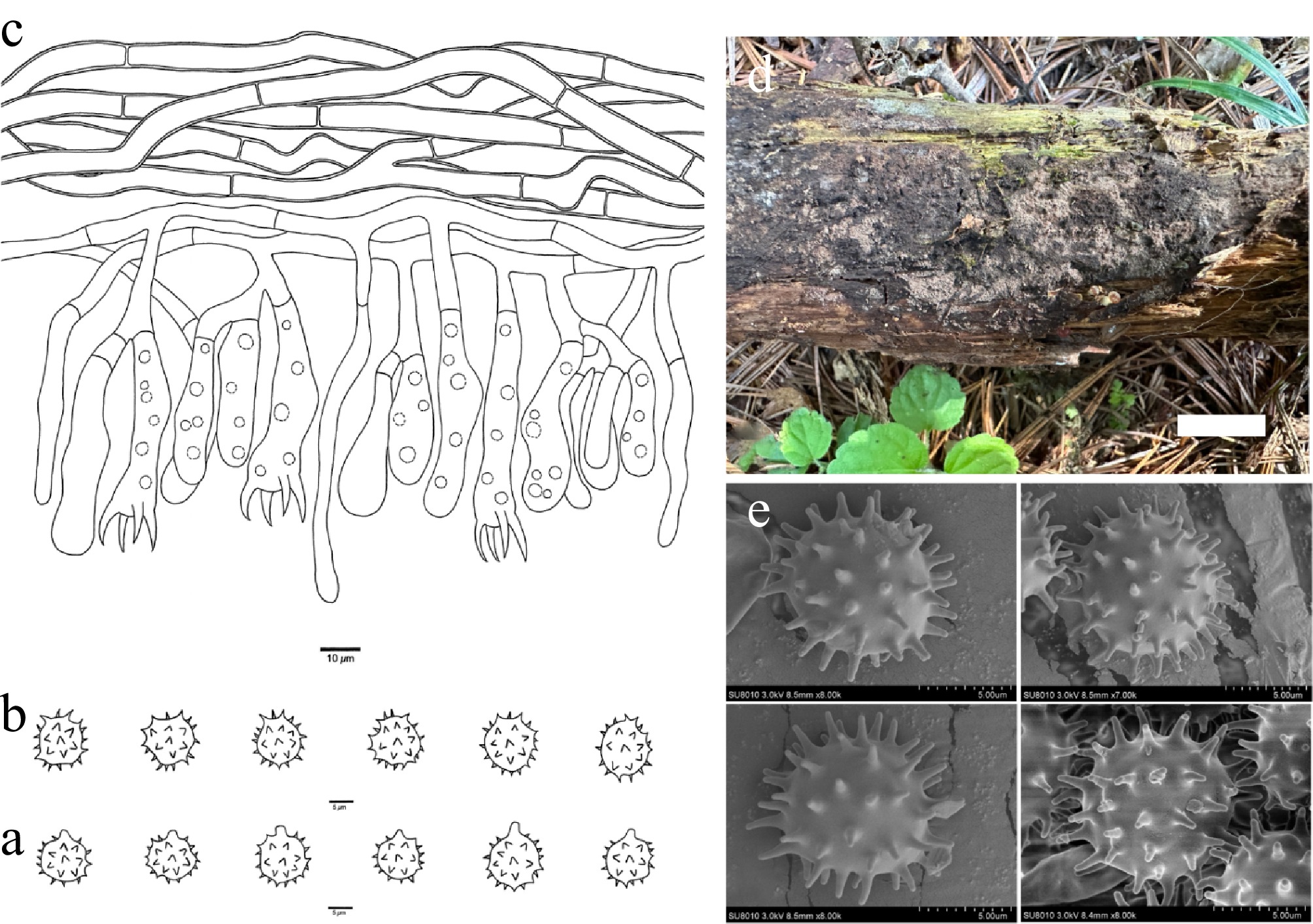
Figure 16.
Basidiomata and microscopic structures of Thelephora roseobubalina (holotype, Dai 37646). (a) Basidiospores in lateral view. (b) Basidiospores in frontal view. (c) A section through a basidioma. (d) Basidiomata on the host substrates. (e) Scanning electron microscope (SEM) of basidiospores. Scale bars: (a), (b) = 5 µm; (c) = 10 µm; (d) = 1 cm.
-
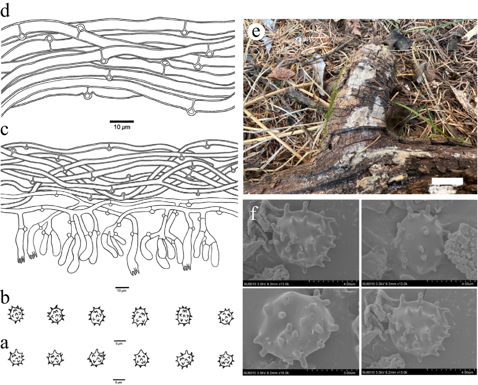
Figure 17.
Basidiomata and microscopic structures of Thelephora submurina (holotype, Dai 38088). (a) Basidiospores in lateral view. (b) Basidiospores in frontal view. (c) A section through a basidioma. (d) Hyphae from a rhizomorph. (e) Basidiomata on the host substrates. (f) Scanning electron microscope (SEM) of basidiospores. Scale bars: (a), (b) = 5 µm; (c), (d) = 10 µm; (e) = 1 cm.
-
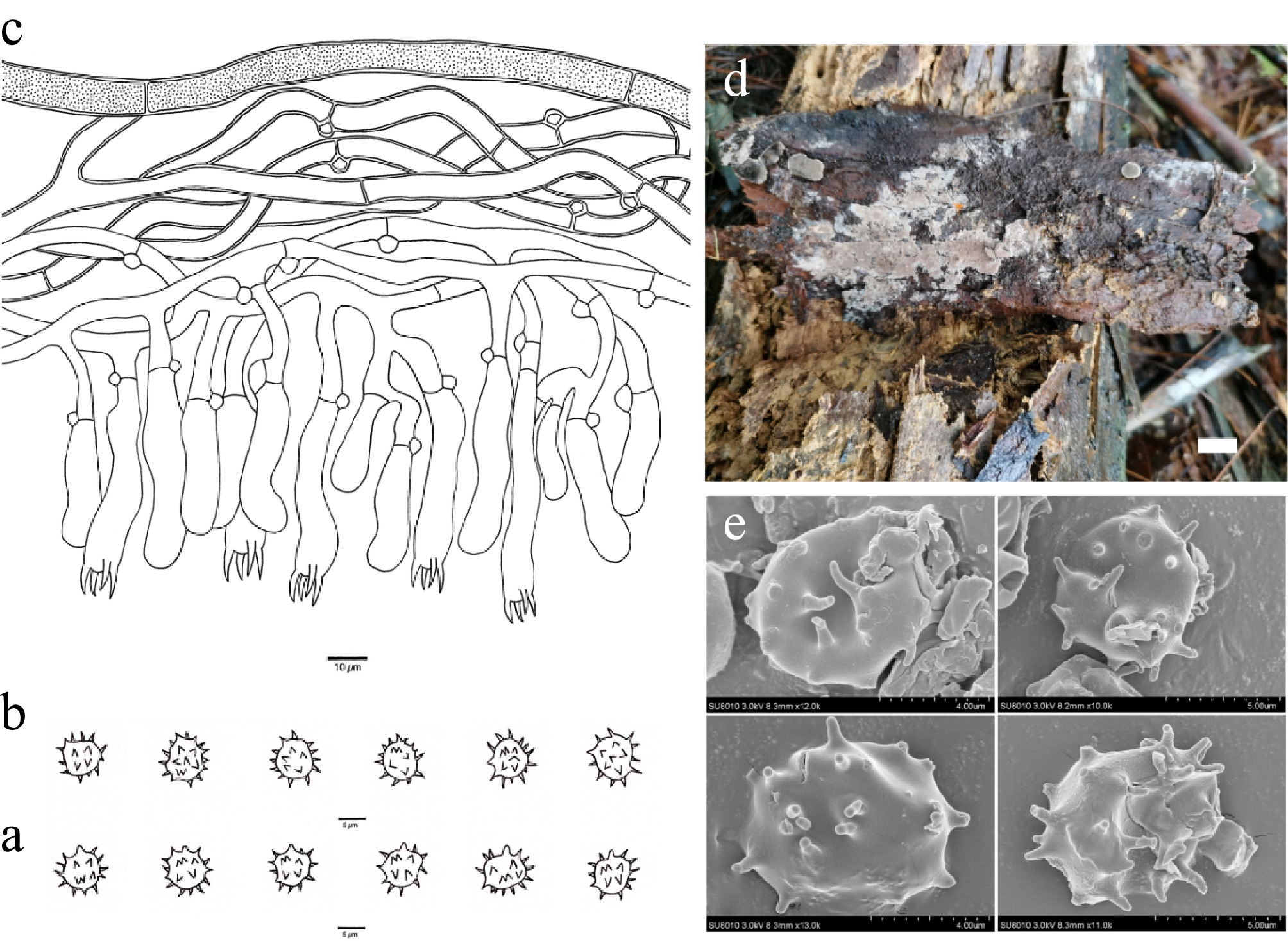
Figure 18.
Basidiomata and microscopic structures of Thelephora subvinosogrisea (holotype, Dai 28754). (a) Basidiospores in lateral view. (b) Basidiospores in frontal view. (c) A section through a basidioma. (d) Basidiomata on the host substrates. (e) Scanning electron microscope (SEM) of basidiospores. Scale bars: (a), (b) = 5 µm; (c) = 10 µm; (d) = 1 cm.
-
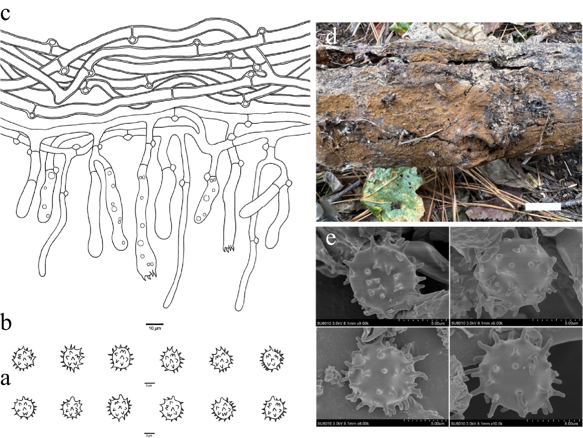
Figure 19.
Basidiomata and microscopic structures of Thelephora tenuicystidiola (holotype, Dai 38254). (a) Basidiospores in lateral view. (b) Basidiospores in frontal view. (c) A section through a basidioma. (d) Basidiomata on the host substrates. (e) Scanning electron microscope (SEM) of basidiospores. Scale bars: (a), (b) = 5 µm; (c) = 10 µm; (d) = 1 cm.
-
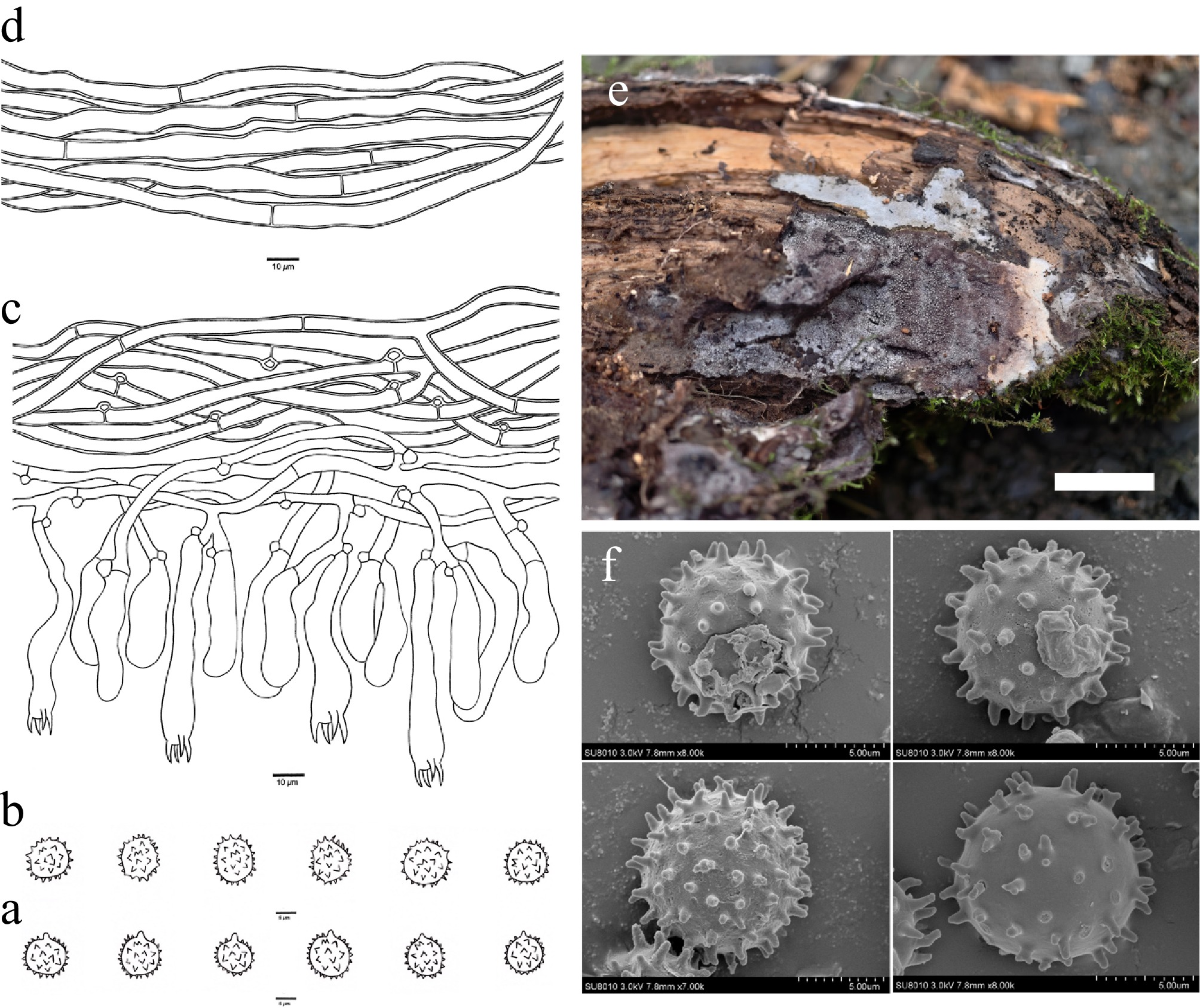
Figure 20.
Basidiomata and microscopic structures of Thelephora vinosogrisea (holotype, Yuan 3514). (a) Basidiospores in lateral view. (b) Basidiospores in frontal view. (c) A section through a basidioma. (d) Hyphae from a rhizomorph. (e) Basidiomata on the host substrates. (f) Scanning electron microscope (SEM) of basidiospores. Scale bars: (a), (b) = 5 µm; (c), (d) = 10 µm; (e) = 1 cm.
-
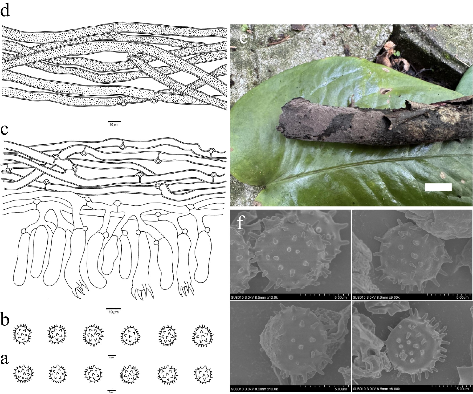
Figure 21.
Basidiomata and microscopic structures of Thelephora violaceardesiaca (holotype, Dai 35184). (a) Basidiospores in lateral view. (b) Basidiospores in frontal view. (c) A section through a basidioma. (d) Hyphae from a rhizomorph. (e) Basidiomata on the host substrates. (f) Scanning electron microscope (SEM) of basidiospores. Scale bars: (a), (b) = 5 µm; (c), (d) = 10 µm; (e) = 1 cm.
Figures
(21)
Tables
(0)