ARTICLE   Open Access    

Assessment for forage grass quality submitted to compaction degrees and nitrogen doses

More Information
  • Received: 01 July 2023
    Revised: 28 February 2024
    Accepted: 01 March 2024
    Published online: 09 April 2024
    Grass Research  4 Article number: e008 (2024)  |  Cite this article
  • The objective of this study was to evaluate the productivity and bromatologic composition of forage species Urochloa brizantha cv. MG-5 Vitória, Urochloa ruzizienses and Megathyrsus maximum cv. Mombaça, submitted to degress of compaction, and increasing doses of nitrogen fertilization. To perform the normal Proctor test, representative samples were collected, determining the density and soil humidity. The design utilized was in randomized blocks in factorial 4 × 4, being 65%, 75%, 85% and 95% of compaction and 0, 200, 250, 300 kg·ha−1 of doses of nitrogen, with four replications, for each forage species. The application of nitrogen linearly increased the production of green mass, dry mass of shoot, height of plants and crude protein content of the respective forages. As you increase nitrogen doses, the contents of neutral and acid detergent fibers decreased in the three studied species. For the root volume, the cultivar that responded linearly and positively to the increasing doses of nitrogen in the compacted soil, was the Urochloa brizantha cv. MG-5 Vitória, showing to be more efficient in compacted soils than Urochloa ruziziensis and Megathyrsus maximum cv. Mombaça.
  • Lotus, Nelumbo nucifera Gaertn. (commonly known as Asian lotus), is one of the most commercially significant aquatic plants in the world. It belongs to the Nelumbonaceae family, which contains a single genus Nelumbo, with the American lotus (Nelumbo lutea Pear) as the only other extant species. The Asian lotus is geographically widely distributed in most Asian countries, as well as in Australia and Russia (Fig. 1). The American lotus, also known as yellow lotus, occurs primarily in eastern regions of North American and the Caribbean[1]. Due to geographical isolation by the Pacific Ocean, the two lotus species exhibit distinct morphologies in plant size, leaf, and petal shape, particularly in their petal colors. The Asian lotus displays diverse flower colors, including white, pink, red, pale yellow, as well as a variety of color patterns, while the American lotus only exhibit yellow flowers (Fig. 1). The two species, however, are genetically crossable and share rather similar aquatic habits and life cycles. Both of the two species have eight pairs of chromosomes (2n = 16), with approximately 800 Mb genome sizes, highly conserved gene contents, and little chromosomal rearrangements[2,3].

    Figure 1.  Geographical distribution of lotus accessions collected in the Wuhan National Lotus Germplasm Bank at the Wuhan Botanical Garden of the Chinese Academy of Sciences. The different colored dots indicate cultivars belonging to different categories.

    While the American lotus predominantly grows naturally in the wild, the Asian lotus has been domesticated and cultivated over 3,000 years, for its nutritional, ornamental, and medicinal attributes[4]. The total lotus cultivation area in China is estimated to cover over 530,000 ha, and so far at least 4,500 lotus cultivar germplasm resources have been conserved[5]. Most of these cultivars are hybrids developed by traditional crossing, while approximately 300 are local genotypes collected from wild lakes all over the world (Fig. 1)[2]. These lotus cultivars are grouped into four categories based on their agricultural uses, including the flower lotus for ornamental purpose; the rhizome lotus for edible rhizome production; the seed lotus for harvesting fresh or mature seeds; and the medicinal lotus for pharmaceutical uses[6].

    As an ancient traditional herb, all lotus plant tissues have been used in the treatment of various diseases, such as fever, sunstroke, sweating, obesity, ischemia, cancer, diabetes, hepatopathy, and hypertension[710]. Its medicinal properties are largely ascribed to the abundantly accumulated active components in different lotus organs, including alkaloids, flavonoids, terpenoids, polysaccharides, saponins, and minerals. Of these compounds, benzylisoquinoline alkaloids (BIAs) are regarded as the most important pharmacological ingredients[11,12]. Here, we summarize the recent advances in the chemical profiles, pharmacological activities, extraction methods, and elucidations of its biosynthesis pathway.

    BIAs are a class of tyrosine-derived nitrogenous secondary metabolites with over 2,500 known structures[13]. The core chemical structure of BIAs consist of a benzene ring, a pyridine ring, and a second aromatic ring (Fig. 2)[14]. Like other plant-derived alkaloids, most BIAs are significantly bioactive[15]. Typical pharmacologically significant BIAs include morphine and codeine with analgesic traits, sanguinarine and berberine with antimicrobial properties, as well as noscapine, papaverine, and tubocurarine with antitussive, vasodilative, and muscle relaxant properties, respectively. The occurrence of BIAs in plants is restricted primarily in the order Ranunculales and eumagnoliids. However, their accumulation has been detected in other families, including Rutaceae, Lauraceae, Cornaceae, and Nelumbonaceae.

    Figure 2.  1-Benzylisoquinoline alkaloids reported in Nelumbo nucifera.

    The lotus plant richly accumulate BIAs in almost all its organs[11]. To date, at least 61 alkaloids (Table 1) have been identified in lotus since the first isolation of nuciferine, roemerine, and O-nornuciferine alkaloids from the plant in the 1960s[16,17]. Notably, all reported lotus alkaloids are BIAs except Oleracein E and Methylcorypalline, which contain only basic isoquinoline structures. Based on their structures, these BIAs can further be divided into three subclasses, including 1-benzylisoquinolines, aporphines, and bisbenzylisoquinolines (Figs 24). 1-Benzylisoquinoline alkaloids are basically intermediates in the biosynthesis of aporphines and bisbenzylisoquinolines, and are mostly accumulated in trace amounts in lotus tissues[18]. In contrast, aporphines and bisbenzylisoquinolines are the end BIA products in lotus, with their accumulation levels accounting for over 95% of the total BIA content[11].

    Table 1.  BIAs detected in the lotus (Nelumbo nucifera) tissues. Alkaloids are assigned with numbers as shown in Figures 25.
    No.AlkaloidFormulaEnantiomerOrganReference
    1-BENZYLISOQUINOLINE
    1LotusineC19H24NO3+E, S, F[25]
    2Methyl lotusineL
    3ArmepavineC19H23NO3(−)-R and (+)-SS, F, L[21]
    4CoclaurineC17H19NO3(+)-RS, L[26]
    5N-norarmepavineC18H21NO3(+)-RL[27]
    6N-methylisococlaurineC18H21NO3L[16,28]
    7N-methylcoclaurineC18H21NO3(−)-RL, S[16]
    8IsococlaurineC19H24NO3+L
    9MethylhigenamineS[17]
    10Norcoclaurine-6-O-glucosideS
    11NorcoclaurineC16H17NO3(+)-R and (+)-SS, L[26,29]
    12ArgemexirineS, L
    136-demethy-4-methyl-N-methylcoclaurineC18H21NO3S[30]
    14Nor-O-methylarmepavineC20H25NO3S[30]
    154’-N-methylcoclaurineC19H23NO3L, S[17]
    164’-methyl coclaurineL, S[17]
    17Bromo methyl armepavineL, S[17]
    18Methoxymethy lisoquinolineL, S[17]
    19HigenamineP[31,32]
    20Higenamine glucosideP[33]
    APORPHINE
    21NuciferineC19H21NO2(−)-RS, F, L[34,35]
    22N-nornuciferineC18H19NO2(−)-RS, L[26]
    23RoemerineC18H17NO2(−)-RS, F, L[16,26,32]
    24O-nornuciferineC18H19NO2(−)-RS, F, L[32,36]
    25AnonaineC17H15NO2(−)-RS, L[16,26]
    26LirinidineC18H19NO2(−)-RS, L[37]
    27Nuciferine-N-MethanolF
    28Nuciferine-N-AcetylF
    29Anonaine-N-AcetylF
    30CaaverineC17H17NO2(−)-RS, L[20,38]
    31Oxidation-nuciferineS, L, F[19,30]
    32AsimilobineC17H17NO2(−)-RS, F, L[17,20]
    33Methyl asimilobineS, L[17]
    34N-methyl asimilobineS, L[16,39]
    35Roemerine-N-oxideS, L[16,40]
    36N-methyl asimilobine-N-oxideS, L, F[16,19]
    37Nuciferine-N-oxideS, L, F[16,19]
    38DehydroanonaineC17H13NO2L[16]
    39DehydronuciferineC19H19NO2L[16]
    40DehydroaporphineC18H15NO2L[41]
    41NelumnucineS, L
    42DehydroroemerineL[16]
    43LiriodenineC17H9NO3L[16,40]
    447-hydroxydehy dronuciferineC19H19NO3L[16]
    45PronuciferineC19H21NO3(+)-R and (−)-SS, F, L[16,26,40]
    46GlaziovineS
    47LysicamineC18H13NO3L[38,42]
    48CepharadioneL[38]
    BISBENZYLISOQUINOLINE
    49NeferineC38H44N2O61R, 1'SS, F, E[17,31]
    50LiensinineC37H42N2O61R, 1'RS, F, E[35]
    51Isoliensinine1R, 1'SS, F, E[25]
    52N-norisoliensinineC36H40N2O6S, F, E[25]
    536-hydroxynorisoliensinineC36H40N2O6S, F, E
    54Methyl neferineS, E[10,17]
    55NelumboferineC36H40N2O6S, E[10,17]
    56NegferineC38H44N2O6L[17,26]
    57NelumborineF[17]
    58DauricineS, F[43]
    TRIBENZYLISOQUINOLINE
    59NeoliensinineC63H70N3O101R, 1'S, 1''RE[44]
    L, Leaf; E, embryo; F, flowers; S, seeds; R, rhizome; LS: leaf sap; NS, not specified.
     | Show Table
    DownLoad: CSV
    Figure 3.  Aporphine-type alkaloids isolated from Nelumbo nucifera.
    Figure 4.  Bis- and tri- benzylisoquinoline alkaloids isolated from Nelumbo nucifera.

    Unlike BIAs in the Ranunculales, lotus BIAs exhibit several unique features. Firstly, lotus BIAs show strict tissue specific accumulation patterns. Although BIAs are found in all the plant tissues, highest levels of approximately 3,000 μg/g fresh weight (FW) are accumulated in the laminae and plumules, followed by petals and petioles with about 500 and 100–200 μg/g FW, respectively. Conversely, lowest BIAs levels (less than 20 μg/g FW) occur in the rhizomes and stamens. Interestingly, the lotus bleeding sap contains extremely high BIAs level of up to 10,000 μg/g FW[11]. In addition, lotus leaves and petals predominantly accumulate aporphine BIAs, including nuciferine, O-nornuciferine, N-nornuciferine, roemerine, and anonaine, while the plumules mostly contain bis-BIAs of liensinine, isoliensinine, and neferine[11]. Secondly, while BIAs prevalently assume the (S)-enantiomer conformation, lotus show enrichment of both (R)- and (S)-conformers. For example, both (R)- and (S)-stereoisomers of armepavine have been isolated from sacred lotus[1921]. To date, most aporphines isolated from lotus belong to the (R)-configurations (Table 1). Thirdly, all aporphine BIAs isolated from lotus lack the hydroxyl and methyl modifications at the C-3' and C-4' positions, suggesting that the (R)-N-methylcoclaurine might be the key precursor for both aporphine and bis-BIA biosynthesis in lotus[22].

    Lotus cultivars exhibit considerably diverse patterns of BIA composition and accumulation. Generally, seed cultivars accumulate the highest concentration of BIAs in leaves, followed by flower cultivars, while lowest levels are observed in the rhizome cultivars[11,23]. Multivariate principal component analysis (PCA) of 92 lotus cultivars showed that the seed cultivars generally accumulate high levels of nuciferine and O-nornuciferine, whereas flower cultivars primarily accumulate high N-nornuciferine, roemerine, and anonaine alkaloids[23]. In contrast, rhizome cultivars contain no dominant BIAs. Cluster analysis of the 92 cultivars also indicated that the American cultivar ‘Meizhouhuanglian’ could group with other Asian lotus cultivars, which suggested the lack of obvious differentiation between the American and the Asian lotus accessions based on BIA content and composition[23]. Developmentally, the lowest BIAs levels are accumulated in the lotus leaves at the bud stages (developmental stage S1). BIAs levels then increase steadily in the leaf growth and expansion stages, peak at S6 stage, then decrease slightly at the S7 senescence stage (Fig. 5a)[11]. Accumulation of BIAs in the lotus plumules is also developmentally regulated[24]. Major BIAs were almost undetectable in the plumules at 12 days post pollination (DAP), which later increased consistently until 21 DAP (Fig. 5b).

    Figure 5.  BIA profiles in the leaf and plumule during development. (a) Lotus leaves showing the seven defined developmental stages. (b) BIA profiles in the seven leaf developmental stages. (c) Lotus plumules showing different developmental stages. (d) BIA content in the plumule at different developmental stages. S, leaf developmental stages; DAP, days post pollination. The figure is modified from previous reports[11,24].

    Extraction of alkaloids from raw materials is the first analytical step in their identification, quantification, and application. Currently, three main alkaloid extraction methods with varied principles are widely used, including solvent extraction, distillation, expeller pressing and sublimation methods. Solvent extraction is presently the most popularly used method. The plant alkaloids occur mostly as salts of organic and inorganic acids[45,46], thus, acidic and basic solvents are initially required to remove the non-alkaloid compounds. Subsequently, appropriate purification methods, primarily acidic extraction is performed by mixing the ground sample materials with weak acid solutions (e.g., acetic acid in water, methanol, or ethanol), to obtain a desired level of purity. The solvent extraction is carried out in four steps: 1) assimilation of solvents into the target solid matrix; 2) alkaloid dissolution in solvents; 3) solvent diffusion out of a solid matrix; 4) collection of extracted solutes[47]. Theoretically, any factors that enhance the above steps can facilitate alkaloid extraction. Key factors that affect the extraction efficiency, include alkaloids-solvent solubility, sample particle size, solvent to solid ratio, extraction temperature, and extraction duration[48,49].

    The lotus BIAs are weak alkalescent compounds, with poor solubility or insolubility in water, but with rapid solubility in organic solvents. Based on the law of similarity and intermiscibility (like dissolves like), crude lotus BIAs extract are usually prepared in alcohol solvents (ethanol or methanol)[10,19]. Other solvents, such as hydrochloric acid, dichloromethane and trichloromethane have also been used for lotus BIA extraction[20]. The solvent extraction efficiency can be enhanced by finely grinding the lotus tissues, solvent refluxing, microwaving, and sonication. A recent comparative study, using three extraction solvent (methanol, 50% methanol, and water) and two different extraction conditions (reflux for 120 min twice or sonication for 30 min twice), suggested that the 'reflux in methanol' method could result in highest BIA recovery from lotus flowers (Table 2)[50]. Moreover, pre-basifying lotus leaves with 10% ammonia water revealed a significant two-fold nuciferine yield relative to that obtained from non-basified leaves[51].

    Table 2.  Extraction efficiency of alkaloids from lotus flower.
    Extraction methodContent (mg/g dry weight)aTotal
    12345678910
    Methanol, reflux1.76 (100)1.75 (100)0.07 (100)0.63 (100)0.69 (100)0.83 (100)1.45 (100)5.73 (100)1.30 (100)0.75 (100)14.96 (100)
    50% Methanol, reflux1.09 (62)1.35 (77)0.05 (71)0.50 (79)0.61 (88)0.78 (94)1.35 (93)3.79 (66)0.94 (73)0.56 (75)11.02 (74)
    H2O, reflux0.24 (14)0.35 (20)nd.b0.21 (33)0.18 (26)0.38 (45)0.78 (54)2.57 (45)0.66 (51)0.29 (38)5.66 (38)
    Methanol, sonication0.88 (50)1.11 (64)0.03 (44)0.39 (62)0.33 (48)0.47 (56)0.97 (67)2.77 (48)0.70 (54)0.42 (56)8.07 (54)
    50% Methanol, sonication0.98 (56)1.27 (73)0.04 (58)0.49 (78)0.47 (69)0.80 (96)1.38 (95)3.93 (69)0.97 (75)0.59 (79)10.92 (73)
    H2O, sonication0.14 (8)0.21 (12)nd.0.12 (20)0.08 (11)0.25 (30)0.53 (37)1.91 (33)0.48 (37)0.19 (26)3.91 (26)
    1. nuciferine; 2. N-nornuciferine; 3. N-methylasimilobine; 4. Asimilobine; 5. Pronuciferine; 6. Armepavine; 7. norarmepavine; 8. N-methylcoclaurine; 9. coclaurine; 10. Norjuziphine. a. relative value (%) against the content obtained by methanol under reflux is given in parentheses. b. less than the quantitation limit.
     | Show Table
    DownLoad: CSV

    The conventional solvent extraction method is typically time consuming, requires large volume of organic solvents, and high cost waste solvent treatment. Consequently, modern or greener extraction methods, such as supercritical fluid extraction (SFC), high-speed counter-current chromatography (HSCCC), and microwave assisted extraction (MAE) techniques have been developed and applied in BIAs extraction. SFE utilizes supercritical fluid as the extraction solvent, which has high solubility as liquid and high diffusivity as gas. SFE is almost pollution-free, and can efficiently extract alkaloids with no residual organic solvents[52]. The solvating properties of supercritical fluid can dramatically rise when the pressure and temperature are near their critical points. The supercritical carbon dioxide (S-CO2) is one of the most widely used supercritical fluids due to its low critical temperature (31 °C), low cost, non-toxicity, chemical inertness, and non-flammability properties[46,5354]. However, due to the relatively low polarity of S-CO2, polar agents, such as methanol and water are used as modifiers to raise its polarity and enhance its efficiency for BIAs extraction from lotus tissues. A previous study showed that the highest nuciferine yield of 325.54 μg/g could be achieved with extraction conditions set at 70 °C, 30 MPa, flow rate 0.2 ml/min, 2 h extraction time, and with 10% (v/v) diethylamine and 1% (v/v) water in methanol as the polarity modifier[51].

    The HSCCC is a liquid-liquid partition chromatography technique that uses no solid support matrix. This method permits the introduction of crude samples directly into the hollow column, and eliminates the adsorbing effects on stationary phase material as well as artifact formation, thus offering maximum sample recovery capacity and a wide range of solvent system selection[55]. Ma et al. used a simple two-phase solvent system comprising of n-hexane-ethyl acetate-methanol-acetonitrile-water (5:3:3:2.5:5, v/v/v/v/v) to successfully purify four main aporphine alkaloids, including 2-hydroxy-1-methoxyaporphine, pronuciferine, nuciferine, and roemerine from a crude extract of lotus leaves[56].

    In addition to the above-mentioned methods, several other extraction methods, such as maceration, percolation, soxhlet, and pressurized liquid extraction are available for selection. Modern and green methods are preferred mostly due to their energy-efficiency, speed, and less solvent requirement, and high extraction yield. However, traditional techniques are still widely used at the industrial level due to their low investment cost. Overall, the selection of suitable extraction techniques depends not only on the physical, chemical, and stability of the target alkaloids, but also on time and cost of the extraction methods.

    Following extraction, the resulting crude BIA extracts are mixtures of a variety of natural products, such as phenolics, carotenes, glycosides, and terpenes, which require further separation and purification. The separation of BIAs depends largely on their physical and chemical characteristics, such as adsorption properties, partition coefficients, molecular sizes, solubility, and ionic strengths[47]. BIAs are mostly water-insoluble but soluble in organic solvents, while their acid salts are water-soluble or soluble in dilute acids, thus, these compounds can be precipitated by adding solvents (e.g., sodium bicarbonate, ammonia, or tartaric acid) that alter the reaction pH levels. Similar theory has been used to develop pH-zone-refining counter current chromatography techniques for lotus BIAs isolation[35,57].

    Isolation of pure lotus BIAs using conventional methods, such as column chromatography and thin-layer chromatography (TLC) is extremely difficult due to their high structural similarity. The structurally similar BIAs can instead be rapidly separated by counter-current chromatography (CCC) according to their different partition coefficients in two immiscible solvents. The CCC is a liquid-liquid partition chromatography technique with no solid support matrixes, and has been widely used for the preparative isolation of lotus BIAs. Accurate selection of the solvent system is highly crucial when using the CCC method. Generally, the two-phase solvent system should have: no decomposition or denaturation effects on the target compounds, sufficient sample solubility and suitable partition coefficient values, and satisfactory retention capacity of the stationary phase[58]. For efficient separation, the partition coefficient (K) value of the target alkaloids and the separation factor between two BIAs (K2/K1) should be close to 1 and greater than 1.5, respectively[55]. Other factors, such as the rotary speed, the mobile phase flow rate, and the column temperature also affect the target alkaloid separation.

    Preparative separation of liensinine and its analogues from lotus plumules using CCC was first reported by Wu et al.[59]. The authors used two-phase solvent systems of light petroleum (b.p. 60–90 °C)–ethyl acetate–tetrachloromethane–chloroform–methanol–water (1:1:4:4:6:2, v/v) and ethyl acetate–tetrachloromethane–methanol–water (1:6:4:1, v/v) (Table 3) to successfully isolate bis-BIAs from lotus plumules in small- and large-scale, respectively. To limit the application of deleterious tetrachloromethane and chloroform solvents, another simplified HSCCC solvent system of n-hexane–ethyl acetate–methanol–water (5:8:4:5, v/v, containing 0.5% NH4OH) was developed and successfully used to separate liensinine, isoliensinine, and neferine from crude lotus extract[60]. Later, several pH-zone-refining CCC solvent systems, which generally use 10 mM triethylamine in the upper organic phase and 5 mM HCl in the aqueous phase for adjustment of pH and K values of target compounds were developed[35,57,61,62]. These two phase solvent systems were used to successfully isolate over 98% pure BIA singletons from both plumules and leaves. Other than the CCC technique, a preparative liquid chromatography has also been developed for high purity laboratory-scale isolation of liensinine, isoliensinine, and neferine[63].

    Table 3.  Major two-phase solvent systems developed for preparative separation of lotus BIAs through counter-current chromatography (CCC) techniques.
    Solvent systemsMix ratios (v/v)Target BIAsReference
    Light petroleum (60–90 °C)–ethyl acetate–tetrachloromethane–chloroform–methanol–water1:1:4:4:6:2Small scale
    Plumule BIAs
    [59]
    Ethyl acetate–tetrachloromethane–chloroform–methanol–water1:6:4:1Small scale
    Plumule BIAs
    [59]
    n-hexane–ethyl acetate–methanol–water5:8:4:5
    0.5% NH4OH
    Plumule BIAs[60]
    n-hexane–ethyl acetate–methanol–water5:5:2:8
    10 mM triethylamine
    5 mM HCl
    Plumule BIAs[57]
    Diethyl ether – Na2HPO4/NaH2PO4 (pH = 7.2 – 7.5)1:1Plumule BIAs[62]
    Petroleum ether (60–90 °C)–ethyl acetate–methanol–water5:5:2:8
    10 mM triethylamine
    5 mM HCl
    Leaf BIAs[61]
    n-hexane-ethyl acetate-methanol-water-[C4mim][PF6]5:2:2:8:0.1
    10 mM triethylamine
    3 mM HCl
    Whole plant BIAs[35]
     | Show Table
    DownLoad: CSV

    After extraction and purification, lotus BIAs are subjected to further identification and quantification processes. High-performance liquid chromatography (HPLC) techniques, particularly the reversed-phase LC methods, were predominantly employed[45]. Identification of known BIA compounds is normally accomplished by HPLC separation and UV detection, followed by electrospray ionization (ESI), and tandem mass spectrometry (MS/MS). The reversed-phase C18 columns are most ideal, and the application of aqueous acetonitrile containing 0.1% triethylamine as mobile phase has been shown to effectively separate both lotus leaf and plumule BIAs with strong peak signals and fine peak shapes[23,63,64]. Other optimized HPLC parameters for lotus BIAs characterization include: column temperature 30C; 270–280 nm UV/photodiode array (DAD) detection wavelength; the gradient elution mode, 40%–80% acetonitrile at 0–15 min, 80% acetonitrile at 15–18 min, 80%–40% acetonitrile at 18–19 min, 40% acetonitrile at 19–25 min; and a flow rate 0.8 ml/min. The MS detection of lotus BIAs are preferably conducted in the positive mode, with cone voltage of 20 V and a nebulizer gas temperature/pressure of 150 °C/21 psi. The needle, shield, and capillary voltage parameters are normally used with default settings. Such HPLC-DAD-ESI-MS/MS methods have been used to identify the major leaf BIAs, including nuciferine, N-nornuciferine, O-nornuciferine, anonaine, and roemerine, as well as plumule BIAs, such as liensine, isoliensinine, and neferine (Table 4; Fig. 6; Fig. 7).

    Table 4.  Identification of major lotus leaf and plumule alkaloids and their HPLC-MS/MS ion characteristics.
    PeaksTRa (min)Molecular weightm/z
    [M + H]+
    Major fragment ionsAlkaloids
    1*7.56281282251/219N-nornuciferine
    2*9.03281282265/250O-nornuciferine
    3*10.17265266249/219Anonaine
    4*12.43295296265/250Nuciferine
    5*13.39279280249Roemerine
    66.61610611503/283/206Liensinine
    79.47610611489/297/192Isoliensinine
    817.72624625503/297/206Neferine
    * The retention time (TRa (min) of peaks 1–5 as obtained by Chen et al.[23].
    ∆ The retention time of peaks 6–9 as reported by Chen et al.[63].
     | Show Table
    DownLoad: CSV
    Figure 6.  Analytical mass spectra of lotus leaf BIAs and their fragmentation pathways. The chemical structures and the corresponding peak numbers 1 to 5 represent signals for N-nornuciferine, O-nornuciferine, anonaine, nuciferine, and Roemerine, respectively. Figures are modified from Luo et al. [65] and Chen et al. [23].
    Figure 7.  Analytical mass spectra of lotus leaf BIAs and their fragmentation pathways. The chemical structures and the corresponding peak numbers 6 to 8 represent signals for liensinine, isoliensinine, and neferine, respectively. Figures are modified from Chen et al. [64], Deng et al. [11], and Lai et al. [66].

    Currently, lotus BIAs are predominantly identified using the ultra-performance liquid chromatography technique equipped with quadrupole time-of-flight mass spectrometry (UPLC-QTof-MS). In contrast to traditional HPLC-MS methods, the UPLC-Q-Tof-MS technique exhibits higher sensitivity and resolution, and mass measurement accuracy, making it a powerful and reliable analytical technique for plant metabolite identification[65,66]. Under optimized UPLC-Q-Tof-MS conditions, over 20 BIAs were identified from lotus leaves and plumules, based on their chromatographic characteristics, UV spectra, exact mass, and MS fragments[31,65,67]. Moreover, these studies further verified previous observations that liensinine, isoliensinine, neferine, and lotusine are the major BIAs in lotus plumules, while those of aporphines, including nuciferine, roemerine, Anonaine, N- and O-nornuciferine are the major BIAs in lotus leaves.

    In addition to their nutritional roles, lotus is also a key source of herbal traditional Chinese medicine. Lotus was originally used to treat various diseases, such as pharyngitis, chest pain, cough, vomiting blood, fever, and heat stroke[68,69]. To date, extensive studies have identified a wide range of lotus bioactive ingredients, their pharmacological efficiencies, and health benefits. As key bioactive lotus constituents, BIA alkaloids have nowadays received significant attention. In the following sections, we present an overview of pharmacological activities of lotus BIAs from various tissues.

    As introduced above, lotus leaves accumulate primarily aporphine-type BIAs, with Nuciferine, N- and O-nornuciferine as the richest components. Diversified pharmacological activities of lotus leaves therefore attribute predominantly to these three BIAs. The nuciferine, for example, has shown anti-obesity, anti-virus, antioxidant, anti-cardiovascular, antimicrobial, and anti-cancer activities.

    Lipids are maintained through dynamic balances within the cell as Triglyceride (TG) by a series of synthesis and catabolism related transcription factors (TFs) and enzymes. Lotus alkaloids can exert lipid-regulating effects in several ways, including mainly inhibition of lipid synthesis and absorption, inhibition of cell proliferation and differentiation, and interaction with proteins. A study on the effects of nuciferine on blood lipids in male golden hamsters, feeding with normal diet, high-fat diet (HFD), or HFD supplemented with nuciferine (10 and 15 mg/kg·BW/day), revealed a reduction in total cholesterol (TC), TG, low-density lipoprotein and free fatty acids in hamsters treated with different doses of nuciferine after eight weeks[70]. In addition, nuciferine could alleviate dyslipidemia, and liver steatosis by inhibiting the expression of hepatic genes related to lipid metabolism in hamsters fed with a high-fat diet[70,71]. Similarly, a recent study demonstrated that lotus leaf extracts could inhibit adipogenesis in 3T3-L1 preadipocytes and suppress obesity in high-fat diet-induced obese rats[72]. Overall, these pharmacological effects demonstrate the potential of lotus leaf BIAs as natural lipid-lowering agents.

    Currently, approximately 90% of global type 2 diabetic patients are characterized by hyperinsulinemia and insulin resistance. Therefore, controlling the blood glucose levels of patients is crucial for diabetes treatment[73]. Huperzine has been reported to lower blood sugar via various mechanism in the body. Conversely, nuciferine can increase the glucose uptake by fat and muscle cells, thereby promoting insulin secretion by pancreatic β-cells[56]. Nuciferine is also directly involved in the process of insulin secretion and has been shown to augment insulin secretion in the pancreatic β-cells by shutting down or stimulating the amplification of adenosine triphosphate pathway[74].

    Free radicals or oxidants that break down cells and tissues can affect metabolic function and cause different health challenges. Antioxidants are substances that effectively inhibit oxidation reaction of free radicals at low concentrations[75]. The antioxidant capacity of aporphine alkaloids isolated from the lotus leaves, including (R)-N-methylasimilobine, lysicamine, and (R)-nuciferine have been screened using antiradical scavenging, metal chelating, and ferric reducing power assays[38]. In addition, a previous study demonstrated that lotus leaf-fermented broth could exhibit 1,1-diphenyl-2-picrylhydrazyl (DPPH) radical scavenging activity, ferric ion reducing power, superoxide dismutase-like activity, tyrosinase inhibition, and nitrite scavenging activity, which provided scientific basis for the application of sugar-fermented lotus leaves as an antioxidant condiment[76]. A recent study reported that, to enhance, achieve, and maintain higher scavenging activity of DPPH radicals and hydroxyl radicals for lotus leaves, the optimal treatment should include oven drying the leaves at ~55–65 °C to maintain alkaloids and amino acids content[77].

    Bacteria is known to be one of the leading causes of numerous diseases. Over exposure or misuse of antibiotics can result in antibacterial resistance. Chinese herbal medicine has been demonstrated to be an excellent alternative for the treatment of such bacterial infections to avoid antibiotic resistance[78]. The n-butanol compound lotus leaves extracts can effectively inhibit peridontitis-causing bacteria, such as Actinobacillus actinomycetemcomitans Y4, Actinomyces viscosus 19246, Porphyromonas gingivalis 33277, Fusobacterium nucleatum 25586, and Actinomyces naeslundii wvl 45[79]. Moreover, the lotus leaf alkaloids not only have anti-mitogenic effects, but also exhibit a stronger antibacterial effect on Escherichia coli. The lotus leaf alkaloids and flavonoids are considered as key bioactive components responsible for the antibacterial activity.

    A key characteristic of cancer is the indefinite cell proliferation, and lotus leaf alkaloids have been shown to exhibit antiproliferative properties against several cancer cells. For example, in the presence of nicotine, nuciferine could significantly inhibit the proliferation of non-small cell lung cancer cells, suppress the activity of Wnt/β-catenin signaling, enhance Axin stability, and induce apoptosis. In addition, nuciferine could also down-regulate the expression levels of β-catenin and its downstream targets, including c-myc, cyclin D, and VEGF-A[80]. Methanolic lotus leaves extract, containing nuciferine, N-methylasimilobine, (-)-lirinidine, 2-hydroxy-1-methoxy-6a, and 7-dehydroaporphine, significantly inhibited melanogenesis in B16 melanoma cells[19]. Furthermore, the inhibitory potential of nuciferine against the growth of human breast cancer cells has been reported[81]. The inhibitory effect of nuciferine is likely due to its ability to block cancer cell division cycle and enhance cancer cell apoptosis.

    A recent study reported that 40 mg/kg nuciferine could not only significantly reduce aortic lesion and vascular plaque in ApoE(-/-) mice fed with a high-fat diet, but also attenuate migration and proliferation of vascular smooth muscle cells in vivo and in vitro through the Calm4/MMP12/AKT signaling pathway[82]. Nuciferine has also been associated with reduced vascular wall inflammation and significant down-regulation of vascular inflammatory factors, such as IL-1β, TNF-α, MCP-1, and NF-κB, with the latter being a major regulator during all phases of the inflammatory response[83]. Additionally, lotus leaf alkaloids are linked with vascular remodeling by down-regulating MMP-2 and 9 expression and inducing TIMP-2 expression. MMP is an important protease that degrades the extracellular matrix (ECM), while TIMP is a matrix metalloproteinase inhibitor, both of which contribute to atherosclerosis development[84].

    In contrast to lotus leaves, lotus plumules accumulate mainly bis-BIAs, with liensinine, isoliensinine, and neferine the most abundant components. The bioactivities of these bis-BIAs contribute largely to the pharmacokinetic properties of lotus plumules.

    Antioxidant compounds, including alkaloids, phenolics, and saponins could be obtained from lotus plumules using 50% ethanol solvent extraction, and DPPH as well as nitric oxide in-vitro assays to test the hydro alcoholic extract of lotus plumules (HANN) revealed excellent free radical scavenging activity[85]. Neferine not only exhibited scavenging activity against ABTS, DPPH, NO, ONOO and O2 radicals, but also showed significant inhibitory properties against lipid peroxidation and protein nitration in the antioxidant assays (cell-free). In vivo assays also revealed that neferine could significantly reduce NFκB activation and moderately inhibit NO synthesis[86]. Neferine could also inhibit various diseases, such as vascular inflammation, hyperglycemia, and liver disease by reducing NO concentration in different cells and inhibiting the elevation of intracellular molecules, such as reactive oxygen species (ROS) and malondialdehyde (MDA)[87]. In addition, total alkaloid from Nelumbinis Plumula (NPA) and its main alkaloids, including liensinine, isoliensinine, and neferine showed significant cytoprotective effect on oxidative stress induced by tert-butyl hydroperoxide (t-BHP) in the human hepatocellular HepG2 cell line, and the protective effect could be associated with reduced ROS formation, thiobarbituric acid-reactive substance production, lactate dehydrogenase release, and elevated GSH levels[88].

    Neferine is a crucial skin anti-inflammatory and anti-aging agent. For example, a previous study demonstrated that neferine could reduce the phosphorylation level of the MAPK/NF-κB pathway, and inhibit mast cell degranulation and cytokine expression by suppressing elevated intracellular calcium in mast RBL-2H3 cells stimulated with A23187/phorbol ester (PMA)[89]. In lipopolysaccharide-induced human endothelial cells, neferine could significantly inhibit the formation of inflammatory mediators, such as NO, tumor necrosis factor-α, cyclooxygenase-2, inducible nitric oxide synthase, and interleukin 1β due to its ability to regulate mitogen-activated protein kinase and nuclear factor-κβ pathways[90]. In addition, neferine could inhibit lipopolysaccharide and dextran sulfate sodium-induced inflammation both in vitro and in vivo, alter protein expression of iNOS, COX-2, receptor-interacting protein 1 (RIP1), RIP3, mixed lineage kinase domain-like protein (MLKL), and increase the protein expression of caspase-8 in colon tissues, all of which affected the incidence and prevalence of ulcerative colitis (UC)[91]. As the combined effects of oxidative stress and inflammation leads to pathogenesis of numerous diseases, the antioxidant and anti-inflammatory mechanisms of lotus seed alkaloids need to be further explored.

    Liensinine and isoliensinine are two main bis-BIAs found in the lotus seed plumules, and both have been reported to have inhibitory effects against cell proliferation. For example, liensinine could functionally regulate the transforming growth factor β1-induced proliferation, migration, and signaling pathways of human tenon fibroblast cells via the mitogen-activated protein kinase 7 gene to enable rapid patients recovery after glaucoma surgery[92]. Liensinine could also not only significantly inhibit the proliferation of GBC cells in vitro by suppressing their G2/M phase growth in a dose- and time-dependent manner, but also induce apoptosis in gallbladder cancer cells by inhibiting the Zinc finger X-chromosomal protein (ZFX)-induced PI3K/AKT pathway[93]. In addition, previous reports have shown that liensinine could inhibit over-proliferation of gastric cancer cells and osteosarcoma cells[94,95]. Isoliensinine is a liensinine isoform, which has also been shown to exhibit inhibitory effects against angiotensin II (Ang II)-induced proliferation of porcine coronary artery smooth muscle cells (CASMCs)[96], as well as HepG2, Huh-7, and H22 hepatocellular carcinoma (HCC) cells[97]. Moreover, BIA alkaloids mixtures, including neferine, liensinine, and isoliensinine extracted from lotus plumules could delay or inhibit the abnormal proliferation and migration of pulmonary artery smooth muscle cell (PASMCs) by regulating the expression of p-SRC and PIM1[98].

    Extensive anti-cardiovascular activities of Neferine, liensinine, and isoliensinine, such as anti-arrhythmic, anti-thrombic, and anti-hypertensive have been documented. Wicha et al.[99] demonstrated that neferine not only had hypotensive effects on NG-nitro-L-arginine methyl ester(L-NAME)-induced rats, but also induced vascular relaxation via the endothelial nitric oxide synthase (ENOS)/nitric oxide (NO)/soluble guanylate cyclase (SGC) pathway. Neferine has shown potential effectiveness in preventing episodes of reentrant ventricular tachycardia and sudden cardiac death after myocardial ischemic injury, and was effective against ischemic arrhythmias. In addition, neferine was shown to significantly inhibit adenosine diphosphate (ADP), collagen, arachidonic acid (AA) and platelet-activating factor (PAF)-induced platelet aggregation in rabbits. A more recent study showed that, unlike neferine, isoliensinine could effectively scavenge early afterdepolarizations (EADs) and delayed afterdepolarizations (DADs) by inhibiting INaL and ICaL in ventricular myocytes, thus, demonstrating its potential anti-arrhythmic effects[100].

    To date, numerous studies have demonstrated the roles of alkaloids from lotus plumules, especially neferine in the treatment of different types of cancer. Neferine is a common chemosensitizer of vincristine that enhances the anti-tumor effect of vincristine by inhibiting gastric cancer cell proliferation (SGC7901) leading to their apoptosis. Neferine has also been shown to inhibit the growth of lung adenocarcinoma A549 cells, arrest their cell cycle in G1 phase, and further induce apoptosis through lipid peroxidation, depletion of the mitochondrial membrane potential, MAPKs activation, DNA degredation, and intracellular calcium accumulation[101]. In addition, neferine could cure liver cancer cells (HepG2) and breast cancer cells (MCF-7) by inhibiting their cell proliferation and growth through different mechanisms[102]. Chang et al.[103] reported that in addition to neferine, liensinine could also act as an anti-tumor agent by inhibiting normal mitochondrial energy supply, impairing lysosomal function, and inhibiting the growth of non-small cell lung cancer both in vitro and in vivo. In addition, liensinine could inhibit the growth of gastric cancer cells by increasing ROS levels and inhibiting the PI3K/AKT pathway[94]. Similarly, isoliensinine could cause apoptosis in HepG2, Huh-7, and H22 hepatocellular carcinoma (HCC) cells by decreasing NF-κB activity and constitutively phosphorylating NF-κB p65 subunit at Ser536 in HCC cells[97]. Overall, lotus seeds are now widely used in various cancer treatments due to their broad antagonistic properties.

    In addition to the pharmacological effects mentioned above, lotus BIAs possess substantial functions in protection against photoaging related skin problems[104], reduced clinical manifestation of Alzheimer disorders[105], suppression of CCl4-induced liver damage, and bleomycin-induced pulmonary fibrosis[106,107]. Other minor BIAs isolated from lotus, such as members of the 1-benzylisoquinolins type BIAs have also shown notable anti-HIV, anti-inflammatory, anti-arrhythmic, and anti-thrombotic properties[17]. Thus, lotus BIAs have significant potential for the development of novel drugs in treating various life-threatening human diseases, such as microbial infection, inflammation, atherosclerosis, cancer, obesity, neurological disorders, and diabetes.

    As mentioned above, lotus BIAs are predominantly aporphines and bis-BIAs, which accumulate mainly in lotus leaves and plumules[11,18]. Notable, 1-benzylisoquinolines, which are mostly biosynthetic intermediates and derivatives of the aporphines and bis-BIAs, have also been detected in lotus tissues in trace amounts. The biosynthetic pathways of lotus BIAs have been proposed based on their identified structures[12,22,29,108]. Noteworthily, lotus BIAs primarily occur as R-enantiomers, which is contrary to the predominant S-enantiomers occurrence in the Ranunculales.

    Similar to other plant BIAs, the biosynthesis of lotus BIAs starts also from the L-amino acid tyrosine. Tyrosine is first converted into L-dopamine (L-DOPA) and 4-hydroxyphenylacetaldehyde (4-HPAA), respectively by the catalyzation of tyrosine decarboxylase (TYDC)[109]. L-DOPA and 4-HPAA are subsequently condensed by norcoclaurine synthase (NCS) into (R)-norcoclaurine, the first BIA scaffold in plants[110] (Fig. 8). Two additional enzymatic steps, catalyzed by norcoclaurine 6-O-methyltransferase (6OMT) and (R)-coclaurine N-methyltransferase (CNMT) respectively, yield the core intermediate (R)-N-methylcoclaurine of almost all BIAs[15]. In other BIA accumulating species, N-methylcoclaurine normally undergoes two further 3'-hydoxylation and 4'-Omethylation reactions, catalyzed by (S)-N-methylcoclaurine 3'-hydroxylase (CYP80B) and 3'-hydroxy-N-methylcoclaurine 4'-O-methyltransferase (4'OMT), respectively, to yield (S)-reticuline, which is another central precursor and a major branch point of numerous BIA structures, including morphine and berberine. Interestingly, neither reticuline nor corytuberine have been isolated from lotus tissues to date. In addition, all identified aporphine-type BIAs in lotus lack the hydroxyl and methyl modifications at the C-4' and C-3' positions, suggesting that the two enzymatic steps catalyzed by CYP80B and 4’OMT are not necessary for aporphine biosynthesis in lotus[22,110]. Consistently, a recent study showed that pronuciferine is the lotus aporphine biosynthesis precursor, and it is synthesized from the core intermediate (R)-N-methylcoclaurine by the enzymatic actions of NnCYP80G and 4-O-methyltransferase (7OMT)[12] (Fig. 8). According to the lotus BIA structures, production of nuciferine from the pronuciferine precursor may arise from an unknown dehydration reaction[18]. Additional modifications, such as N-demethylation, O-demethylation, and methylenedioxy-bridge formation, catalyzed by the N-demethlase (NDM), O-demethlase (NDM), and CYP719A, respectively[22,111], transforms nuciferine into other diverse aporphines (Fig. 8).

    Figure 8.  Proposed BIAs biosynthetic pathway in lotus. Steps marked with red, green, and purple background represent the common reactions for the biosynthesis of (R)-N-Methylcoclaurine, the lotus aporphine branch, and the bis-BIA biosynthesis branch, respectively. All biosynthetic enzymes are shown in red. The (R)-N-Methylcoclaurine is the branch point for aporphine and bis-BIA biosynthesis in lotus. Dotted arrows indicate multiple enzymatic or unknown steps.

    The lotus bis-BIAs can also directly be synthesized from the (R)-N-methylcoclaurine precursor (Fig. 8). The 7-O3' coupling reaction, catalyzed by CYP80A enzyme, transfers two molecules of (R)-N-methylcoclaurine into Nelumboferine, which is the first bis-BIAs in the bis-BIA biosynthesis pathway[12,108]. Two further enzymatic steps, catalyzed by the 4'OMT and 7OMT enzymes respectively, yield the three major bis-BIAs, including liensinine, isoliensinine, and neferine. Other enzymatic modifications, including N-demethylation, double 4'-Omethylation, 8-O3' and 3'-3' intermolecular couplings, result in the production of other diverse bis-BIA structures (Fig. 4).

    The currently advanced next generation sequencing techniques have facilitated the availability of at least seven high quality lotus genome versions as references for isolation of BIAs biosynthetic genes[2,112115]. The previously functionally characterized BIAs pathway genes in the Ranunculales[116] have been retrieved and used to query and predict most of their corresponding homologs from the lotus genomic and transcriptomic data based on sequence similarities. As a result, at least 5, 4, and 1 TYDC, NCS, and CNMT candidate gene copies, respectively have been predicted in the early lotus BIA biosynthesis pathway[22]. Similarly, seven OMT genes have been identified, with four and three of which forming clusters with the 6OMT/4’OMT and 7OMT clades, respectively. In addition, downstream lotus BIA biosynthetic gene sequences, including CYP80A, CYP80G, CYP719A, NDM, and ODM have been isolated[22,29].

    The lack of stable lotus transformation system has hindered functional characterization of most its BIA pathway enzymes. Consequently, functional studies have mostly involved correlation analysis between gene expression levels and spatial temporal BIA accumulation patterns in different lotus tissues or developmental stages[22,29,117,118]. In vitro enzymatic assays were recently used to functionally characterize four lotus OMTs, with NnOMT1 exhibiting 6OMT activity and accepting both S- and R-substrates, while NnOMT5 mainly showing 7-O-methyltransferase activity with strong S-enantiomer stereospecific preference[119]. In contrast, Nn6OMT and Nn7OMT have both been shown to display 6-O and 7-O methyltransferase activities[108,120]. Notably, none of these NnOMTs accepted aporphine substrates, indicating that O-methylation reactions proceed primarily from 1-benzylisoquinoline intermediates.

    The core intermediate (R)-N-Methylcoclaurine is the lotus BIA biosynthesis branch point, from which intermolecular C-O phenol coupling reaction catalyzed by NnCYP80A produces diverse bis-BIAs, while the intramolecular C-C phenol coupling reaction catalyzed by NnCYP80G yields aporphines (Fig. 8). Two recent studies[12,108] have functionally characterized the enzymes responsible for the lotus CYP80A and CYP80G members. The expression of NnCYP80G in yeast could efficiently convert the (R)-N-Methylcoclaurine substrate into a pronuciferine glaziovine in lotus, while in the presence of NnOMT5, glaziovine could efficiently be transferred into pronuciferine[12]. Similarly, the expression of NnCYP80A augmented with CPR and CYB5 in yeast could catalyze the production of nelumboferine from (R)-N-Methylcoclaurine substrate[108]. In addition, NnCYP719A, which is the third P450 in lotus could efficiently convert caaverine and lirinidine substrates into anonaine and roemerine, respectively.

    Although the sequences of other lotus BIA biosynthetic genes, such as NnTYDCs, NnNCSs, NnCNMT, NnODMs, and NnNDMs have already been isolated based on similarities with homologs from other BIA producing species, they are still yet to be functionally validated. Since none of the isolated lotus NCSs exhibit NCS activity, a non-enzymatic Pictet-Spengler condensation of dopamine and 4-HPAA might yield norcoclaurine in lotus[108]. However, since both R and S enantiomers of norcoclaurine have been detected in lotus, it is unlikely to entirely rule out the possibility of additional enzyme requirement for the Pictet-Spengler condensation and the formation of (S)-norcoclaurine in lotus, as well as a second enzyme for stereochemical inversion of (S)-norcoclaurine to (R)-norcoclaurine.

    Unlike their characterization, little knowledge is still available on the regulation of lotus BIA genes. A previous comparative transcriptomic analysis of high and low BIA accumulating lotus cultivars showed that the expression levels of most BIA biosynthetic genes were significantly higher in the former than the latter cultivar[22], suggesting transcriptional regulation of lotus BIA biosynthesis. Correlation analysis between gene expression profiles and BIA contents revealed 16 candidate TFs, such as WRKYs, MYBs, ERFs, and bHLHs that potentially regulate lotus BIAs biosynthesis. As an important group of secondary metabolites, lotus BIA biosynthesis is regulated by mechanical wounding and jasmonate (JA) treatment[11,121]. To date, the only two functionally characterized BIA regulator TFs are NnWRKY70a and NnWRKY70b, which both belong to the group III WRKY TFs and are JA responsive. Overexpression of NnWRKY70a and NnWRKY70b in the lotus petals significantly enhanced BIA biosynthesis. Functional validation assays showed that NnWRKY70a and NnWRKY70b could directly bind and activate one or more BIA biosynthetic gene promoters, while NnWRKY70b could physically interact with NnJAZ1 and two other WRKY TFs (NnWRKY53b and NnWRKY70a), thus suggesting its potential interaction with other WRKY TFs to regulate lotus BIA biosynthesis via the JA signaling pathway[122].

    Lotus contains a vast number of BIAs, most of which are R-conformers. Aporphines and bisbenzylisoquinoline alkaloids are the two major lotus BIAs types, and are predominantly accumulated in the leaves and plumules, respectively. The lotus BIAs are generally weak alkalescent compounds with poor solubility or insoluble properties in water, but soluble in organic solvents. The crude lotus BIAs are commonly extracted with ethanol or methanol solvents, assisted with reflux, sonication, and microwave techniques. Critical fluid extraction and high-speed counter-current chromatography are relatively greener, energy-efficient, time- and solvent-saving, as well as high yielding BIA extraction techniques. The UPLC-Tof-MS is currently the most popular method for BIA isolation, characterization, and quantification. Lotus BIAs harbors significant medicinal properties with potential application in the treatment of various life-threatening diseases, such as obesity, inflammation, cancer, HIV, and aniocardiopathy.

    The availability of high-quality lotus genome sequences have facilitated the isolation of most genes or enzymes involved in lotus BIA biosynthesis and regulation. However, functional characterization specifically through protein expression in engineered yeast strain and downstream enzyme activity assays have only been successfully conducted for NnOMTs, NnCYP80A, and NnCYP80G. Future intensive work on functional characterization of putative genes and enzymes is needed to fully elucidate the lotus BIA biosynthetic pathways. In addition, although the tissue specific accumulation of lotus BIAs has been clarified, the exact cell types involved in lotus biosynthesis and storage remain largely unknown. Combining cutting edge spatial metabolomics with single-cell RNA sequencing would not only give valuable clues on the localization of BIAs and their biosynthetic enzymes, but also facilitate the isolation of BIA biosynthesis regulators as well as possible intermediate transporters. The unique stereochemistry of BIAs in this basal eudicot species is equally worthy of research both at the molecular and biochemical levels.

    The ultimate aim studying lotus BIAs is of course to develop clinical drugs based on these molecules. There is no doubt that BIAs in lotus bear significant pharmacological activities. It should be noticed, however, unmodified lotus BIAs possess suboptimal efficiency in absorption, metabolism, excretion and toxicity properties. Research should be directed on total chemical synthesis, structural modifications, creation of BIA hybrids or new BIA analogs, with an aim to develop lotus BIA-based novel drugs for treatment of degenerative diseases, cancer, and the continuous threat of novel infections like COVID-19.

    The authors confirm contribution to the paper as follows: study design: Li J, Deng X; wrote the manuscript: Wei X, Deng X; revised the manuscript: Zhang M, Ogutu C, Yang M. All authors reviewed the results and approved the final version of the manuscript.

    The datasets generated during and/or analyzed during the current study are available from the corresponding author on reasonable request.

    This project was supported by funds received from the National Natural Science Foundation of China (Grant no. 32070336 and 32370428) and the Natural Science Foundation of Shandong Province (Grant no. ZR2021MC163).

  • The authors declare that they have no conflict of interest.

  • [1]

    Santos MHF, Ribon AA, Fernandes KL, Fernandes KL, Silva OCC, et al. 2015. Estimativa da compactação através da resistência do solo à penetração em solo com diferentes culturas e mata nativa. Revista Científica Eletrô nica de Agronomia 14:49−62

    Google Scholar

    [2]

    Santana SC. 2009. Indicadores físicos da qualidade de solos no monitoramento de pastagens degradadas na região sul do Tocantins. Tese de Doutorado. Universidade Federal de Tocantins, Gurupi. 76 pp.

    [3]

    Souza IA. 2012. Avaliação do capim-braquiária e dos atributos físicos do solo sob doses de nitrogênio. Dissertação Mestrado. Universidade Federal dos Vales do Jequitinhonha e Mucuri, Diamantina, MG. 53 pp.

    [4]

    Luczyszyn VC, Rossi P Jr. 2007. Composição bromatológica de pastagens de inverno submetidas a pastejo por ovinos, obtidas por fístulas esofágicas. Revista Acadêmica Ciências Agrárias e Ambientais 5:345−51

    Google Scholar

    [5]

    de Alencar CAB, Cóser AC, Martins CE, de Oliveira RA, da Cunha FF, et al. 2010. Altura de capins e cobertura do solo sob adubação nitrogenada, irrigação e pastejo nas estações do ano. Acta Scientiarum. Agronomy 32:21−27

    doi: 10.4025/actasciagron.v32i1.319

    CrossRef   Google Scholar

    [6]

    Calonego JC, Raphael JPA, Rigon JPG, de Oliveira Neto L, Rosolem CA. 2017. Soil compaction management and soybean yields with cover crops under no-till and occasional chiseling. European Journal of Agronomy 85:31−37

    doi: 10.1016/j.eja.2017.02.001

    CrossRef   Google Scholar

    [7]

    De Moura MS, Silva BM, Mota PK, Borghi E, de Resende AV, et al. 2021. Soil management and diverse crop rotation can mitigate early-stage no-till compaction and improve least limiting water range in a Ferralsol. Agricultural Water Management 243:106523

    doi: 10.1016/j.agwat.2020.106523

    CrossRef   Google Scholar

    [8]

    Stumpf L, dos Anjos Leal O, Pauletto EA, Pinto LFS. 2018. Roots of perennial grasses in the recovery of soils degraded by coal mining in southern Brazil. Grasses as Food and Feed

    doi: 10.5772/intechopen.79153

    CrossRef   Google Scholar

    [9]

    Jank L, Martuscello JA, Euclides VPB, Valle CB, Resende RMS. 2010. Panicum maximum. In Plantas Forrageiras, eds. Fonseca DM, Martuscello JA. Viçosa-MG: Editora UFV. pp. 166−96

    [10]

    Policarpo VHC, da Costa RD, Nascimento HLB, Tavares RLM, Ferreira CJB, et al. 2023. Root parameters and physical soil atributes with forage growth of Panicum and Urochloa grasses forage. Evidência 23:1−16

    Google Scholar

    [11]

    Ministerio da Agricultura, Pecuária e Abastecimento. 2009. Regras para análise de sementes. Secretaria de Defesa Agropecuaria. Brasilia, DF: Mapa/ACS. 395 pp. www.gov.br/agricultura/pt-br/assuntos/lfda/arquivos-publicacoes-laboratorio/regras-para-analise-de-sementes.pdf/view

    [12]

    Embrapa. Empresa Brasileira de Pesquisa Agropecuária. 1997. Centro Nacional de Pesquisa de Solos (Rio de Janeiro, RJ). Manual de métodos de análises de solos, 2nd edition. Rio de Janeiro. 212 pp.

    [13]

    Stancati G, Nogueira JB, Villar OM. 1981. Compactação do solo. In Ensaios de laboratório em mecânica dos solos. São Paulo: Universidade de São Paulo. pp. 81–93.

    [14]

    Falker. 2008. Manual do medidor eletrônico de teor de clorofila (ClorofiLOG/CFL 1030). Porto Alegre, Falker Automação Agrícola. 33 pp.

    [15]

    Silva DJ, Queiroz AC. 2002. Análises de alimentos (métodos químicos e biológicos). 3rd Edition. Viçosa, MG: Editora UFV. 235 pp.

    [16]

    Detmann E, Souza MA, Valadares Filho SC, Queiroz AC, Berchielli TT, et al. 2012. Métodos para análise de alimentos, INCT – Ciência animal. Visconde do Rio Branco, MG: Suprema. 214 pp.

    [17]

    Monzani EE. 2014. Padronização de método analítico de fibra em alimentos volumosos. Dissertação Mestrado. SP/UNICASTELO, São José dos Campos. 75 pp.

    [18]

    Ferreira DF. 2010. Sisvar - Sistema de análise de variância. Versão 5.3. Lavras-MG: UFLA.

    [19]

    Brasil. Instrução Normativa n° 30, de 21 de maio de 2008. Estabelece normas e padrões para produção e comercialização de sementes de espécies forrageiras de clima tropical, na forma dos Anexos I a VII desta Instrução, que terão validade em todo o Território Nacional. Diário Oficial da União: Brasília, de 20/12/2011. seção 1, 6 pp.

    [20]

    Brasil. Instrução Normativa n° 33, de 04 de novembro de 2010. Estabelece, na forma desta Instrução Normativa, as normas de produção de sementes de espécies forrageiras de clima temperado, bem como seus padrões de identidade e qualidade. Diário Oficial da União: Brasília, de 05/11/2010. seção 1, 9 pp.

    [21]

    Van Soest PJ. 1994. Nutritional ecology of the ruminant. 2nd Edion. Ithaca: Cornell Universtity Press. 476 pp. https://doi.org/10.7591/9781501732355

    [22]

    Rivero LM, Espinosa J. 1988. Duração da dormência de Brachiaria decumbens. Pastagens Tropicais 10:20−23

    Google Scholar

    [23]

    González E, Mendoza F, Torres R. 1994. Efeito do armazenamento e escarificação química e mecânica em sementes de Brachiaria decumbens cv. Basilisk. Pastagens e Forragens35−43

    Google Scholar

    [24]

    Garcia J, Cícero SM. 1992. Superação de dormência em sementes de Brachiaria brizantha cv. Marandu. Scientia Agrícola 49:9−13

    doi: 10.1590/S0103-90161992000400003

    CrossRef   Google Scholar

    [25]

    De Vitis M, Hay FR, Dickie JB, Trivedi C, Choi J, et al. 2020. Seed storage: maitaining seed viability and vigor for restoration use. Restoration Ecology 28:S249−S255

    doi: 10.1111/rec.13174

    CrossRef   Google Scholar

    [26]

    Freitas KR, Rosa B, Ruggiero JA, do Nascimento JL, Heinemam AB, et al. 2005. Avaliação do capim mombaça (Panicum maximum Jacq.) submetido a diferentes doses de nitrogênio. Acta Scientiarum. Agronomy 27:83−89

    doi: 10.4025/actasciagron.v27i1.2154

    CrossRef   Google Scholar

    [27]

    Galindo FS, Buzetti S, Filho MCMT, Dupas E, Ludkiewicz MGZ. 2017. Application of different nitrogen doses to increase nitrogen efficiency in mombasa guinegrass (Panicum maximum cv. mombasa) at dry and rainy seasons. Australian Journal of Crop Science 11:1657−64

    doi: 10.21475/ajcs.17.11.12.pne907

    CrossRef   Google Scholar

    [28]

    Borges BMMN, Silveira ML, Cardoso SS, Moline EFV, Coutinho Neto AM, et al. 2017. Growth, herbage accumulation, and nutritive value of 'Tifton 85' bermudagrass as affected by nitrogen fertilization strategies. Crop Science 57:3333−42

    doi: 10.2135/cropsci2016.10.0890

    CrossRef   Google Scholar

    [29]

    Viçosi KA, Amorim NB, Brito MAS, Pelá A. 2020. Características bromatológicas e produtividade do capim Tifton 85 submetido a fontes de adubos nitrogenados. Revista Cultura Agronômica 29:106−17

    doi: 10.32929/2446-8355.2020v29n1p106-117

    CrossRef   Google Scholar

    [30]

    Marschner H. 1995. Mineral nutrition of higher plants, 2nd edition. London: Academic Press. 889 pp.

    [31]

    Sarmento P, de Andrade Rodrigues LR, Lugão SMB, da Cruz MCP, de Campos FP, et al. 2008. Sistema radicular do Panicum maximum Jacq. cv. IPR-86 Milênio adubado com nitrogênioe submetidoà lotação rotacionada. Revista Brasileira de Zootecnia 37:27−34

    Google Scholar

    [32]

    Giacomini AA, de Mattos WT, de Mattos HB, Werner JC, da Cunha EA, et al. 2005. Crescimento de raízes dos capins Aruana e Tanzânia submetidos a duas doses de nitrogênio. Revista Brasileira de Zootecnia 34:1109−20

    doi: 10.1590/S1516-35982005000400004

    CrossRef   Google Scholar

    [33]

    Lopes WB. 2010. Níveis de água disponível no solo e adubação nitrogenada sobre as características morfogênicas e estruturais da Brachiaria brizantha cv. Mg-5. Dissertação Mestrado. Itapetinga-BA: Universidade Estadual do Sudoeste da Bahia - UESB, Campus de Itapetinga. 55 pp.

    [34]

    Machado AO, Cecato U, Mira RT, Pereira LAF, Damasceno JC. 1998. Avaliação da composição química e digestibilidade in vitro da matéria seca de cultivares e acessos de Panicum maximum Jacq sob duas alturas de corte. Revista Brasileira de Zootecnia 27:1057−63

    Google Scholar

    [35]

    Taffarel LE, Mesquita EE, Castagnara DD, Galbeiro S, Costa PB, et al. 2016. Tifton 85 grass responses to different nitrogen levels and cutting intervals. Semina: Ciências Agrárias 37:2067−84

    doi: 10.5433/1679-0359.2016v37n4p2067

    CrossRef   Google Scholar

  • Cite this article

    Pires de Sousa-Baracho I, Nery MC, Rocha WW, de Melo Farzeni MM, Valeriano FR, et al. 2024. Assessment for forage grass quality submitted to compaction degrees and nitrogen doses. Grass Research 4: e008 doi: 10.48130/grares-0024-0004
    Pires de Sousa-Baracho I, Nery MC, Rocha WW, de Melo Farzeni MM, Valeriano FR, et al. 2024. Assessment for forage grass quality submitted to compaction degrees and nitrogen doses. Grass Research 4: e008 doi: 10.48130/grares-0024-0004

Figures(1)  /  Tables(10)

Article Metrics

Article views(2536) PDF downloads(308)

ARTICLE   Open Access    

Assessment for forage grass quality submitted to compaction degrees and nitrogen doses

Grass Research  4 Article number: e008  (2024)  |  Cite this article

Abstract: The objective of this study was to evaluate the productivity and bromatologic composition of forage species Urochloa brizantha cv. MG-5 Vitória, Urochloa ruzizienses and Megathyrsus maximum cv. Mombaça, submitted to degress of compaction, and increasing doses of nitrogen fertilization. To perform the normal Proctor test, representative samples were collected, determining the density and soil humidity. The design utilized was in randomized blocks in factorial 4 × 4, being 65%, 75%, 85% and 95% of compaction and 0, 200, 250, 300 kg·ha−1 of doses of nitrogen, with four replications, for each forage species. The application of nitrogen linearly increased the production of green mass, dry mass of shoot, height of plants and crude protein content of the respective forages. As you increase nitrogen doses, the contents of neutral and acid detergent fibers decreased in the three studied species. For the root volume, the cultivar that responded linearly and positively to the increasing doses of nitrogen in the compacted soil, was the Urochloa brizantha cv. MG-5 Vitória, showing to be more efficient in compacted soils than Urochloa ruziziensis and Megathyrsus maximum cv. Mombaça.

    • Pastures play an important role in cattle production in Brazil due, among other factors, to their high productive potential and low production cost[1] despite this, most of these areas are degraded or in a state of degradation[2]. Intensive cultivation and the trampling of animals, the excessive use of agricultural machinery and implements, or even the incorrect use of the soil, results in a decrease in the volume of unsaturated soils[1].

      To circumvent the processes and causes of pasture degradation, the search for methods to recover and leverage productivity, are essential to maintain agribusiness and preserve the environment[3]. The physical structure and fertility of the soil are considerable factors for good formation and maintenance of pastures.

      The quality of a forage plant is the combination of chemical composition, digestibility and voluntary consumption by the animal. Thus, it is important to know the levels of crude protein, neutral detergent fiber, acid detergent fiber and dry matter, in order to achieve the benefits that a quality forage will bring to animal feed[4].

      Nitrogen is the nutrient that has the greatest and fastest effect on vegetative development, related to shoot tillering and root system development[3].

      Grasses of the genus Urochloa, due to their high productive potential and quality, are considered an essential food support in raising cattle for beef or milk production[5]. Additionally, grasses in this genus exhibit a root system with a superior capacity to penetrate compacted soil layers when compared to other grasses. The positive effects observed in improving the physical quality of the soil tend to be more long-lasting compared to mechanical subsoiling interventions[68].

      Forage plants of the Megathyrsus species have stood out due to the high productivity of the aerial part, large size, good quality and acceptability by animals[9]. The root system of Megathyrsus also contributes to improving the physical structure of the soil, particularly favoring the establishment of upcoming crops due to the formation of stable aggregates and macropores, which act as aeration channels[10].

      Although the literature presents data, the lack of information for some foragers is notable. Thus, an important new step would be the characterization of widely cultivated forage grasses subjected to different degrees of soil compaction and different doses of nitrogen fertilizers. In this work, we hypothesize that soil compaction can induce different responses among the three tropical grasses. In addition, nitrogen fertilization can mitigate the effects of this stress through better development of the root system. Thus, in the present study, we characterized the morphology and chemical responses of three important forage grasses: Urochloa brizantha cv. MG-5 Vitória, Urochloa ruzizienses and Megathyrsus maximum cv. Mombaça.

    • The experiment was carried out in a greenhouse at Universidade Federal dos Vales do Jequitinhonha e Mucuri (UFVJM), campus JK, located at an altitude of 1,365 m in the city of Diamantina - MG, Brasil (18°12' south and 43°35' west). For the study, three forage species were selected: U. brizantha cv. MG-5 Vitória, U. ruziziensis, and M. maximum cv. Mombaça. The seeds originated from the Mato Grosso region, 2013/2014 crop.

      At the Seed Laboratory of the Department of Agronomy (UFVJM), the profile of seed lots was characterized by the germination test. The germination test was conducted using four replicates of 50 seeds per species on germitest paper substrate, moistened with an amount of water equivalent to 2.5 times the dry weight of the substrate in acrylic gerbox containers. The containers were kept in a B.O.D. germinator with an alternating temperature of 20−35 °C and a photoperiod of 16−8 h. Evaluations were concluded at 21 d after installation for U. brizantha cv. MG-5 Vitória and U. ruzizienses species, and at 28 d for M. maximum cv. Mombaça. The results were expressed as a percentage of normal seedlings[11]. For the characterization of grasses under the effects of treatments, a randomized block design was used in a double factorial scheme (4 × 4), with four degrees of compaction being tested (65%, 75%, 85% and 95%) and four doses of nitrogen (N) fertilizer (0, 200, 250, 300 kg·ha−1 of N) using ammonium sulfate as a source, and four replications.

    • Approximately 5 kg of soil samples were collected from the AB horizon of an Oxisol Red Yellow[12], to carry out the normal Proctor test. To obtain the soil compaction curve, at least five specimens were compacted with increasing moisture content. The compaction of the specimens took place in three layers of soil, which received 25 blows from the hammer used in the normal Proctor test[13], weighing the specimen with known volume. In each specimen, a sample was collected for moisture determination. With the values ​​of moisture and soil density, the points were plotted, obtaining, through Excel for Windows® software, the regressions that best fit these points determined at the maximum point of the function, obtaining the maximum soil density (DsMax) and the optimum humidity (UÓt) for compaction through the expressions, DsMax = −B/2A and UÓt = −(B2 − 4AC)/4a, where A, B and C are the coefficients of fitting the equations.

      The degree of compaction (DC) is the ratio between the natural Density of the soil or the desired one and the maximum Density, obtained by the normal Proctor test, multiplied by 100, that is:

      DC=SoildensityinthefieldMaxproctordensity×100

      Once the degree of compaction was stipulated, knowing the maximum soil density and the pot volume, it was possible to calculate the soil mass to be placed inside the pots. The pots used obtained a known volume of 8 L. After filling the pots, and compacting the soil until reaching the weight for each degree of compaction, sowing was carried out.

      Three independent experiments were set up with the respective forages: U. brizantha cv. MG-5 Vitória, U. ruzizienses and M.maximum cv. Mombaça. The N doses were applied in coverage at 30 and 60 d after planting. Sowing was carried out, and after emergence, thinning was carried out, leaving the two seedlings. Irrigation was carried out according to the humidity at field capacity, considering a water retention potential of 6 Kpa, and managed with the aid of an electronic soil moisture meter, brand FALKER 2030, operated according to the manufacturer's instructions[14].

    • At 90 d after sowing, the height of the plant was measured with the aid of a metal measuring tape, measuring from the base to the curvature of the leaves of each plant. Subsequently, cuts were made at a height of 20 cm from ground level with the aid of scissors. The samples were placed in identified paper bags and sent to the laboratory to obtain the green mass (GM) and dry mass (DM) of the aerial part.

      The samples were weighed, obtaining the weight of the GM. Then, they were taken to the forced air circulation oven at 55 °C until constant weight for determination of the air dry material (ASA), weighed and ground in an analytical mill model Q298A and stored in sealed plastic pots, for analysis of the bromatological composition. Root volume (RV) was obtained by displacing the volume of water in a graduated cylinder.

    • Crude protein (CP) analysis was performed by obtaining the total N content by the elemental analyzer. With this value, the calculation was made by multiplying the N content found by the factor 6.25[15].

      Neutral detergent fiber (NDF) was obtained by the difference in weight after digestion with neutral detergent solution in non-woven fabric (TNT) bags taken to an autoclave following the methodology described by Detmann et al.[16]. After obtaining the NDF value, with the same samples the acid detergent fiber content (ADF) was obtained, which consisted of placing the samples in an acid detergent solution following the methodology described by Detmann et al.[16]. Through the difference, the ADF value was found for the respective treatments. After the sequential determination of the NDF and ADF fraction, the TNT bags containing the samples were placed in plastic bottles, where a 72% sulfuric acid solution was added to determine the lignin content in acid detergent, following the methodology described by Monzani[17]. Then, the CP, NDF, ADF and lignin contents were corrected for the dry matter basis.

    • The data obtained from the experimental units were submitted to analysis of variance to determine differences between treatments for each of the evaluated variables and, when significant differences were found, the Tukey test was used at the 5% level to compare means. Regression studies were performed for nitrogen doses. Analyzes were performed using the SISVAR® statistical program[18].

    • The forage seeds U. brizantha presented a germination percentage of 73%, while M. maximum and U. ruziziensis corresponded to 61% and 56% respectively. In the case of U. brizantha and M. maximum, a germination percentage higher than the minimum standard recommended for seed production and commercialization was observed, in accordance with Normative Instruction n° 30/2008[19] and Normative Instruction n° 33/2010[20]. The recommended minimum is 60% for Urochloa and 50% for Megathyrsus. However, for U. ruziziensis, the germination percentage was below the recommended minimum for Urochloa. The lower germination of U. ruziziensis can be attributed to the reduced viability of its seeds, possibly due to the fact that the batch in question was one year older compared to the others. Therefore, special attention was given to this cultivar during seeding, ensuring an ideal quantity of seeds to guarantee a uniform stand. With this purpose, 15 seeds of U. ruzizienses were planted in pots, while 10 seeds were used for U. brizantha and M. maximum. After germination, thinning was performed, leaving only two seedlings per pot for each species.

    • Table 1 presents the analysis of variance for the average levels of green mass (GM) and dry mass (DM), production of the aerial part height (H), root volume (RV), crude protein (CP), neutral detergent fiber (NDF), acid detergent fiber (ADF) and lignina (LIG). No significant difference was observed for any of the variables evaluated in the different degrees of compaction for forage grasses U. brizantha cv. MG-5 Vitória, M. maximum cv. Mombaça and U. ruziziensis. And neither significant interaction was found for the different degrees of compaction and nitrogen fertilization for the forage M. maximum cv. Mombaça and for U. brizantha cv. MG-5 Vitória.

      Table 1.  Analyses of variance for the average contents of GM and DM production of the aerial part, H, RV, CP, NDF, ADF and LIG for the forage plants Megathyrsus maximum cv. Mombaça, Urochloa brizantha cv. MG-5 Vitória and Urochloa ruziziensis. UFVJM, Diamantina, 2016.

      Pr > Fc
      VFDFGM (%)DM (%)H (cm)RV (cm3)CP (% DM)NDF (% DM)ADF (% DM)LIG (% DM)
      Megathyrsus maximum cv. Mombaça
      Block30,03*0,054ns0,48ns0,89ns0,00*0,29ns0,37ns0,23ns
      ND30,00*0,00*0,00*0,00*0,00*0,00*0,00*0,18ns
      DC30,64ns0,38ns0,74ns0,11ns0,66ns0,60ns0,58ns0,90ns
      ND × DC90,45ns0,59ns0,82ns0,46ns0,33ns0,91ns0,59ns0,25ns
      Waste45
      Urochloa brizantha cv. MG-5 Vitória
      Block30,32ns0,70ns0,07ns0,81ns0,00*0,84ns0,80ns0,45ns
      ND30,00*0,00*0,00*0,00*0,00*0,00*0,00*0,23ns
      DC30,49ns0,26ns0,72ns0,56ns0,31ns0,69ns0,78ns0,97ns
      ND × DC90,46ns0,26ns0,76ns0,63ns0,77ns0,31ns0,45ns0,38ns
      Waste45
      Urochloa ruziziensis
      Block30,56ns0,36ns0,83ns0,64ns0,02*0,64ns0,85ns0,82ns
      ND30,00*0,00*0,23ns0,00*0,00*0,00*0,00*0,88ns
      DC30,76ns0,22ns0,71ns0,10ns0,52ns0,63ns0,35ns0,33ns
      ND × DC90,34ns0,18ns0,95ns0,04*0,63ns0,66ns0,55ns0,55ns
      Waste45
      * Significant and ns (not significant) at 5% probability. ND = Nitrogen Dose; DC = Degree of Compaction; VF = Variation Fator; DF = Degree of Freedom.

      Each nitrogen dose was subject to compaction levels (65%, 75%, 85%, and 95%), but the compaction levels did not interfere with any of the variables analyzed for the studied species. In other words, even at high compaction levels that could potentially hinder plant growth, the studied species excelled by appropriately utilizing available resources, resulting in satisfactory growth, except for the RV in U. ruziziensis.

      Tables 2 & 3 display the mean values of GM and DM production, respectively from the aerial part of the forage plants M. maximum cv. Mombaça, U. brizantha cv. MG-5 Vitória, and U. ruziziensis based on N doses. It can be observed that the GM and DM production from the aerial part increased when N doses were increased to the respective forage plants. This is attributed to the positive influence that this nutrient has on the forage grasses.

      Table 2.  Average values of green mass (GM) in g·plant−1 for the forage plants Megathyrsus maximum cv. Mombaça, Urochloa brizantha cv. MG-5 Vitória and Urochloa ruziziensis under different doses of nitrogen (N) kg·ha−1 in UFVJM, Diamantina, 2016.

      Forage speciesDoses of N (kg·ha−1)
      0200250300
      Megathyrsus maximum cv. Mombaça7.51b26.84a27.19a28.83a
      Urochloa brizantha cv. MG-5 Vitória10.79b35.79a38.09a38.46a
      Urochloa ruzizienses10.53c32.21b33.80b41.71a
      Averages followed by the same lowercase letter in the row do not differ by the Tukey test at 5% probability.

      Table 3.  Average dry mass (DM) values in g·plant−1 for the forage plants Megathyrsus maximum cv. Mombaça, Urochloa brizantha cv. MG-5 Vitória and Urochloa ruziziensis under different doses of nitrogen (N) kg·ha−1 in UFVJM, Diamantina, 2016.

      Forage speciesDoses of N (kg·ha−1)
      0200250300
      Megathyrsus maximum cv. Mombaça1.79b5.53a5.45a5.65a
      Urochloa brizantha cv. MG-5 Vitória2.59b7.83a8.26a8.51a
      Urochloa ruzizienses2.09c6.53b6.69b8.35a
      Averages followed by the same lowercase letter in the row do not differ by the Tukey test at 5% probability.

      As can be observed in Table 4, when applying higher N doses to the studied forage crops, there was an increase in plant height, which is directly related to the increase in DM and GM of the aerial part. This behavior demonstrates that nitrogen availability in the soil and its absorption by plants reflect the growth of grasses. For the M. maximum, without nitrogen fertilization, the average height was found to be 66.70 cm, while at the highest dose (300 kg·ha−1 of N), the average height was 94.71 cm. Regarding U. brizantha cv. MG-5 Vitória, at the nitrogen dose of 200 kg·ha−1, the forage gained greater height. For the U. ruziziensis, no significant differences were observed between the control and the applied nitrogen doses.

      Table 4.  Average plant height values (H) in cm for the forage plants Megathyrsus maximum cv. Mombaça, Urochloa brizantha cv. MG-5 Vitória and Urochloa ruziziensis under different doses of nitrogen (N) kg·ha−1 in UFVJM, Diamantina, 2016.

      Forage speciesDoses of N (kg·ha−1)
      0200250300
      Megathyrsus maximum cv. Mombaça66.76b91.53a89.74a94.71a
      Urochloa brizantha cv. MG-5 Vitória76.16b103.68a91.36a101.17a
      Urochloa ruzizienses62.81a72.63a70.71a74.06a
      Averages followed by the same lowercase letter in the row do not differ by the Tukey test at 5% probability.

      Table 5 presents the average values of RV based on N doses for the three studied forage species. It is noteworthy that the maximum value of RV in the forage plant M. maximum cv. Mombaça was achieved with the application of nitrogen fertilizer at the dose of 250 kg·ha−1, below which the RV continuously increased; then there was a decrease at the highest dose of 300 kg·ha−1. For the species U. brizantha cv. MG-5 Vitória, as the N dose to the plant increased, the RV also increased. In the case of U. ruziziensis, the RV reached its maximum when the N fertilizer dose was 200 kg·ha−1, before which the root volume showed a growing trend and then decreased at the dose of 250 kg·ha−1 of N.

      Table 5.  Average values of root volume (RV) in cm3 for the forage plants Megathyrsus maximum cv. Mombaça, Urochloa brizantha cv. MG-5 Vitória and Urochloa ruziziensis under different doses of nitrogen (N) kg·ha−1 in UFVJM, Diamantina, 2016.

      Forage speciesDoses of N (kg·ha−1)
      0200250300
      Megathyrsus maximum cv. Mombaça7.44b13.63a14.13a11.56a
      Urochloa brizantha cv. MG-5 Vitória9.31b16.56a17.25a17.75a
      Urochloa ruzizienses6.69b16.50a14.13a15.50a
      Averages followed by the same lowercase letter in the row do not differ by the Tukey test at 5% probability.

      An interaction between N dose and DC is observed in the RV of U. ruziziensis (Fig. 1). For the 65% DC, as the N dose increases, the RV increases considerably. As for the 75%, 85% and 95% DC, it is observed that there is an increase in the RV up to the dose of 250 kg·ha−1 of N and a decrease in the RV above that dose.
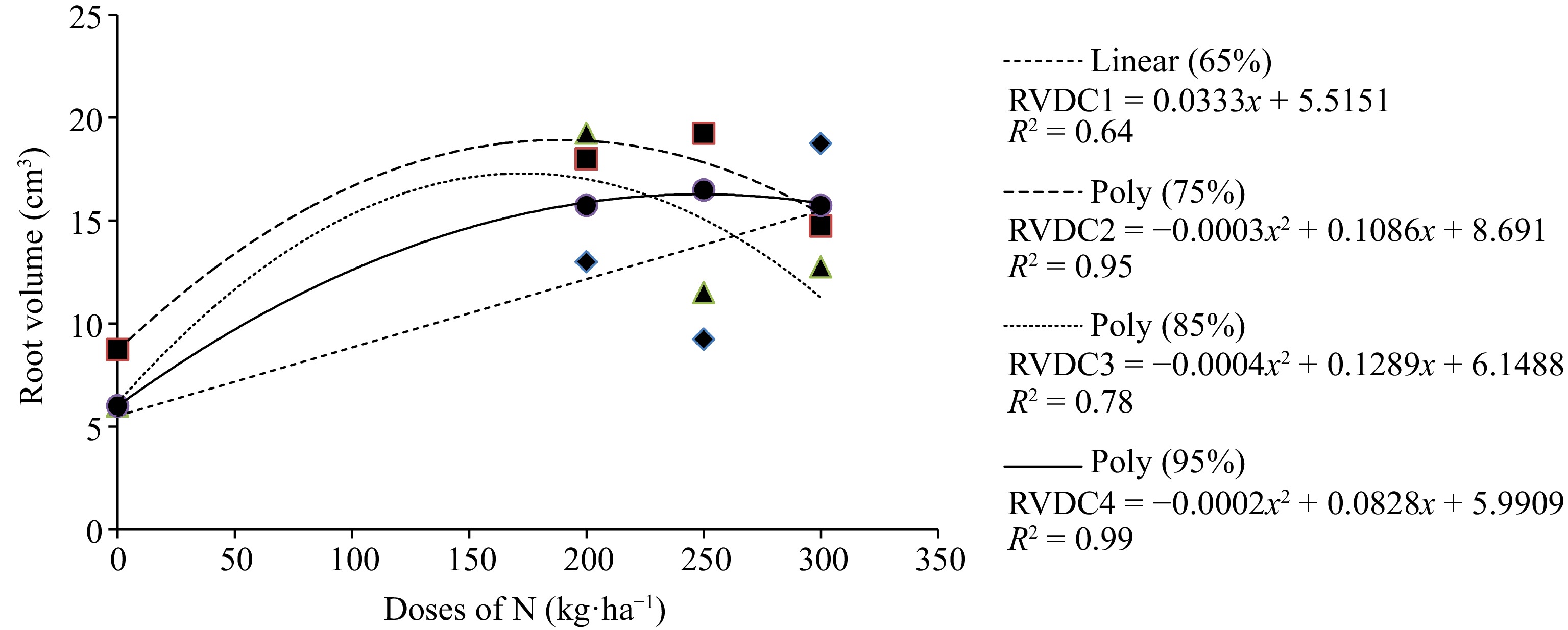
      Figure 1. 

      Average value of RV in Urochloa ruziziensis under increasing doses of N and different DC. DC1 ♦ = 65%; DC2 ■ = 75%; DC3 ▲ = 85% and DC4 ● = 95%.

      The average values of RV for U. ruziziensis fertilized with increasing N doses under different compaction levels are presented in Table 6. It is noticeable that there is a difference between the average values only for the N application dose of 250 kg·ha−1 at the respective compaction levels. This indicates that at the compaction level of 75% with the N dose of 250 kg·ha−1, the plant obtained the highest average RV.

      Table 6.  Average value of root volume in cm3 for the forage plant Urochloa ruziziensis as affected by nitrogen (N) fertilization under different degrees of compression (DC).

      DC (%)Doses of N (kg·ha−1)
      0200250300
      656,0a13,0a9,3b18,8a
      758,8a18,0a19,3a14,8a
      856,0a19,3a11,5ab12,8a
      956,0a15,8a16,5ab15,8a
      Means followed by the same lowercase letter in the column do not differ by Tukey's test at 5% probability. DC (%) = (Soil density in the field/Max proctor density) × 100.
    • Table 7 shows the average contents of crude protein (CP) of the forage species M. maximum cv. Mombaça, U. brizantha cv. MG-5 Vitória and U. ruziziensis. It is observed for the three forage species that as the N dose increased, the CP value increased. The average CP values for the forage M. maximum cv. Mombaça, ranged from 7.36% to 21.76%, showing that even in the control (zero dose), this cultivar obtained a CP value of over 7%. This value is considered the minimum for the plant to have good digestibility by the animals.

      Table 7.  Average crude protein (CP) values in % DM for the forage plants Megathyrsus maximum cv. Mombaça, Urochloa brizantha cv. MG-5 Vitória and Urochloa ruziziensis under different doses of nitrogen (N) kg·ha−1 in UFVJM, Diamantina, 2016.

      Forage speciesDoses of N (kg·ha−1)
      0200250300
      Megathyrsus maximum cv. Mombaça7.36b20.29a21.08a21.76a
      Urochloa brizantha cv. MG-5 Vitória4.73c17.40b18.93ab21.29a
      Urochloa ruzizienses6.23c23.14b25.14ab26.45a
      Averages followed by the same lowercase letter in the row do not differ by the Tukey test at 5% probability.

      The forage U. brizantha cv. MG-5 Vitória showed an increase in CP content as N doses increased. It is observed, in the control (zero dose), at CP content of 4.73%. For the other doses, this value reached was greater than 7%, reaching 21.29% when the dose of 300 kg·ha−1 was applied (Table 7). There was an effect of N doses on CP levels for U. ruziziensis, with a linear increase as N doses increased, reaching 26.45% when the dose was applied of 300 kg·ha−1 of N (Table 7).

    • Tables 8 & 9 show the average values of NDF and ADF in relation to N doses for the forage plants M. maximum cv. Mombaça, U. brizantha cv. Vitória, and U. ruziziensis. The NDF and ADF levels were reduced with higher N doses. Despite the decrease in NDF levels for the analyzed forages, these values were still higher than the critical range of 55%−60%[21]. This indicates that these forages may not be highly palatable to animals, meaning the plant becomes less attractive to the animal. As for ADF values, all analyzed species showed levels below 40%, suggesting that these forages have good digestibility.

      Table 8.  Average values of neutral detergent fiber (NDF) in % DM for the forage plants Megathyrsus maximum cv. Mombaça, Urochloa brizantha cv. MG-5 Vitória and Urochloa ruziziensis under different doses of nitrogen (N) kg·ha−1 in UFVJM, Diamantina, 2016.

      Forage speciesDoses of N (kg·ha−1)
      0200250300
      Megathyrsus maximum cv. Mombaça73.72a66.21b64.07b64.13b
      Urochloa brizantha cv. MG-5 Vitória75.18a67.41b64.56b64.34b
      Urochloa ruzizienses66.55a55.32b55.75b56.56b
      Averages followed by the same lowercase letter in the row do not differ by the Tukey test at 5% probability.

      Table 9.  Average values of acid detergent fiber (ADF) in % DM for the forage plants Megathyrsus maximum cv. Mombaça, Urochloa brizantha cv. MG-5 Vitória and Urochloa ruziziensis under different doses of nitrogen (N) kg·ha−1 in UFVJM, Diamantina, 2016.

      Forage speciesDoses of N (kg·ha−1)
      0200250300
      Megathyrsus maximum cv. Mombaça36.06a31.71b30.09b30.67b
      Urochloa brizantha cv. MG-5 Vitória36.59a32.97b30.58b30.81b
      Urochloa ruzizienses31.36a24.37b24.07b25.22b
      Averages followed by the same lowercase letter in the row do not differ by the Tukey test at 5% probability.
    • Table 10 presents the average values of lignin in relation to N doses for the analyzed forage plants. There was no significant difference in lignin content among different N doses for any of the studied forage species.

      Table 10.  Average values of lignin in % DM for the forage plants Megathyrsus maximum cv. Mombaça, Urochloa brizantha cv. MG-5 Vitória and Urochloa ruziziensis under different doses of nitrogen (N) kg·ha−1 in UFVJM, Diamantina, 2016.

      Forage speciesDoses of N (kg·ha−1)
      0200250300
      Megathyrsus maximum cv. Mombaça4.50a3.96a3.08a3.17a
      Urochloa brizantha cv. MG-5 Vitória3.11a4.61a3.51a5.29a
      Urochloa ruzizienses2.70a3.01a2.67a3.08a
      Averages followed by the same lowercase letter in the row do not differ by the Tukey test at 5% probability.

      The compaction levels did not affect the bromatological analyses. Despite the high compression, at 95%, the plant was able to absorb the available nutrient.

    • The low germination rate of forage seeds is considered one of the main obstacles to the establishment of pastures. However, for U. brizantha and for M. maximum, a percentage of germination was higher than the minimum standard recommended for production and commercialization of seeds according to Normative Instruction n° 30/2008[19] and Normative Instruction n° 33/2010[20], which corresponds to 60% for species of the genus Urochloa and 50% for Megathyrsus. As for U. ruziziensis, the percentage of germination was lower than the recommended minimum, which is 60%. In this way, care was taken with this genotype at the time of sowing, sowing an ideal amount of seeds was conducted to ensure a uniform stand.

      The low germination rate of U. ruziziensis may be caused by a physiological phenomenon known as dormancy, which is common in tropical pastures. Freshly harvested seeds exhibit low germination rates in the early stages of storage and show deep dormancy during this period. As the storage time increases, this dormancy tends to decrease[22, 23]. However, care must be taken when storing the seeds. Seeds stored for long periods may lose their vigor[24]. This loss of vigor is due to the deterioration of the metabolic system caused by this long storage time, leaving these seeds with slow development or prevented from germinating, leading to the development of poorly formed seedlings and a reduction in the number of seeds that germinate. Therefore, satisfactory seed storage is based on slowing down the normal metabolism of seeds as much as possible, causing as little damage as possible[25].

    • Considering the biometric evaluations carried out in forage species. The direct relationship that exists between the addition of N via fertilization with the increase in shoot GM and DM and consequently in forage height in M. maximum cv. Mombaça, U. brizantha cv. MG-5 Vitória and U. ruziziensis demonstrates the positive effect that N fertilization promotes on growth, increase in the number of tillers and expansion of the aerial part of the plant[26]. Nitrogen has a rapid effect on plant development. With the addition of N doses, there is an increase in shoot tillering, which results in efficient vegetative development[3]. When N is made available in the soil, it is soon absorbed by the forage, which is associated with the carbon chains, promoting an increase in cellular constituents and, consequently, a significant increase in the vigor and total production of dry matter of the plants[27]. Furthermore, the accumulation of carbohydrates, amino acids, enzymes and phytohormones is affected, thus promoting an increase in biomass[28]. The use of N fertilizer is considered a promising alternative to be implemented in the field, in which it promotes an increase in total productivity from 102% to 269%, which leads to greater availability of biomass for food and greater stocking capacity animal in the area[29].

      In the root system of the forage M. maximum cv. Mombaça, there was a positive response with an increase in the doses of N fertilizer up to a dose of 250 kg·ha−1 of N, promoting a growth of this system, with a decrease in the dose of 300 kg·ha−1 of N. With the application of N, the aerial part of the plants responds more significantly than the root system[30]. In a study evaluating the effect of increasing doses of N (0, 150, 300 and 450 kg·ha−1) on the root system of M. maximum subjected to rotational stocking, the researchers found a decrease in root length of 3.3% with N fertilization close to 300 kg·ha−1[31]. For U. brizantha vc. MG-5 Vitória as the doses of N fertilizer increased, there was a positive linear growth in the root system of this forage. When the plants are not using reserve compounds for shoot growth, these can be used by the root system, favoring its growth[32]. As for the forage U. ruziziensis, an apex of root development was observed in the application of the N fertilizer dose of 200 kg·ha−1, with an increasing behavior of the RV up to that point and then there was a decrease in the dose of 250 kg·ha−1 of N.

      However, it was possible to observe an interaction between N fertilizer dose and DC in the RV of U. ruziziensis. For the 65% DC, as the N dose increased, the RV increased considerably. This is due to the soil structure being looser, providing better absorption of this nutrient by the roots, resulting in a better development of the root system.

      For compaction degrees 75%, 85% and 95%, there was an increase in RV up to the application of 250 kg·ha−1 of N. The root system of plants needs a large amount of oxygen due to its high respiration rate. With the reduction of pore spaces between soil particles, there will be a reduction in the respiration of this system, harming its development[30]. For N fertilization to be efficient for the plant, it needs large volumes of water, which favors the production of roots, promoting the absorption of nutrients[33]. When the soil is compacted, there is a decrease in macro and microporosity, which makes it difficult for water and nutrients to infiltrate the soil profile, limiting its availability to the plant, resulting in poor development of the root system, which reflects on the production of aerial part.

    • Nitrogen fertilizer promoted a considerable increase in CP in forage M. maximum cv. Mombaça, U. brizantha cv. MG-5 Vitória and U. ruziziensis. This value is higher than what is considered the minimum when it comes to plant digestibility. CP contents of less than 7% limit DM consumption and reduce the digestibility of the plant[34], since the inadequate levels of N cause the population of microorganisms in the rumen to decrease and, in response, digestibility and intake of dry mass are reduced[21], causing the animal to consume less feed[34]. The absence or low dose of N fertilization in pastures reduces the levels of CP and rumen degradable protein, which can limit the performance of ruminants with higher protein requirements[35].

      The doses of N fertilizer promoted a decrease in NDF and ADF values ​​for the three forage grasses studied. However, even with a decrease in NDF values, they were higher than what is considered critical. Since the NDF is considered a more limiting factor in the consumption of roughage, it is related to the consumption of the plant by the animal, and when it is higher than 55%−60% of dry mass, they are negatively correlated[21], showing that it is not a very attractive forage for the animal. NDF values ​​found in the present work were higher than the critical value of 55% for the three studied forages, thus the voluntary consumption of these forages in grazing could be limited, resulting in loss of animal gain.

      ADF is negatively correlated with plant digestibility. ADF levels found in forage grasses above 40% are considered limiting for the digestibility of dry mass, compromising animal performance[21]. It is observed that, with the increasing application of N doses in the soil, average values ​​of ADF below 40% were found for the three studied forages. Thus, it can be stated that M. maximum cv. Mombaça, U. brizantha cv. MG-5 Vitória and U. ruziziensis, proved to be forages with good chemical composition in ADF, being considered plants with good digestibility.

    • The increase in N doses improves the nutritional quality of the evaluated species, since it promotes decreases in NDF and ADF contents and increases in green mass, shoot dry mass, height and protein contents. It is of great importance to study the cost-benefit ratio so that the appropriate dosage of N can be recommended.

      Even in compacted soil, the development of the forage root system U. brizantha cv. MG-5 Vitória responded gradually to N doses, proving to be a forage resistant to soils with reduced pore spaces.

    • The authors confirm their contribution to the article as follows: conception and elaboration of the research: Pires de Sousa Baracho I, Nery MC, Rocha WW; setting up, conducting the experiment and collecting the data: Pires de Sousa Baracho I, Valeriano FR, Maria da Cruz Bento B, de Souza Rocha A, de Melo Farnezi MM; analysis and interpretation of results: Pires de Sousa Baracho I, Nery MC, Rocha WW, de Melo Farnezi MM, de Cássia Ribeiro Carvalho R; preparation of the draft manuscript: Pires de Sousa Baracho I, Valeriano FR. All authors reviewed the results and approved the final version of the manuscript.

    • The datasets generated during and/or analyzed during the current study are available from the corresponding author on reasonable request.

      • The Universidade Federal dos Vales do Jequitinhonha e Mucuri (UFVJM), where he took the postgraduate course in plant production, in which he led this study. The Fundação de Amparo à pesquisa do Estado de Minas Gerais (Fapemig), for granting the research grant.

      • The authors declare that they have no conflict of interest.

      • Copyright: © 2024 by the author(s). Published by Maximum Academic Press, Fayetteville, GA. This article is an open access article distributed under Creative Commons Attribution License (CC BY 4.0), visit https://creativecommons.org/licenses/by/4.0/.
    Figure (1)  Table (10) References (35)
  • About this article
    Cite this article
    Pires de Sousa-Baracho I, Nery MC, Rocha WW, de Melo Farzeni MM, Valeriano FR, et al. 2024. Assessment for forage grass quality submitted to compaction degrees and nitrogen doses. Grass Research 4: e008 doi: 10.48130/grares-0024-0004
    Pires de Sousa-Baracho I, Nery MC, Rocha WW, de Melo Farzeni MM, Valeriano FR, et al. 2024. Assessment for forage grass quality submitted to compaction degrees and nitrogen doses. Grass Research 4: e008 doi: 10.48130/grares-0024-0004

Catalog

  • About this article

/

DownLoad:  Full-Size Img  PowerPoint
Return
Return