-
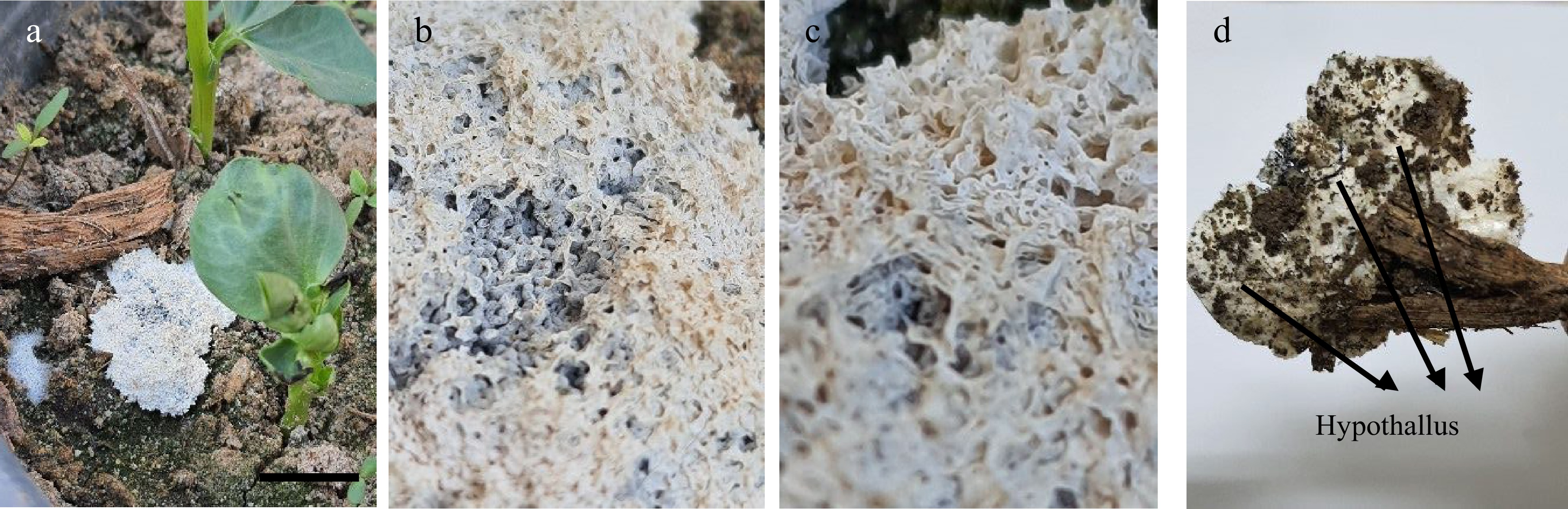
Figure 1.
Sporocarp of L. carestianum. (a) On the soil surface. (b) & (c) Lime scales covered the peridium of sporocarp (3X). (d) Hypothallus (2X).
-
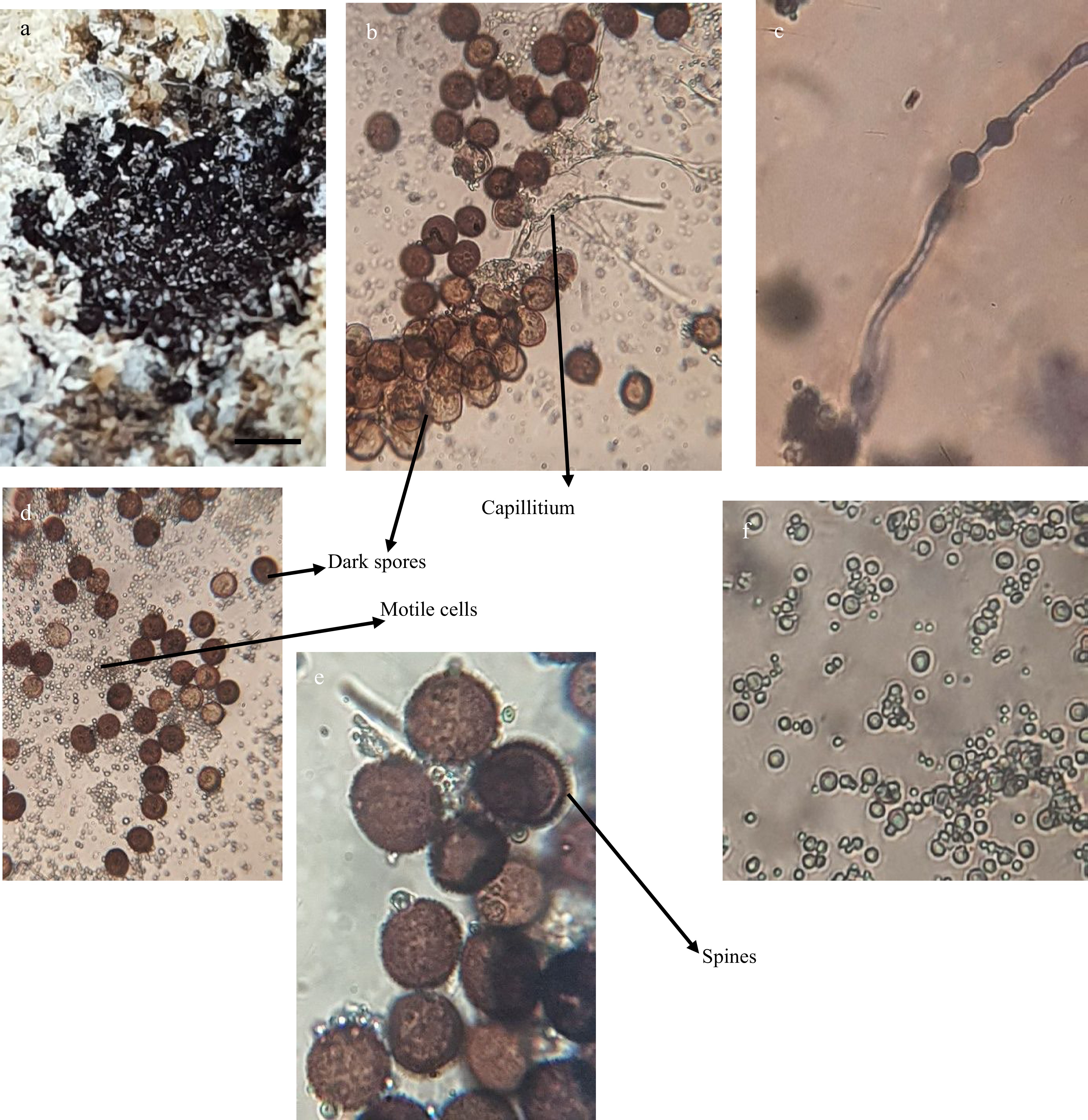
Figure 2.
A black powder inside sporocarp of L. carestianum (2X). (b) Dark spores. (c) Capillitium thread with swellings. (d) Dark spores and masses of motile cells. (e) Enlarged spores with spinose walls. (f) Enlarged motile cells. (b, c, d and f: 400X. e: 1,000X)
-
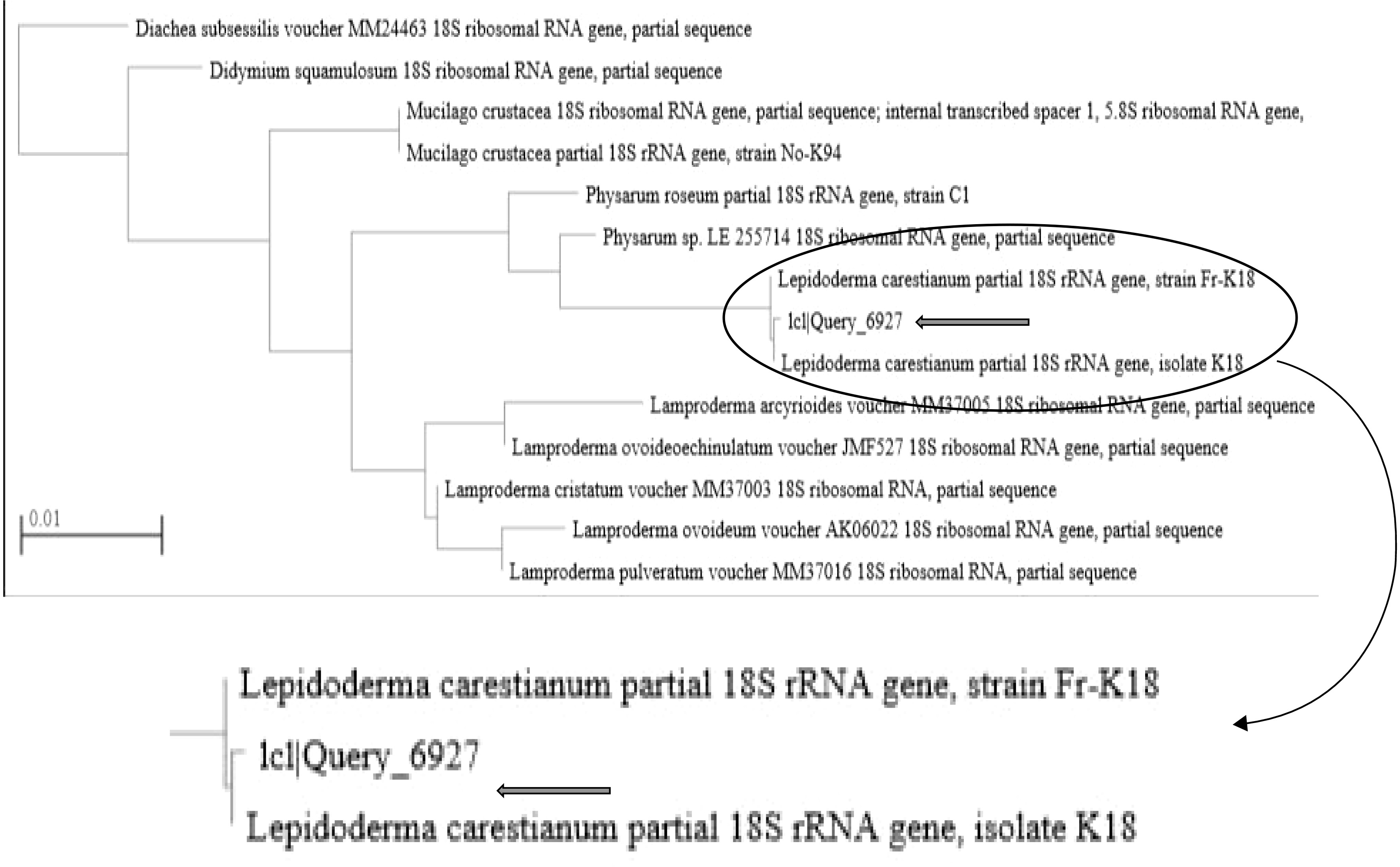
Figure 3.
Phylogenetic tree for tested myxomycetes.
-

Figure 4.
Morphology and microscopic examination of A. bambusicola: (a) White colony. (b) Reverse with black spots due to conidiomata (2X). (c) & (d) Conidiomata surrounded by septate hyphae (100X & 400X). (e) & (f) Conidia. (400X & 1,000X).
-

Figure 5.
Phylogenetic tree based on ITS sequences, showing the relationship between the tested isolate (Arth) and other species belong to the genus Arthrinium. The tree was constructed using the MEGAX and neighbor-joining method.
-
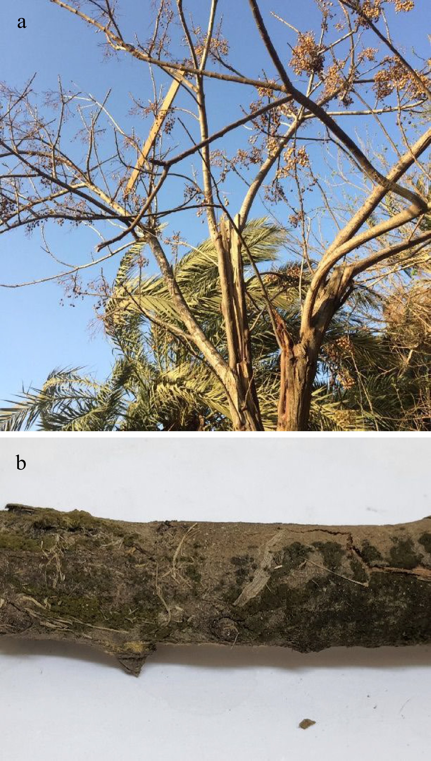
Figure 6.
Melia azedarach tree. (a) Healthy tree. (b) Olivaceous green growth of A. bambusicola on the dead branch (2X).
-
Tested strain Name Accession No. Query
cover
(%)Percent identity
(%)1c1|Query_6927 Lepidoderma carestianum HE614609.1 94 93.95 Lepidoderma carestianum AM231296.1 94 93.95 Physarum vernum KC759102.1 49 95.33 Physarum vernum KC759101.1 49 95.33 Physarum nivale DQ903680.2 49 95.33 Diderma crustaceum JQ277927.1 49 95.02 Mucilago crustacea MH348907.1 50 94.12 Table 1.
Molecular data of (1c1|Query_6927) strain with other previously reported myxomycetous strains.
Figures
(6)
Tables
(1)