-

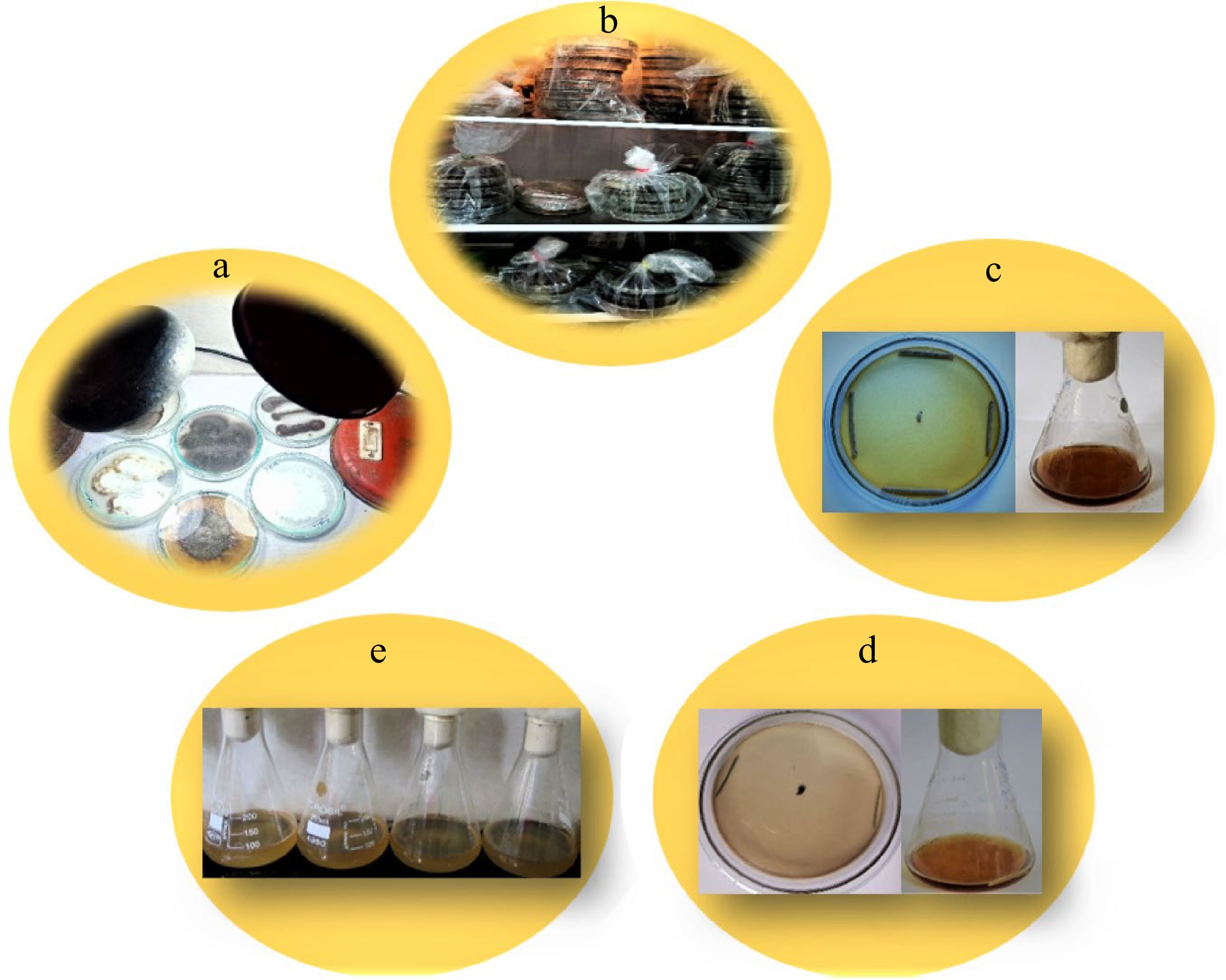
Figure 1.
Treatments for non-sporulating endophytic fungi. (a) Alternate light and dark cycles. (b) Cold treatment in a refrigerator. (c) Treatment with autoclaved host tissue on solid and liquid media. (d) Treatment with pine needles on solid and liquid media. (e) Low strength media.
-

Figure 2.
(a) Scientific diagrams showing the number of sterile endophytic fungal isolates recovered from E. gerardiana. (b) Number of corresponding sterile isolates from roots and stem.
-

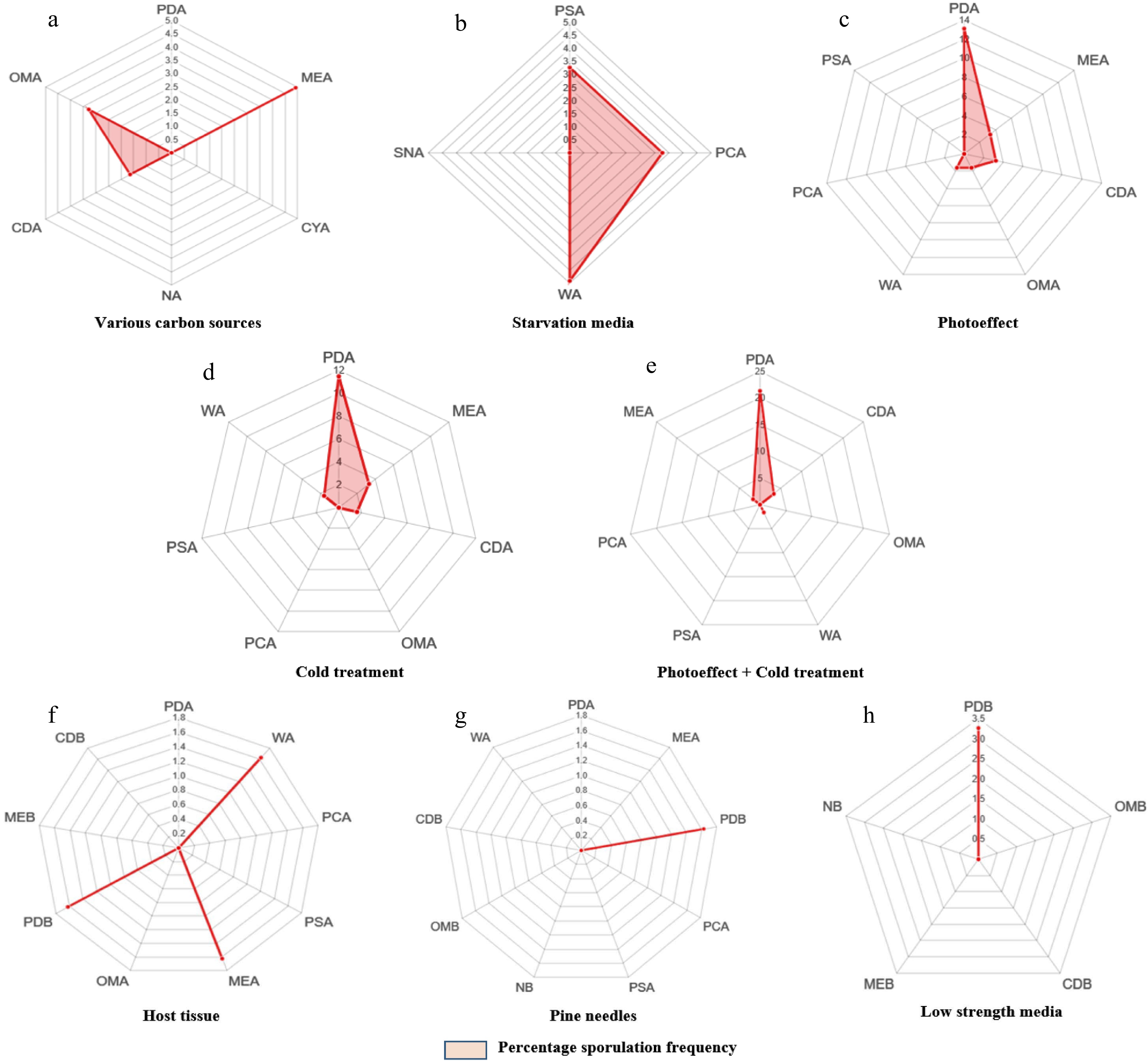
Figure 3.
(a)-(h) Radarcharts showing percentage of sporulation in various morphospecies with respect to different methods and media.
-

Figure 4.
Diagrammatic representation showing differences between different media revealed by post Hoc analysis. 2, 3, 5 & 6 exhibit the significant differences between PDA and CDA, PDA and OMA, PDA and PSA, PDA and PCA with values higher than Tukey's value (5.53).
-
Method Media Name of the
morphospeciesMaximum number
of morphospecies sporulated Nmax (%)Various carbon sources PDA 0 MEA MN5, MN9, MN23 4.91 CYA 0 NA 0 CDA MN8 1.63 OMA MN10, MN34 3.27 Starvation media PSA MN15, MN33 3.27 PCA MN7, MN32 3.27 WA MN41, MN58, MN62 4.91 SNA 0 Photo-effect PDA MN20, MN27, MN42, MN45, MN18, MN52, MN66, MN64 13.11 MEA MN21, MN14 3.27 CDA MN24, MN49 3.27 OMA MN65 1.63 WA MN56 1.63 PCA 0 PSA 0 Cold treatment PDA MN2, MN3, MN6, MN19, MN25, MN40, MN61 11.47 MEA MN4, MN12 3.27 CDA MN59 1.63 OMA 0 PCA 0 PSA 0 WA MN55 1.63 Cold treatment + photo-effect PDA MN11, MN16, MN28, MN29, MN30, MN31, MN36, MN44, MN46, MN47, MN35, MN37,
MN5121.31 CDA MN53, MN63 3.27 OMA 0 WA MN57 1.63 PSA 0 PCA 0 MEA MN17 1.63 Host tissue PDA 0 WA MN60 1.63 PCA 0 PSA 0 MEA MN38 1.63 OMA 0 PDB MN22 1.63 MEB 0 CDB 0 Pine needles PDA 0 MEA 0 PDB MN43 1.63 PCA 0 PSA 0 NB 0 OMB 0 CDB 0 WA 0 Low strength media PDB MN39, MN67 3.27 OMB 0 CDB 0 MEB 0 NB 0 Table 1.
Maximum number of morphospecies Nmax (%) sporulated after applying diverse methods.
Figures
(4)
Tables
(1)