-
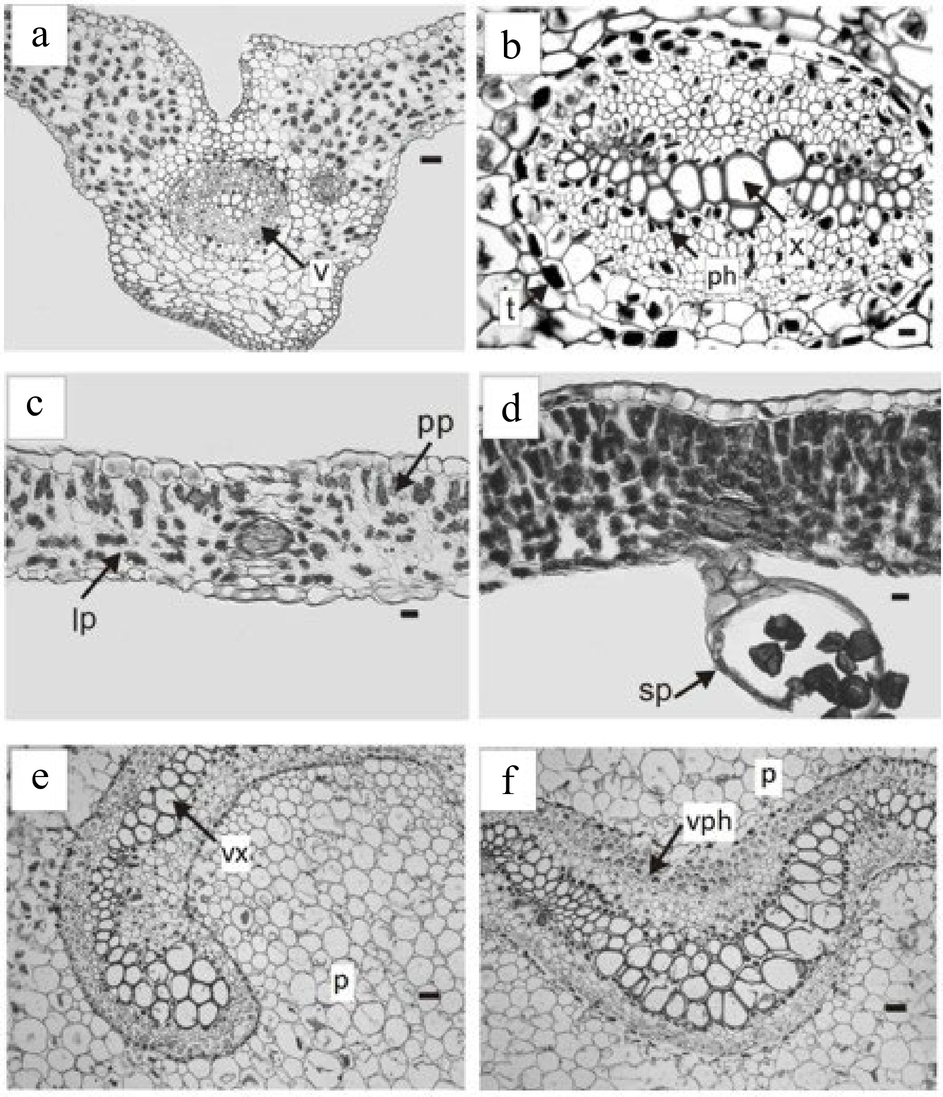
Figure 1.
Trismeria trifoliata. (a) Cross-section of the main middle nerve of the central leaflet and (b) detail of the vascular bundle. (c) & (d) cross-sections of the intercostal areas. (d) Detail of the sporangia on the lower face of the leaflet. (e) & (f), cross-sections of the petiole with the phloem and xylem presenting a central curvature. ph, phloem; p, parenchyma; pp, palisade parenchyma; lp, lacunose parenchyma; sp, sporangia; t, tannins; v, vascular bundle; vph, vessel phloem; vx, vessel xylem. Scales: (a), (e), (f) = 180 μm, (b) = 45 μm, (c) & (d) = 100 μm.
-
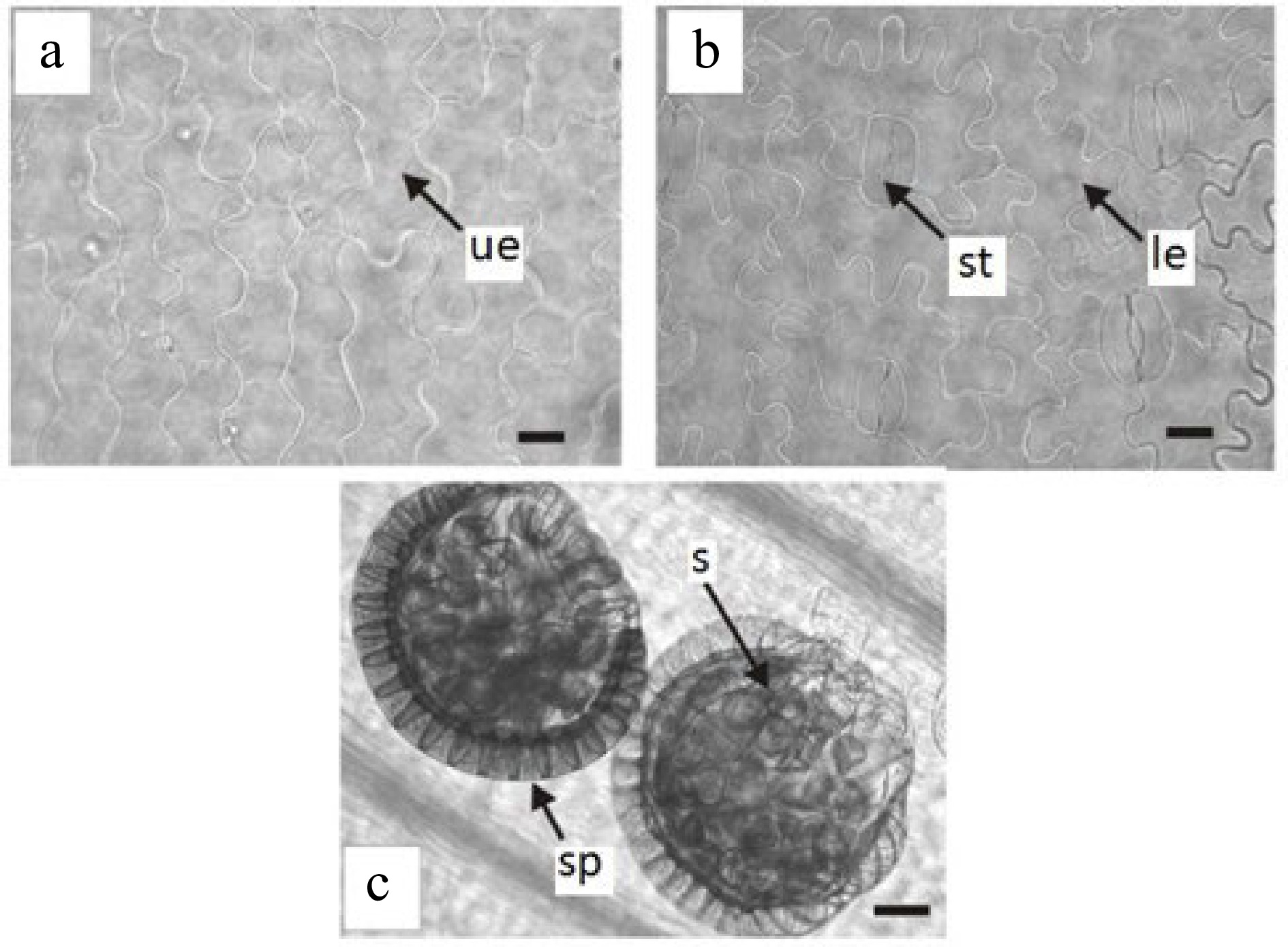
Figure 2.
Epidermis in surface view of Trismeria trifoliata. (a) and (b) upper and lower epidermis of the central leaflet, respectively. (c) view of sporangia on the lower face of the central leaflet. le, lower epidermis; s, spores; sp, sporangia; st, stomata; ue, upper epidermis. Scales: (a) and (b) = 45 μm and (c) = 100 μm.
Figures
(2)
Tables
(0)