-
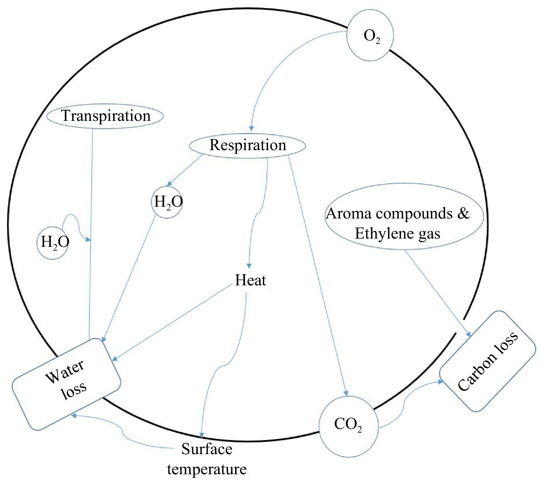
Figure 1.
The concept of the respiration process in a plant cell showing the different mass and heat components involved.
-
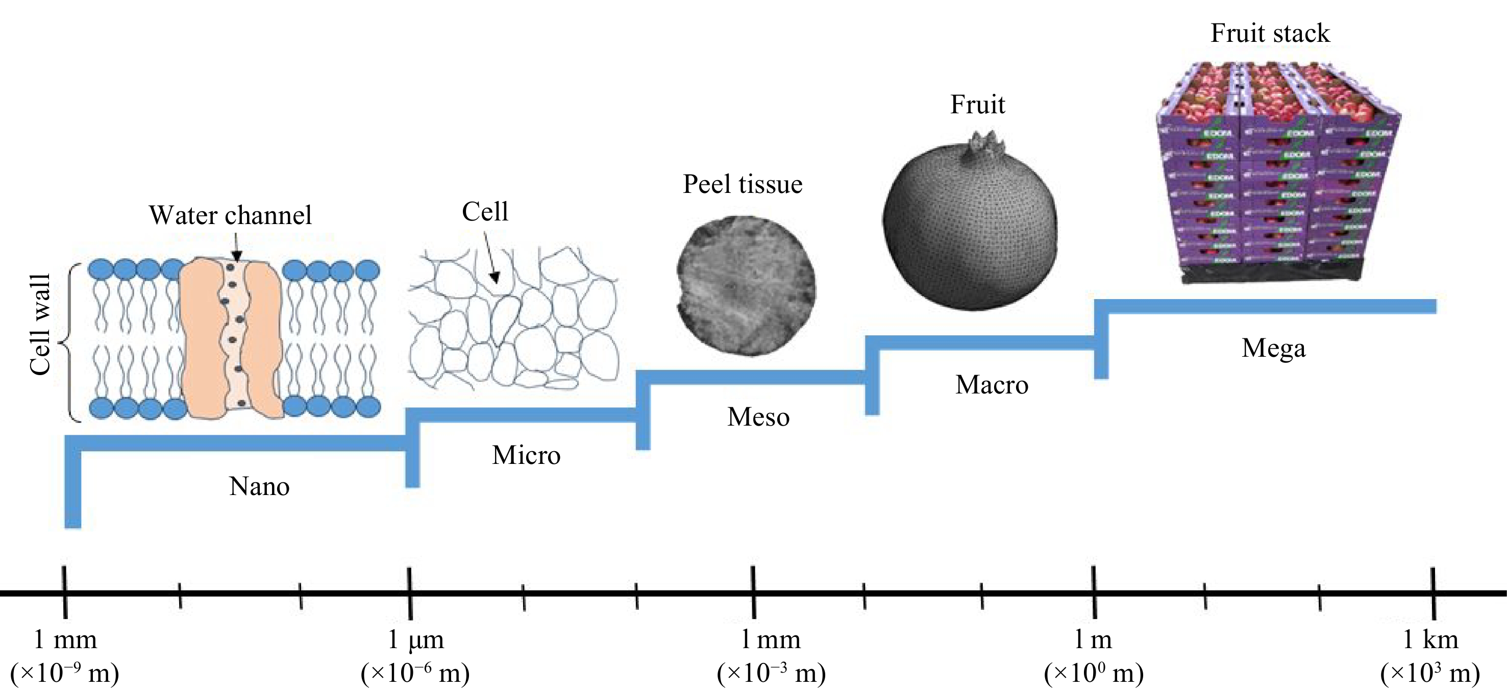
Figure 2.
Spatial scales of water transport modelling in the fresh fruit industry.
-
Fruit Conditions of determination Part of fruit Diffusion coefficient (m2·s−1) Ref. Pear
(cv. Conference)Mesocarp (flesh) 1.520E-11 to 1.730E-11 [51] Cuticle 1.100E-13 to 1.200E-13 Pear
(cv. Conference)273.15 K Inner cortex tissue 1.230E-11 [18] 293.15 K 4.359E-11 273.15 K Outer cortex tissue 5.300E-13 293.15 K 1.050E-12 273.15 K Cuticle 5.500E-14 293.15 K 1.280E-13 Apple
(cv. Jonagold)273.15 K,
92% RHTissue 1.120E-11 [26] Cutin 6.420E-14 Wax 2.100E-15 Cuticle 8.740E-15 Apple
(cv. Estar)273.15 K,
92% RHTissue 4.330E-12 Cutin 7.160E-14 Wax 3.030E-14 Cuticle 5.080E-14 Apple
(cv. Jonagored)273.15 K,
92% RHMesocarp tissue 1.380E-11 Cutin 4.030E-14 Wax 1.920E-15 Cuticle 9.100E-15 Apple
(cv. Jonagold)Flesh 1.030E-10 [52] Skin 1.320E-13 Table 1.
Moisture diffusivity across fruit tissues.
-
Model scale Fresh produce Structural/geometrical details Details of water transport model Ref. Nanoscale Artificial plant cell walls The objective was to study the effect of cell wall composition and temperature on the structure, desorption isotherms and water conductivity of artificial cell walls. [59] Microscale Pears (cv. Conference) 2D geometric model of cortex tissue, composed of cells of random sizes and shapes, cell walls and intercellular spaces. A steady state model applied,
Modelling of water transport in the intercellular space, the cell wall network and cytoplasm was done using diffusion laws and irreversible thermodynamics.[58] Microscale Pear (cv. Conference) cortex tissue intercellular space, cell wall network and cytoplasm A 2D geometrical model was obtained with a virtual fruit tissue generator, based on cell growth modelling. To account for the microstructure, a microscopic layout was introduced into the modelling as the computational geometry of the model. Water transport was described using a coupled approach incorporating the microscale water transport model and the cell mechanics model that predicts cell and tissue deformation resulting from hydrostatic stress caused by moisture loss. [60] Microscale & Mesoscale Apple (cv. Jonagold and Elstar) cuticle: Tissue, cutin and wax Water transport is modelled at a mesoscale (tissue) level by considering moisture loss across the cuticle.
However, a more detailed microscopic approach was applied to account for the microstructural features of surface cracks, open and closed lenticels.
In the geometrical basis of the moisture diffusion model, lenticel and crack structures, total crack area, and effect of wax smoothing were incorporated.The actual diffusion properties of the cuticle, cutin and wax were derived from apparent diffusion properties determined experimentally using gravimetric procedures. [23] Mesoscale Apple fruit tissues (cv. Elstar, Jonagold and Jonagored). A mesoscale geometry of apple tissue cylinder was constructed to represent fruit tissue, cuticle and cutin and wax layer. Water transport across fruit tissue, cutin and surface wax layer was simulated.
Diffusion coefficients of apple tissue and cuticle were experimentally determined using apple tissue cylinders with intact cuticle, without cuticle or without wax.[26] Mesoscale Apple fruit (cv. Jonagold): Cylindrical apple tissue A coupled mass transfer and mechanics model was used to describe water transport and associated deformation of apple tissue during dehydration.
The model was one-dimensional.[61] Mesoscale Conference pears tissues (cuticle, inner and outer cortex) Water transport in the different fruit tissues was described using sorption isotherm experimental data, fitted with Ferro Fontan model. [62] Mesoscale Conference pears tissues (cuticle, inner and outer cortex) Water transport in fruit tissues was described based on effective diffusivity of water. Modelling was done using Fick's first and second laws of diffusion on the cuticle and cortex tissues, respectively. A chemical potential gradient was employed as the driving force. [18] Mesoscale and Macroscale Pears (cv. Conference): cuticle, inner and outer cortex A computer vision-based modelling system was used. Eight digital images of the fruit taken from different directions. A 3D shape of the whole fruit was reconstructed from contours extracted from the images. The diffusion model was based on Fick's second law, to simulate water transport in whole fruit at shelf (293.15 K, 75% RH) and cold storage (274.15 K, 60% RH) conditions.
Different pear tissues with varying diffusion properties, were used to describe water transport at a mesoscale level.[63] Macroscale Apple (cv. Elstar and Jonagold) A wedge-shaped geometrical model was used to predict moisture loss over a whole apple during 6 months of controlled atmosphere storage. Actual diffusion coefficients of apple tissue, cutin, wax and cuticle were integrated with different geometric sub-models in order to predict moisture loss of whole fruit under specific storage conditions. [29] Macroscale Apple whole fruit
Macro-scaleMacroscale water transport model for the entire fruit A microscale model was used to compute the water transport properties of the apple skin. Then the water transport model for the entire fruit was computed from the water transport properties of the skin. [26,29] Multiscale (Combined microscale and macroscale) Apple tissue (cv. Jonagold) A 2D multiscale water transport and mechanical model was used to predict water loss and viscoelastic deformation.
The apparent parameters of the macroscale model were computed from a microscale model. At a microscopic level, water movement across tissue microstructures: cell wall network, cytoplasm and intercellular space were considered.[64] Table 2.
Water transport models in fresh fruit applied at microscale, mesoscale, macroscale and multiscale.
Figures
(2)
Tables
(2)