-
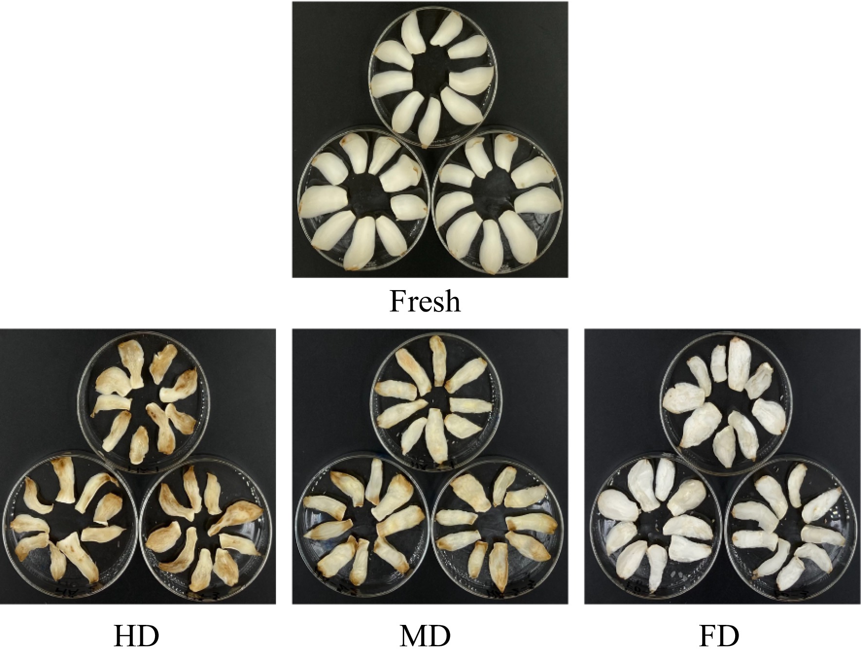
Figure 1.
The appearance of dried bulbs processed by different drying methods.
-
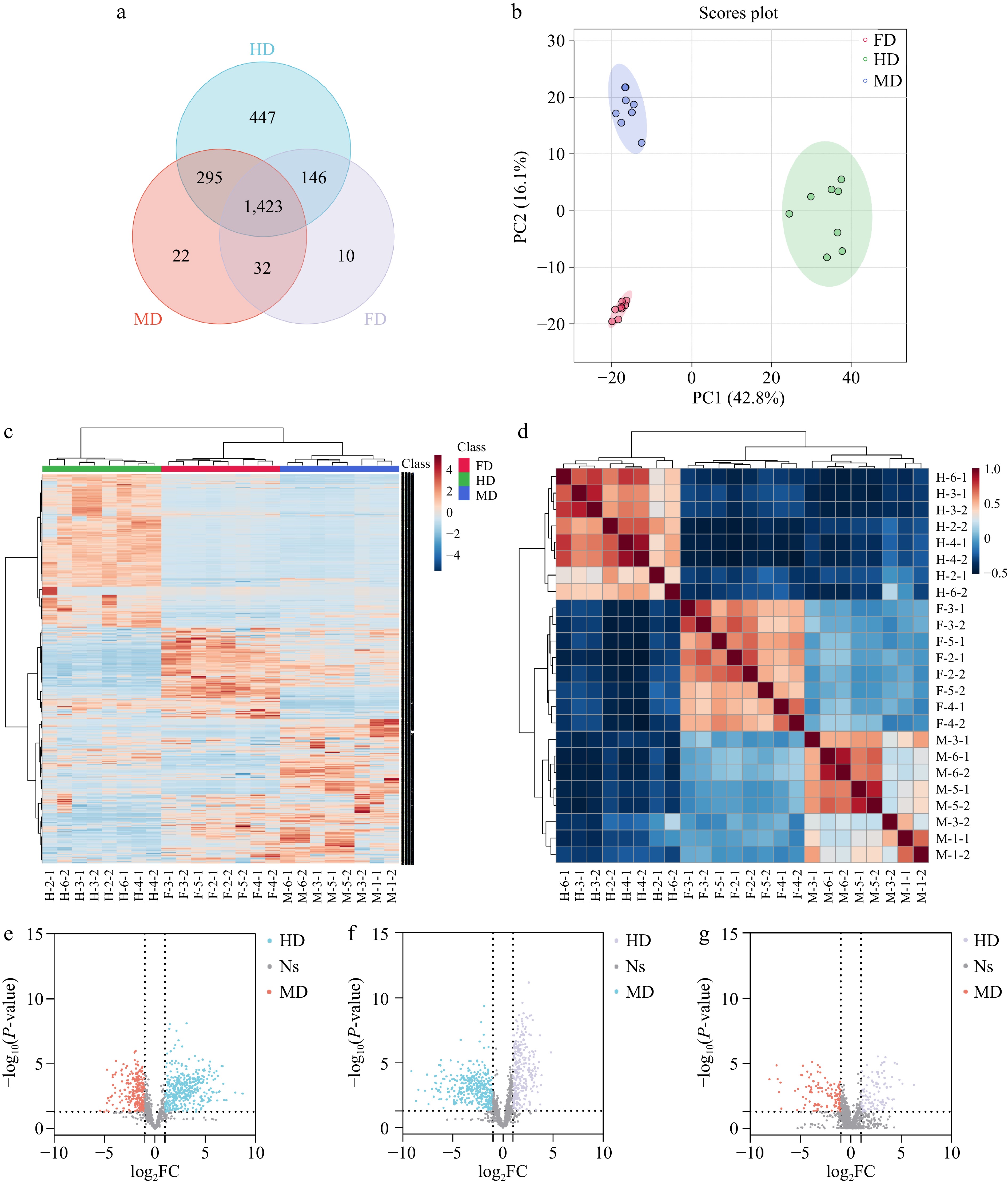
Figure 2.
Differentiation of different dried lily bulbs in positive mode. (a) Venn plot. (b) PCA scores plot. (c) heatmap. (d) Pearson correlation. (e), (f) Volcano plot of features in dried bulbs processed by different drying methods: HD & MD, FD & HD, FD & MD.
-

Figure 3.
The annotated markers among different drying methods. The left part are mirror plots and chemical structures, and the right part are box-plots. (a) Tryptophan, (b) phenylalanine, (c) glutamine (d) N-fructosyl-phenylalanine, (e) riboflavin, (f) rosmarinic acid, (g) caffeic acid, (h) coumarin, (i) p-coumaric acid, and (j) eleutheroside E.
-
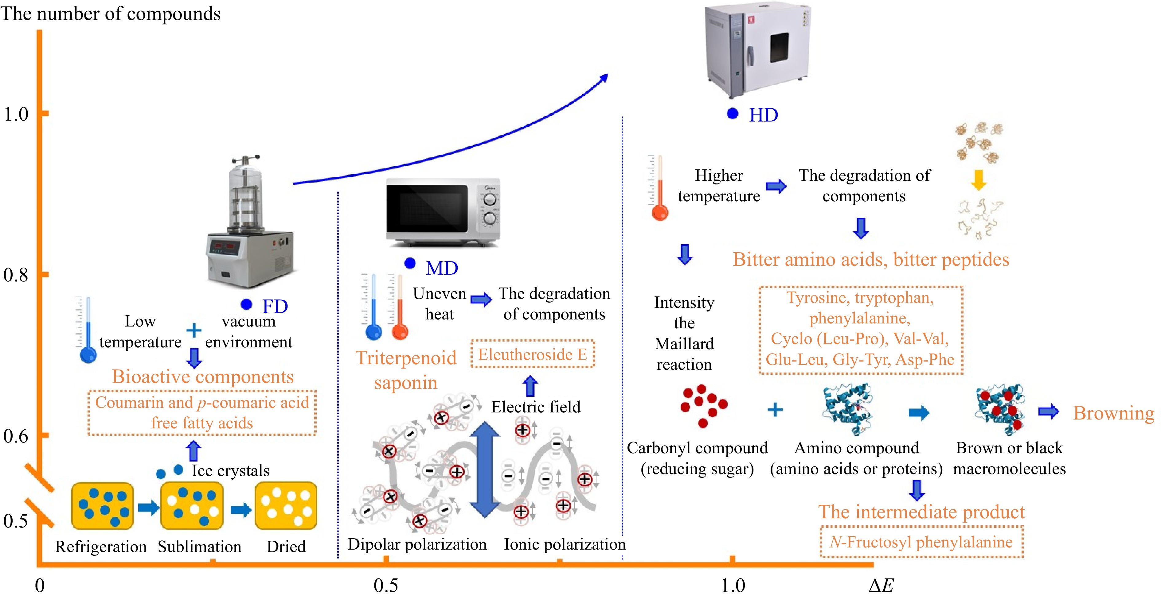
Figure 4.
The tentative mechanism of markers in different drying methods. Note: The number of compounds and ΔE of HD were considered as 1, and the values of FD and MD were compared with HD.
-

Figure 5.
Extracted ion chromatogram of (a) standard and samples and (b) the content of eleutheroside E in samples. Note: The precursor ion and the product ion of eleutheroside E were 787.2/579.3 Da.
-
Drying
methodsΔL* Δa* Δb* ΔE HD −23.60 ± 2.37c 4.06 ± 0.77a 11.69 ± 2.15a 26.79 ± 1.75a FD 6.22 ± 1.57a 0.48 ± 0.10c 3.75 ± 0.73b 7.39 ± 1.05c MD −9.58 ± 1.92b 2.71 ± 0.32b 9.55 ± 1.71a 14.00 ± 1.35b ΔL*, Δa*, and Δb* represent the difference between dried bulbs and fresh samples. Values followed by lowercase letters indicate the significant difference at the 0.05 level in the same column. Table 1.
The color index of dried bulbs processed by different drying methods.
-
No Compounds Classification m/z Ionization model Retention
time (min)Formula Error
(ppm)HD 1 Tryptophan Amino acid 205.0970 [M+H]+ 4.187 C11H12N2O2 −1.511 2 Phenylalanine Amino acid 166.0877 [M+H]+ 2.769 C9H11NO2 8.550 3 Val-val Dipeptide 217.1554 [M+NH4]+ 2.461 C10H20N2O3 −0.092 4 Glu-Leu Dipeptide 259.1283 [M-H]− 3.375 C11H20N2O5 −2.933 5 Gly-Tyr Dipeptide 239.1032 [M+H]+ 2.313 C11H14N2O4 −3.179 6 Asp-Phe Dipeptide 281.1137 [M+H]+ 4.294 C13H16N2O5 −4.802 7 N−Fructosyl phenylalanine Other 328.1412 [M+H]+ 2.692 C15H21NO7 7.344 8 Rosmarinic acid Hydroxycinnamic acid 163.0411 [M+H-C9H10O5]+ 8.453 C18H16O8 8.280 9 Caffeic acid Hydroxycinnamic acid 181.0450 [M+H]− 4.599 C9H8O4 4.748 10 (-)-Riboflavin Vitamin 163.0411 [M+H]+ 6.514 C17H20N4O6 −1.538 FD 11 Glutamine Amino acid 147.0757 [M+H]+ 7.208 C5H10N2O3 −5.031 12 Coumarin Coumarin 147.0443 [M+H]+ 10.316 C9H6O2 1.700 13 p−Coumaric acid Hydroxycinnamic acid 147.0459 [M+H]+ 8.393 C9H8O3 10.677 14 Palmitic acid Free fatty acid 255.2360 [M-H]− 14.792 C16H32O2 11.558 15 Heptadecanoic acid Free fatty acid 269.2468 [M-H]− 15.208 C17H32O2 −5.868 16 γ−Linolenic acid Free fatty acid 277.2164 [M-H]− 13.868 C18H30O2 −3.319 17 Vaccenic acid Free fatty acid 281.2472 [M-H]− 14.873 C18H34O2 −5.653 18 Stearic acid Free fatty acid 283.2637 [M-H]− 15.754 C18H36O2 −2.048 MD 19 Eleutheroside E Triterpenoid saponin 787.2607 [M+FA-H]− 8.024 C34H46O18 −7.520 Table 2.
Tentative markers in dried lily bulbs processed by different methods.
Figures
(5)
Tables
(2)