-
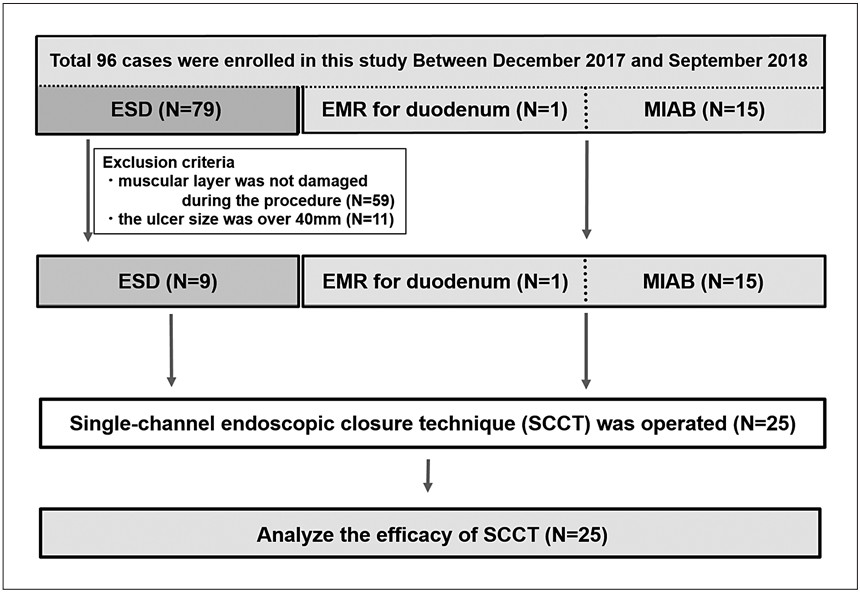
Figure 1.
Flowchart of patients with lesions enrolled in this study.
-
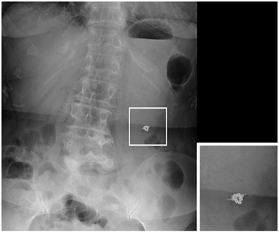
Figure 2.
Representative abdominal X-ray image, which shows clips remaining in the proper position in the stomach.
-
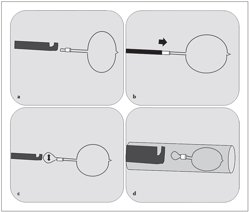
Figure 3.
A schematic illustration depicting the preparations for the SCCT. a, b Moving the stopper forward to the top of the loop. c Releasing the loop from the ligation device and widening the loop tail sufficiently. d Putting the loop directly into the outer sheath. The modified loop is stored in the outer sheath.
-
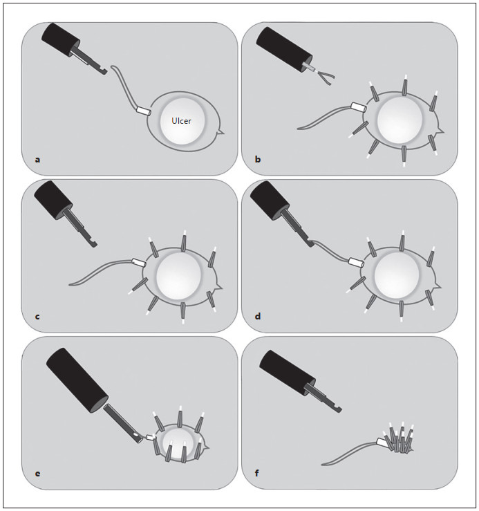
Figure 4.
A schematic illustration of the SCCT. a Dropping the tail of the loop. b Fixing the loop to the mucosa surrounding the ulcer by clips. c, d Catching the tail of the loop with the ligation device. e, f Closing the loop tightly.
-
Age, years 65 (range 36–81) Males/females 12/13 Ulcer location, n casesa Stomach (upper) 8 (1/7/0) Stomach (middle) 6 (0/6/0) Stomach (lower) 5 (3/2/0) Duodenum 1 (0/0/1) Colon (ascending) 1 (1/0/0) Colon (transverse) 2 (2/0/0) Colon (descending) 1 (1/0/0) Rectum 1 (1/0/0) Type of treatment, n cases ESD 9 MIAB 15 EMR 1 Median diameter of ulcer, mm 20 (range 10–40) ESD 26 (range 15–40) MIAB 15 (range 10–40) EMR 10 a Total (ESD/MIAB/EMR). Table 1.
Characteristics of the patients enrolled in this study
-
Technical success rate (complete closure) 100% (25/25) Procedure time, min 16 (5–49) Number of clips 8 (5–12) Complication rate 0% (0/25) Clinical success on day 1 100% (19/19) Clinical success on day 5 100% (9/9) Severe stenosis 0% (0/0) Table 2.
Efficacy and safety of the modified single-channel endoscopic closure technique
Figures
(4)
Tables
(2)