-
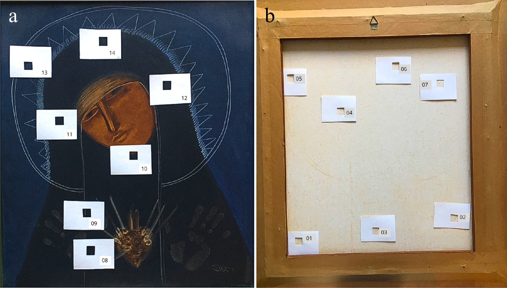
Figure 1.
Madona oil painting on canvas, by Siron Franco. Location of the sampling areas (4 cm2 ) on the obverse and reverse sides of the oil painting canvas used for the study. (a) 8 to 14, bright areas of the obverse side; (b) 1 to 7, areas of the reverse side.
-
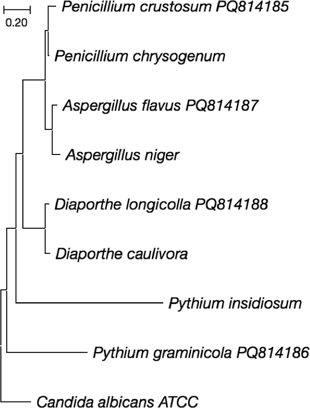
Figure 2.
Phylogenetic tree constructed based on the sequencing of the ITS1 and ITS4 region of fungal isolates using the maximum composite likelihood method and 1000 Bootstrap tests in the MEGA v.11.0 program.
-
Species of fungi Colony Macro morphology Micro morphology of structures Aspergillus flavus Mycelium floccose with sporulation and yellowish-green and olive conidia. Colonies with white border. No exudates produced. The reverse sides of the colonies show furrowed and slightly pale brown. The colony diameter range between 55 and 75 mm. Conidiophores colorless, thick-walled, roughed, and bearing vesicles, with diameter 900 to 1,100 μm. The vesicle shape globose to sub-globose, 1,700 to 1,900 μm. The metulae surround the vesicles' surface and emitted in all directions. The conidia globose, thin-walled, slightly roughed, and from 240 to 400 μm in diameter. Diaporthe longicolla Fluffy and dense aerial mycelium in white colonies with greenish yellow areas. From the reverse side, colony color appears initially greenish, yellow and black spots developed later. Reproduced asexually with α-conidia with enormous stromata with long pycnidial beaks and soybean stems in culture. contain oval shaped, hyaline and biguttulate αconidia (6.4 μm × 2.2 μm) exuding from the pycnidial ostiole in a yellowish, creamy drop. Penicillium crustosum Mycelium white, and colonies blue-green with abundant sporulation. Colonies plane with a granular texture. Mean colony diameter is 29.2−30.4 mm. Conidiophores terverticillate, stipes septate with rough walls, and conidia, borne in columns, smooth and spherical to subglobose. Conidial diameter 2.1−3.8 μm. Pythium graminicola Light grayish mycelium, aerial, fast growing (7−10 days), in which the sporangia for asexual reproduction and ornate oospores. Coenocytic hyphae with 2.7 to 4.5 μm. Sporangia lobulate. Oogonia globose. Oospores globose, antheridia diclinous. Table 1.
Micro and macro morphological characteristics of the structures of the isolated fungal species.
-
Sampling area Fungal species Top BLAST search results (GenBank accession number) Query cover (%) Identity (%) No. of bp analysed GenBank accession number 03 reverse side Penicillium crustosum PQ606659.1 99 99 540 PQ814185 05 reverse side Pythium graminicola AB562908.1 100 100 770 PQ814186 07 reverse side Aspergillus flavus OK314989.1 99 100 555 PQ814187 12 observe side Diaporthe longicolla HM347700.1 100 100 541 PQ814188 Table 2.
Results of molecular identification based on rDNA sequencing and matching with the NCBI GenBank database of fungi species isolated from painting.
-
Essentials oils tested MIC (μg ml−1) Penicillium crustosum Phythium graminicola Aspergillus flavus Diaporthe longicolla Curcuma longa 22.5 45.0 22.5 22.5 Thymus vulgaris 22.5 45.0 22.5 22.5 Melaleuca alternifolia 22.5 90.0 22.5 45.0 MIC: Minimal Inhibitory Concentration Table 3.
MIC values of essentials oils in vapor phase for isolate species of fungi.
Figures
(2)
Tables
(3)