


-
Siron Franco, born in 1947 in Goiás Velho, Brazil, is a prominent contemporary artist known for his work in painting, sculpture, illustration, and installation. He gained international recognition after winning major awards at the São Paulo Biennials in 1974 and 1975 and was named Best Painter of the Year in 1980. His artworks are displayed in prestigious museums in Brazil and worldwide, including The Met in New York (USA) and the Museum of Contemporary Art of Monterrey (Mexico).
Contemporary artworks represent an invaluable cultural heritage, serving not only as expressions of artistic creativity but also as critical ways for preserving and conveying history, culture, and traditions to future generations[1,2]. Associated with this, many artistic collections are subject to negative microbial action, especially from filamentous fungi[3]. Fungal decay is a serious problem for the preservation of oil paintings on canvas because it threatens both their structural integrity and aesthetic appeal[4].
Fungal species use the organic materials of paintings, such as binders, pigments, and canvas fibers, as nutrients, and their colonization can result in discoloration, staining, and surface changes, which can degrade priceless cultural artifacts[5]. Thus, fungal activity can also cause structural damage, such as flaking and cracking of paint layers, as well as deterioration of canvas fibers due to enzymatic activity. Some fungal species also release acidic metabolites that worsen material deterioration[6].
The conservation procedure is made more difficult by the health dangers posed by volatile organic chemicals and fungal spores[7]. Numerous fungus species are accountable for the biodeterioration of oil paintings, like as Alternaria spp., Cladosporium spp., Aspergillus spp., and Penicillium spp. are often implicated taxa[8,9]. These fungi can produce enzymes that break down the organic and structural elements of artwork, such as cellulases and proteases[10]. The initial look of the artwork can be further altered by the chemical interactions of their metabolic wastes with paints and varnishes[11].
Chemical biocides, which are effective but may endanger the environment and the artwork, have been the mainstay of traditional treatments for fungal rot. Researchers have looked into alternate strategies to address these issues, including using natural therapies like essential oils with antifungal qualities. According to Martins and collaborators[12], essential oils derived from plants such as oregano and tea tree have demonstrated encouraging outcomes in regulating fungal development while preserving material safety[13].
In this context, natural EOs emerge as a promising alternative to conventional antifungal agents. The compounds extracted from plants have antimicrobial properties, as well as efficacy against filamentous fungi in in vitro assays. Additionally, they present a lower environmental impact and reduced toxicity, making them safer for use in conservation environments[14].
Biotechnological breakthroughs provide alternative solutions for sustainable conservation. The utilization of biocompounds generated by helpful microorganisms, such as Bacillus species, has proven potential to prevent fungal colonization without hurting the artwork[15]. These cutting-edge techniques seek to preserve cultural assets over the long term by fusing effectiveness with material and environmental compatibility[16].
Integrating essential oils into the antifungal treatment for contemporary artworks represents a sustainable and effective approach to addressing biodeterioration. The significance of interdisciplinary efforts in tackling the problems of fungal biodeterioration is shown by these integrated methods. Thereby, this thorough knowledge of fungal decay and creative conservation techniques emphasizes the need for continued study and the application of sustainable practices in the preservation of cultural heritage.
-
Made in the 70s, the painting by Brazilian plastic artist Siron Franco was titled Madona. The object of art has dimensions of 74 cm × 82 cm × 3 cm by a technique of oil on canvas (Fig. 1a, b).
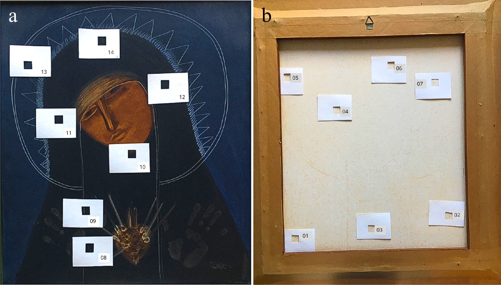
Figure 1.
Madona oil painting on canvas, by Siron Franco. Location of the sampling areas (4 cm2 ) on the obverse and reverse sides of the oil painting canvas used for the study. (a) 8 to 14, bright areas of the obverse side; (b) 1 to 7, areas of the reverse side.
Sampling
-
Biological sampling was performed using a sterile swab to collect material from the painting's surface (4 cm2) at 14 distinct locations, ensuring extensive coverage (Fig. 1a, b). Sampling sites were selected based on the presence of biological growth detectable both macroscopically and under magnification[17]. The collected specimens were then transferred into sterile microtubes and transported under controlled conditions to the Laboratory at the Brazilian University for further analysis.
Culture media and fungal cultivation
-
Biological material isolated from all areas of the artwork was subjected to serial dilutions ranging from 10−1 to 10−4 in a 0.85% saline solution supplemented with 0.001% Tween®-20. A 100 μl aliquot of each diluted sample was aseptically spread onto Sabouraud Dextrose Agar (SDA) plates (Difco Laboratories, Detroit, MI, USA) supplemented with chloramphenicol (100 μg·ml−1; Sigma-Aldrich, St. Louis, MO, USA) to inhibit bacterial contamination. The plates were subsequently incubated at 27 °C for 14 d under controlled conditions to promote fungal growth and colony development[18].
Colony-forming units (CFUs) were enumerated for each sample to assess fungal load. The taxonomic identification of filamentous fungi was performed using classical methodologies, integrating macroscopic and microscopic morphological analyses of the cultured isolates.
Macroscopic features, including colony texture, shape, and surface and reverse coloration, were analyzed alongside microscopic traits such as reproductive structures, chlamydospores, hyphae morphology, and the coloration of hyphae and spores. These characteristics were compared with the classification criteria established by Hoog et al.[19]. To enhance the identification and visualization of filamentous fungal structures, a slide culture technique on a glass slide was performed following the method described by Riddell[20] and is described in Table 1. For long-term preservation, fungal isolates were stored at −80 °C in cryotubes containing 20% glycerol as a cryoprotectant.
Table 1. Micro and macro morphological characteristics of the structures of the isolated fungal species.
Species of fungi Colony Macro morphology Micro morphology of structures Aspergillus flavus Mycelium floccose with sporulation and yellowish-green and olive conidia. Colonies with white border. No exudates produced. The reverse sides of the colonies show furrowed and slightly pale brown. The colony diameter range between 55 and 75 mm. Conidiophores colorless, thick-walled, roughed, and bearing vesicles, with diameter 900 to 1,100 μm. The vesicle shape globose to sub-globose, 1,700 to 1,900 μm. The metulae surround the vesicles' surface and emitted in all directions. The conidia globose, thin-walled, slightly roughed, and from 240 to 400 μm in diameter. Diaporthe longicolla Fluffy and dense aerial mycelium in white colonies with greenish yellow areas. From the reverse side, colony color appears initially greenish, yellow and black spots developed later. Reproduced asexually with α-conidia with enormous stromata with long pycnidial beaks and soybean stems in culture. contain oval shaped, hyaline and biguttulate αconidia (6.4 μm × 2.2 μm) exuding from the pycnidial ostiole in a yellowish, creamy drop. Penicillium crustosum Mycelium white, and colonies blue-green with abundant sporulation. Colonies plane with a granular texture. Mean colony diameter is 29.2−30.4 mm. Conidiophores terverticillate, stipes septate with rough walls, and conidia, borne in columns, smooth and spherical to subglobose. Conidial diameter 2.1−3.8 μm. Pythium graminicola Light grayish mycelium, aerial, fast growing (7−10 days), in which the sporangia for asexual reproduction and ornate oospores. Coenocytic hyphae with 2.7 to 4.5 μm. Sporangia lobulate. Oogonia globose. Oospores globose, antheridia diclinous. DNA extraction and polymerase chain reaction (PCR)
-
DNA extraction from filamentous fungi followed established protocols[21]. The internal transcribed spacer (ITS) region was amplified using universal primers ITS1 and ITS4[22], and the amplicon was purified with the ExoSAP-IT PCR Clean-up Kit (GE Healthcare, Sunnyvale, CA, USA) per the manufacturer's instructions.
Sequencing reaction
-
Purified DNA bands were sequenced using a Sequencer DNA Analyzer (Applied Biosystems, Carlsbad, CA, USA). Sequence analysis was performed with Lasergene (DNASTAR Inc., Madison, WI, USA), and a consensus sequence was generated using BioEdit v7.0.5.3 (Ibis Biosciences, Carlsbad, CA, USA). Similarity searches were conducted using the BLASTn algorithm (NCBI,
www.ncbi.nlm.nih.gov ). The identified isolate's sequence was deposited in GenBank (www.ncbi.nlm.nih.gov/genbank ) under the accession number provided in Table 2.Table 2. Results of molecular identification based on rDNA sequencing and matching with the NCBI GenBank database of fungi species isolated from painting.
Sampling area Fungal species Top BLAST search results (GenBank accession number) Query cover (%) Identity (%) No. of bp analysed GenBank accession number 03 reverse side Penicillium crustosum PQ606659.1 99 99 540 PQ814185 05 reverse side Pythium graminicola AB562908.1 100 100 770 PQ814186 07 reverse side Aspergillus flavus OK314989.1 99 100 555 PQ814187 12 observe side Diaporthe longicolla HM347700.1 100 100 541 PQ814188 Phylogenetic analysis
-
Sequences were deposited in the GenBank database, and evolutionary analyses were performed using MEGA v11.0[23]. Evolutionary distances were estimated using the maximum composite likelihood method, with bootstrap values calculated from 1,000 replicates to ensure statistical robustness. To enhance comparative and phylogenetic assessments, Aspergillus niger, Diaporthe caulivora, Penicillium chrysogenum, and Pythium insidiosum were included to evaluate their phylogenetic proximity to the species identified in this study.
Assessment of antimicrobial activity of essential oils (EO)
-
This study evaluated the antifungal activity of commercially available essential oils: Melaleuca alternifolia, Thymus vulgaris, and Curcuma longa certified by the Health Surveillance Agency (ANVISA) and sourced from Laszlo Aromaterapia (Minas Gerais, Brazil). The antimicrobial efficacy of these EOs was assessed in vitro using the agar disc diffusion method[24].
For the assay, a 100 μl fungal inoculum, prepared in 0.09% saline solution with an optical transmittance density of 75%−77%, was evenly spread onto Sabouraud Dextrose Agar (SDA) plates (90 mm Petri dishes). Sterile blank filter discs (6 mm diameter) were impregnated with 10 μl of EO solutions at ethanol-based concentrations of 6.25%, 12.5%, 25%, and 50%[25]. Control discs were treated with 10 μl of 70% ethanol (Sigma-Aldrich, St. Louis, MO, USA)[24].
To prevent evaporation of volatile compounds, plates were sealed with Parafilm® and incubated at 27 °C for seven days. The minimal inhibitory concentration (MIC) values, summarizing the antifungal effects, are presented in Table 3.
Table 3. MIC values of essentials oils in vapor phase for isolate species of fungi.
Essentials oils tested MIC (μg ml−1) Penicillium crustosum Phythium graminicola Aspergillus flavus Diaporthe longicolla Curcuma longa 22.5 45.0 22.5 22.5 Thymus vulgaris 22.5 45.0 22.5 22.5 Melaleuca alternifolia 22.5 90.0 22.5 45.0 MIC: Minimal Inhibitory Concentration EO chemical characterization
-
The chemical composition of essential oils was analyzed using high-resolution gas chromatography (Agilent 7820A) following the methodology described by Boniek et al.[26].
-
Four filamentous fungal strains were isolated from the artwork and identified through a combination of classical morphological analysis (macroscopic and microscopic) and molecular techniques (Tables 1 & 2). Species identification was confirmed based on sequence similarity of ≥ 99% with reference sequences in the NCBI GenBank database. The identified fungi included Penicillium crustosum, Pythium graminicola, Aspergillus flavus, and Diaporthe longicolla (Table 2).
The cultivable fungal species isolated from Franco's artwork were P. crustosum, P. graminicola, and A. flavus on the reverse side of the artwork. The only detected isolate of the observe side was D. longicolla. The counts in log10CFU ml−1 were 1.0 ± 0.01 for the isolated fungal species on the obverse and reverse side.
Only two cultivable fungal species detected in the artwork studied demonstrated tropism for the painting's tissue support, which is of organic origin[2], except the species P. graminicola[27] and D. longicolla[28] that colonized the reverse and observe side of the painting analyzed, respectively.
Phylogenetic analysis
-
The phylogenetic tree presented illustrates the evolutionary relationships among various species of filamentous fungi, including D. longicolla, P. crustosum, A. flavus, P. graminicola, and C. albicans (serving as the outgroup). The arrangement of branches within the tree reflects the genetic proximity of these species, with those positioned closer together sharing a more recent common ancestor. The evolutionary distance among these organisms is represented by a value of 0.10, indicating the scale of genetic divergence. Therefore, the filamentous fungi D. longicolla, P. crustosum, and A. flavus appear to exhibit greater evolutionary proximity, as they share common morphological and metabolic characteristics. P. graminicola, on the other hand, is an oomycete and may have been included in the tree due to genetic similarities, despite not being a true fungus (Fig. 2).
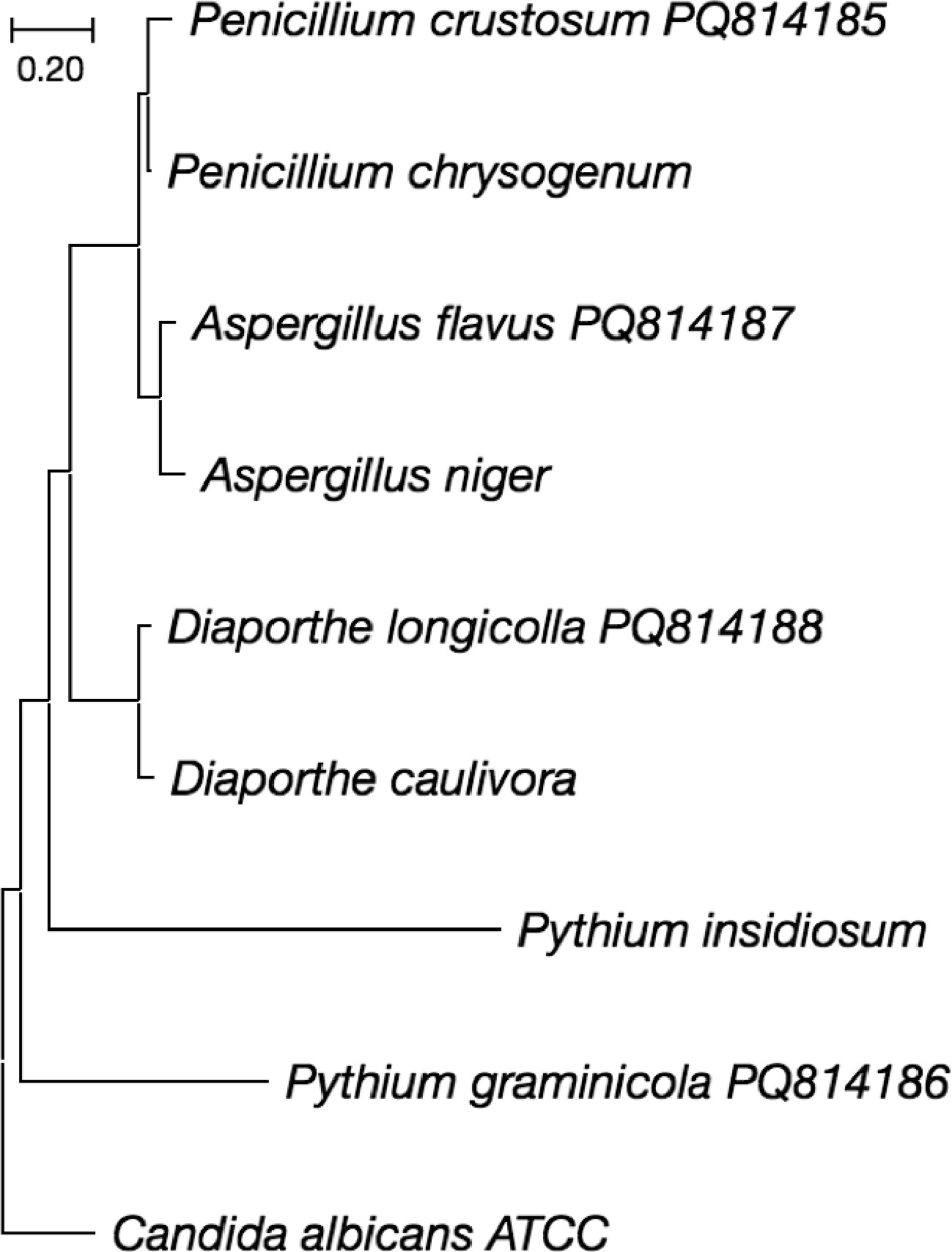
Figure 2.
Phylogenetic tree constructed based on the sequencing of the ITS1 and ITS4 region of fungal isolates using the maximum composite likelihood method and 1000 Bootstrap tests in the MEGA v.11.0 program.
EOs antimicrobial activity
-
The chemical composition of essential oils, as determined by gas chromatography, is detailed in Boniek et al.[26]. The antifungal activity of EOs is attributed to their bioactive compounds, including monoterpenes, sesquiterpenes, phenols, aldehydes, and ketones, which exert synergistic, additive, or complementary effects[29].
Following incubation, inhibition zones formed around EO-impregnated discs, indicating fungal susceptibility. The diameter of these zones qualitatively reflected the sensitivity of the fungal strains to the tested EOs. At the inhibition zone limit, the antifungal concentration in the agar corresponds to the MIC. A logarithmic relationship exists between inhibition zone diameter and MIC in diffusion-based assays.
As presented in Table 3, the MIC values for M. alternifolia, C. longa, and T. vulgaris EOs were 22.5, 45.0, and 90.0 μg·ml−1, respectively. Among the tested fungal species, A. flavus and P. crustosum exhibited the lowest MIC values, indicating greater susceptibility to the antifungal compounds present in these natural extracts (Table 3).
-
Institutions related to cultural heritage, such as cathedrals, museums, and technical reserves are advised to ensure ideal temperature and relative humidity conditions to ensure that collections composed of organic materials are protected against the negative effects of microbial biodeterioration[30,31]. However, in most current cases, even when maintaining relative humidity below 60%, collections are not free from microbial contamination and the proliferation of filamentous fungi, such as A. flavus[32] and P. crustosum[33]. Thus, the proposal of less toxic and more efficient microbial growth control measures against microorganisms that deteriorate artworks becomes imminent[34].
Pythium spp., a prominent genus within the family Pythiaceae (order Pythiales, class Oomycetes), comprises over 300 globally distributed species. These species exhibit diverse ecological roles, functioning as saprophytes, mycoparasites, and pathogens of both plants and animals[35].
Species of Diaporthe are widely distributed and exhibit diverse ecological roles, functioning as plant pathogens, endophytes, or saprobes[36]. They are responsible for diseases affecting a broad range of plant hosts, including economically significant crops, causing root and fruit rots, dieback, cankers, leaf spots, blights, decay, and wilt[37].
Furthermore, the occurrence of the species D. longicolla in a pigmented area (observe side), from sample 12, may also indicate tropism for the composition of the pigments used in the sampled area. Detailed analyses of the composition of the painting's pictorial layer, as well as tests of affinity of the fungal species with the identified pigment (data not shown), could elucidate the hypothesis raised with greater accuracy.
The occurrence of both species with phytopathogenic potential can be justified by the stowage of the artwork, whose surroundings had wooded environments and natural ventilation in the building where it was located, in addition to the lack of monitoring of relative air humidity and internal temperature. Thus, the colonization of these fungi on the painting becomes imminent and inevitable, due to the organic contribution as a natural resource for fungal conidia.
The remaining points sampled did not show cultivable fungal growth. The stains observed with the naked eye on the paint at these sampled points may be due to a late and undetermined microbial colonization in the temporal scale, and which, in principle, does not present risks to the integrity of the artwork or to human health.
Furthermore, this possible late microbial colonization could be confirmed by using total genetic material detection tools, including the non-cultivable or metabolically inactive microbial population[38]. However, this method will only reveal the identity of the microorganism, with little information about the metabolic physiology of this filamentous fungus.
Regarding fungi with phytopathogenic potential, the MIC values obtained for D. longicolla and P. graminicola were higher, indicating a resistance mechanism to the chemical compounds present in the EOs tested. However, Krupalini et al.[39] demonstrated, in vitro, the positive antifungal effects of five EOs: clove, lemon grass, citronella, peppermint, and tea tree oils on Diaporthe phaseolorum.
Previous studies conducted in Brazil have demonstrated the effectiveness of essential oils from Corymbia citriodora, Mentha arvensis, and Mentha spicata in inhibiting the mycelial growth of Pythium spp.[40]. These findings suggest that EOs have the potential to be utilized in the development of biofumigants for soil treatment, either for seedling production or application in agricultural fields.
Essential oils and their key chemical constituents exert antifungal activity through multiple cellular mechanisms, as described in previous studies[41]. One of the most widely accepted hypotheses suggests that EOs interact with ergosterol, a crucial component of the fungal cell membrane, compromising its integrity[26]. This disruption leads to plasma membrane destabilization and the leakage of essential cellular ions, including K+, Ca2+, and Mg2+[42]. Additionally, EOs can interfere with nucleic acids and gene expression, disrupting fungal development and metabolic pathways by altering transcriptional and translational processes[43].
In conclusion, despite the relatively low CFU log10 count values and the detection of cultivable filamentous fungi at only four sampling points, antimicrobial assays demonstrated the effectiveness of certain in vitro fungal control methods. The use of EO vapor from M. alternifolia, T. vulgaris, and C. longa showed potential as an alternative approach for inhibiting the growth of A. flavus and P. crustosum. Based on the MIC values obtained, alternative control strategies may be required for D. longicolla and P. graminicola, including the application of an anoxic atmosphere[44,45] or the use of other essential oils not evaluated in this study.
Regardless of the microbial growth control method used, pre-testing is essential to ensure it does not negatively impact the Madona oil painting. Therefore, clean and natural alternatives can be employed to eliminate fungal populations, aiming to slow the biological deterioration process and preserve the artwork for decades.
-
The authors confirm contribution to the paper as follows: conceptualization: Boniek D, Bonadio L, Batista dos Santos AF, de Resende Stoianoff MA; methodology, validation, writing-original draft preparation: Boniek D; formal analysis: Boniek D, de Resende Stoianoff MA; writing-review and editing: Boniek D, Batista dos Santos AF, de Resende Stoianoff MA. All authors have read and agreed to the published version of the manuscript.
-
The data generated and analyzed during this study are available in this article. DNA sequence data are available in the GenBank database, and the accession numbers are provided in Table 1.
-
The authors sincerely thank Miguel Rosselini, the private owner of Madona, for granting permission to conduct scientific bioprospecting on the artwork and for authorizing the publication of its images.
-
The authors declare that they have no conflict of interest.
- Copyright: © 2025 by the author(s). Published by Maximum Academic Press, Fayetteville, GA. This article is an open access article distributed under Creative Commons Attribution License (CC BY 4.0), visit https://creativecommons.org/licenses/by/4.0/.
-
About this article
Cite this article
Boniek D, Bonadio L, Batista dos Santos AF, de Resende Stoianoff MA. 2025. Prospecting for populations of filamentous fungi in a Brazilian oil painting and susceptibility to natural antifungals. Studies in Fungi 10: e003 doi: 10.48130/sif-0025-0004 

Prospecting for populations of filamentous fungi in a Brazilian oil painting and susceptibility to natural antifungals
- Received: 31 December 2024
- Revised: 20 February 2025
- Accepted: 21 February 2025
- Published online: 21 March 2025
Abstract: The preservation of contemporary artwork is a growing problem in conservation due to their susceptibility to biodeterioration. Fungal colonization regularly targets these cultural assets, which can have great artistic and historical significance. Filamentous fungi implicated in the deterioration of oil paintings have a strong enzymatic degradation capacity, targeting components, such as vegetable oils and resin. This process is enhanced by interactions with pigments and chemicals, which can act as nutrient sources for fungi. Traditional antifungal treatments have been extensively explored as a technique to counteract biodeterioration; however, their efficiency is frequently restricted by their toxicity to conservators, environmental impact, and the ability to create chemical changes in the materials of the artworks. Natural antifungal compounds, specifically essential oils (EOs) extracted from Melaleuca alternifolia, Curcuma longa, and Thymus vulgaris were assessed for their antifungal efficacy against metabolically active fungal isolates. Furthermore, in vitro assays demonstrated that the interaction between these fungal strains and the tested EOs resulted in significant growth inhibition, indicating their potential as effective antifungal agents, and all EOs tested showed low minimum inhibitory concentration (MIC) values for Penicillium crustosum and Aspergillus flavus. On the other hand, Pythium graminicola and Diaporthe longicolla showed higher MIC values for all EOs tested. Fungal identification was conducted through the examination of both macroscopic and microscopic morphological characteristics as well as the Internal Transcribed Spacer (ITS) region of the rDNA. This study highlights the efficacy of three essential oils as a sustainable alternative to conventional antifungal agents, which are often associated with toxicity in artwork preservation.
-
Key words:
- Eukaryotic microorganism /
- Deterioration /
- Artwork /
- Heritage /
- Natural treatment