-
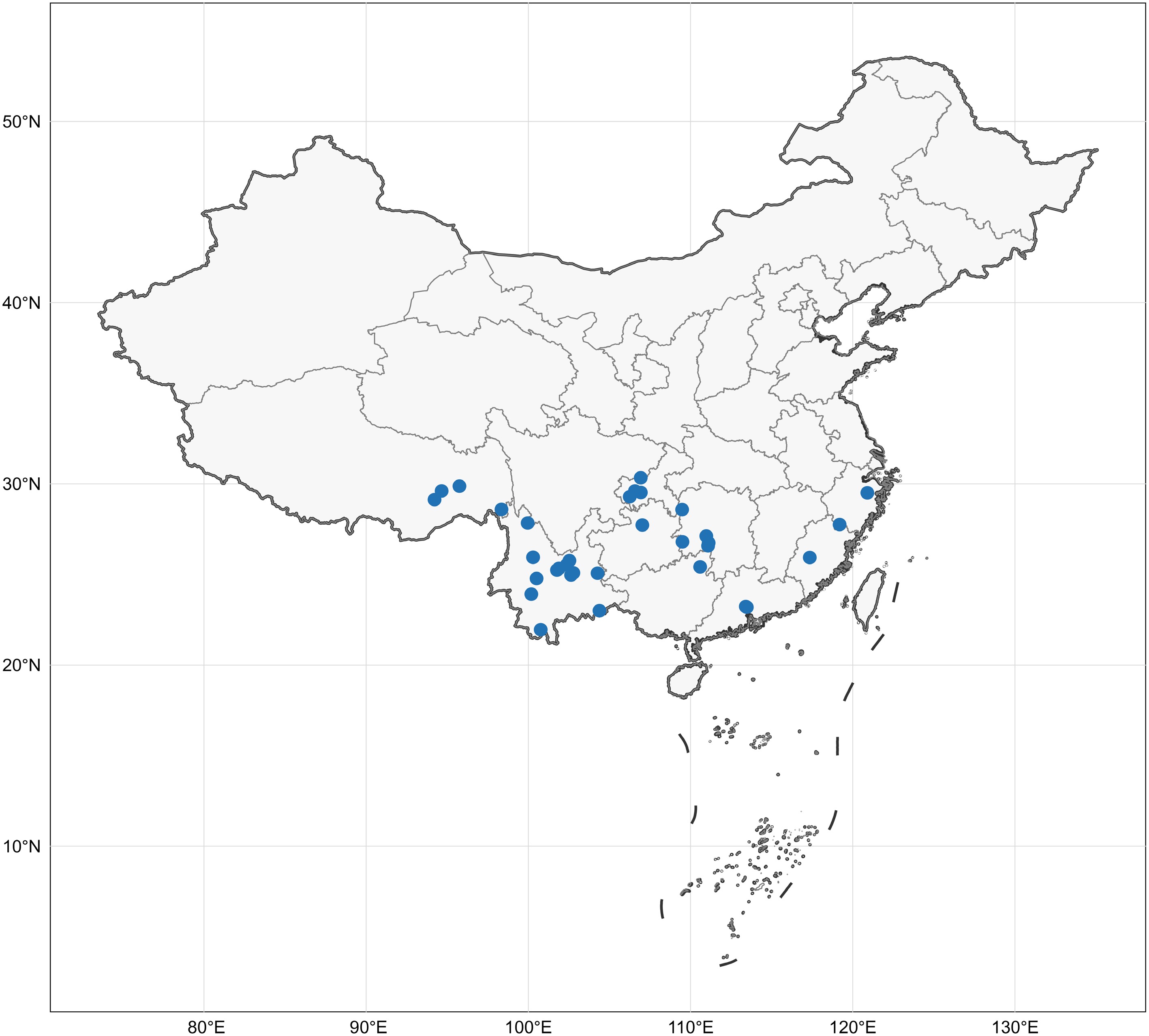
Figure 1.
The collection localities of all specimens in this study. Map source: Standard map approved by the Ministry of Natural Resources of China, Approval No. GS(2024)0650.
-
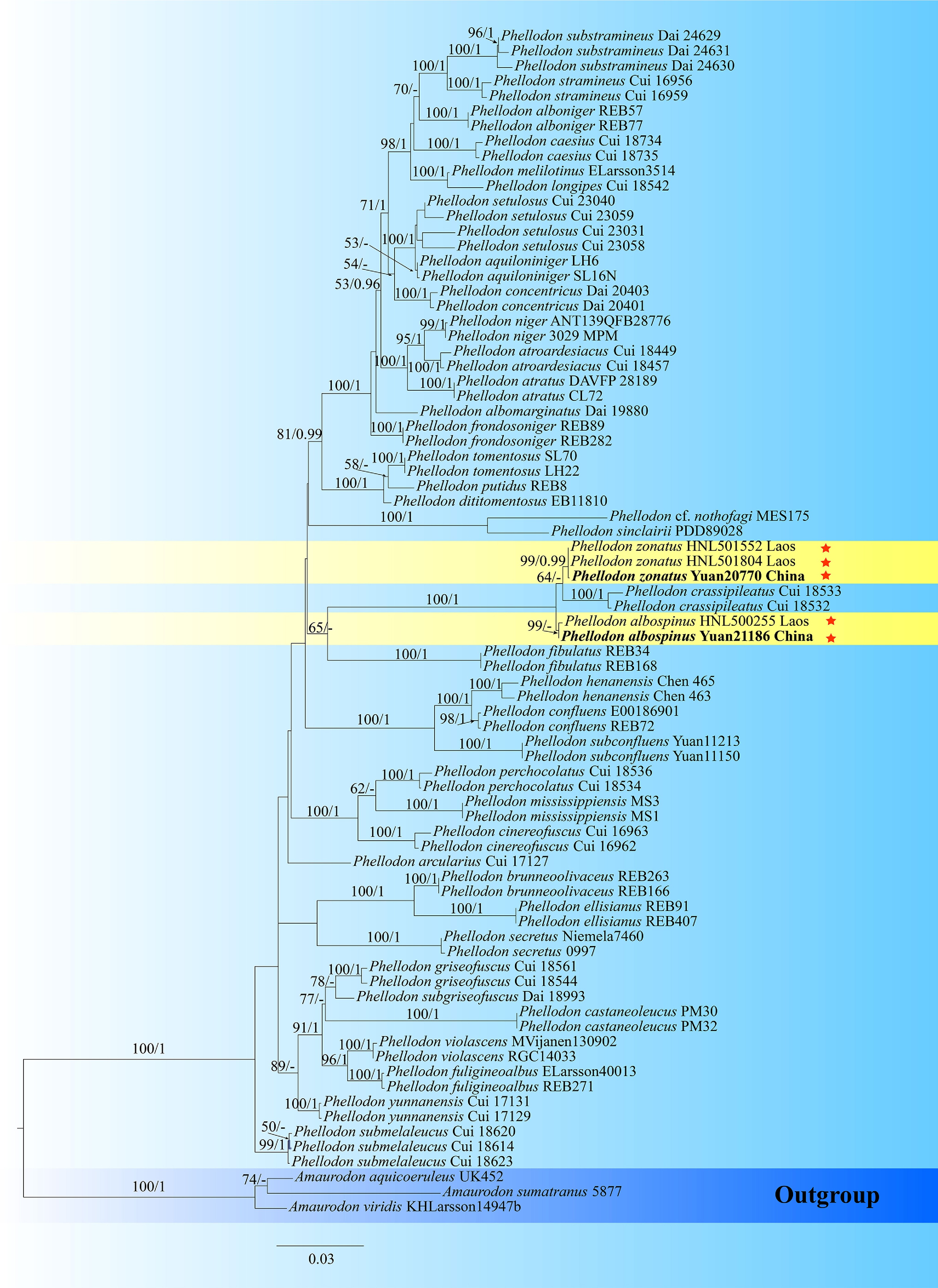
Figure 2.
Maximum likelihood tree illustrating the phylogeny of Phellodon and related genera in Thelephorales based on ITS + nLSU + nSSU sequences. Branches are labeled with maximum likelihood bootstrap values higher than 50%, and Bayesian posterior probabilities more than 0.95 respectively. Specimens examined are in bold, and new species are marked with red stars.
-
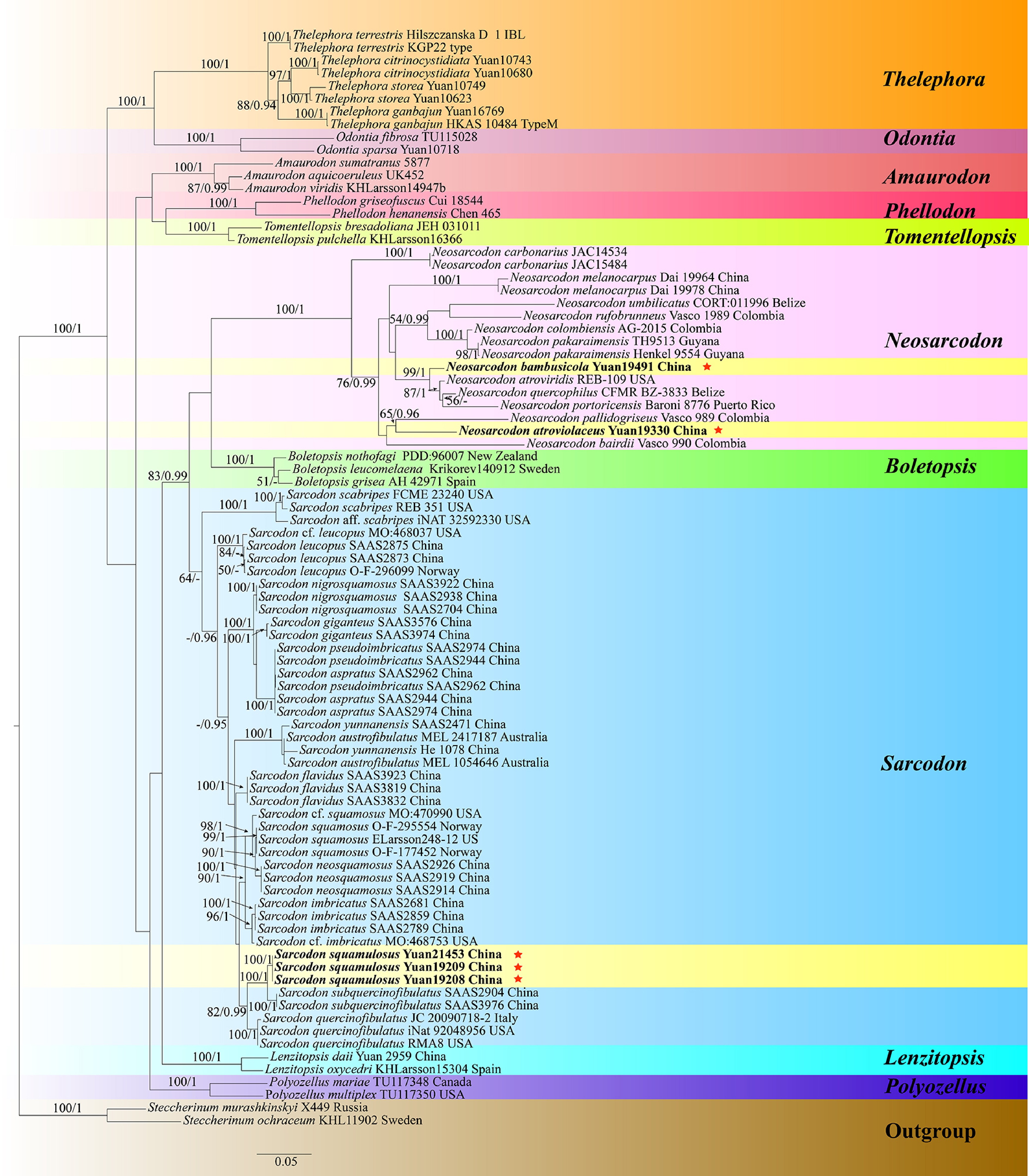
Figure 4.
Maximum likelihood tree illustrating the phylogeny of Neosarcodon, Sarcodon, and related genera in Thelephorales based on ITS + nLSU + nSSU sequences. Branches are labeled with maximum likelihood bootstrap values higher than 50%, and Bayesian posterior probabilities more than 0.95 respectively. Specimens examined are in bold, and new species are marked with red stars.
-
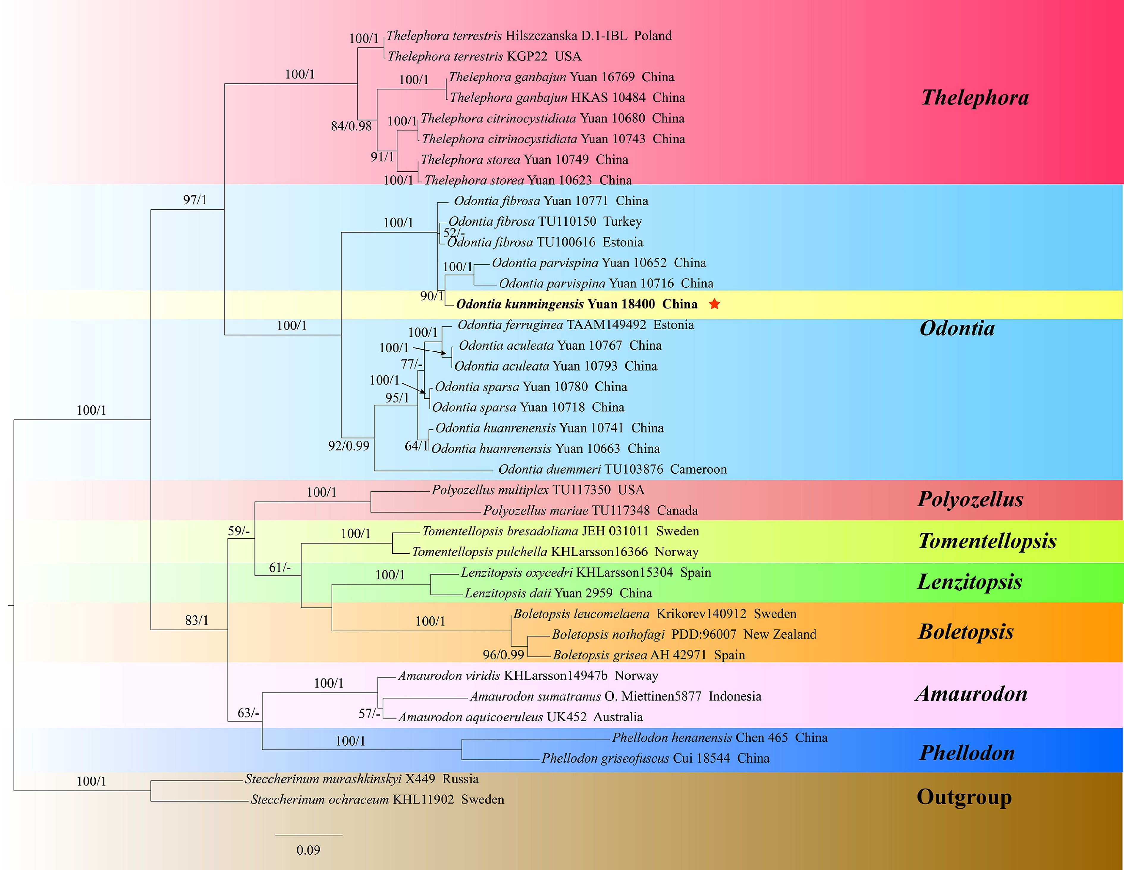
Figure 5.
Maximum likelihood tree illustrating the phylogeny of Odontia, and related genera in Thelephorales based on ITS + nLSU + nSSU sequences. Branches are labeled with maximum likelihood bootstrap values higher than 50%, and Bayesian posterior probabilities more than 0.95 respectively. Specimens examined are in bold, and new species are marked with red stars.
-
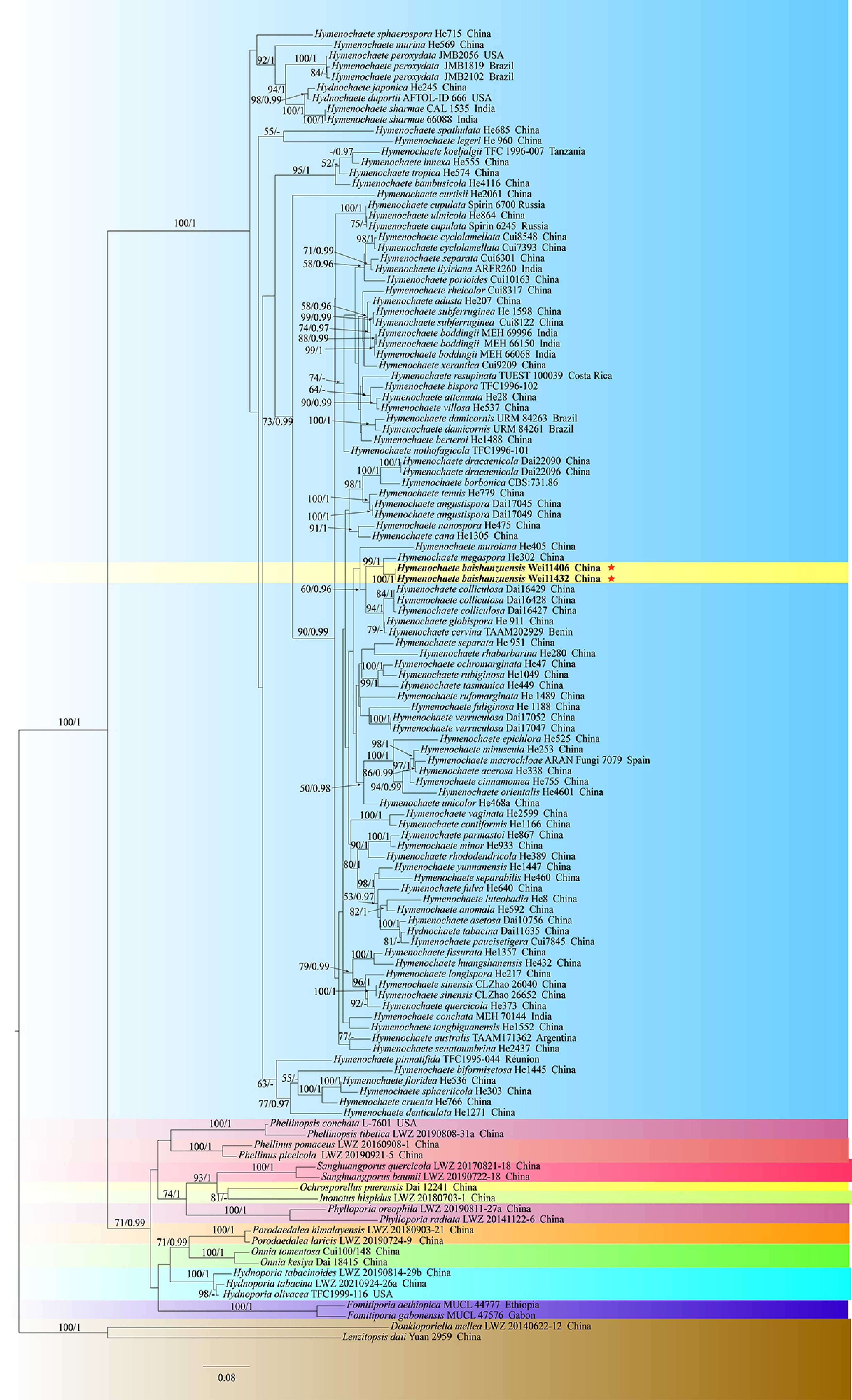
Figure 7.
Maximum likelihood tree illustrating the phylogeny of Hymenochaete, and related genera in Hymenochaetales based on ITS + nLSU sequences. Branches are labeled with maximum likelihood bootstrap values higher than 50%, and Bayesian posterior probabilities more than 0.95 respectively. Specimens examined are in bold, and new species are marked with red stars.
-

Figure 9.
Maximum likelihood tree illustrating the phylogeny of Lyomyces, and related genera in Hymenochaetales based on ITS + nLSU sequences. Branches are labeled with maximum likelihood bootstrap values higher than 50%, and Bayesian posterior probabilities more than 0.95 respectively. Specimens examined are in bold, and new species are marked with red stars.
-
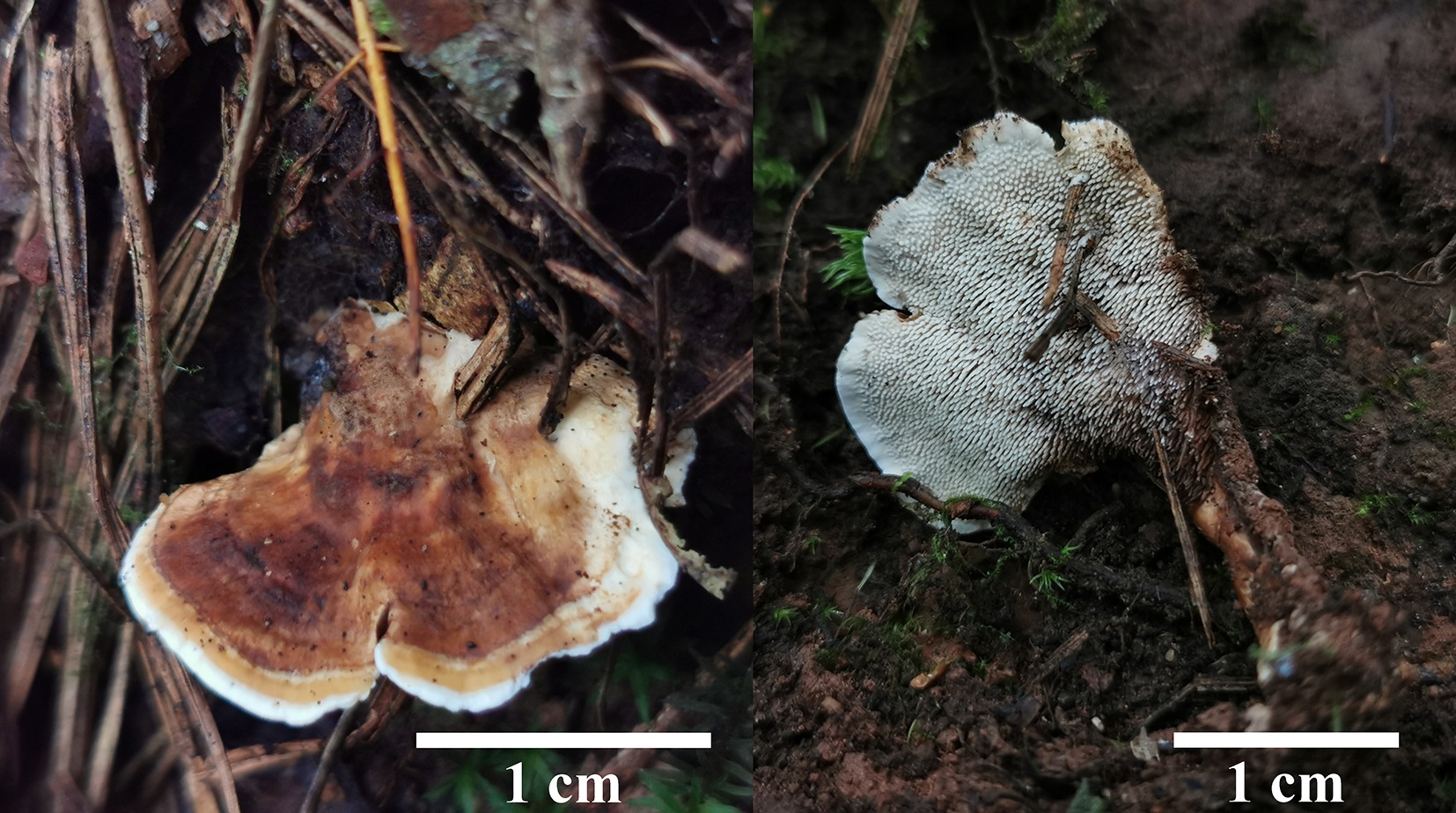
Figure 10.
Basidiomata of Phellodon albospinus (holotype IFP 020036).
-
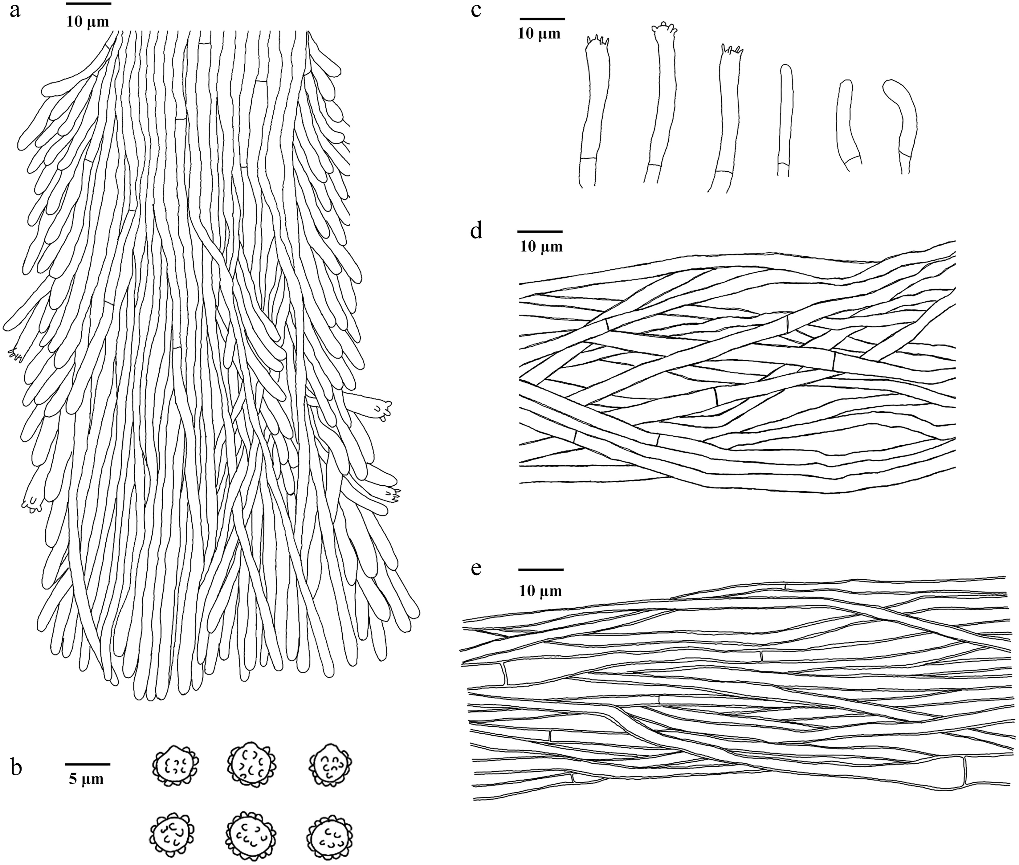
Figure 11.
Microscopic structures of Phellodon albospinus (drawn from the holotype IFP 020036). (a) Section through spines. (b) Basidiospores. (c) Basidia and basidioles. (d) Hyphae from pileus. (e) Hyphae from stipe.
-
Figure 12.
Basidiomata of Phellodon zonatus (holotype IFP 020035).
-
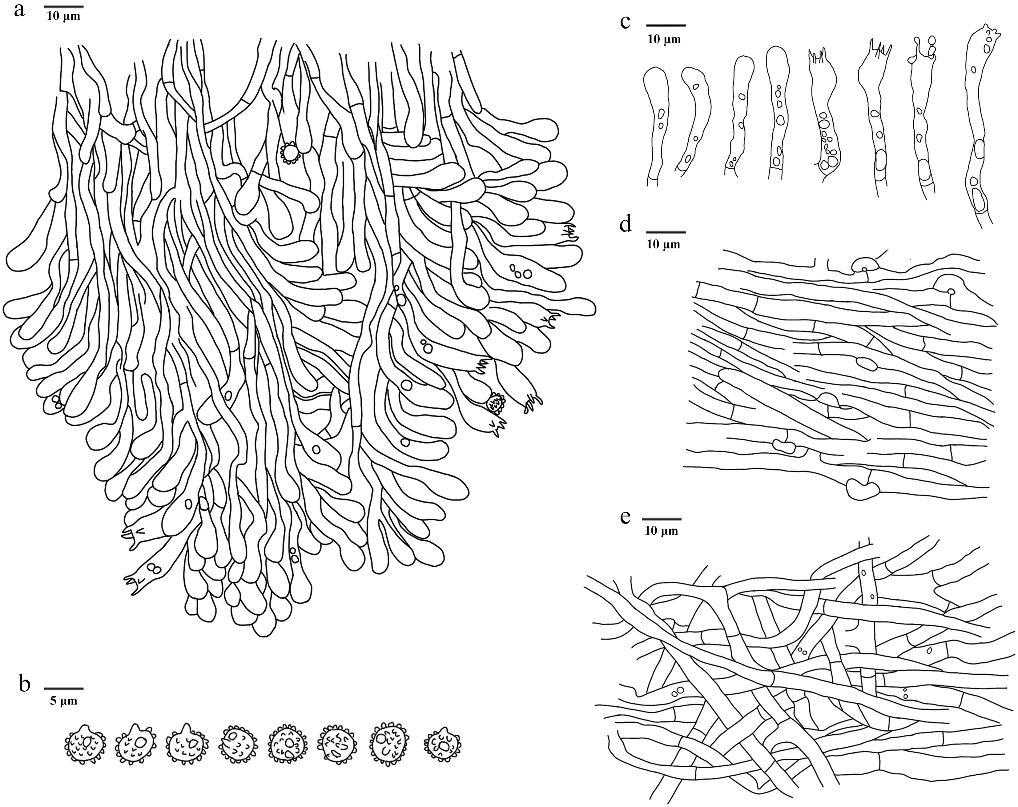
Figure 13.
Microscopic structures of Phellodon zonatus (drawn from the holotype IFP 020035). (a) Section through spines. (b) Basidiospores. (c) Basidia and basidioles. (d) Hyphae from pileus. (e) Hyphae from stipe.
-
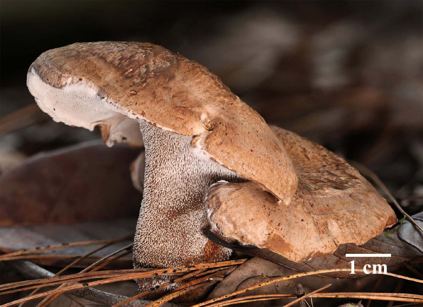
Figure 14.
Basidiomata of Hydnellum carnosum (holotype IFP 020026). Photo by Xue-Lian Gao.
-
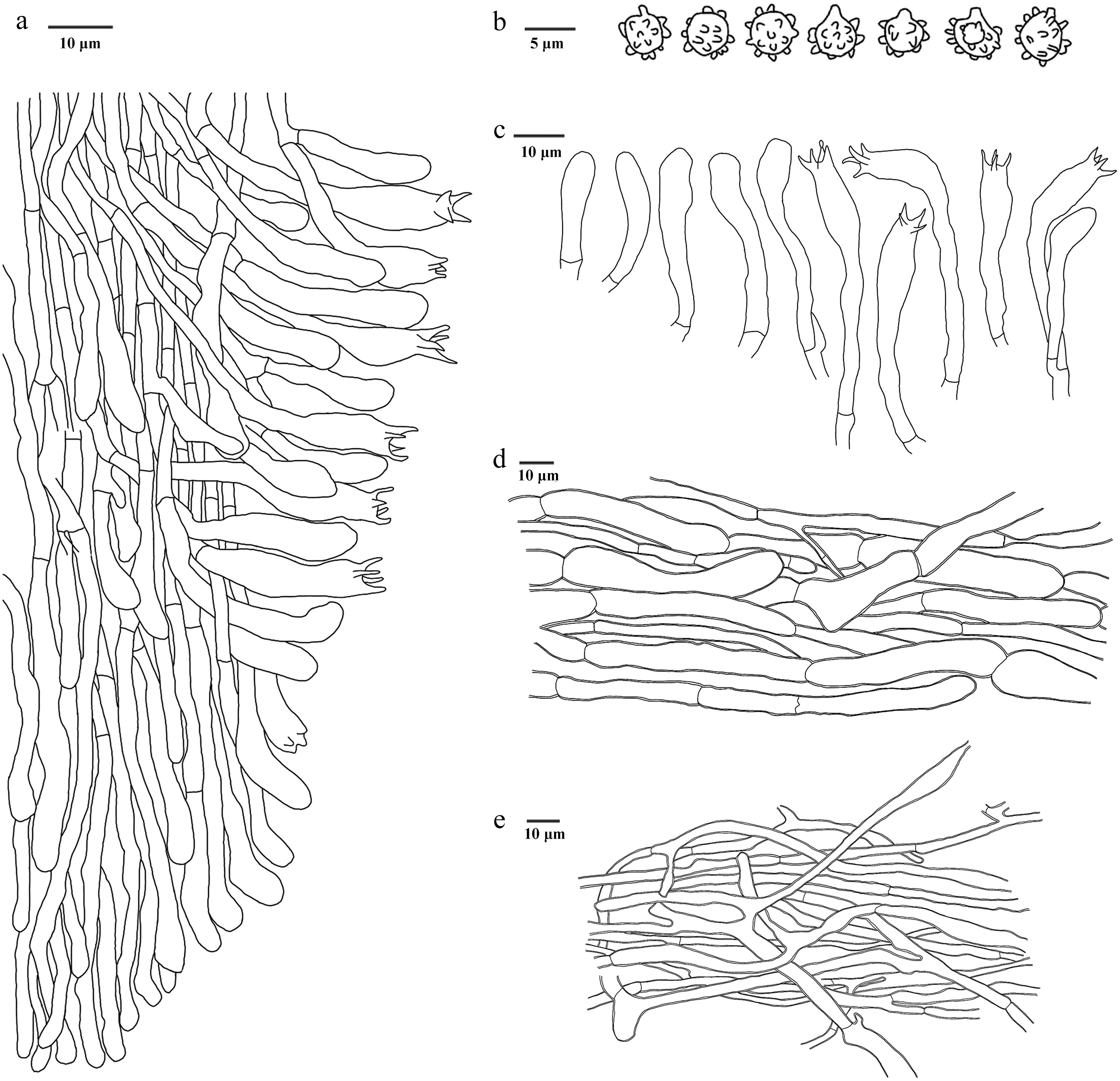
Figure 15.
Microscopic structures of Hydnellum carnosum (drawn from the holotype IFP 020026). (a) Section through spines. (b) Basidiospores. (c) Basidia and basidioles. (d) Hyphae from pileus. (e) Hyphae from stipe.
-
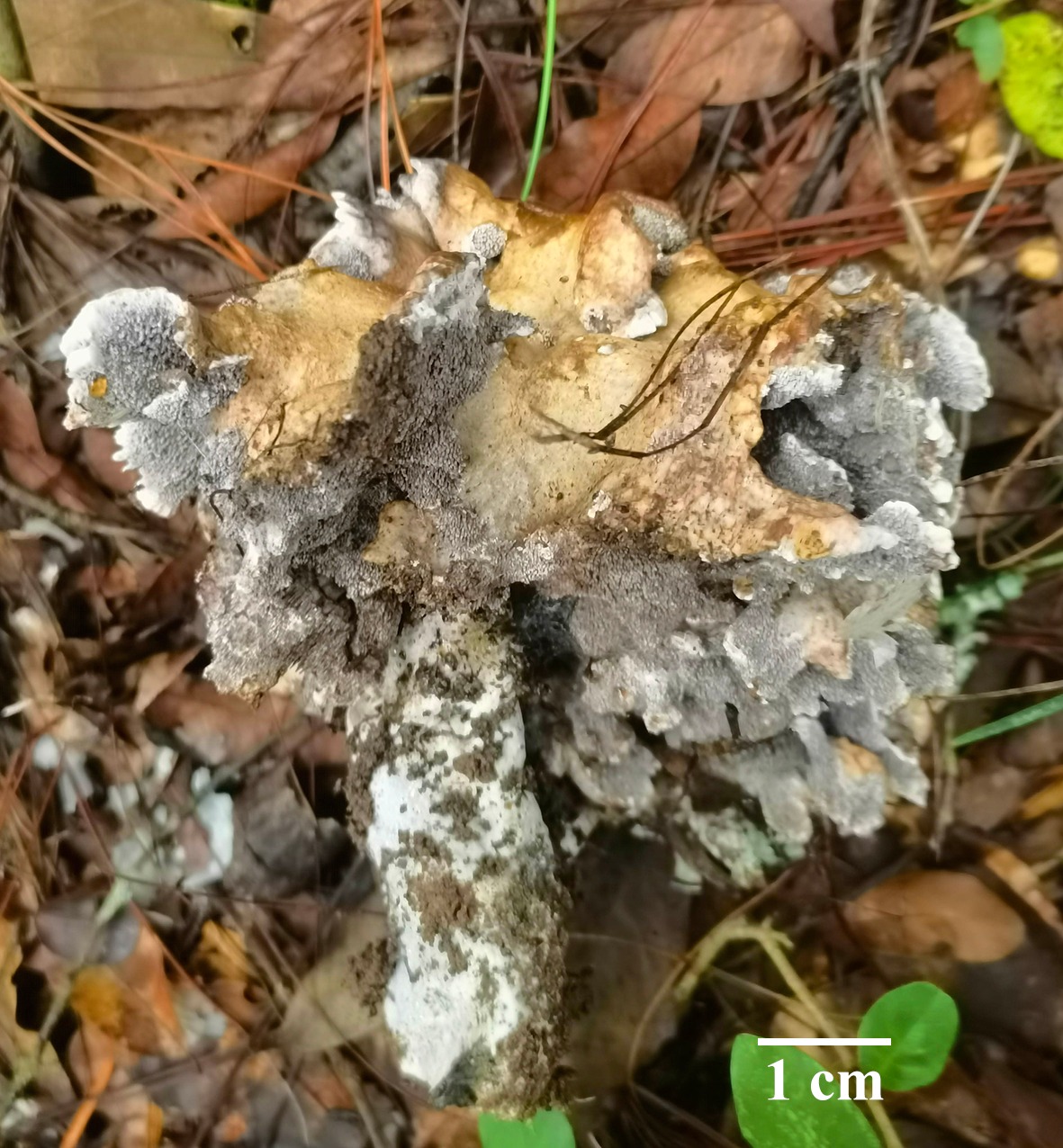
Figure 16.
Basidiomata of Hydnellum hydrangeoides (holotype IFP 020029).
-
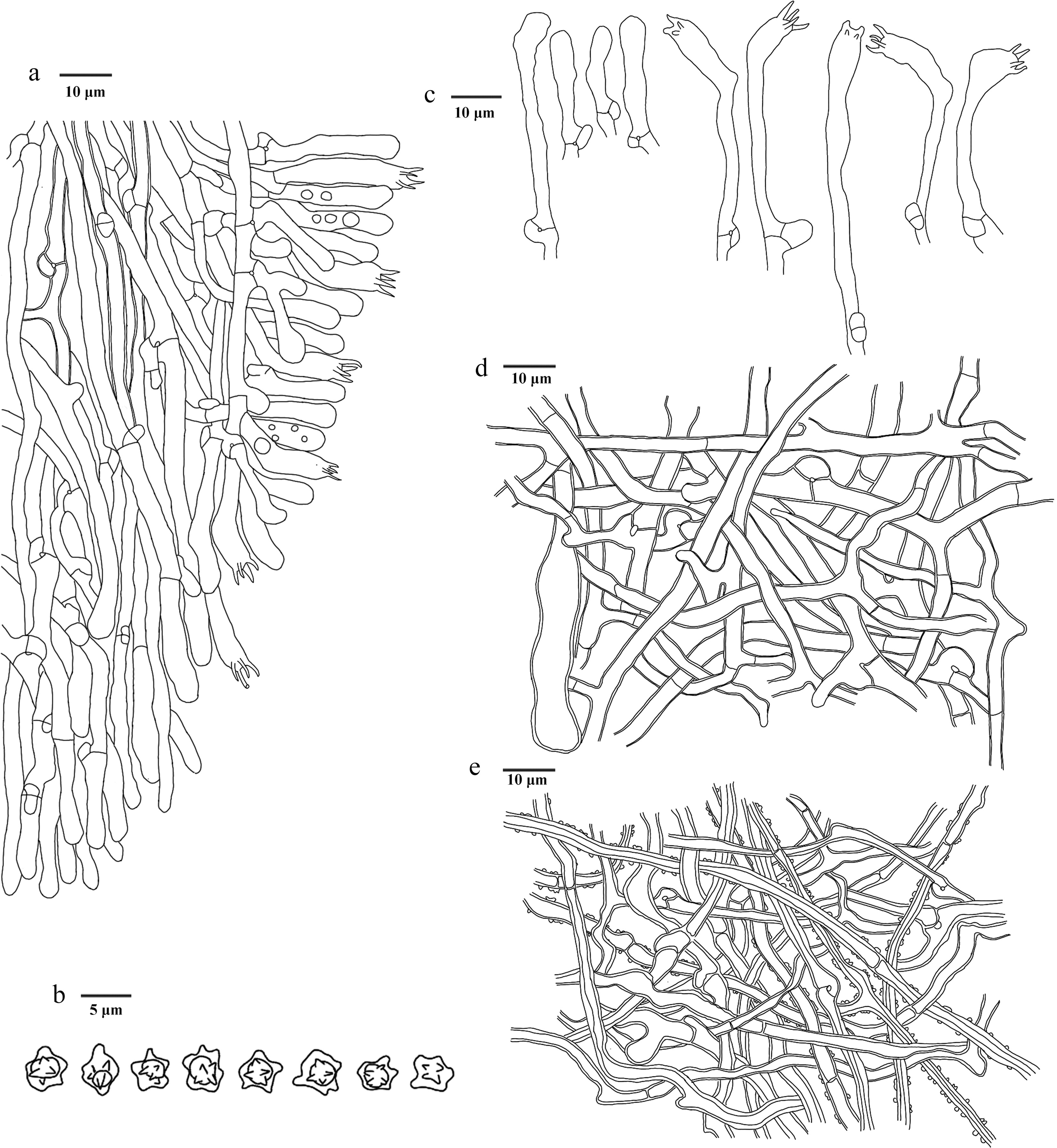
Figure 17.
Microscopic structures of Hydnellum hydrangeoides (drawn from the holotype IFP 020029). (a) Section through spines. (b) Basidiospores. (c) Basidia and basidioles. (d) Hyphae from pileus. (e) Hyphae from stipe.
-
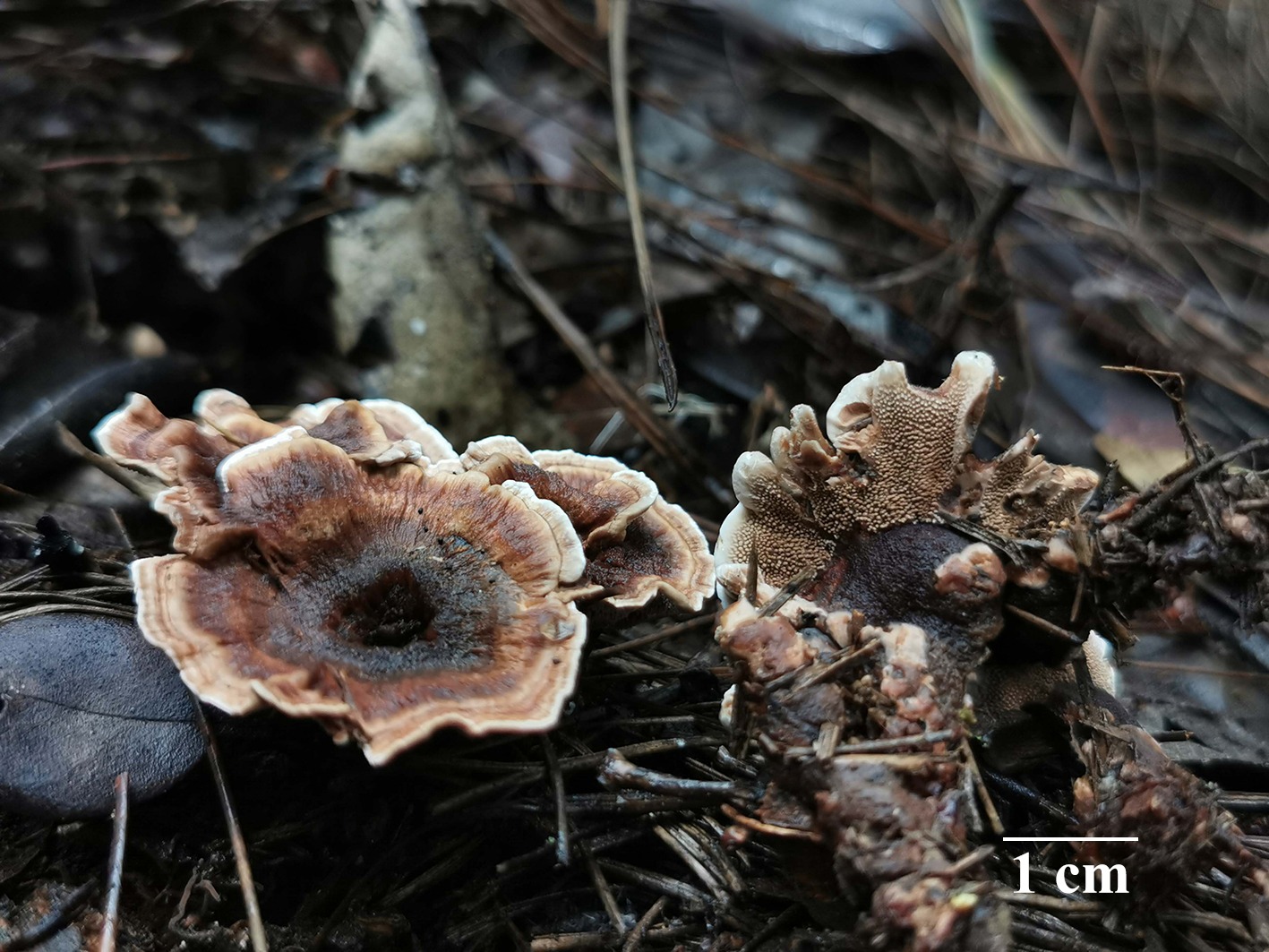
Figure 18.
Basidiomata of Hydnellum infundibuliforme (holotype IFP 020021).
-
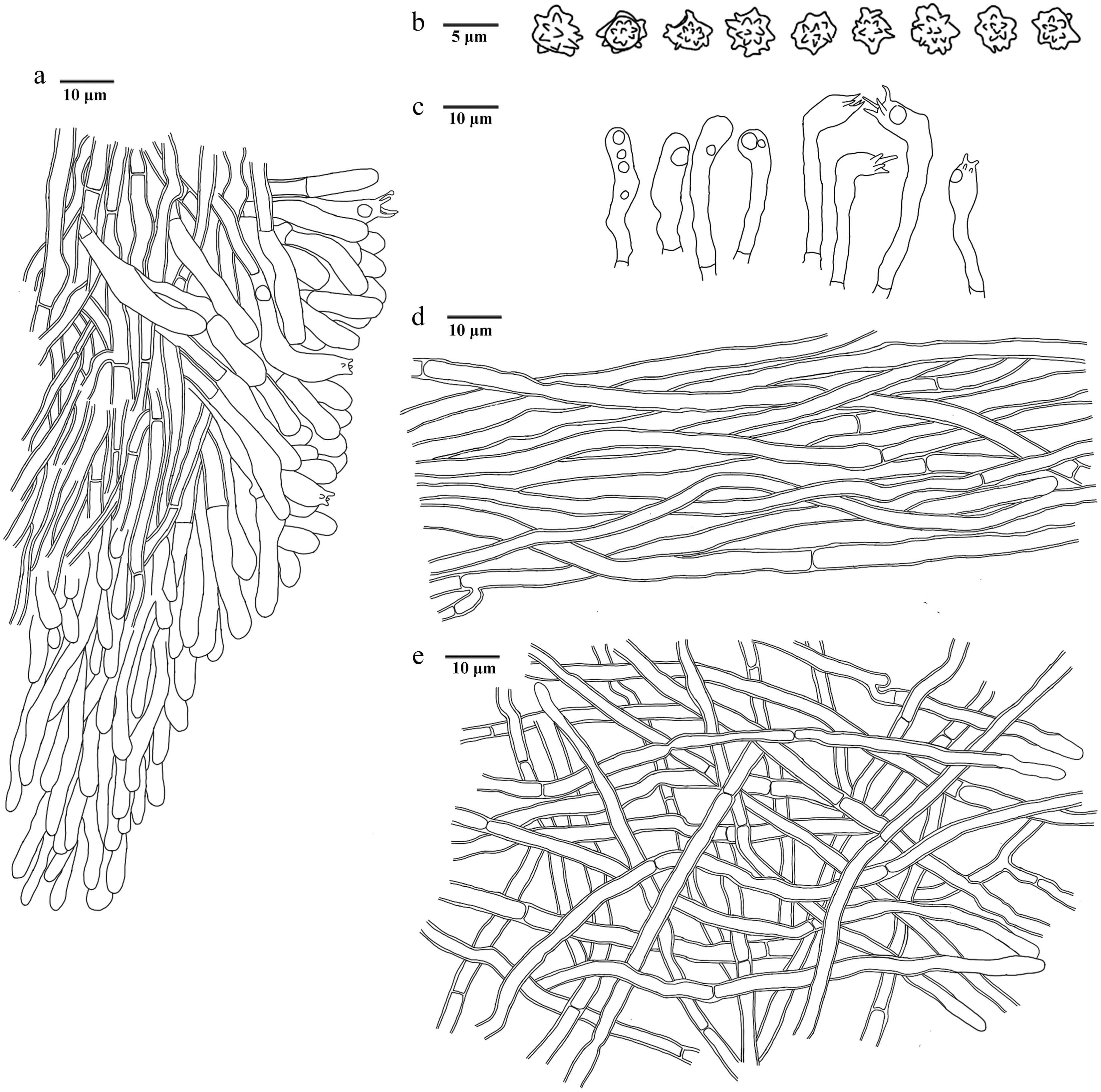
Figure 19.
Microscopic structures of Hydnellum infundibuliforme (drawn from the holotype IFP 020021). (a) Section through spines. (b) Basidiospores. (c) Basidia and basidioles. (d) Hyphae from pileus. (e) Hyphae from stipe.
-
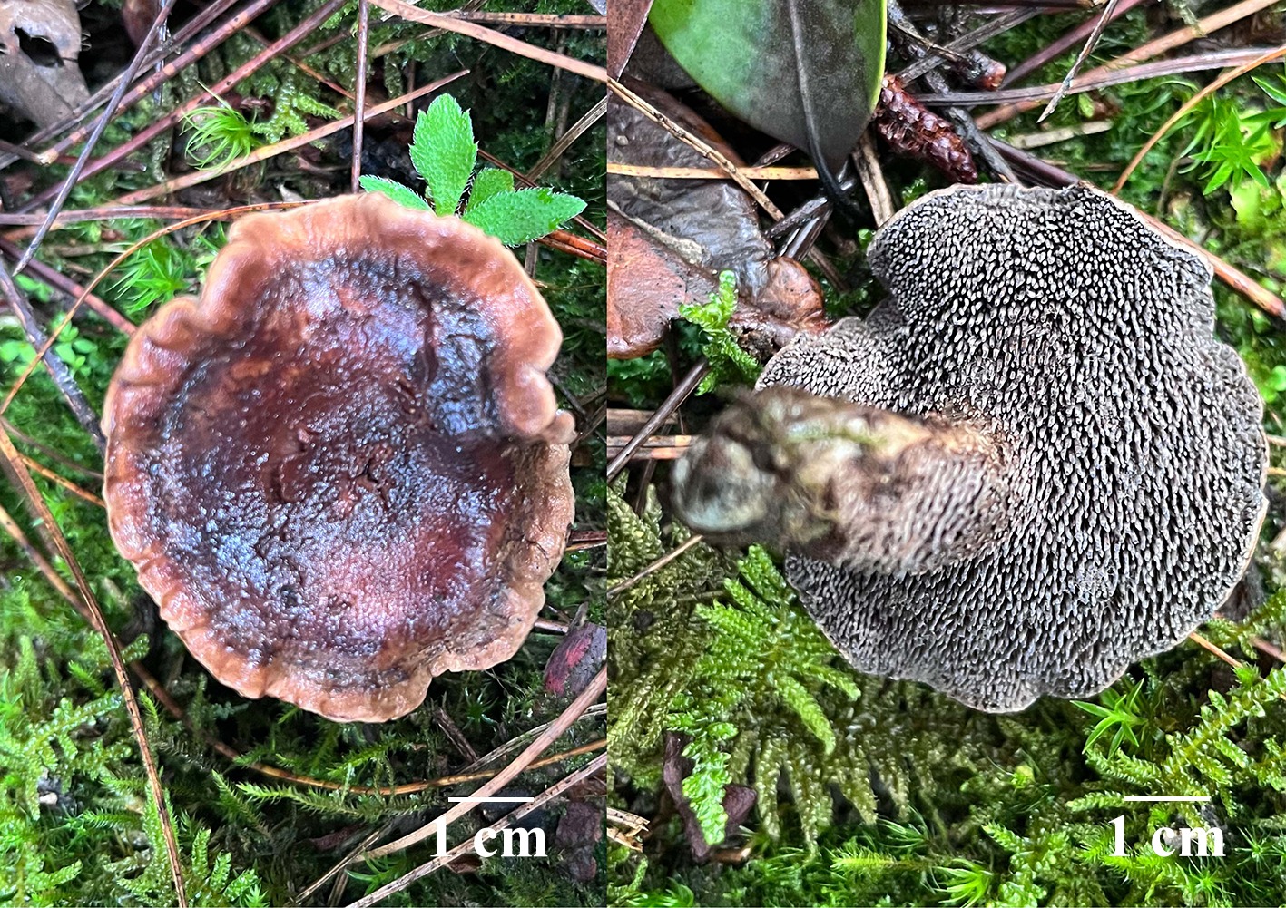
Figure 20.
Basidiomata of Hydnellum liantaishanense (holotype IFP 020024).
-
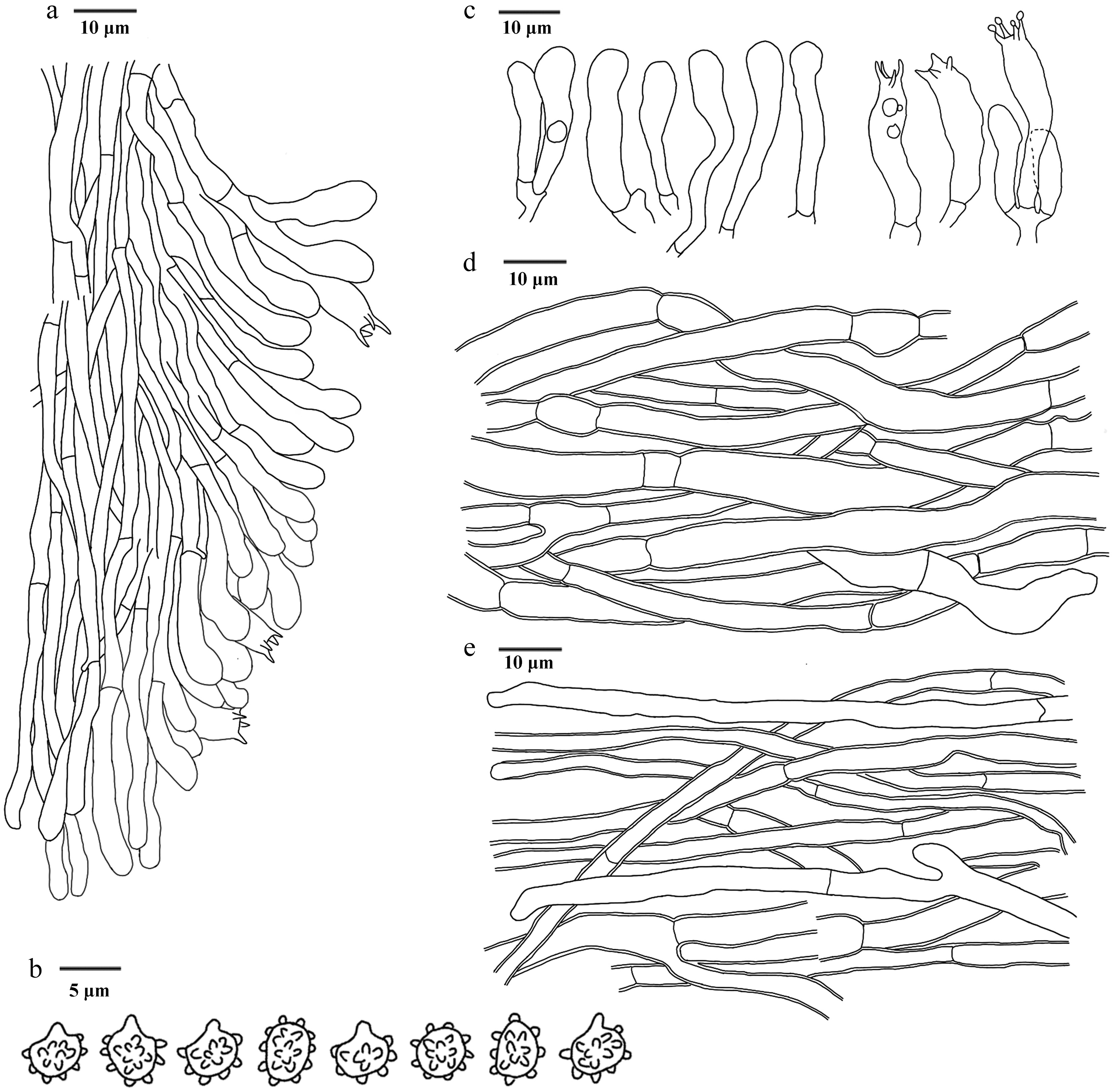
Figure 21.
Microscopic structures of Hydnellum liantaishanense (drawn from the holotype IFP 020024). (a) Section through spines. (b) Basidiospores. (c) Basidia and basidioles. (d) Hyphae from pileus. (e) Hyphae from stipe.
-
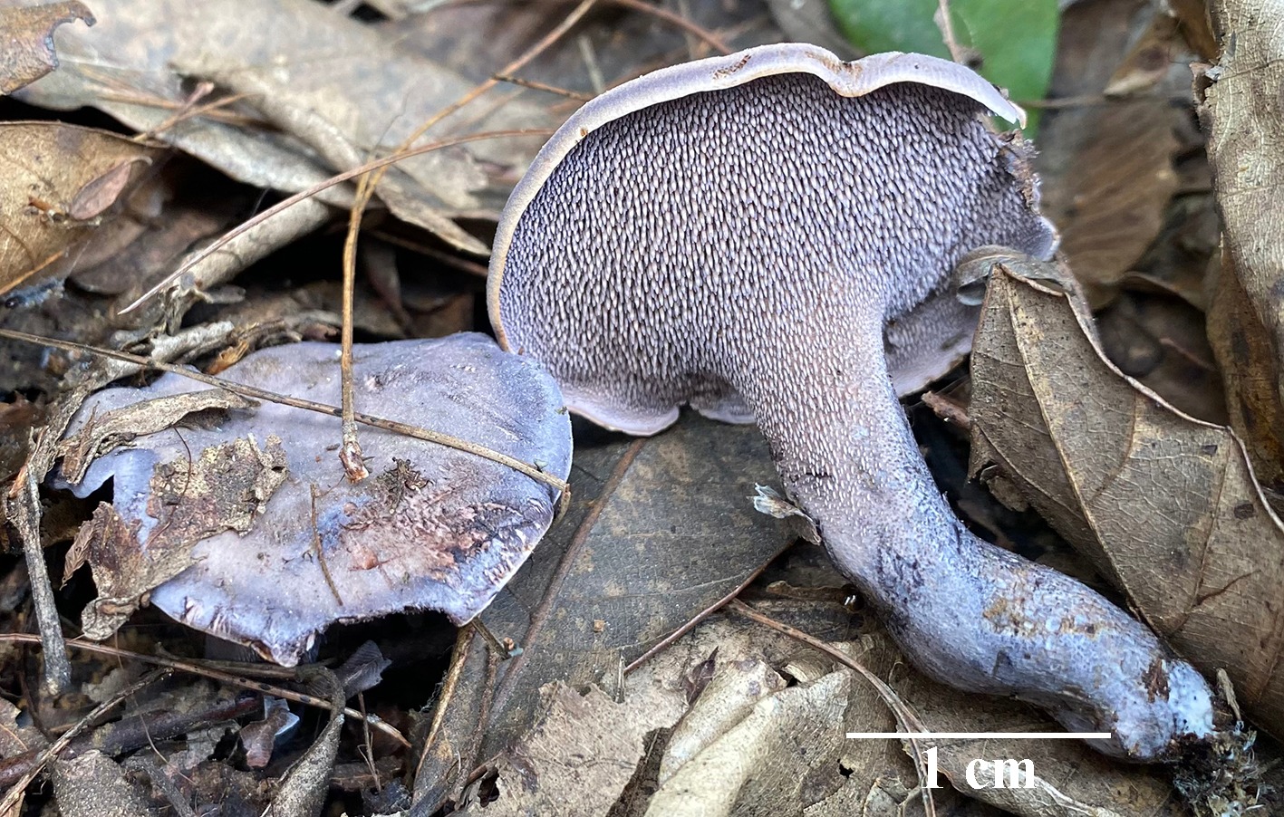
Figure 22.
Basidiomata of Hydnellum porphyreum (holotype IFP 020027). Photo by Yan-Yan He.
-
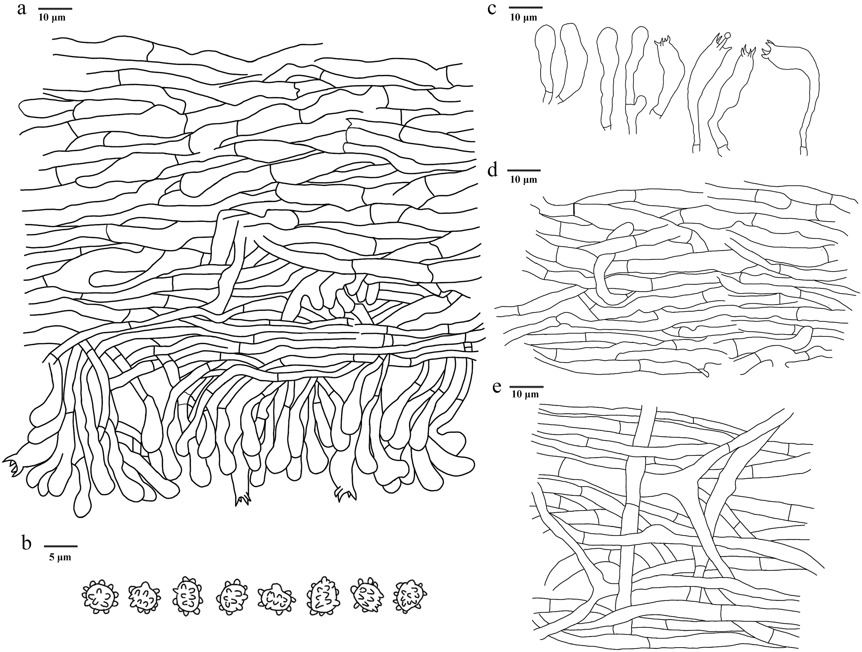
Figure 23.
Microscopic structures of Hydnellum porphyreum (drawn from the holotype IFP 020027). (a) Section through spines. (b) Basidiospores. (c) Basidia and basidioles. (d) Hyphae from pileus. (e) Hyphae from stipe.
-
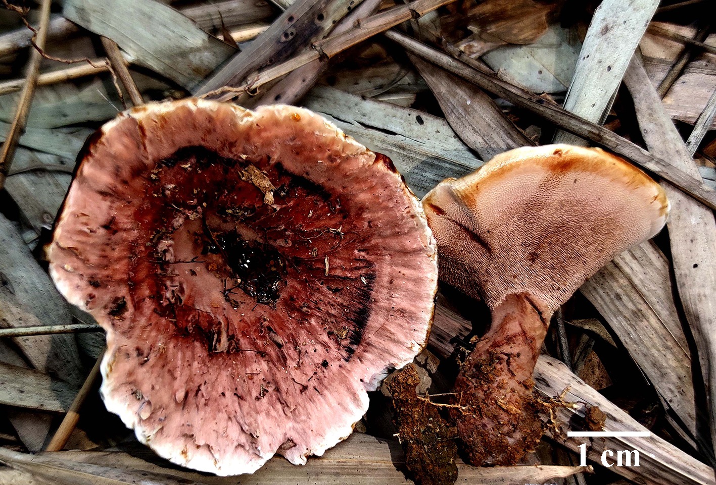
Figure 24.
Basidiomata of Hydnellum testaceum (holotype IFP 020030).
-
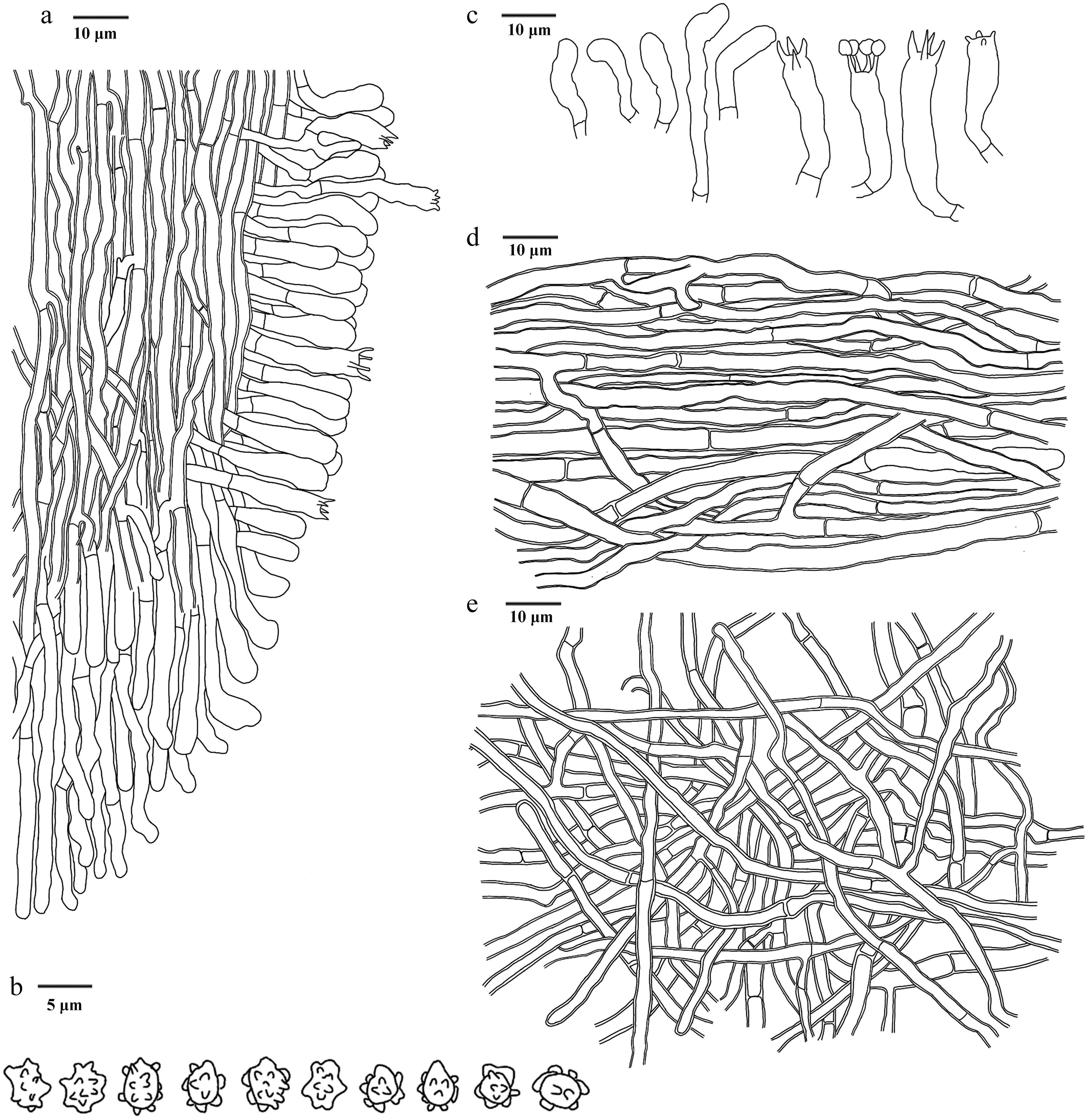
Figure 25.
Microscopic structures of Hydnellum testaceum (drawn from the holotype IFP 020030). (a) Section through spines. (b) Basidiospores. (c) Basidia and basidioles. (d) Hyphae from pileus. (e) Hyphae from stipe.
-
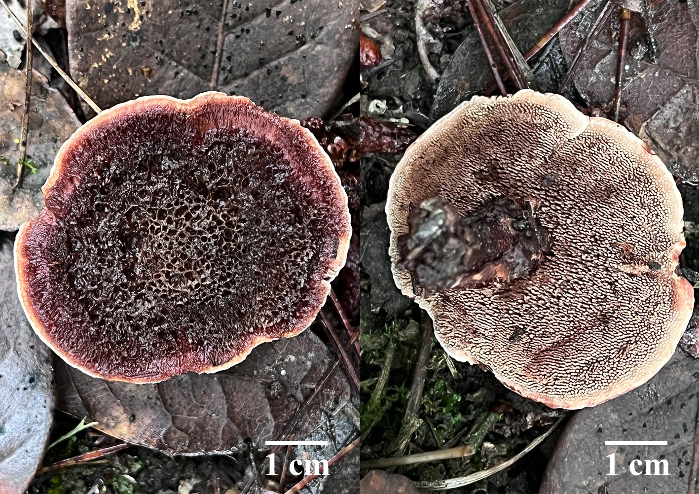
Figure 26.
Basidiomata of Hydnellum tomentosum (holotype IFP 020019).
-
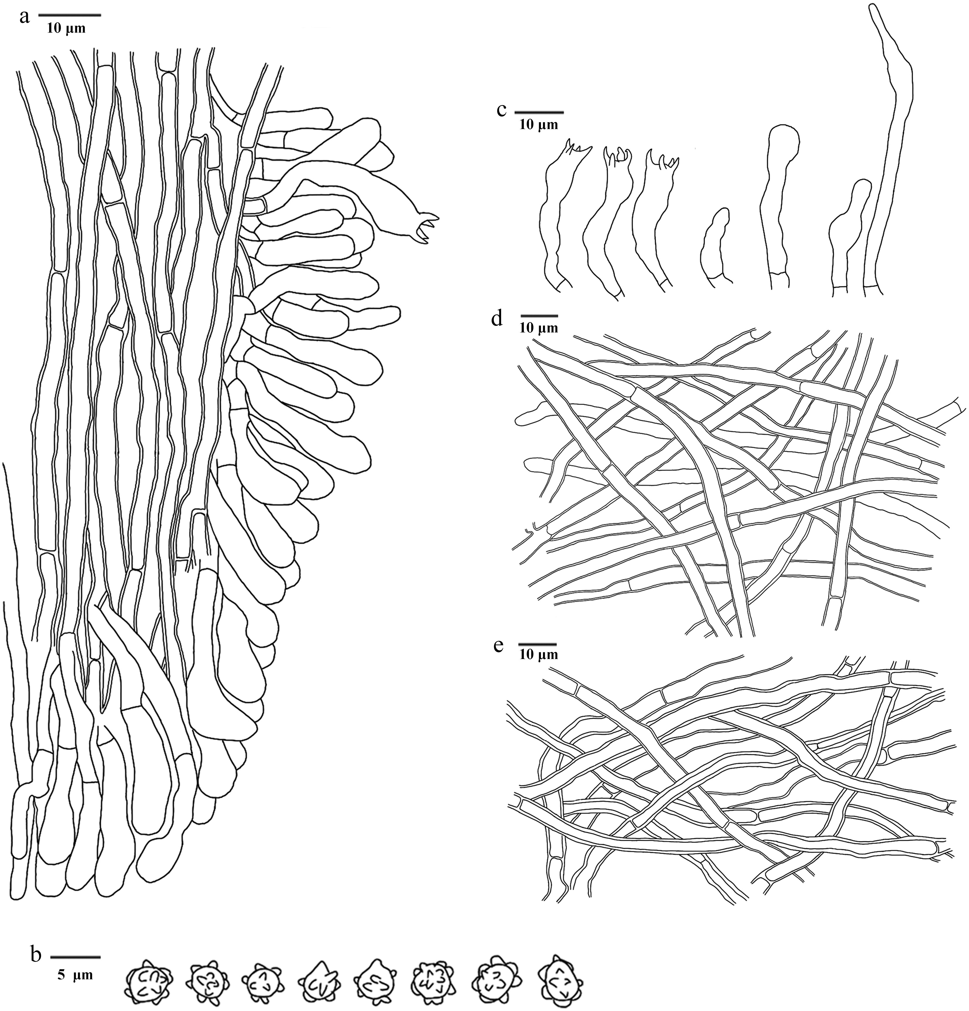
Figure 27.
Microscopic structures of Hydnellum tomentosum (drawn from the holotype IFP 020019). (a) Section through spines. (b) Basidiospores. (c) Basidia and basidioles. (d) Hyphae from pileus. (e) Hyphae from stipe.
-
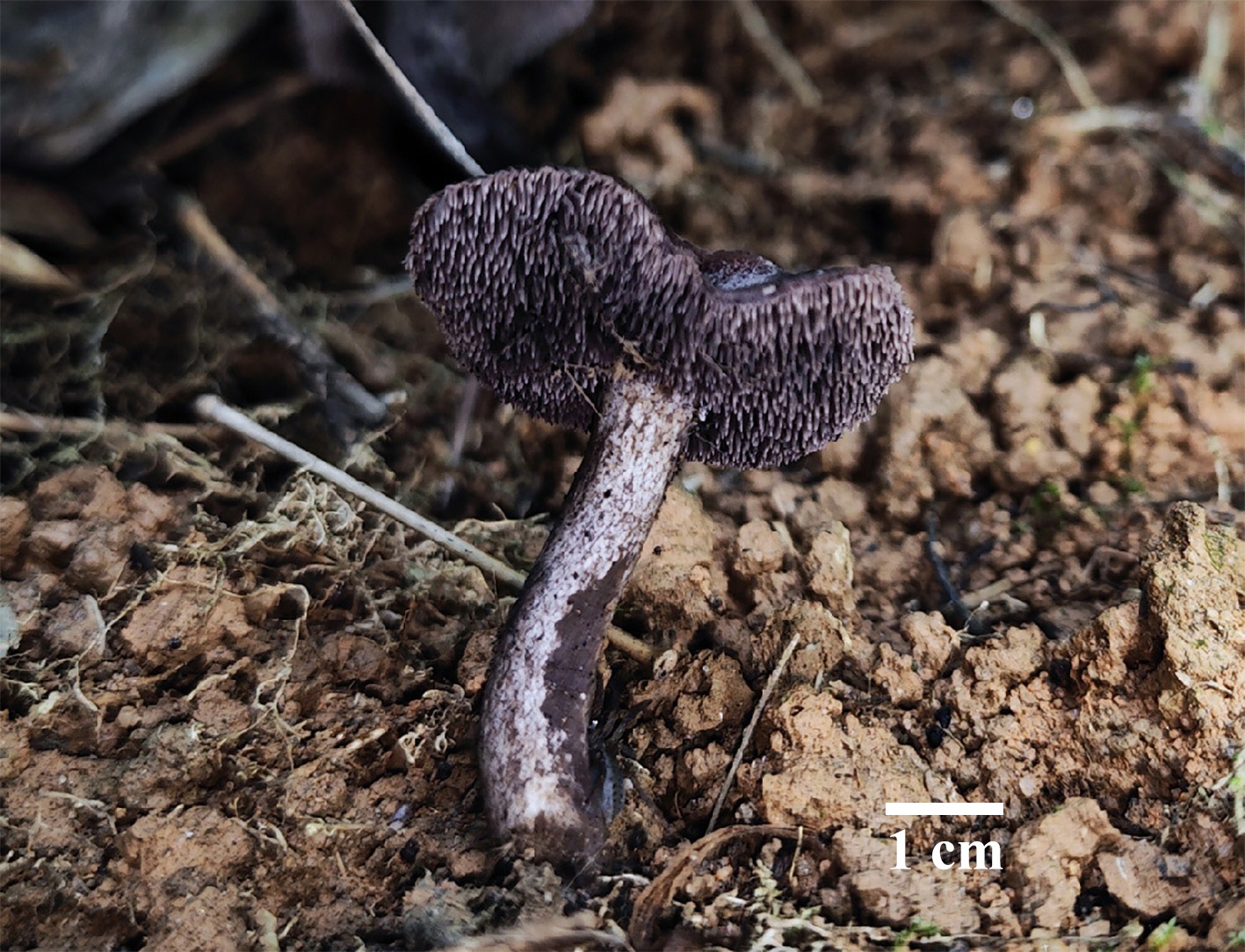
Figure 28.
Basidiomata of Neosarcodon atroviolaceus (holotype IFP 020031). Photo by Jian-Feng Tan.
-
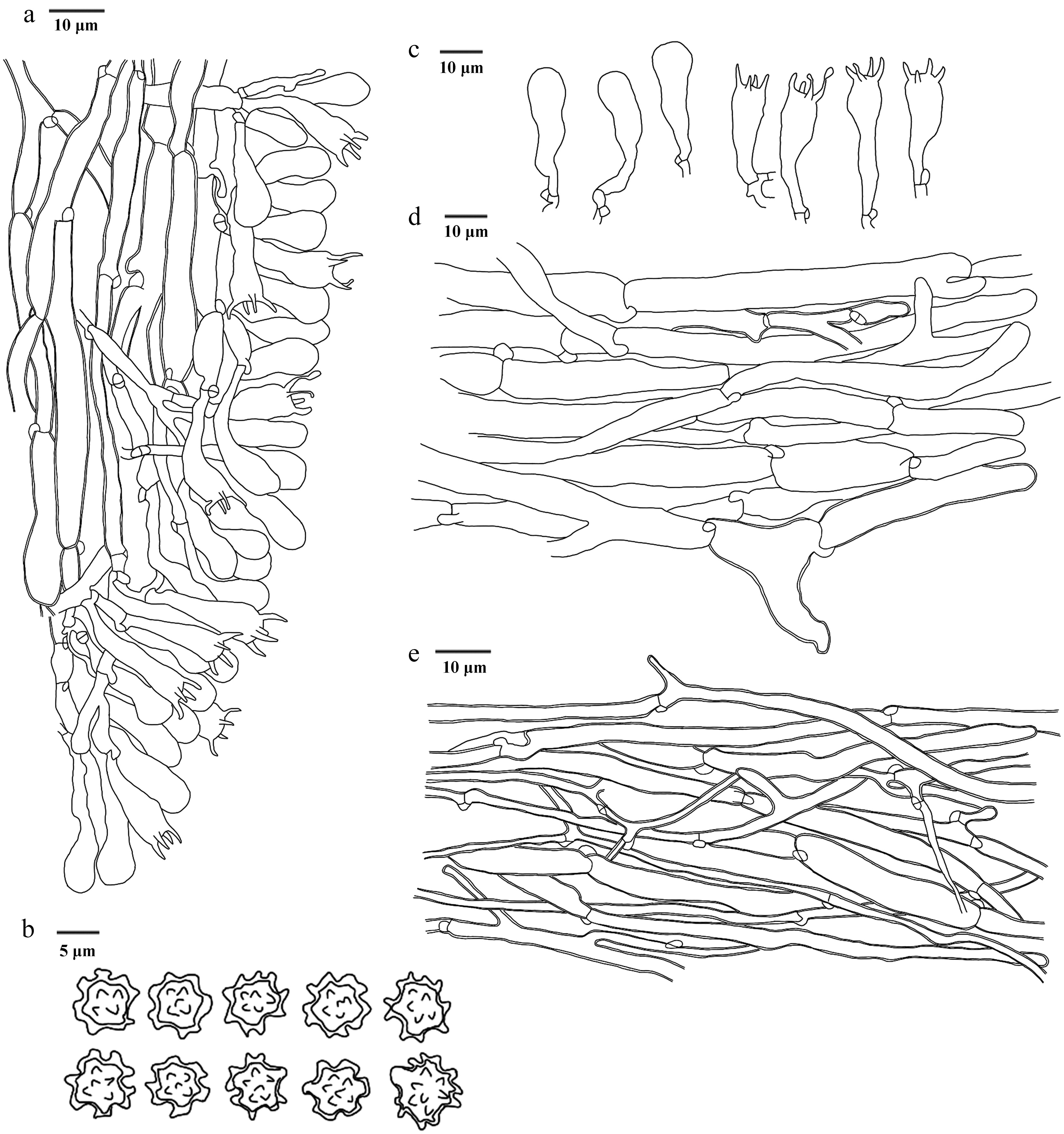
Figure 29.
Microscopic structures of Neosarcodon atroviolaceus (drawn from the holotype IFP 020031). (a) Section through spines. (b) Basidiospores. (c) Basidia and basidioles. (d) Hyphae from context. (e) Hyphae from stipe.
-
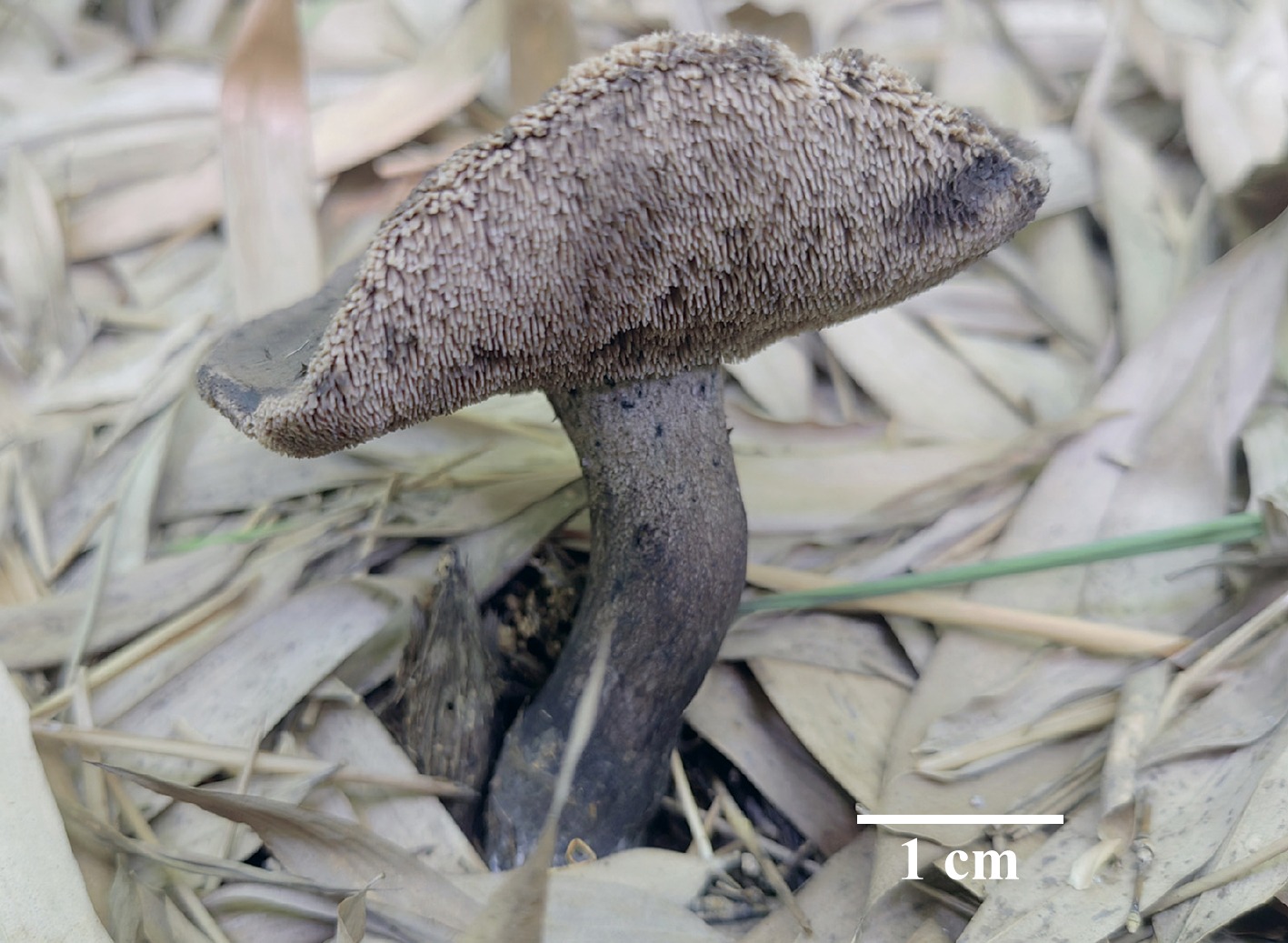
Figure 30.
Basidiomata of Neosarcodon bambusicola (holotype IFP 020033). Photo by Zhong-Ping Feng.
-
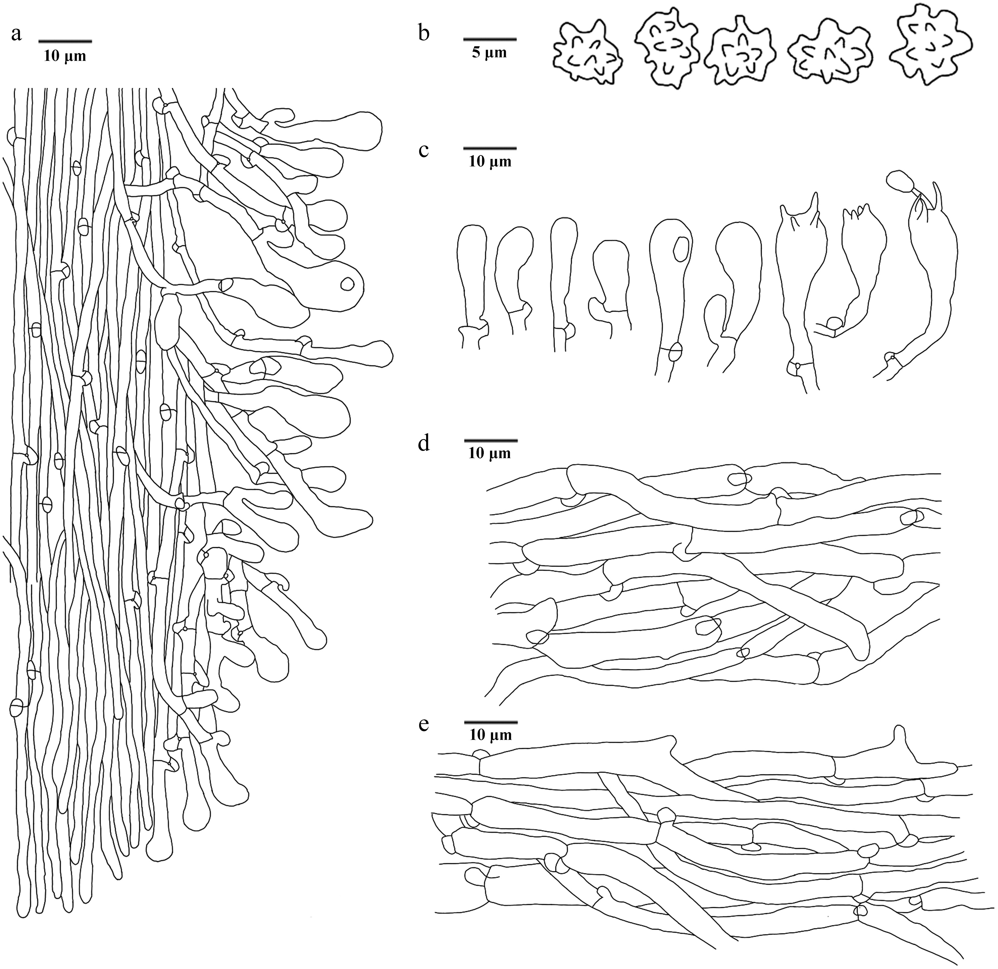
Figure 31.
Microscopic structures of Neosarcodon bambusicola (drawn from the holotype IFP 020033). (a) Section through spines. (b) Basidiospores. (c) Basidia and basidioles. (d) Hyphae from pileus. (e) Hyphae from stipe.
-
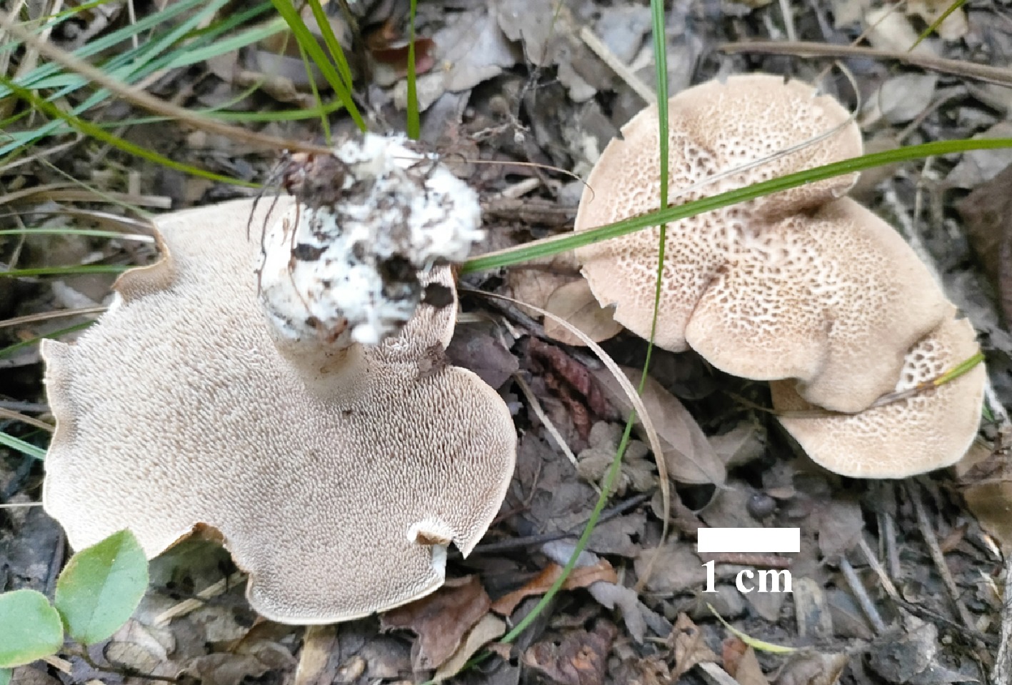
Figure 32.
Basidiomata of Sarcodon squamulosus (holotype IFP 020045). Photo by Yang-Ling Deng.
-
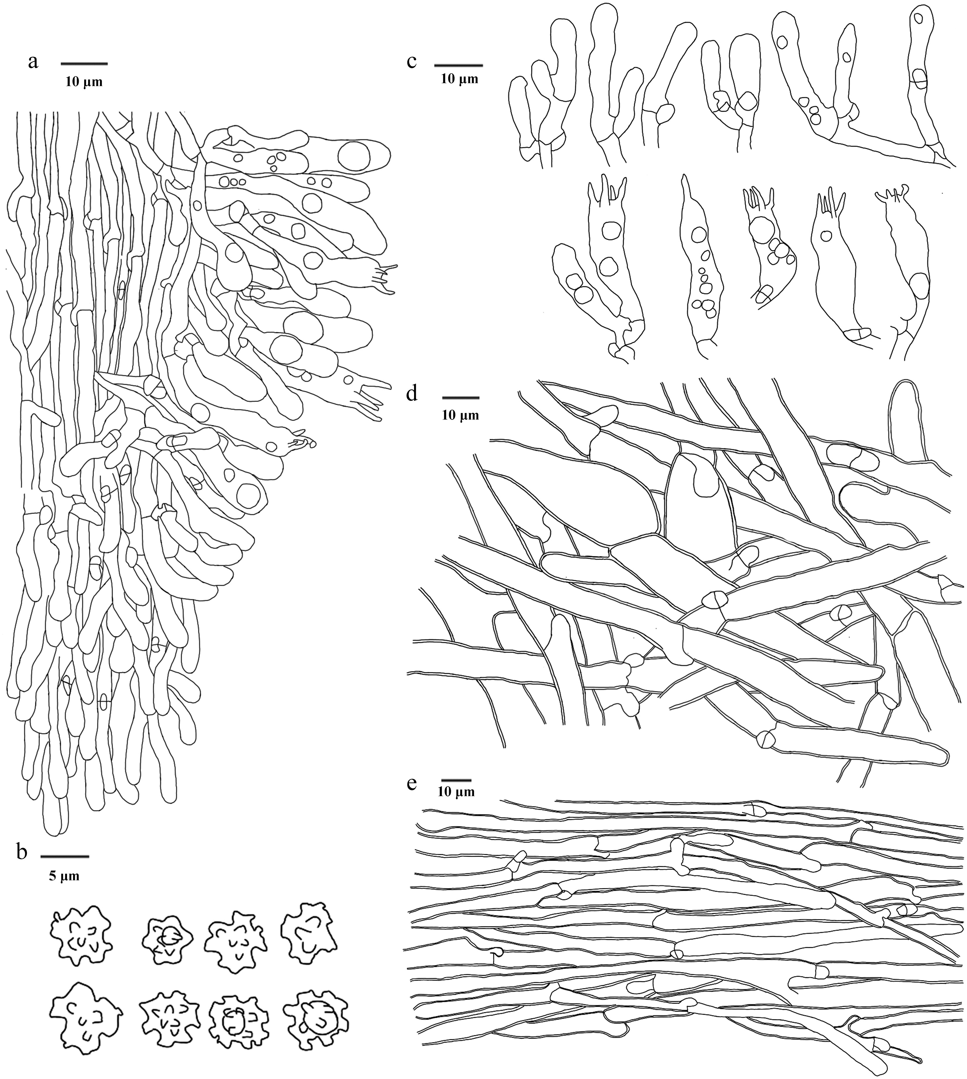
Figure 33.
Microscopic structures of Sarcodon squamulosus (drawn from the holotype IFP 020045). (a) Section through spines. (b) Basidiospores. (c) Basidia and basidioles. (d) Hyphae from pileus. (e) Hyphae from stipe.
-
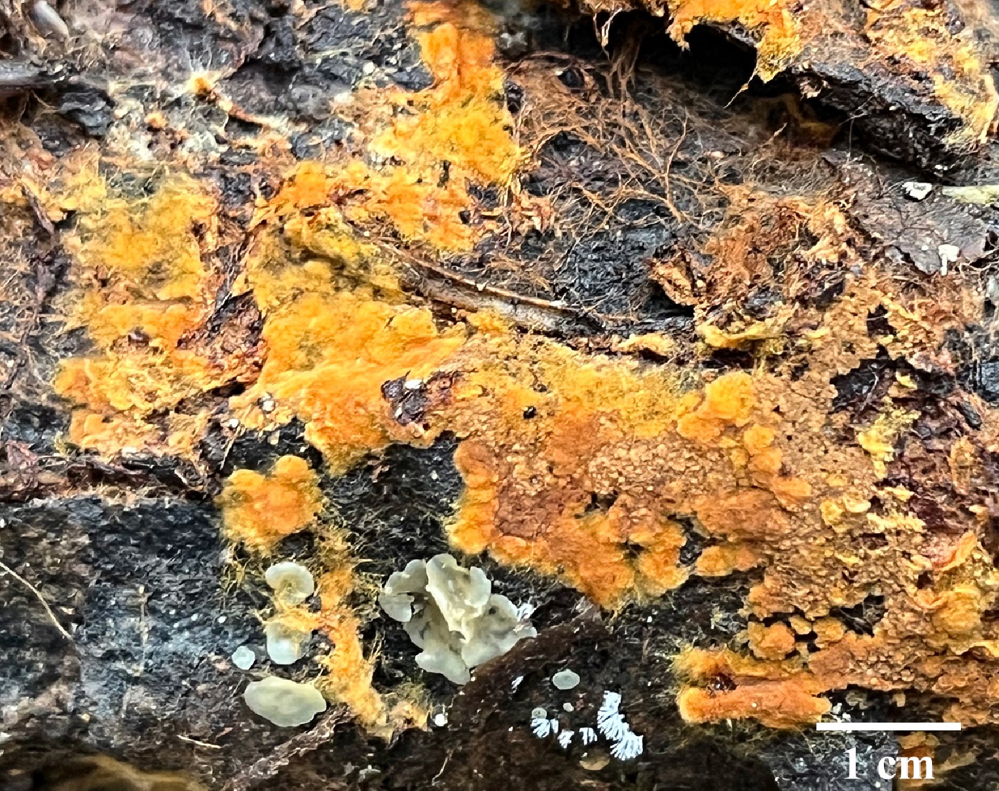
Figure 34.
Basidiomata of Odontia kunmingensis (holotype IFP 020034).
-
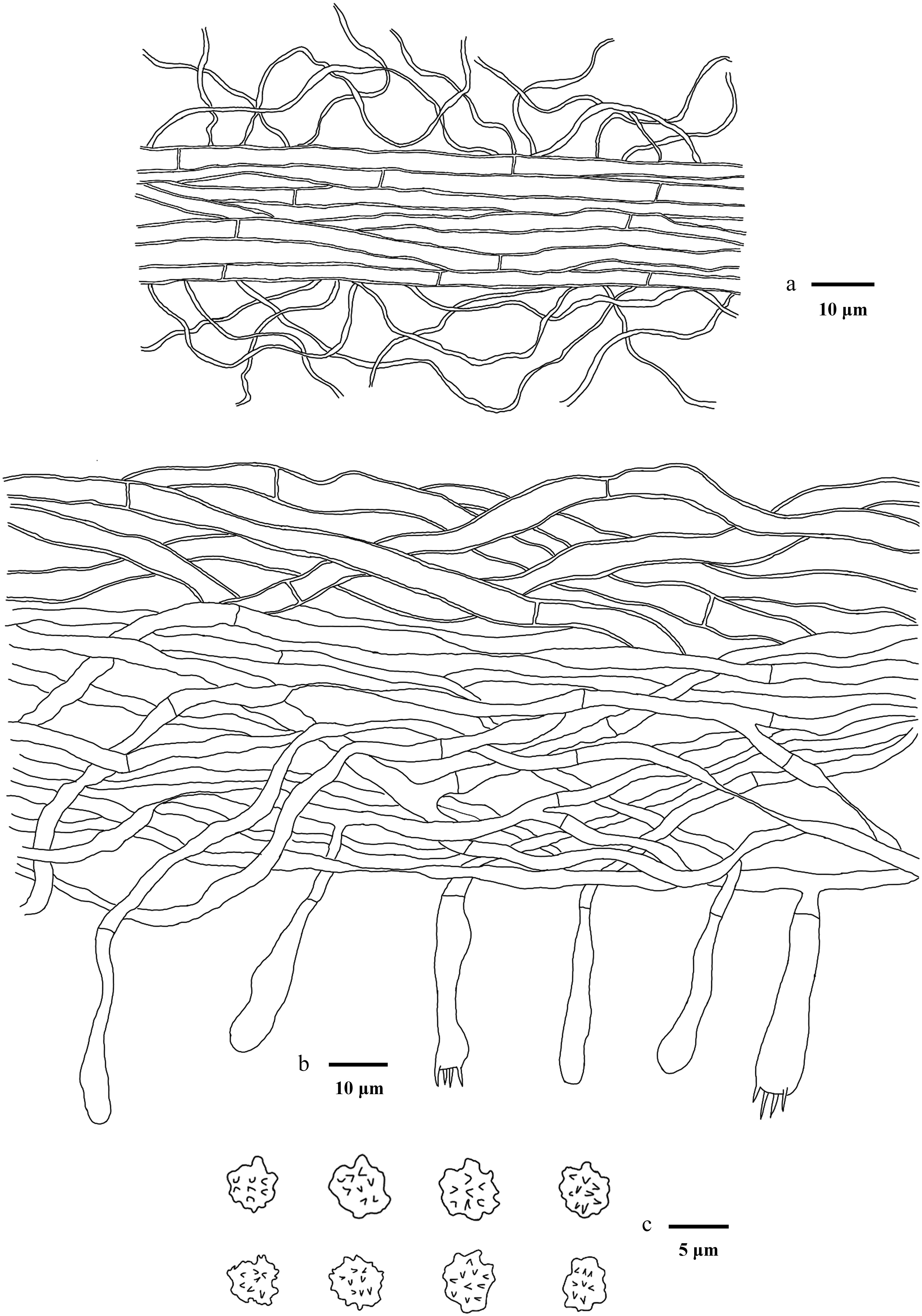
Figure 35.
Microscopic structures of Odontia kunmingensis (drawn from the holotype IFP 020034). (a) Section through rhizomorph. (b) Section through basidiomata. (c) Basidiospores in frontal and lateral view.
-
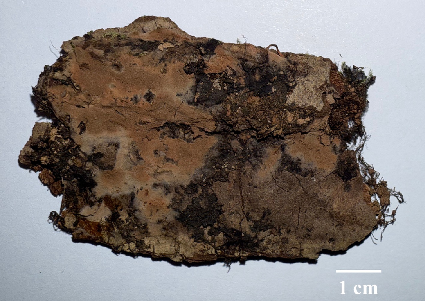
Figure 36.
Basidiomata of Thelephora angusta (holotype IFP 020075).
-
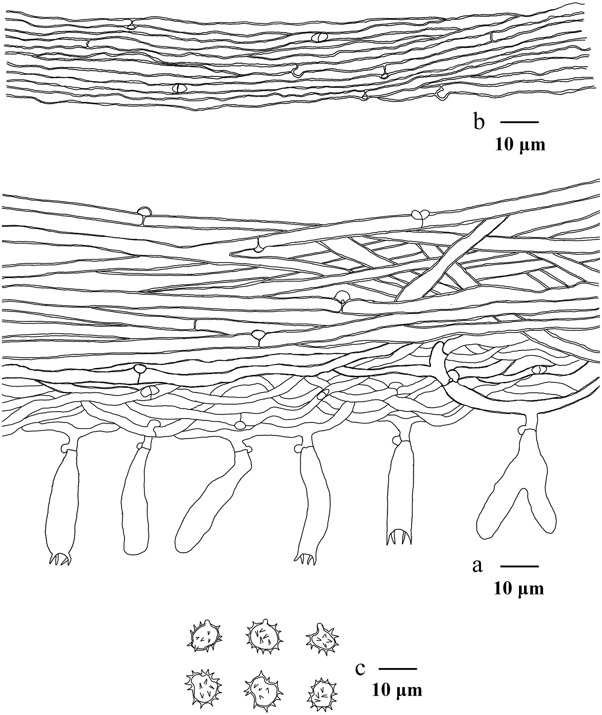
Figure 37.
Microscopic structures of Thelephora angusta (drawn from the holotype IFP 020075). (a) Section through rhizomorph. (b) Section through basidiomata. (c) Basidiospores in frontal and lateral view.
-
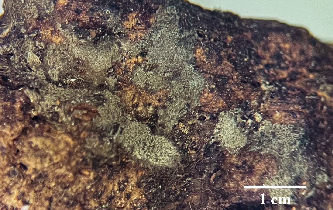
Figure 38.
Basidiomata of Thelephora bomiensis (holotype IFP 020056).
-
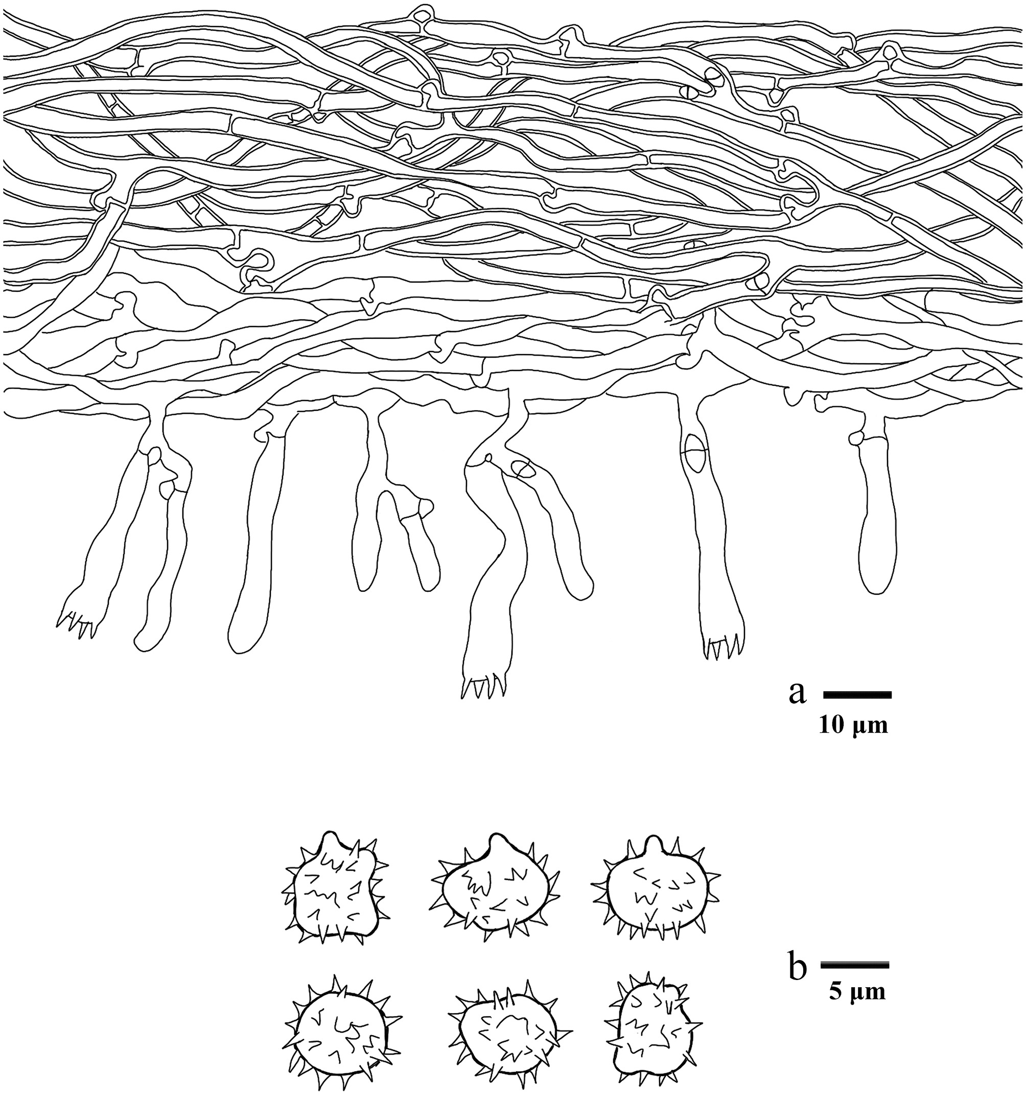
Figure 39.
Microscopic structures of Thelephora bomiensis (drawn from the holotype IFP 020056). (a) Section through basidiomata. (b) Basidiospores in frontal and lateral view.
-
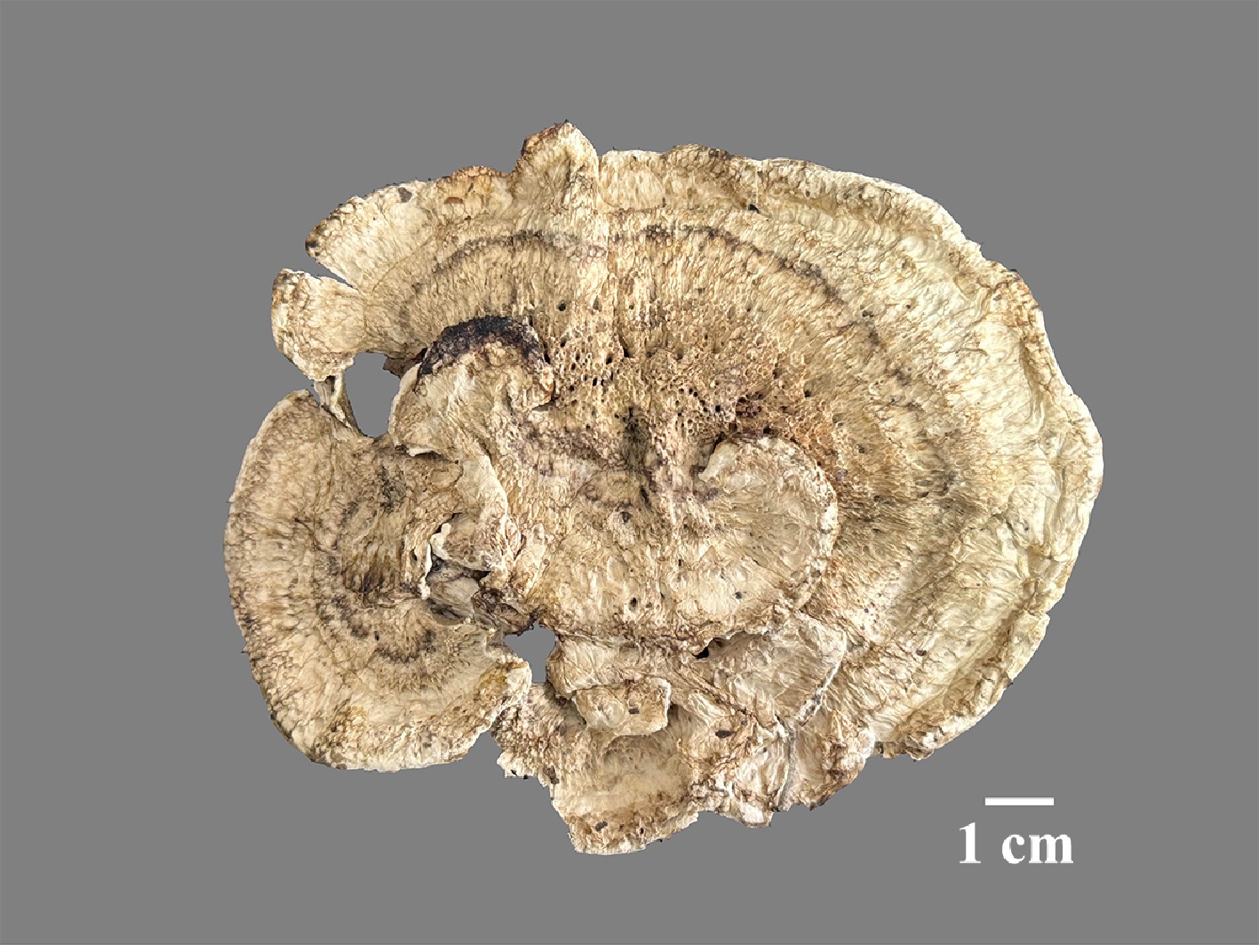
Figure 40.
Basidiomata of Thelephora cacao (holotype IFP 020077).
-
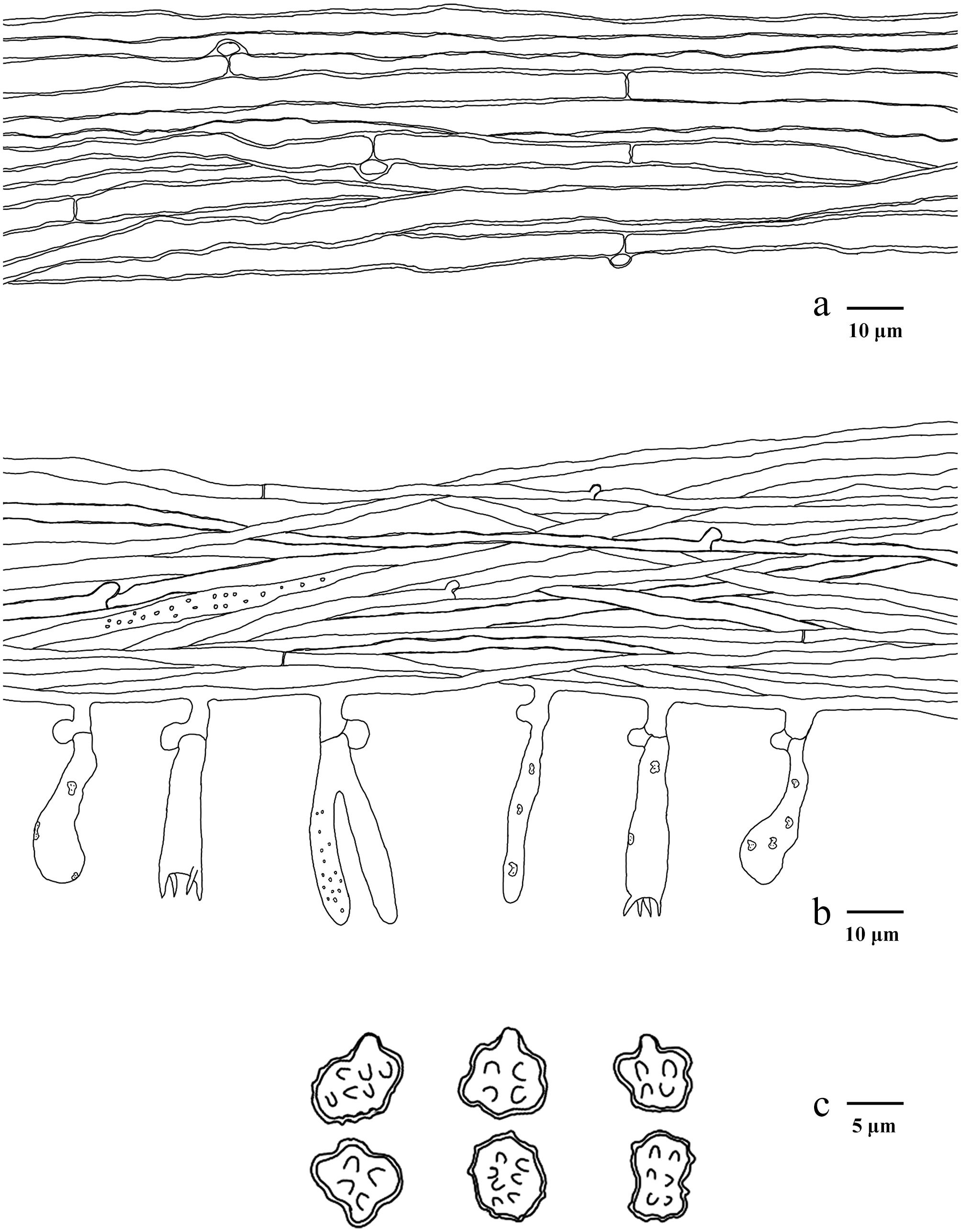
Figure 41.
Microscopic structures of Thelephora cacao (drawn from the holotype IFP 020077). (a) Hyphae from pileal context. (b) Section of hymenium and subhymenium. (c) Basidiospores in frontal and lateral view.
-
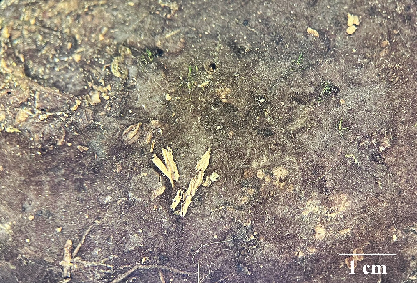
Figure 42.
Basidiomata of Thelephora chayuensis (holotype IFP 020064).
-
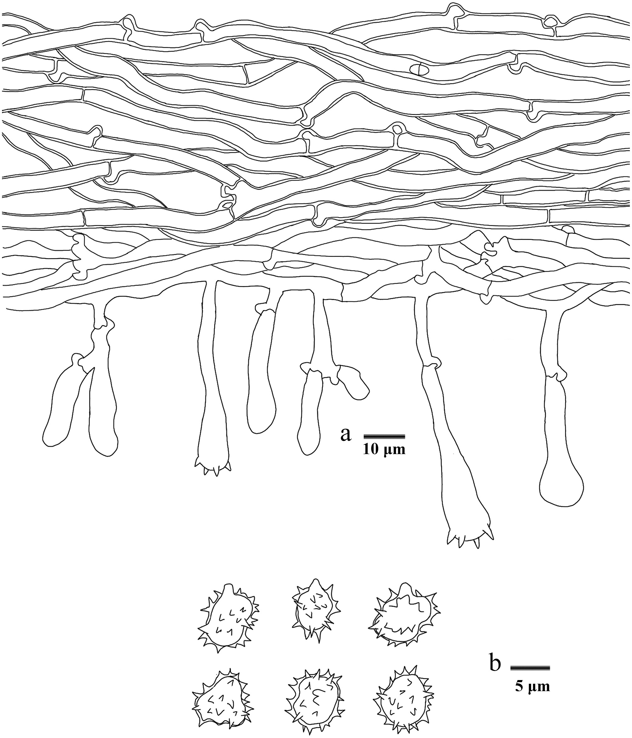
Figure 43.
Microscopic structures of Thelephora chayuensis (drawn from the holotype IFP 020064). (a) Section through basidiomata. (b) Basidiospores in frontal and lateral view.
-
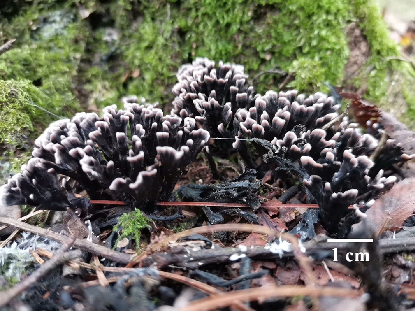
Figure 44.
Basidiomata of Thelephora fasciculata (holotype IFP 020052).
-
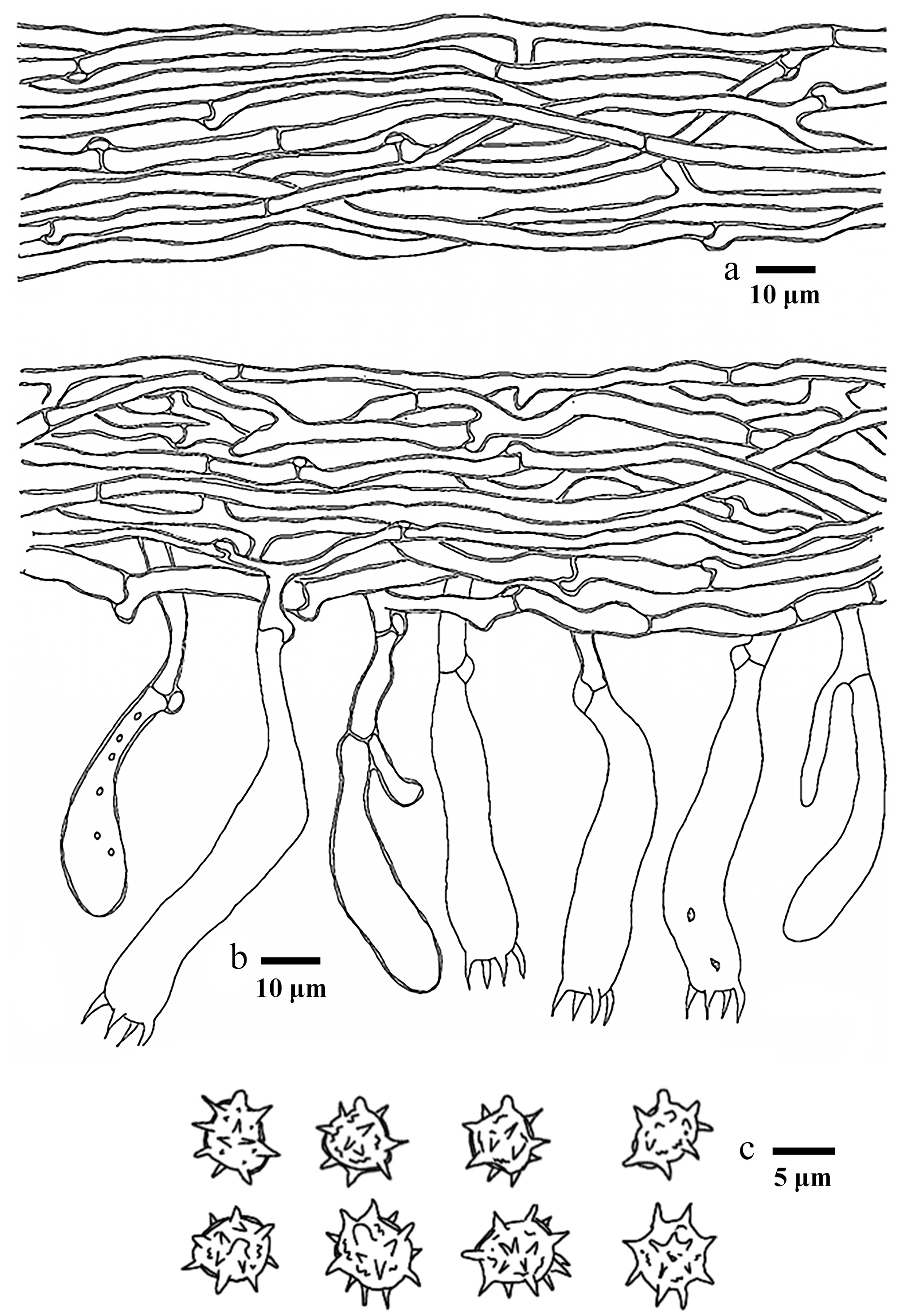
Figure 45.
Microscopic structures of Thelephora fasciculata (drawn from the holotype IFP 020052). (a) Hyphae from pileal context. (b) Section of hymenium and subhymenium. (c) Basidiospores in frontal and lateral view.
-
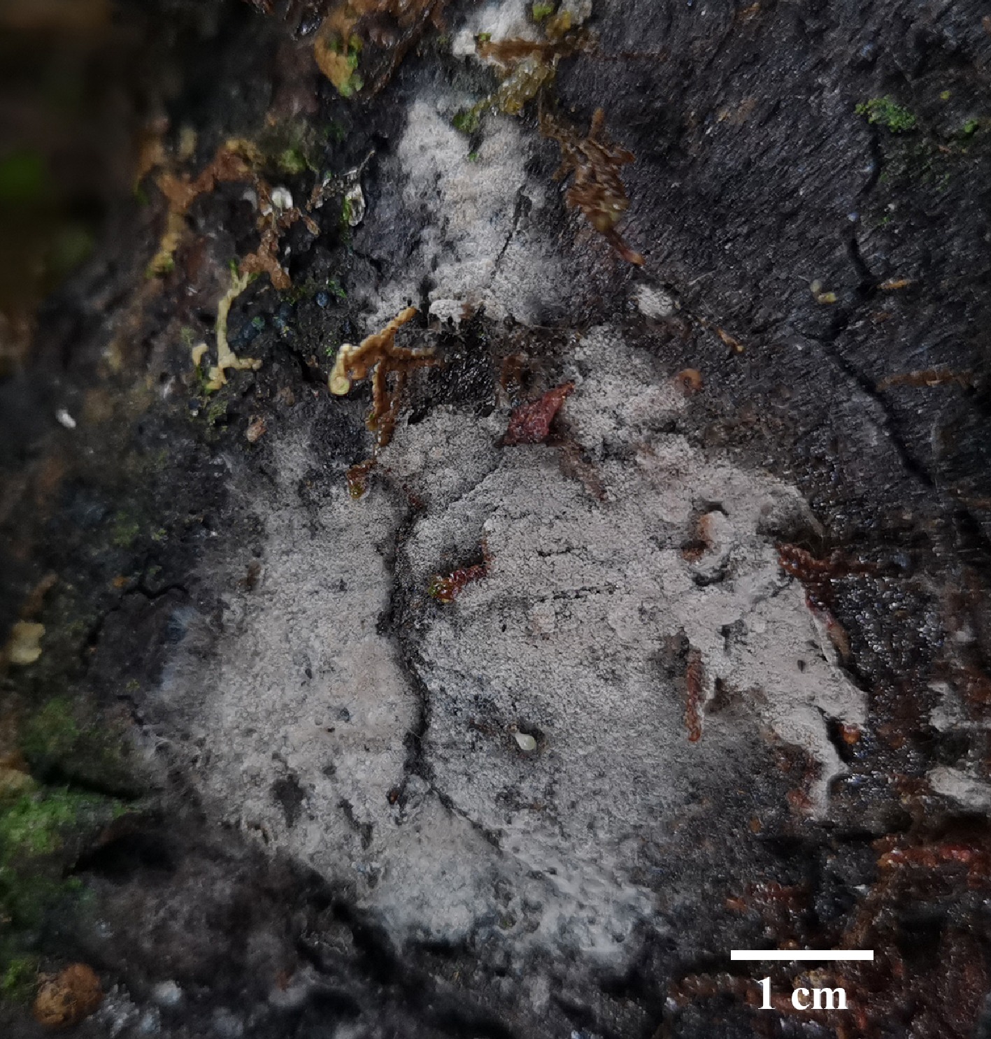
Figure 46.
Basidiomata of Thelephora latihypha (holotype IFP 020079).
-
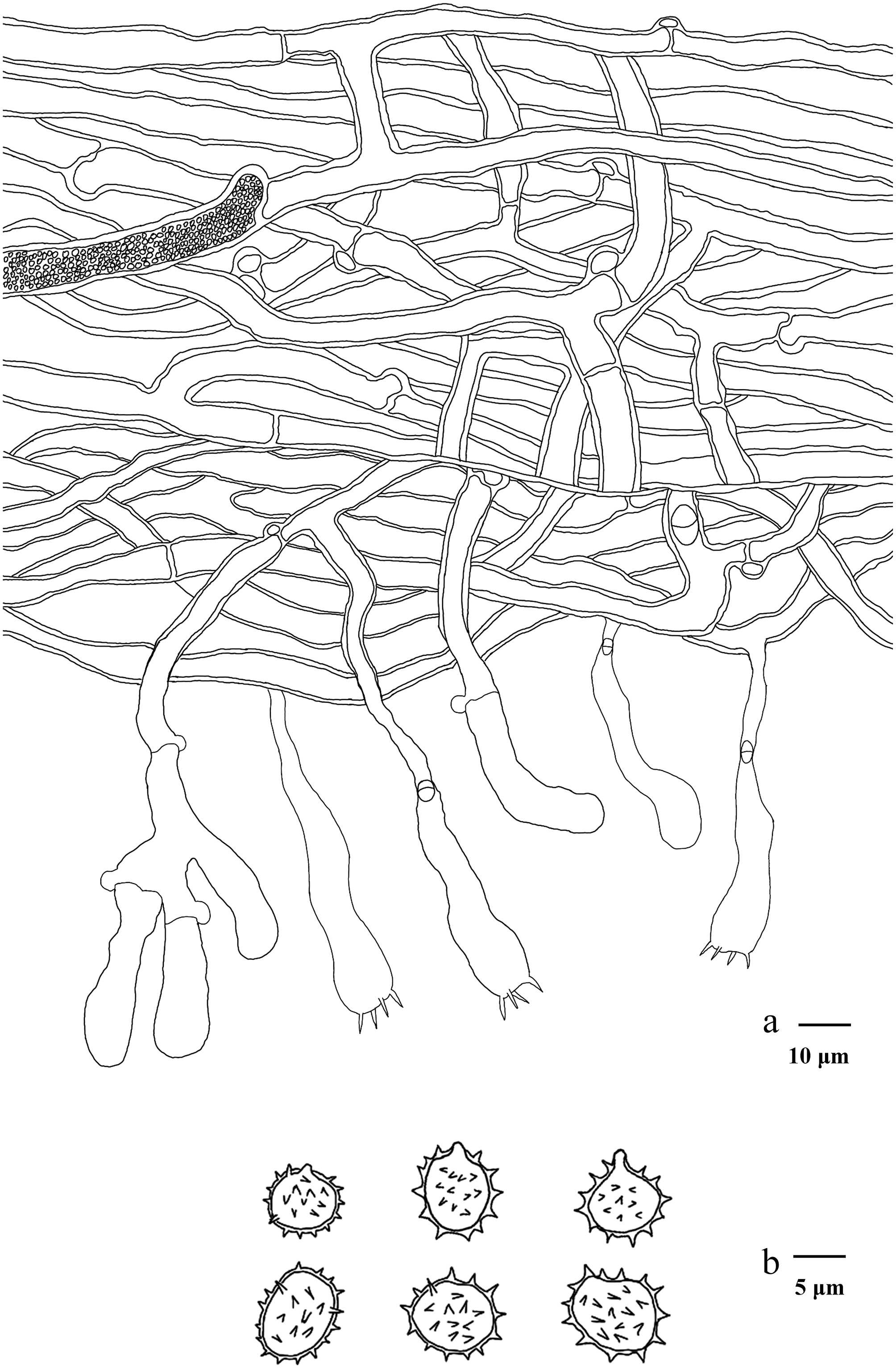
Figure 47.
Microscopic structures of Thelephora latihypha (drawn from the holotype IFP 020079). (a) Section through basidiomata. (b) Basidiospores in frontal and lateral view.
-
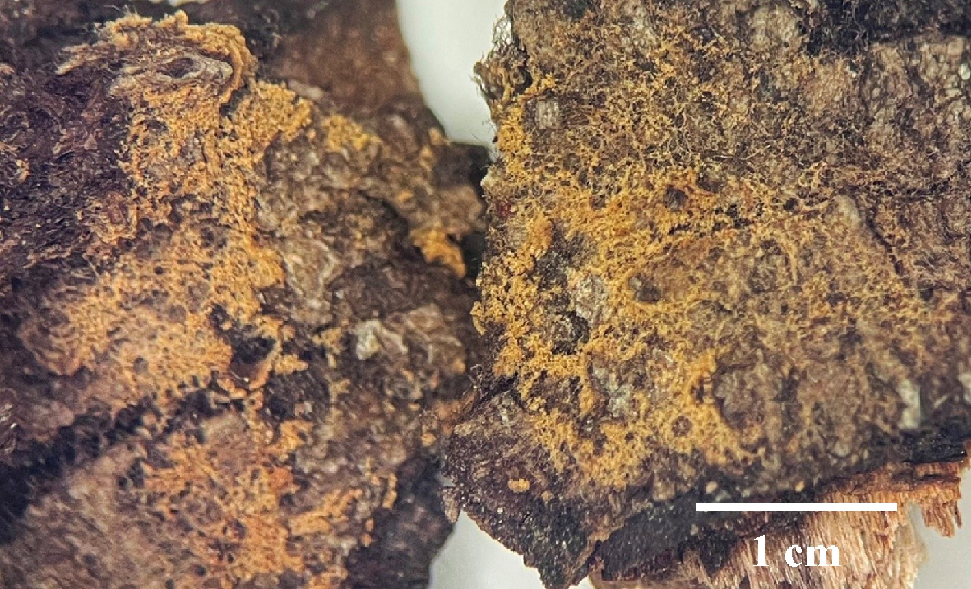
Figure 48.
Basidiomata of Thelephora linzhiensis (holotype IFP 020058).
-
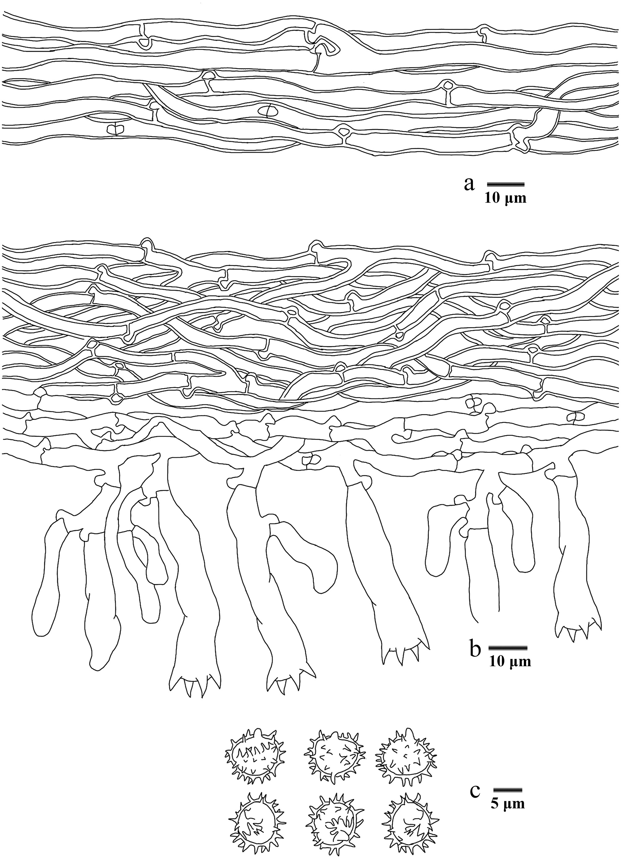
Figure 49.
Microscopic structures of Thelephora linzhiensis (drawn from the holotype IFP 020058). (a) Hyphae from a rhizomorph. (b) Section through basidiomata. (c) Basidiospores in frontal and lateral view.
-
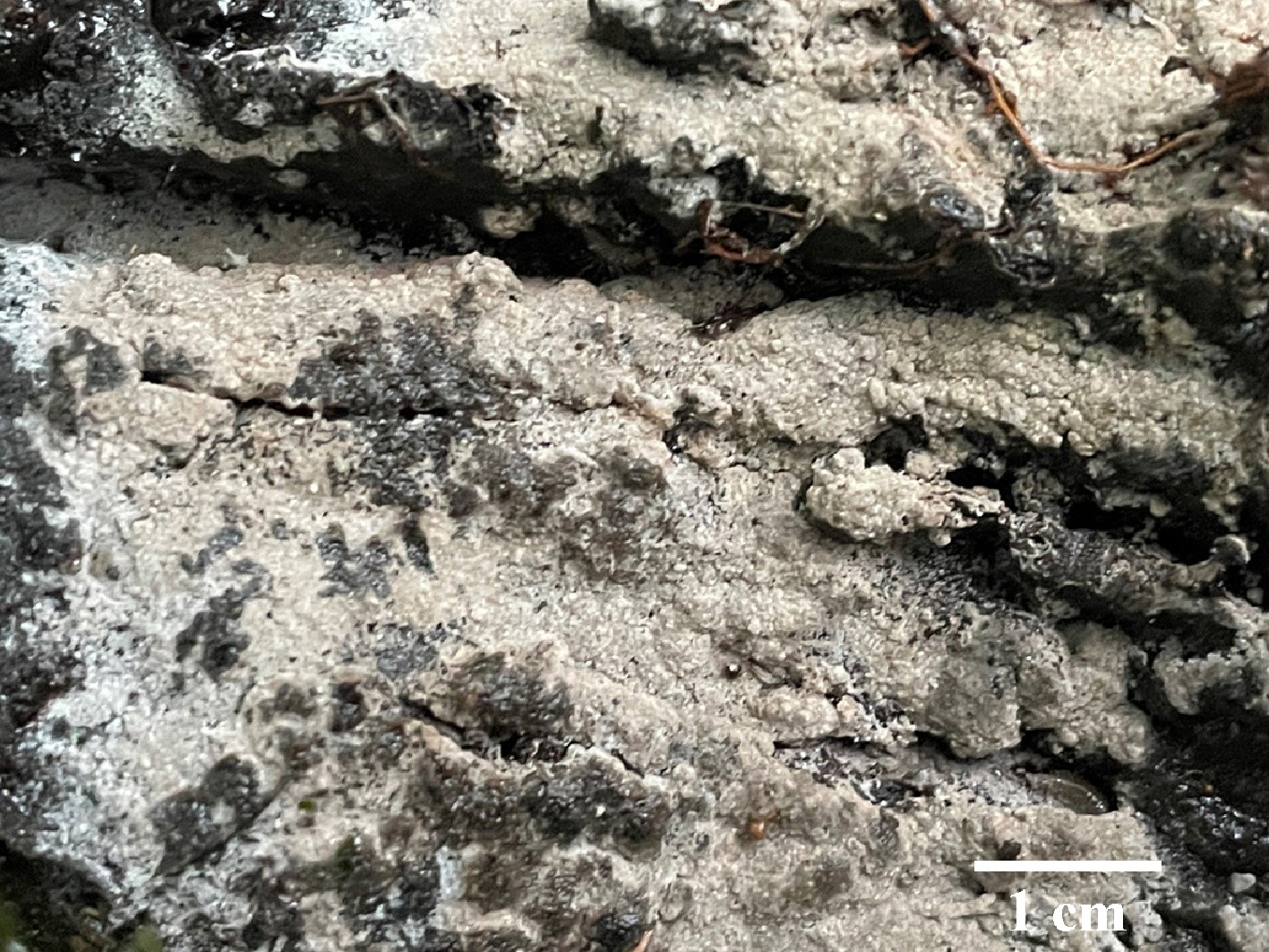
Figure 50.
Basidiomata of Thelephora longicystidiata (holotype IFP 020081).
-
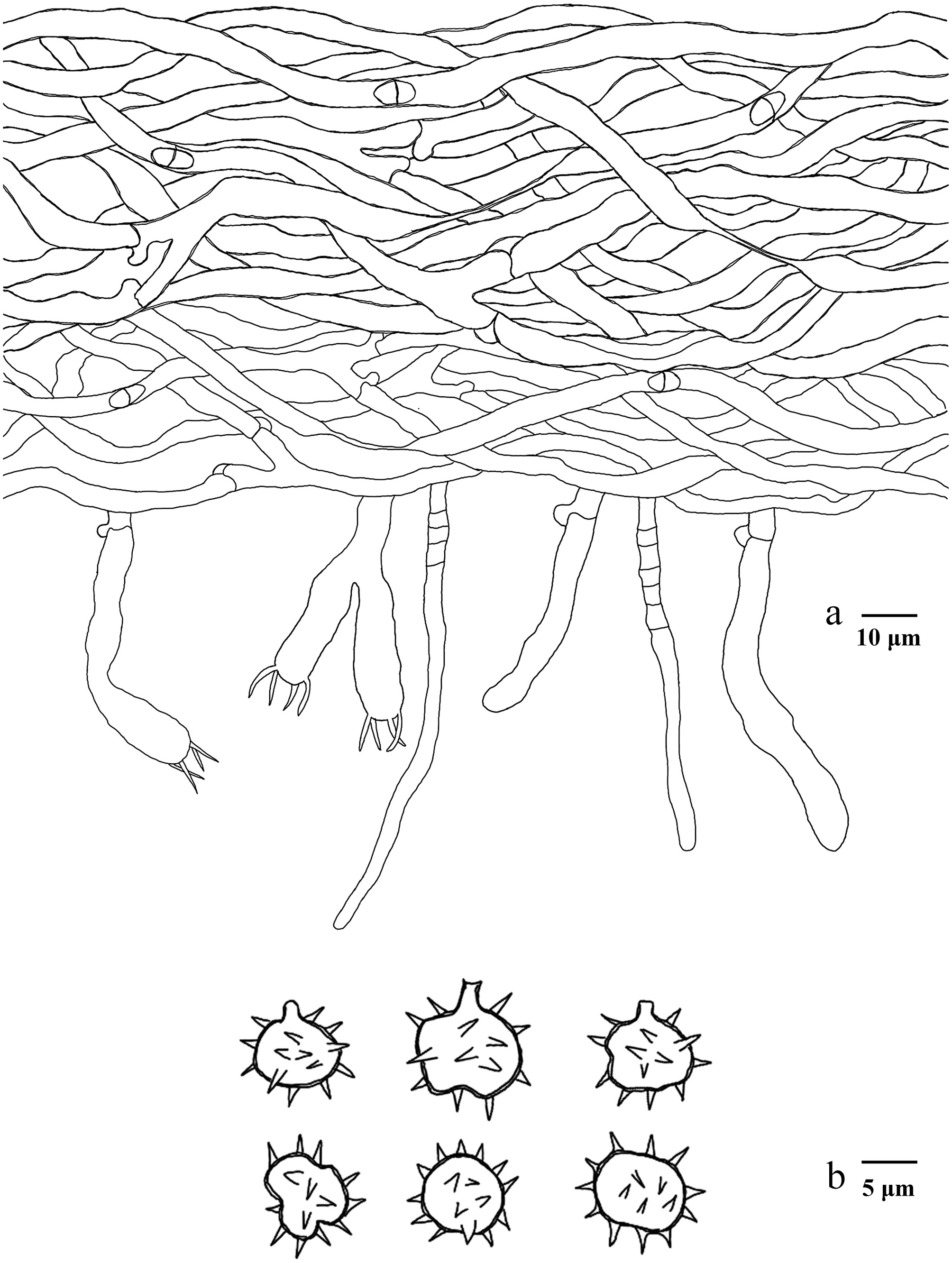
Figure 51.
Microscopic structures of Thelephora longicystidiata (drawn from the holotype IFP 020081). (a) Section through basidiomata. (b) Basidiospores in frontal and lateral view.
-
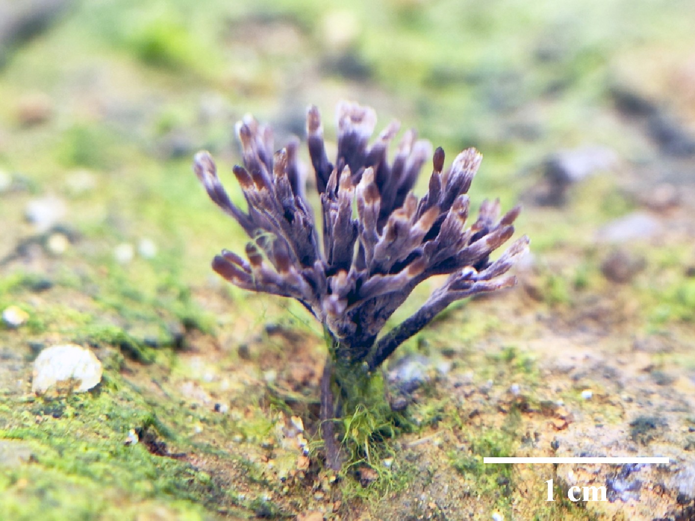
Figure 52.
Basidiomata of Thelephora microcarpa (paratype IFP 020055). Photo by Yu-Rong Liang.
-
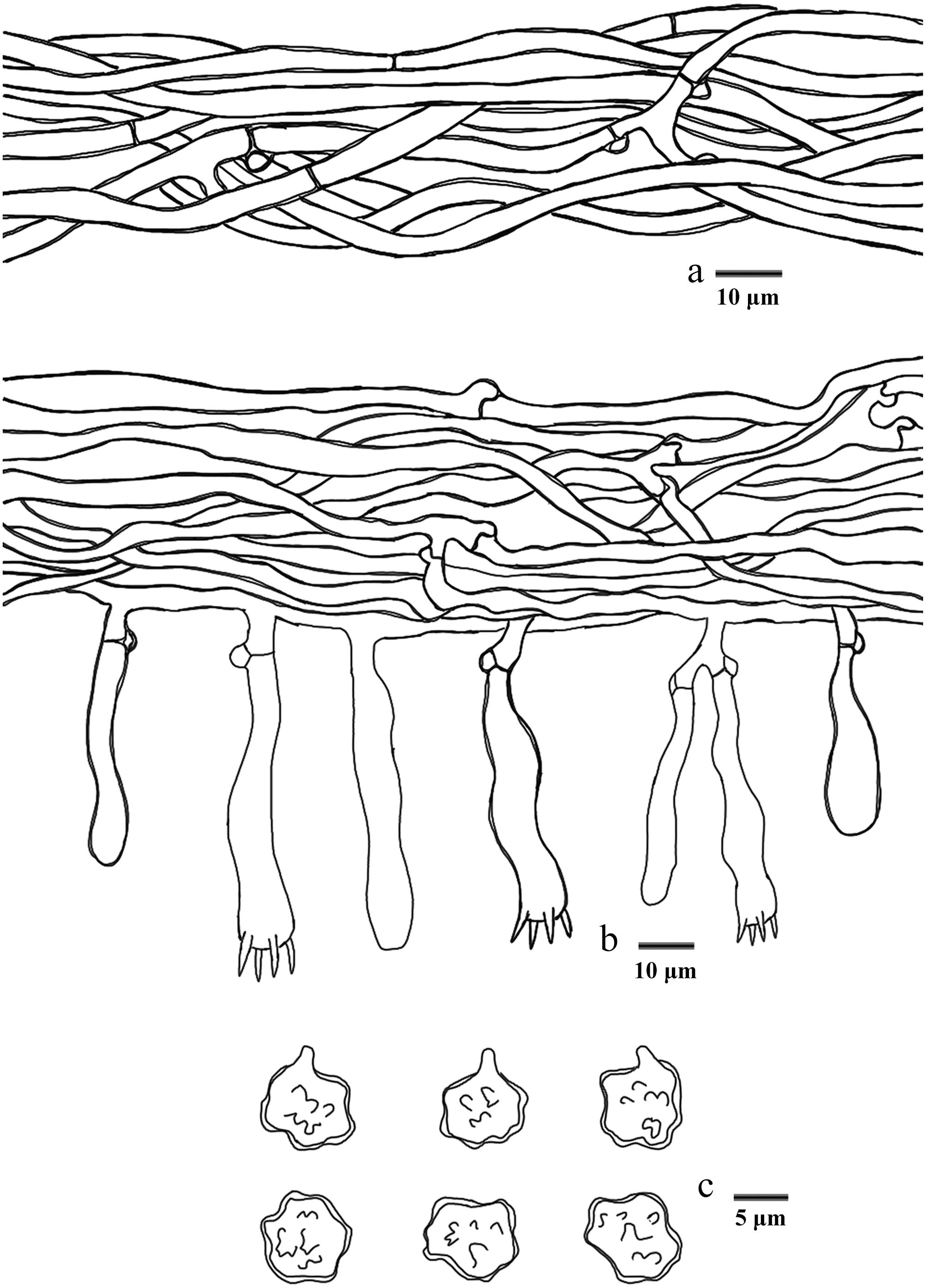
Figure 53.
Microscopic structures of Thelephora microcarpa (drawn from the holotype IFP 020054). (a) Hyphae from pileal context. (b) Section of hymenium and subhymenium. (c) Basidiospores in frontal and lateral view.
-
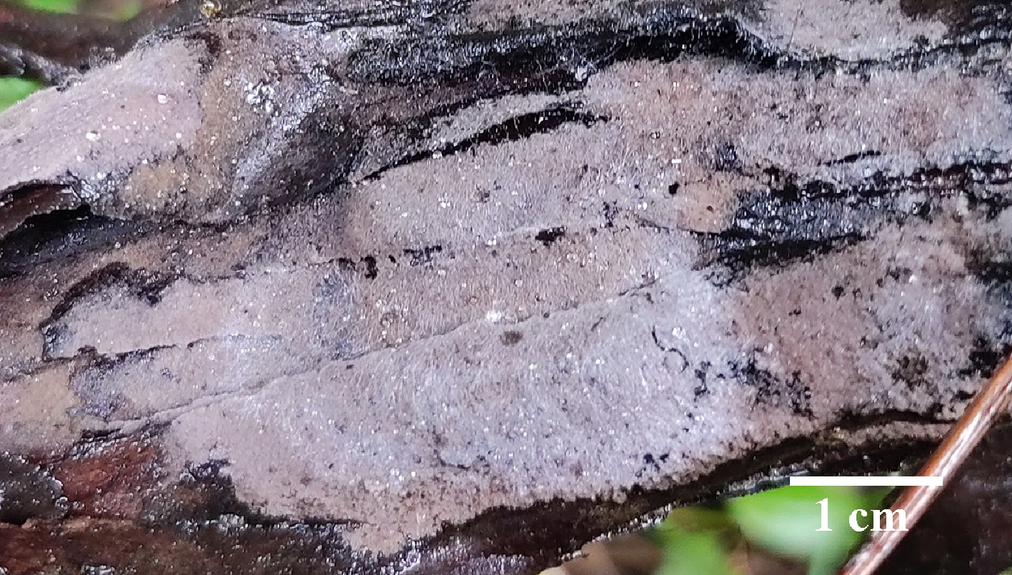
Figure 54.
Basidiomata of Thelephora nanyigouensis (holotype IFP 020060).
-
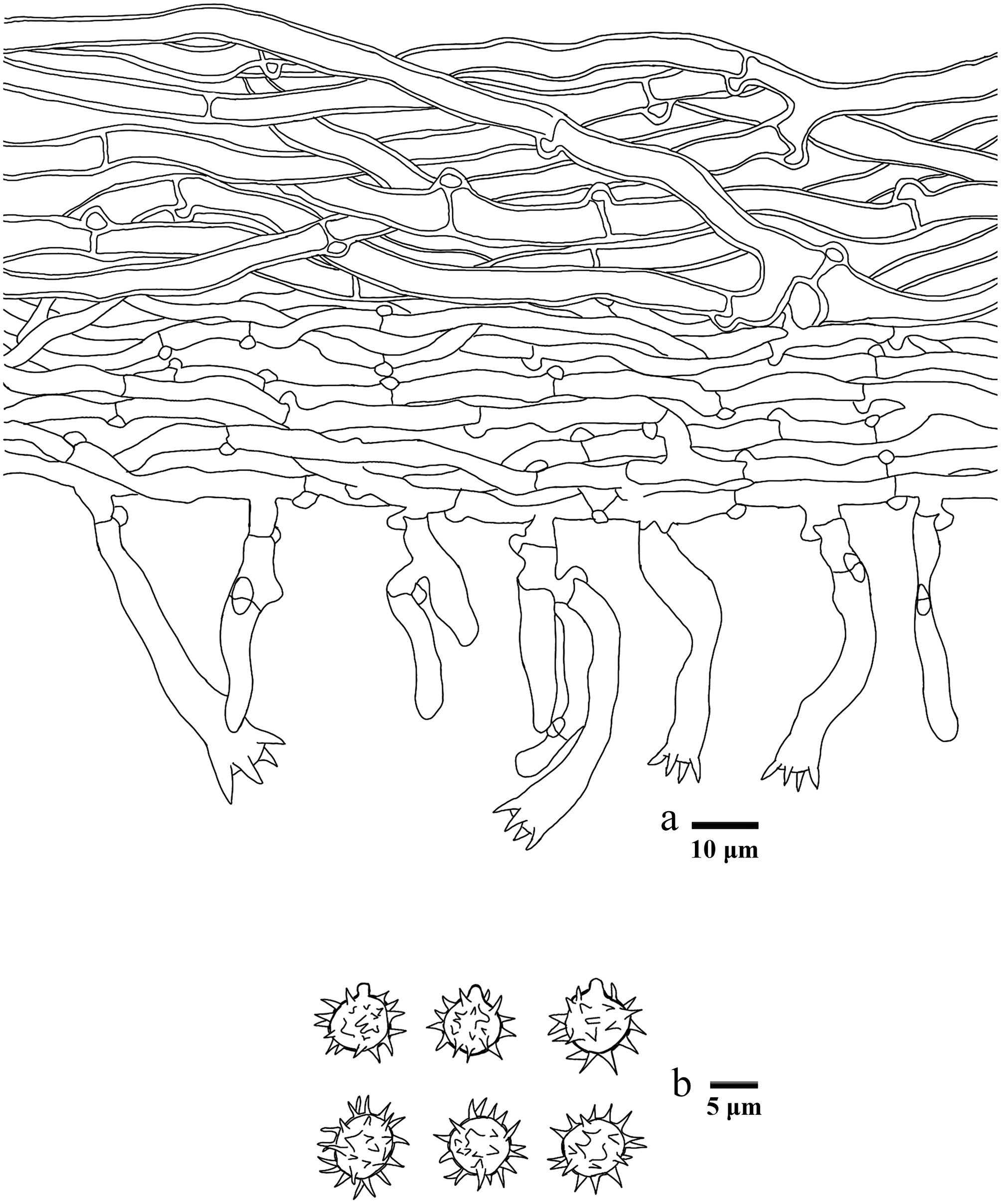
Figure 55.
Microscopic structures of Thelephora nanyigouensis (drawn from the holotype IFP 020060). (a) Section through basidiomata. (b) Basidiospores in frontal and lateral view.
-
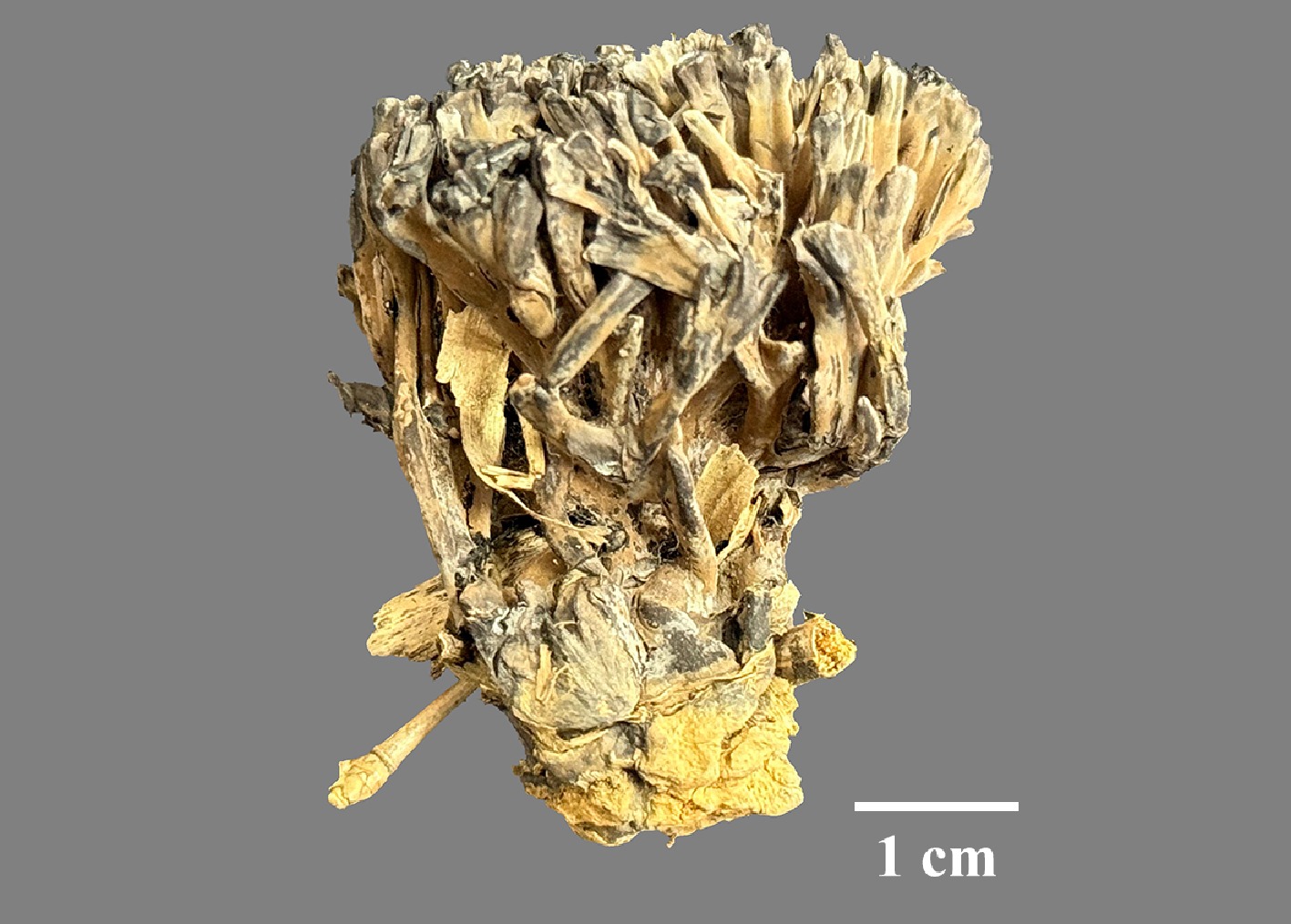
Figure 56.
Basidiomata of Thelephora nigromarginata (holotype IFP 020083).
-
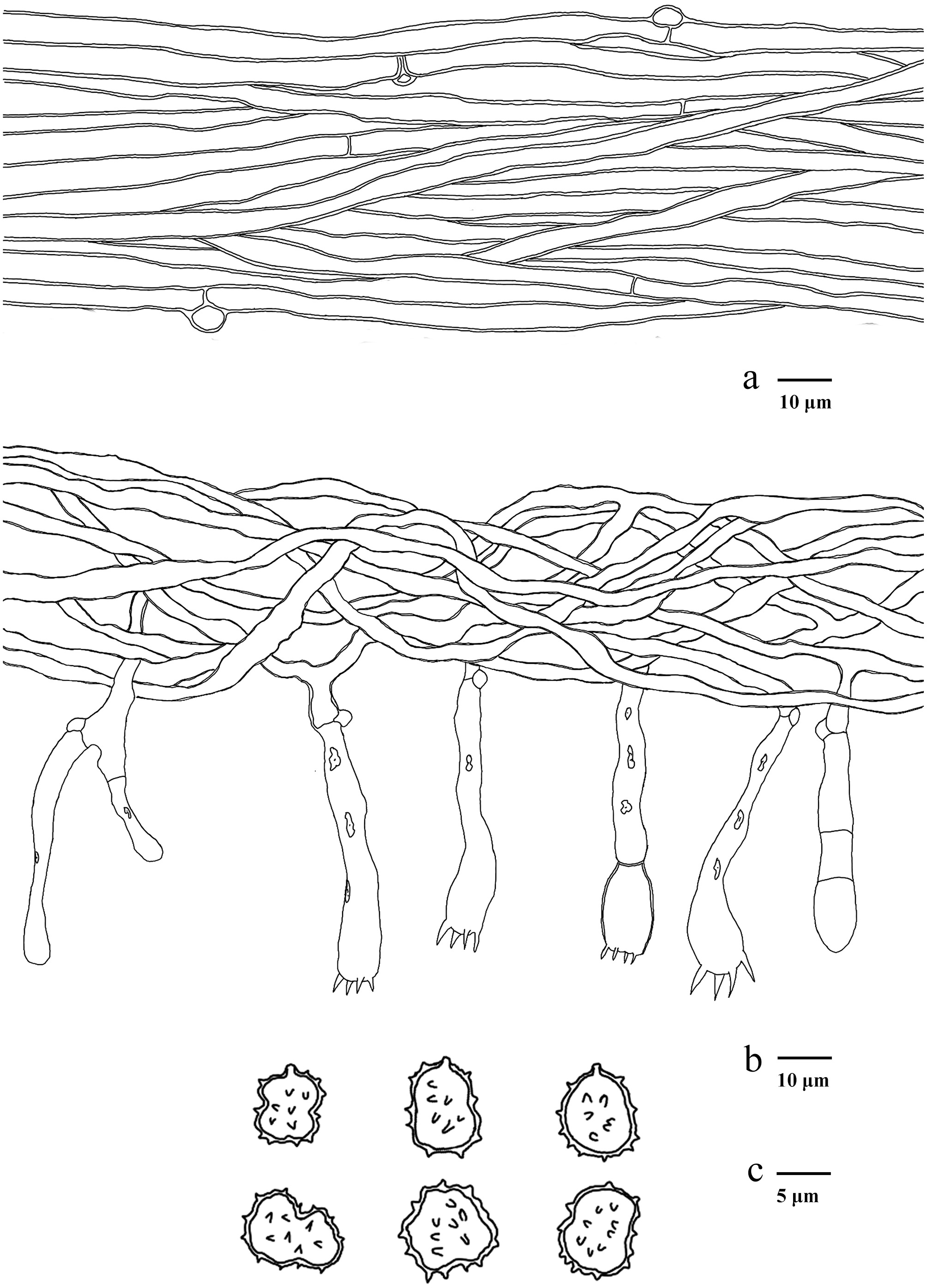
Figure 57.
Microscopic structures of Thelephora nigromarginata (drawn from the holotype IFP 020083). (a) Hyphae from pileal context. (b) Section of hymenium and subhymenium. (c) Basidiospores in frontal and lateral view.
-
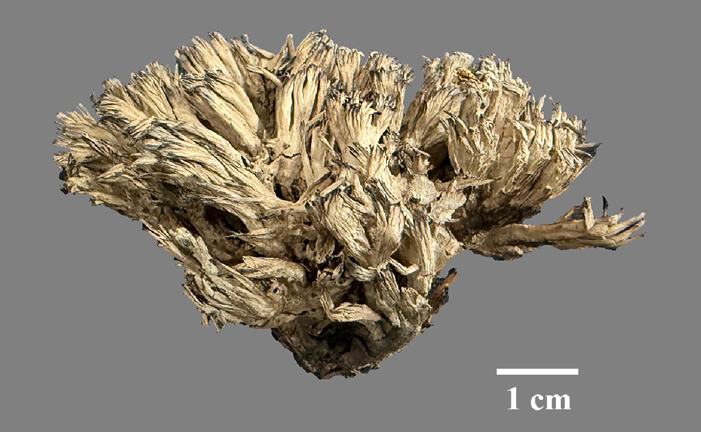
Figure 58.
Basidiomata of Thelephora scopiformis (holotype IFP 020086).
-
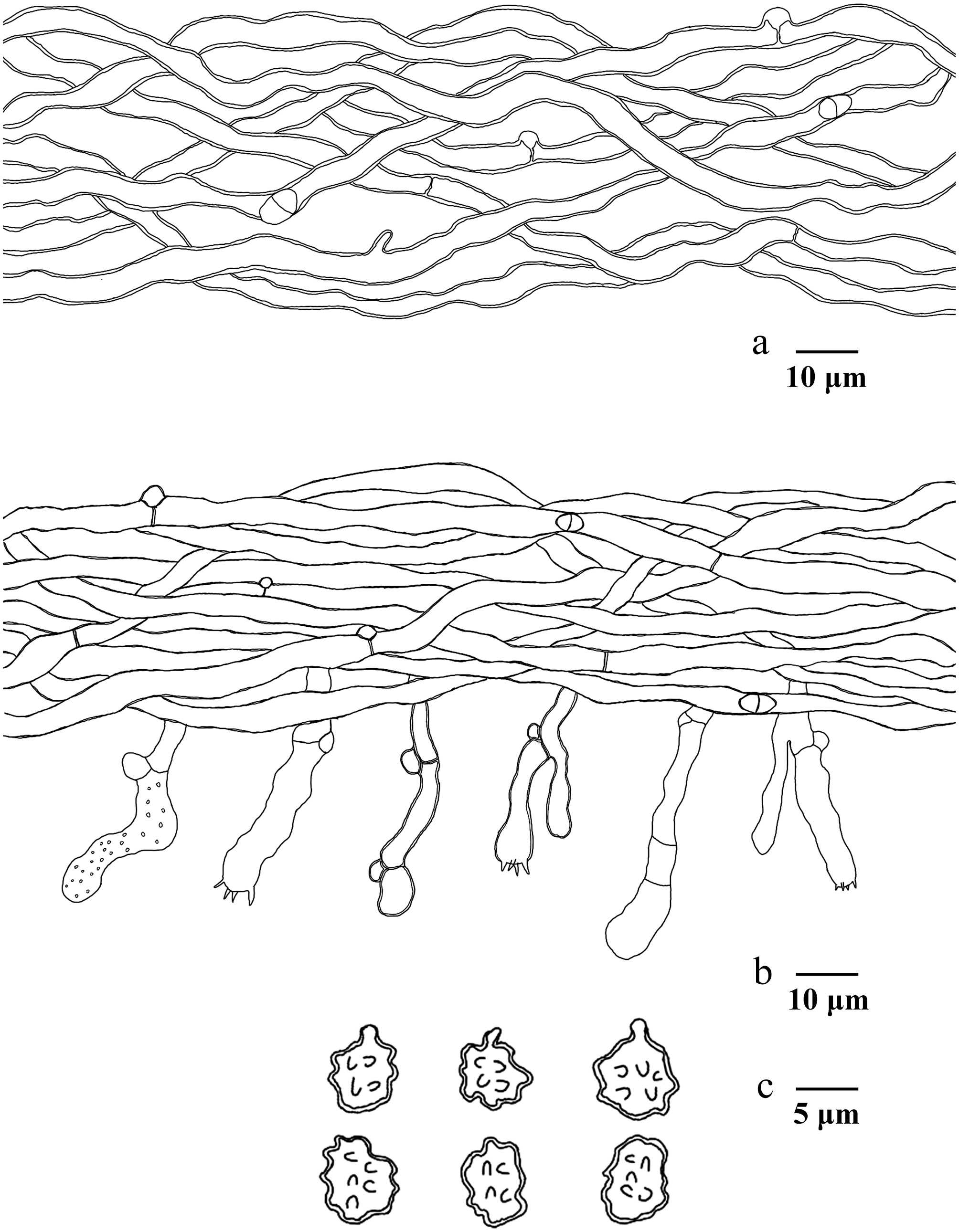
Figure 59.
Microscopic structures of Thelephora scopiformis (drawn from the holotype IFP 020086). (a) Hyphae from pileal context. (b) Section of hymenium and subhymenium. (c) Basidiospores in frontal and lateral view.
-
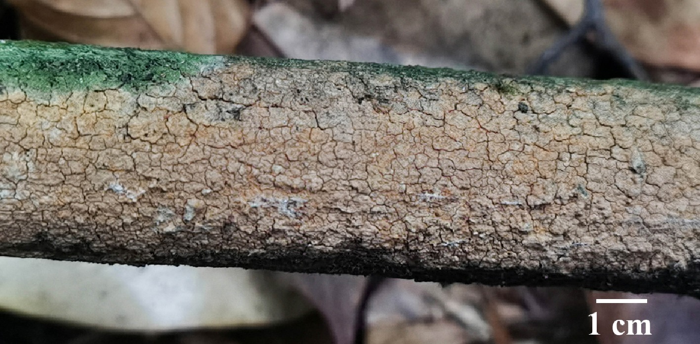
Figure 60.
Basidiomata of Hymenochaete baishanzuensis (holotype IFP 020012).
-
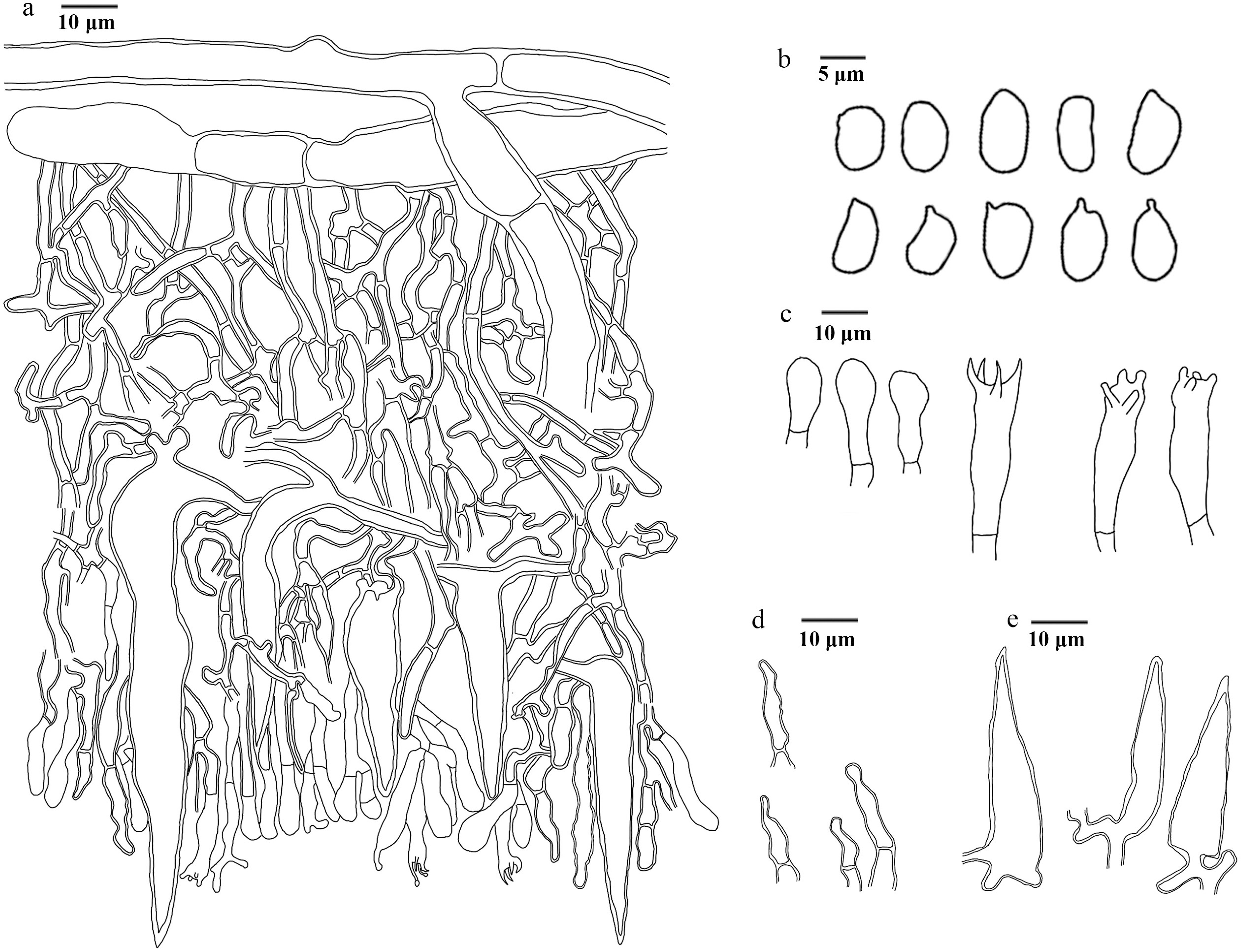
Figure 61.
Microscopic structures of Hymenochaete baishanzuensis (drawn from the holotype IFP 020012). (a) Section of hymenium. (b) Basidiospores. (c) Basidia and basidioles. (d) Hyphidia. (e) Setae.
-
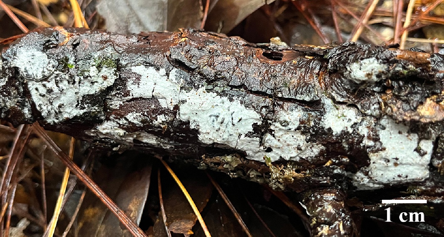
Figure 62.
Basidiomata of Peniophorella alba (holotype IFP 020014).
-
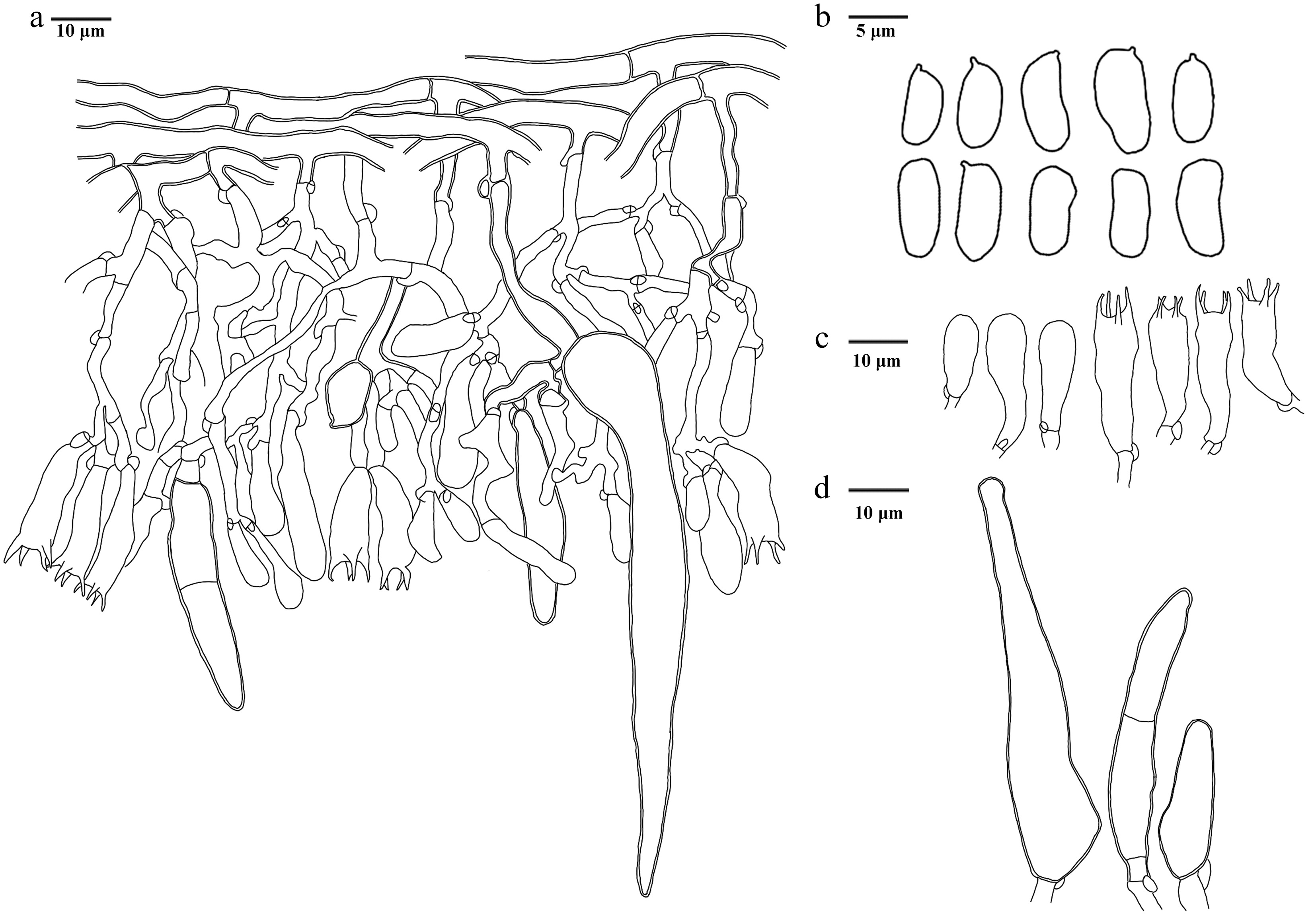
Figure 63.
Microscopic structures of Peniophorella alba (drawn from the holotype IFP 020014). (a) Section of hymenium. (b) Basidiospores. (c) Basidia and basidioles. (d) Cystidia.
-
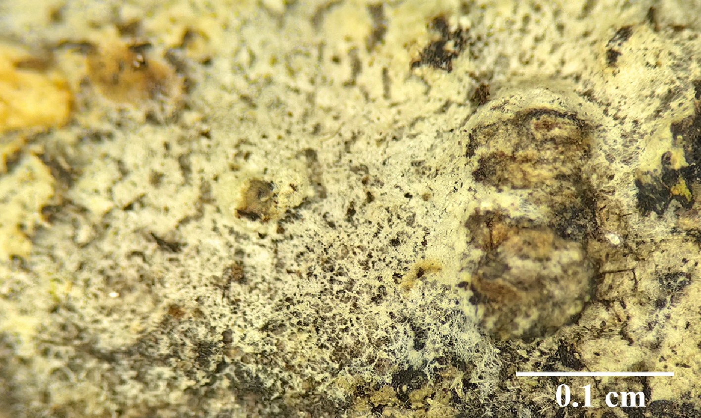
Figure 64.
Basidiomata of Lyomyces membranaceus (holotype IFP 020089).
-
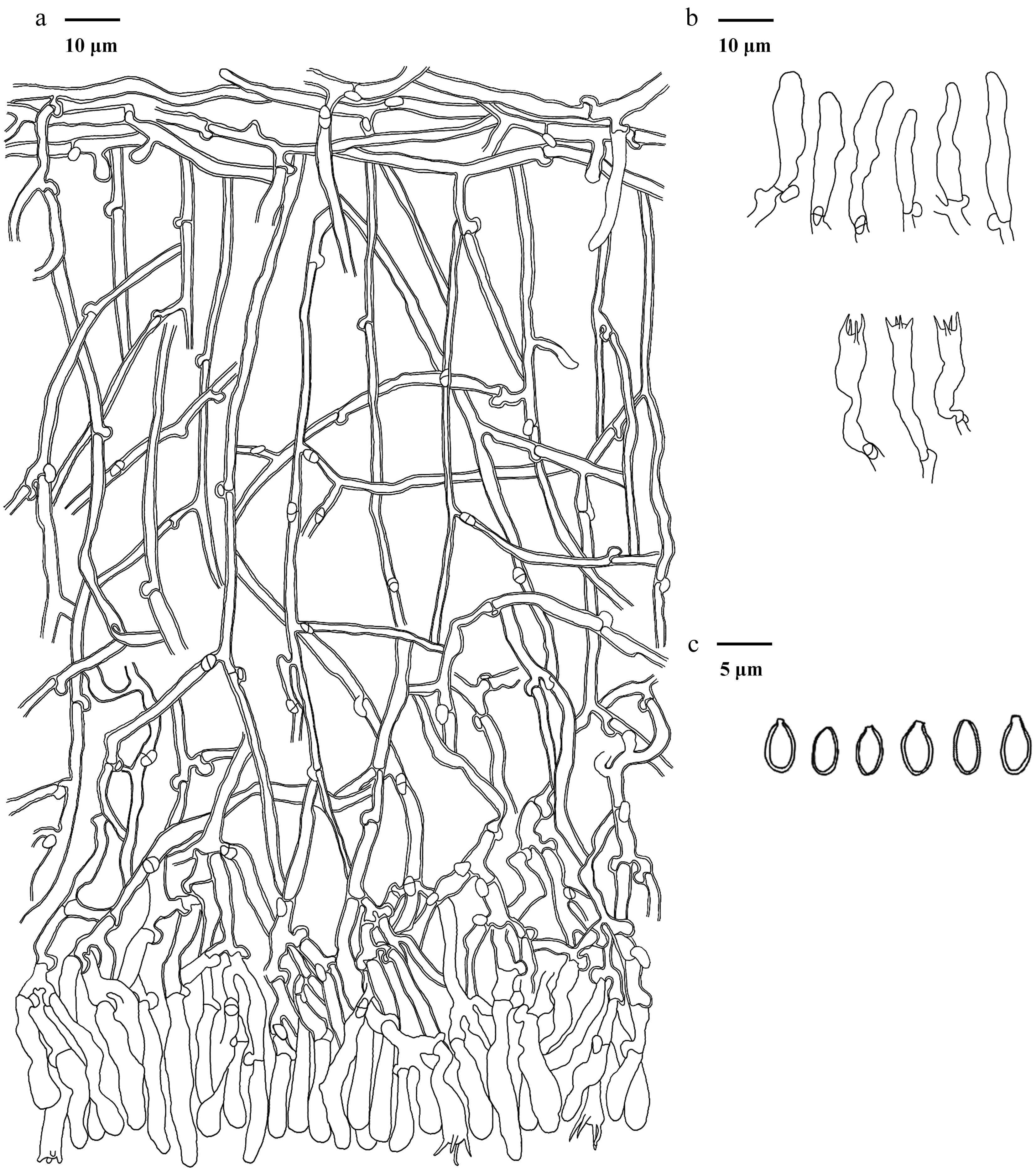
Figure 65.
Microscopic structures of Lyomyces membranaceus (drawn from the holotype IFP 020089). (a) Section of hymenium. (b) Basidia and basidioles. (c) Basidiospores.
-
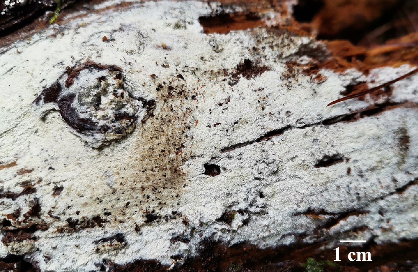
Figure 66.
Basidiomata of Xylodon albus (holotype IFP 020016).
-
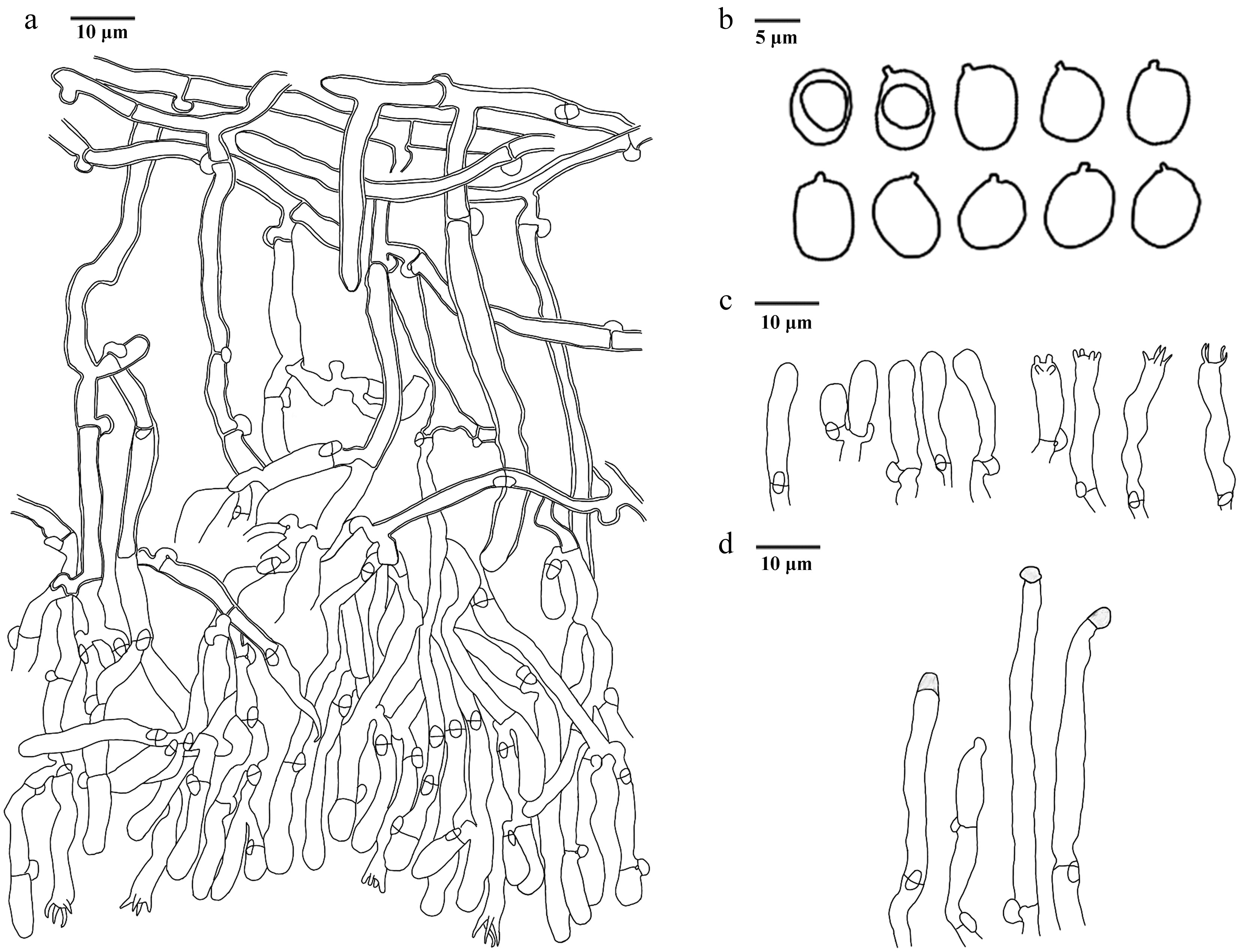
Figure 67.
Microscopic structures of Xylodon albus (drawn from the holotype IFP 020016). (a) Section of hymenium. (b) Basidiospores. (c) Basidia and basidioles. (d) Cystidia.
-
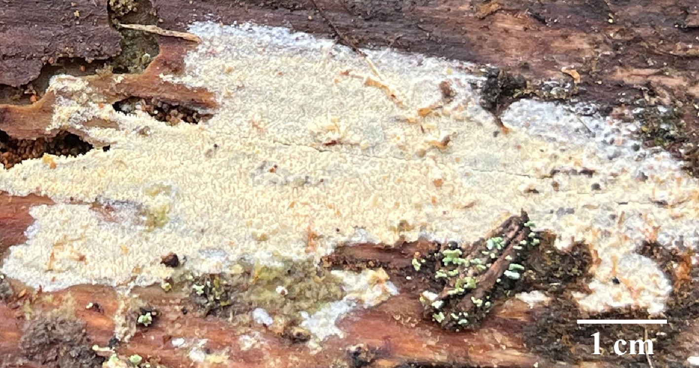
Figure 68.
Basidiomata of Xylodon bicystidiatus (holotype IFP 020017).
-
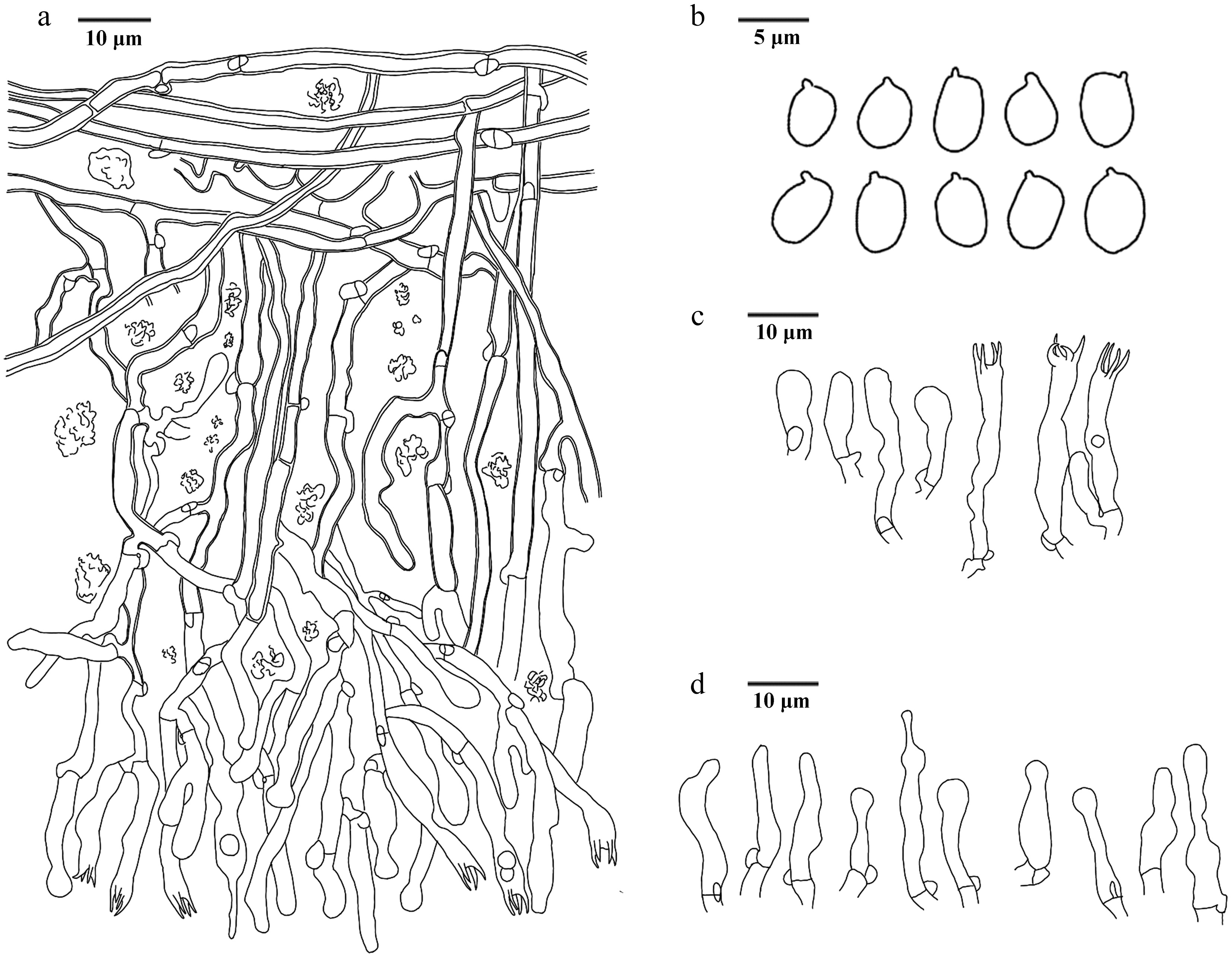
Figure 69.
Microscopic structures of Xylodon bicystidiatus (drawn from the holotype IFP 020017). (a) Section of hymenium. (b) Basidiospores. (c) Basidia and basidioles. (d) Cystidia.
-
Key to species of Hydnellum from China. 1 Basidiospores hyaline 2 1 Basidiospores brown 12 2 Basidia clavate or sinuous 3 2 Basidia clavate 4 3 Pileus round to circular Hydnellum liantaishanense 3 Pileus flabelliform to subcircular Hydnellum tomentosum 4 Generative hyphae with mostly simple-septa, occasionally clamped 5 4 Generative hyphae with simple-septa 7 5 Stipe central 6 5 Stipe central to lateral Hydnellum melanocarpum 6 Pileus infundibuliform Hydnellum concentricum 6 Pileus irregularly flabelliform Hydnellum hydrangeoides 7 Stipe central 8 7 Stipe lateral 10 8 Pileal margin white Hydnellum testaceum 8 Pileal margin not white 9 9 Taste mild Hydnellum infundibuliforme 9 Taste bitter Hydnellum carnosum 10 Basidiospores subglobose to ellipsoidal 11 10 Basidiospores subglobose to globose Hydnellum chocolatum 11 Pilieal surface smooth Hydnellum crassipileatum 11 Pilieal surface fibrillose Hydnellum radiatum 12 Generative hyphae with simple-septa 16 12 Generative hyphae not with simple-septa 13 13 Basidiomata fleshy Hydnellum versipelle 13 Basidiomata woody 14 14 Stipe central 15 14 Stipe lateral Hydnellum atrospinosum 15 Pilieal surface glabrous Hydnellum diabolus 15 Pilieal surface velutinous to tomentose Hydnellum suaveolens 16 Generative hyphae only with simple-septa 20 16 Generative hyphae mostly with simple-septa, occasionally clamped 17 17 Stipe surface glabrous Hydnellum cinnamomea 17 Stipe surface not glabrous 18 18 Stipe surface plushy Hydnellum caeruleum 18 Stipe surface tomentose 19 19 Taste mild Hydnellum fibulatum 19 Taste acrid Hydnellum peckii 20 Pileal surface scaled 21 20 Pileal surface not scaled 28 21 Taste none 22 21 Taste like something 24 22 Pileus depressed Hydnellum subscabrosellum 22 Pileus planar 23 23 Pileus circular Hydnellum subailaoensis 23 Pileus ellipsoid to round Hydnellum grosselepidotum 24 Taste mild Hydnellum edulium 24 Taste bitter 25 25 Pileal margin white Hydnellum fagiscabrosum 25 Pileal margin not white 26 26 Spines up to 5 mm long 27 26 Spines up to 1 mm long Hydnellum lidongensis 27 Pileus planar to plano-convex Hydnellum illudens 27 Pileus depressed Hydnellum glaucopus 28 Stipe lateral 29 28 Stipe central or central to lateral 30 29 Pileal margin incurved, occasionally incised Hydnellum ailaoense 29 Pileal margin lobed Hydnellum sulcatum 30 Stipe central 31 30 Stipe central to lateral 39 31 Pilieal surface glabrous 32 31 Pilieal surface not glabrous 34 32 Stipe surface tomentose to matted 33 32 Stipe surface spongy Hydnellum succulentus 33 Pilieal surface azonate Hydnellum coactum 33 Pilieal surface zonate Hydnellum ferrugineum 34 Pilieal surface azonate 35 34 Pilieal surface zonate 36 35 Taste bitter Hydnellum atrorubrum 35 Taste none Hydnellum porphyreum 36 Taste disagreeable 37 36 Taste none 38 37 Spines up to 5 mm Hydnellum aurantiacum 37 Spines up to 9 mm Hydnellum pineticola 38 Stipe surface subtomentose to matted Hydnellum chrysinum 38 Stipe surface glabrous Hydnellum tarda 39 Pileal margin white 44 39 Pileal margin not white 40 40 Taste none 41 40 Taste bitter 42 41 Basidiospores irregularly Hydnellum martioflavum 41 Basidiospores subglobose Hydnellum earlianum 42 Stipe rugose 43 42 Stipe velutinous to matted Hydnellum spongiosipes 43 Spines up to 4 mm Hydnellum inflatum 43 Spines up to 2 mm Hydnellum granulosum 44 Basidiospores subglobose to globose 45 44 Basidiospores irregular 46 45 Spines up to 2 mm Hydnellum xanthopus 45 Spines up to 4 mm Hydnellum nitida 46 Basidiomata fleshy Hydnellum subalpinum 46 Basidiomata woody 47 47 Stipe surface tomentose 49 47 Stipe surface not tomentose 48 48 Taste acrid Hydnellum bomiense 48 Taste none Hydnellum qinghaiense 49 Taste none Hydnellum squamulosum 49 Taste mild 50 50 Pileal margin lobed 51 50 Pileal margin eroded Hydnellum yunnanense 51 Pileus infundibuliform to flabelliform Hydnellum brunneorubrum 51 Pileus depressed to flabelliform or irregularly circular Hydnellum rubidofuscum -
Key to resupinate species of Thelephora from China. 1 Rhizomorphs present 2 1 Basidiomata absent 29 2 Rhizomorphs differentiated (with a distinct central core and outer layer) 3 2 Rhizomorphs undifferentiated (uniform structure) 8 3 Rhizomorphs dimitic 4 3 Rhizomorphs monomitic 5 4 Rhizomorphs type G; central hyphae usually simple septate Thelephora dimidiata 4 Rhizomorphs type G; central hyphae clamped Thelephora incrustata 5 Cystidia present 6 5 Cystidia absent 7 6 Spores aculeate (up to 2 5 µm long) Thelephora cystidiata 6 Spores echinulate to aculeate (up to 1 5 µm long) Thelephora citrinocystidiata 7 Basidiomata arachnoid, continuous Thelephora qingyuanensis 7 Basidiomata mucedinoid, discontinuous Thelephora brevis 8 Rhizomorphs of type A 9 8 Rhizomorphs of type B 11 9 Rhizomorph hyphae thin-walled Thelephora tenuirhizomorpha 9 Rhizomorph hyphae thick-walled 10 10 Subicular hyphae thick-walled, 3.5–6.5 μm Thelephora fuscocrustosa 10 Subicular hyphae slightly thick- to thick-walled Thelephora pallidomarginata 11 Hyphal septation in rhizomorphs exclusively by simple-septa 12 11 Hyphal septation in rhizomorphs with clamp connections (or both clamps and simple-septa) 14 12 Rhizomorph hyphae ≥ 3 µm 13 12 Rhizomorph hyphae < 3 µm Thelephora efibulata 13 Subicular hyphae thick-walled, smooth, 3–5 μm Thelephora efibulis 13 Subicular hyphae slightly thick walled, with encrustation, 4–8 µm Thelephora olivaceomarginata 14 Rhizomorph hyphae with both clamp connections and simple-septa 15 14 Rhizomorph hyphae possessing exclusively clamp connections 20 15 Spores thick-walled 16 15 Spores slightly thick-walled 17 16 Basidiomata mucedinoid Thelephora angusta 16 Basidiomata arachnoid Thelephora fuscoaraneosa 17 Spores ≤ 7 µm Thelephora aureomarginata 17 Spores > 7 µm 18 18 Sterigmata > 5 µm Thelephora separata 18 Sterigmata ≤ 5 µm 19 19 Cystidia present Thelephora capitatocystidiata 19 Cystidia absent Thelephora brunneoflava 20 Basidiomata discontinuous 21 20 Basidiomata continuous 22 21 Basidia utriform, not stalked Thelephora linzhiensis 21 Basidia clavate, stalked Thelephora interrupta 22 Spores wall-thined Thelephora casiae 22 Spores wall-thicked 23 23 Subglobose to bi-, tri- or quadra-lobed 24 23 Irregularly globose or lobed 25 24 Spores echinulate, up to 1 μm long Thelephora flavidobadia 24 Spores echinulate to aculeate, up to 2 µm long Thelephora guiyangensis 25 Cystidia present Thelephora gloeocystidiata 25 Cystidia absent 26 26 Basidia not stalked Thelephora inconspicua 26 Basidia stalked 27 27 Rhizomorph ≥ 20 µm Thelephora fuscogranulosa 27 Rhizomorph ≤ 20 µm 28 28 Basidia not sinuous, Subhymenial frequently branched Thelephora olivacea 28 Basidia sinuous, Subhymenial occasionally branched Thelephora parvispora 29 Cystidia present Thelephora longicystidiata 29 Cystidia absent 2 30 Spore thin or thin- to slightly thick-walled 3 30 Spore slightly thick-walled or thick-walled 5 31 Spore thin-walled Thelephora rotundata 31 Spore thin- to slightly thick-walled 4 32 Hyphal septation with both simple-septa and clamp connections Thelephora coffeae 32 Hyphal septation exclusively with clamp connections Thelephora globospora 33 Spores slightly thick-walled 6 33 Spores thick-walled 16 34 Spore ornamentation ≥ 2 µm long 7 34 Spore ornamentation < 2 µm long 11 35 Spores > 10 µm long Thelephora nanyigouensis 35 Spores ≤ 10 µm 8 36 Basidia utriform 9 36 Basidia clavate 10 37 Spores with aculeate (isolated), basidia not sinuous Thelephora griseofusca 37 Spores with echinulate (sometimes grouped in 2 or more), basidia sinuous Thelephora segregata 38 Basidia stalked, sterigmata > 10 µm long, subicular hyphae with crystal Thelephora longiaculeifera 38 Basidia not stalked, sterigmata < 10 µm long, subicular hyphae smooth Thelephora duplexa 39 Spores > 7 µm Thelephora bomiensis 39 Spores ≤ 7 µm 12 40 Basidia utriform Thelephora schrenkiana 40 Basidia clavate 13 41 Basidiomata granulose 14 41 Basidiomata smooth 15 42 Sterigmata ≤ 5 µm long Thelephora griseocastanea 42 Sterigmata > 5 µm long Thelephora olivaceobrunnea 43 Spores > 6 long and > 5 µm wide Thelephora griseomarginata 43 Spores ≤ 6 long and ≤ 5 µm wide Thelephora pallidocastanea 44 Spore ornamentation wart-like (verrucose or nodulose) 17 44 Spore ornamentation spine-like (echinulate or aculeate) 21 45 Spore broadly ellipsoid Thelephora velutina 45 Spore subglobose to globose 18 46 Hyphal > 5 µm in diam 19 46 Hyphal ≤ 5 µm in diam 20 47 Basidiospores membranaceous Thelephora wumenshanensis 47 Basidiospores farinaceous Thelephora tenuifarinacea 48 Basidia clavate (< 50 µm long) Thelephora olivaceobasidiosa 48 Basidia cylindrical to subclavate (> 50 µm long) Thelephora yunnanensis 49 Basidia clavate 22 49 Basidia utriform 25 50 Basidiospores > 7 µm long, > 6 µm wide 23 50 Basidiospores < 7 µm long, < 6 µm wide 24 51 Basidiomata arachnoid, continuous; subicular hyphae frequently branched Thelephora asiae-orientalis 51 Basidiomata mucedinoid, uncontinuous; subicular hyphae occasionally branched Thelephora aurimucida 52 Subicular hyphae frequently branched
> 5 μm; hymenophoral surface farinoseThelephora farinosa 52 Subicular hyphae rarely branched
≤ 5 μm; hymenophoral surface granuloseThelephora storea 53 Spore spine-like ornamentation > 1.5 µm long 26 53 Spore spine-like ornamentation ≤ 1.5 µm long 34 54 Spore echinuli usually isolated 27 54 Spore echinuli usually isolated, sometimes grouped in two or more 29 55 Spore echinuli subglobose to bi-lobed Thelephora longiechinuli 55 Spore echinuli subglobose to globose 28 56 Sterigmata > 10 μm long Thelephora conclusa 56 Sterigmata < 10 μm long Thelephora atrocastanea 57 Sterigmata > 6 μm long 30 57 Sterigmata ≤ 6 μm long 32 58 Subicular hyphae occasionally branched
≤ 5 μmThelephora fuscopelliculosa 58 Subicular hyphae frequently branched
> 5 μm31 59 Basidiomata dark blonde to yellowish brown Thelephora stipitata 59 Basidiomata brownish grey to dark brown Thelephora brunneogrisea 60 Hyphal ≤ 5 μm in diam Thelephora exiguelata 60 Hyphal > 5 μm in diam 33 61 Spore > 9 μm long > 8 μm wide Thelephora megaspora 61 Spore < 9 μm long < 8 μm wide Thelephora pallidobrunnea 62 Basidiomata granulose Thelephora fuscofarinosa 62 Basidiomata smooth 35 63 Subhymenial hyphae slightly thick-walled or thick-walled 36 63 Subhymenial hyphae thin-walled 38 64 Basidiomata crustose Thelephora atrobadia 64 Basidiomata mucedinoid 37 65 Subhymenial hyphae slightly thick-walled, occasionally branched > 5 μm Thelephora pertenuis 65 Subhymenial hyphae thick-walled, frequently branched ≤ 5 μm Thelephora latihypha 66 Spore globose or lobed 39 66 Spore subglobose to globose 40 67 Spore irregularly globose or lobed, aculeate, up to 1.5 μm long Thelephora liaoningensis 67 Spore subglobose to bi-, tri-, or quadra-lobed, echinulate, up to 1 μm long Thelephora chayuensis 68 Sterigmata > 5.5 μm Thelephora kanasensis 68 Sterigmata ≤ 5.5 μm 41 69 Spore ornamentation echinulate to aculeate, up to 1 µm long Thelephora aurantispora 69 Spore ornamentation echinulate, up to 1.5 μm long Thelephora changbaiensis -
Key to upright species of Thelephora from China. 1 Cystidia present 2 1 Cystidia absent 4 2 Cystidia one type 3 2 Cystidia two types Thelephora grandinioides 3 Cystidia clavate Thelephora ganbajun 3 Cystidia tubular Thelephora wuliangshanensis 4 Sterigmata 2–4 5 4 Sterigmata 4 9 5 Hyphae commonly clamped and
simple-septateThelephora austrosinensis 5 Hyphae clamped 6 6 Basidiospores umber purple Thelephora caryophyllea 6 Basidiospores fuscous purple 7 7 Basidiomata imbricate 8 7 Basidiomata clavarioid Thelephora palmata 8 Pileus spathulate to flabelliform Thelephora japonic 8 Pileus dimidiate or spuriously
infundibuliformThelephora terrestris 9 Hyphae clamped 10 9 Hyphae clamped and simple-septate 16 10 Basidiospores lobed 12 10 Basidiospores not lobed 11 11 Basidia subclavate Thelephora subtropica 11 Basidia barreled Thelephora yunnanensis 12 Hymenial surface concolorous with
abhymenial surface13 12 Hymenial surface not concolorous with
abhymenial surface14 13 Pileus spathulate to narrow petaloid Thelephora dactyliophora 13 Pileus clavate to pinnatifid or ramiform Thelephora pinnatifida 14 Smell strong Thelephora aurantiotincta 14 Smell not strong 15 15 Basidiospores umber brown Thelephora penicillata 15 Basidiospores olive buff Thelephora vialis 16 Stipe central 18 16 Stipe central to lateral 17 17 Basidiospores bluish green Thelephora pseudoganbajun 17 Basidiospores yellowish brown Thelephora petaloides 18 Basidiospores hyaline Thelephora sikkimensis 18 Basidiospores not hyaline 19 19 Abhymenial surface azonate 20 19 Abhymenial surface zonate 22 20 Pileus imbricate Thelephora lacunosa 20 Pileus coralloid 21 21 Pileal margin deeply lacerate Thelephora nigromarginata 21 Pileal margin tips needle-like Thelephora scopiformis 22 Smell mild 23 22 Smell no odor 25 23 Stipe subconical to broadened or flatted Thelephora cacao 23 Stipe clavillose to flatted or broadened 24 24 Pileus infundibuliform to ligulate Thelephora fasciculata 24 Pileus clavate to coralliform Thelephora microcarpa 25 Abhymenial surface radially rugulose or
wrinkled26 25 Abhymenial surface wrinkled Thelephora nebula 26 Pileal margin imperceptibly wavy Thelephora glaucoflora 26 Pileal margin irregularly lobed to wavy Thelephora aquila
Figures
(66)
Tables
(3)