-
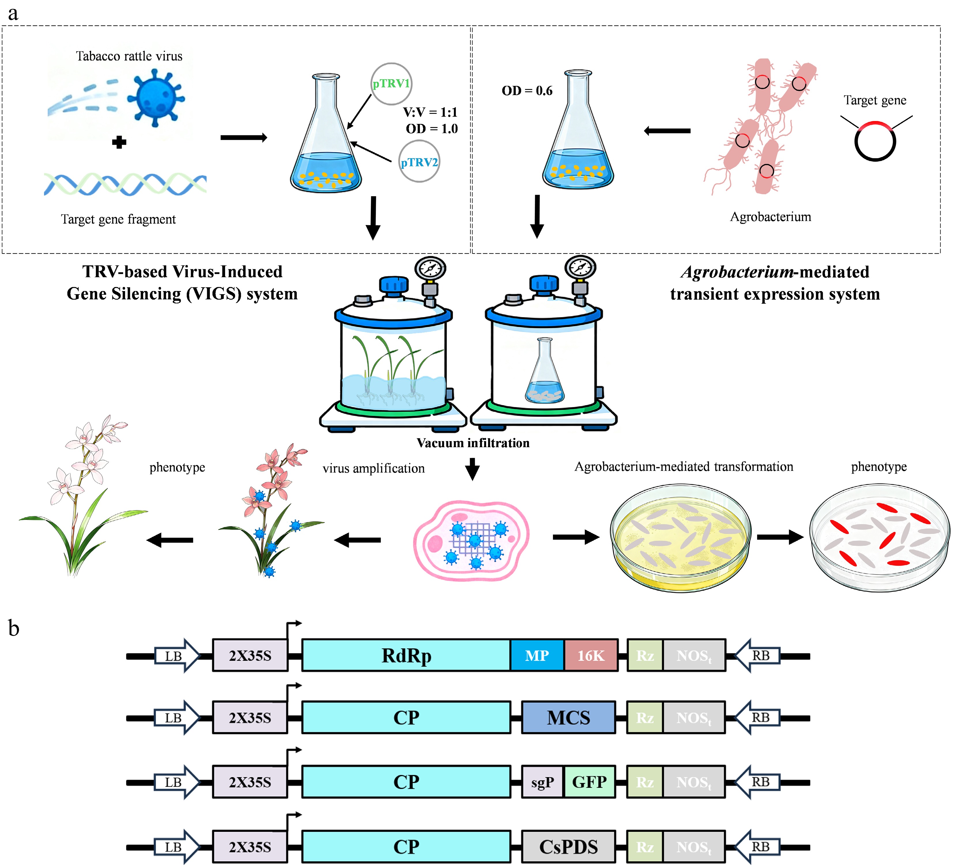
Figure 1.
Virus-induced gene silencing (VIGS) and Agrobacterium-mediated transient expression in Cymbidium. (a) A workflow for VIGS and Agrobacterium-mediated transient expression infiltration in Cymbidium. (b) Schematics of pTRV1, pTRV2, pTRV-GFP, and pTRV2-CsPDS. RdRp, RNA-dependent RNA polymerase; MP, movement protein; 16k, 16-kD protein; CP, coat protein; sgP, subgenomic promoter; MCS, multiple cloning site; LB, left border; RB, right border; Rz, self-cleaving ribozyme; NOSt, NOS terminator.
-
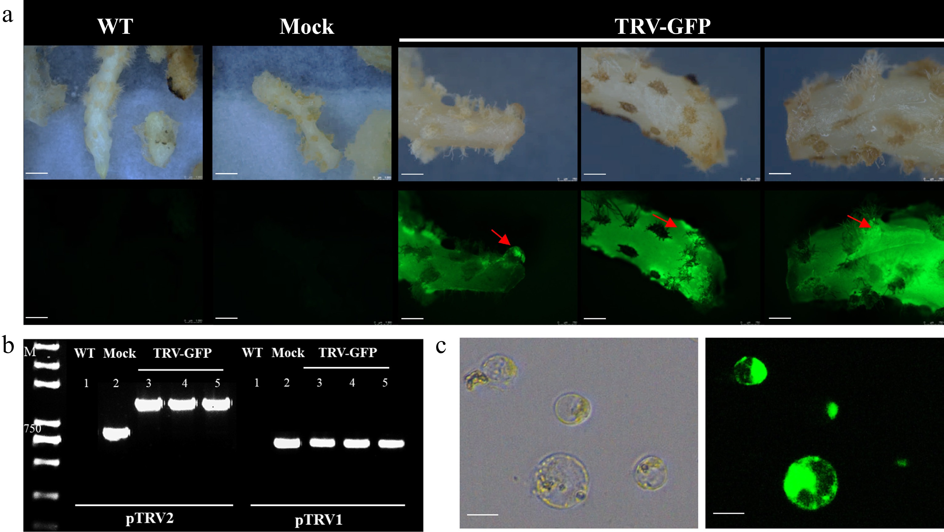
Figure 2.
Tobacco rattle virus (TRV) infection and systemic movement in Cymbidium tissues. (a) VIGS of the rhizomes of Cymbidium with pTRV2-GFP. Green fluorescent protein was detected in the infected rhizomes, and this fluorescence was transmitted to the uninfected newly emerging bud sites. WT, wild-type; Mock, rhizomes injected with pTRV1 + pTRV2; TRV-GFP, rhizomes injected with pTRV1 + pTRV2-GFP. Scale bar: 1,000 μm. (b) RT-PCR detection of TRV2 and TRV2-GFP vectors in infected tissues. M, marker. (c) Transient GFP expression in Cymbidium protoplasts transfected with pTRV2-GFP. Cells were examined 20 h after inoculation under the fluorescence microscope. Scale bar: 50 μm.
-
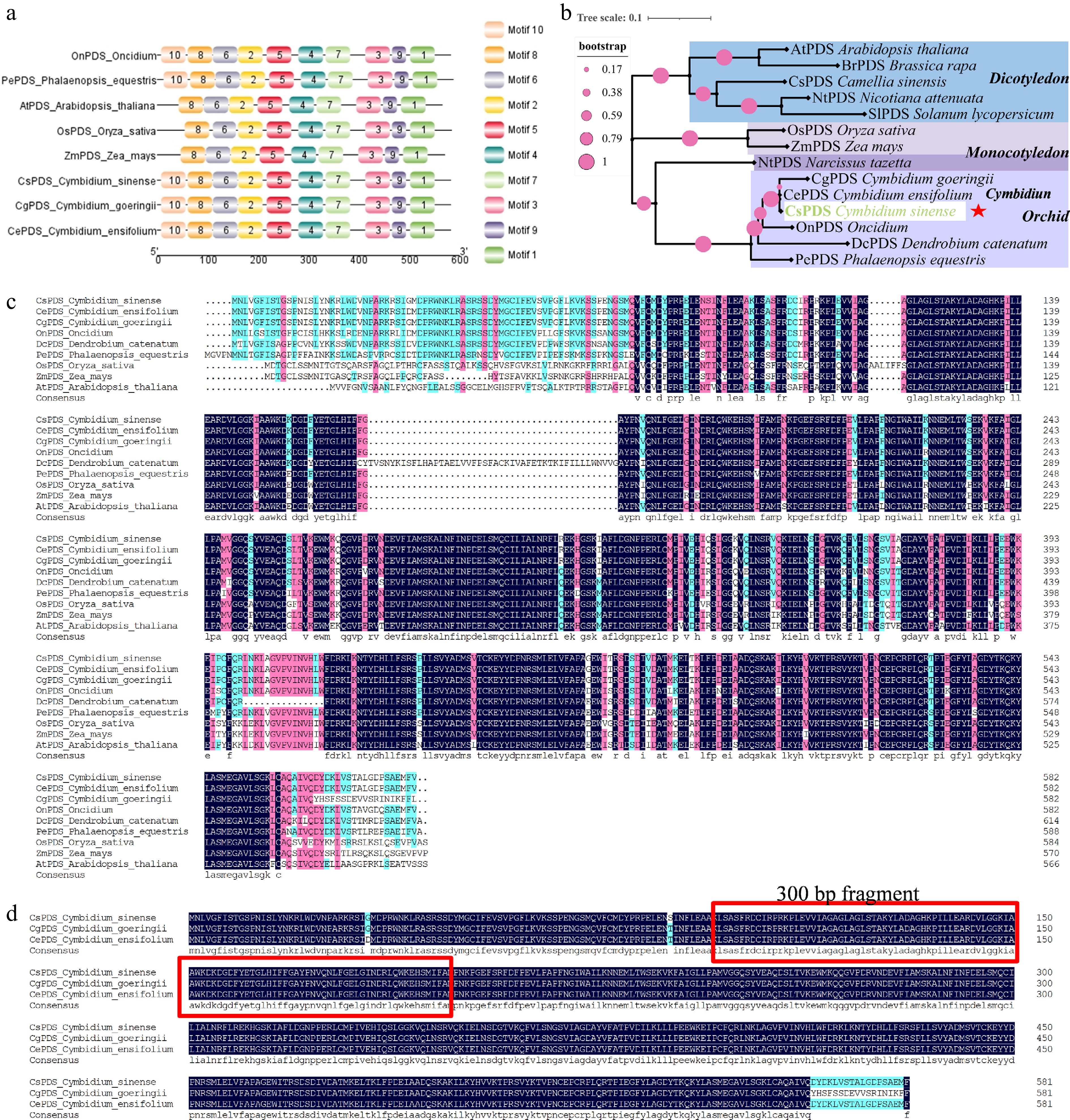
Figure 3.
Identification of the PDS gene in C. sinense. (a) Conserved protein motifs identified in CsPDS and orthologs from other plant species. (b) Maximum likelihood phylogenetic tree of PDS proteins from C. sinense and selected angiosperms. The CsPDS clade is highlighted with a red star. (c) Multiple sequence alignment of PDS protein sequences from C. sinense and other plants. (d) Protein sequence alignment of PDS from three Cymbidium species.
-
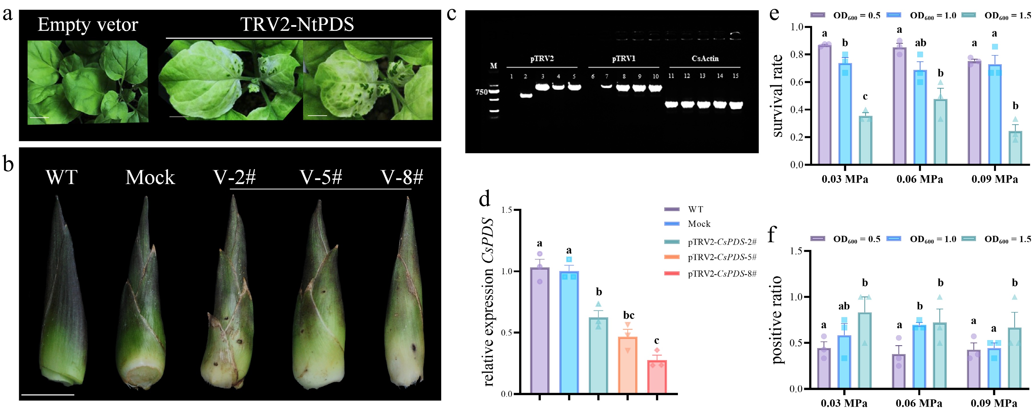
Figure 4.
Functional analysis of CsPDS silencing in C. sinense. (a) Positive control for the VIGS system: Nicotiana benthamiana leaves infiltrated with pTRV2-NtPDS exhibit photobleaching. Scale bar: 10 mm. (b) Photobleaching phenotype in C. sinense leaf buds after treatment with pTRV1 + pTRV2-CsPDS. (c) PCR-based tracking of TRV vectors confirms successful infection and replication in C. sinense tissues. WT: 1, 6, and 11; Mock: 2, 7, and 12; pTRV2-CsPDS: 3, 4, 5, 8, 9, 10, 13, 14, and 15. (d) Expression of CsPDS was downregulated in photobleached leaf buds in C. sinense. Significance was evaluated by one-way analysis of variance. The survival rates (e) and positive rates (f) of the plants in the infection solutions with different levels of negative pressure and different OD600 values after infection. Data represent the mean ± standard error of the mean (SEM) of three biological replicates. The transient transformation rate = number of positive plants / total number of infiltrated plants.
-
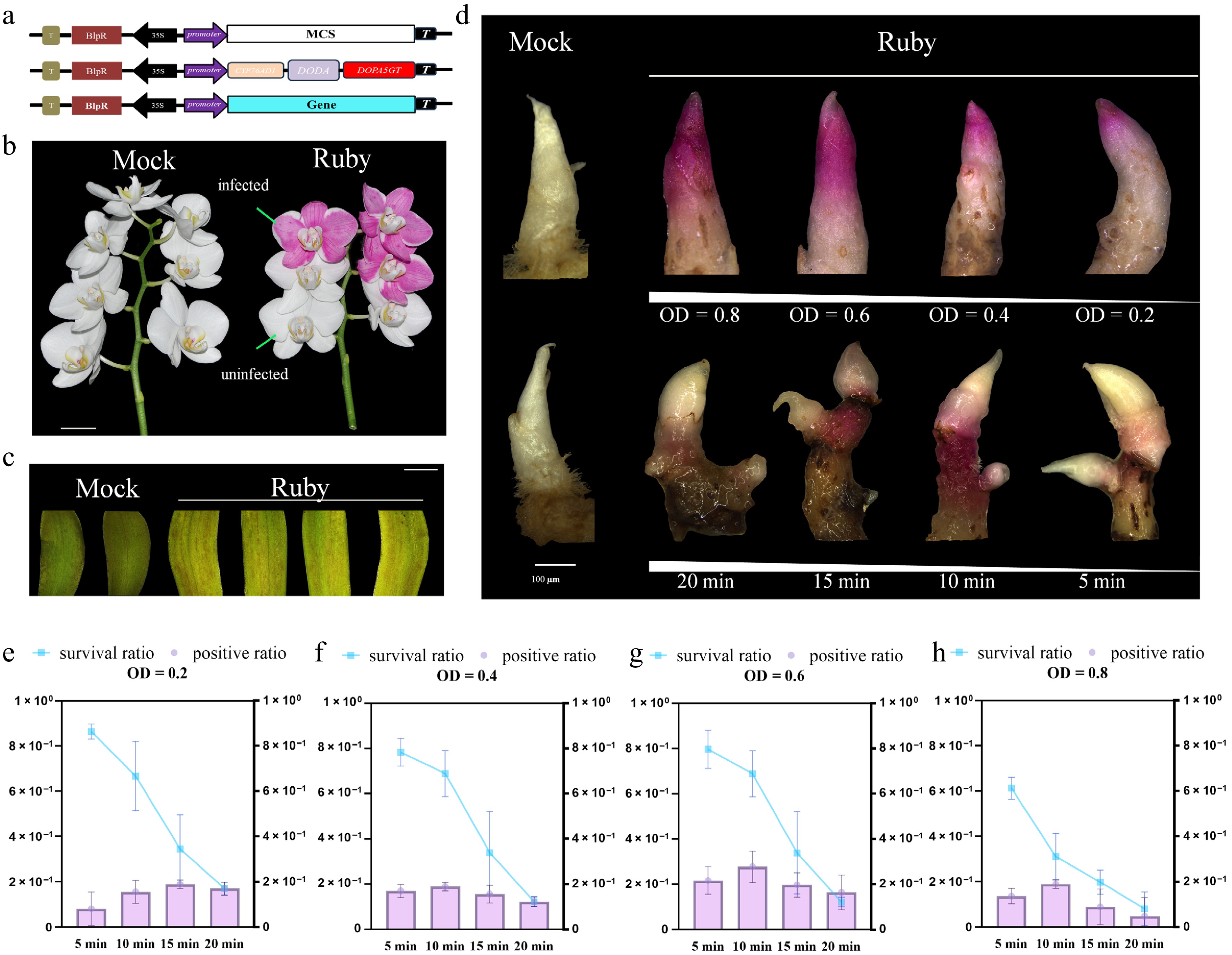
Figure 5.
Establishing the Ruby reporter for transient transformation in Cymbidium. (a) Schematics of the Ruby expression vector. (b) Phenotype of the Ruby reporter in the petals of Phalaenopsis. Scale bar: 10 mm. (c) Agrobacterium-mediated transient expression of the Ruby reporter in the petals of Cymbidium. Scale bar: 5 mm. (d) Distinct red-purple pigmentation was observed on Cymbidium rhizomes following infiltration with different Agrobacterium concentrations (upper). The growth phenotype of rhizomes was also recorded after various vacuum infiltration durations (lower). Scale bar: 100 μm. (e)–(h) Survival rates and positive rates of Cymbidium rhizomes in the infection solutions with different times of vacuum infiltration and different OD600 values after infection. Data represent the mean ± SEM of three biological replicates. The transient transformation rate = number of positive plants/total number of infiltrated plants.
Figures
(5)
Tables
(0)