-
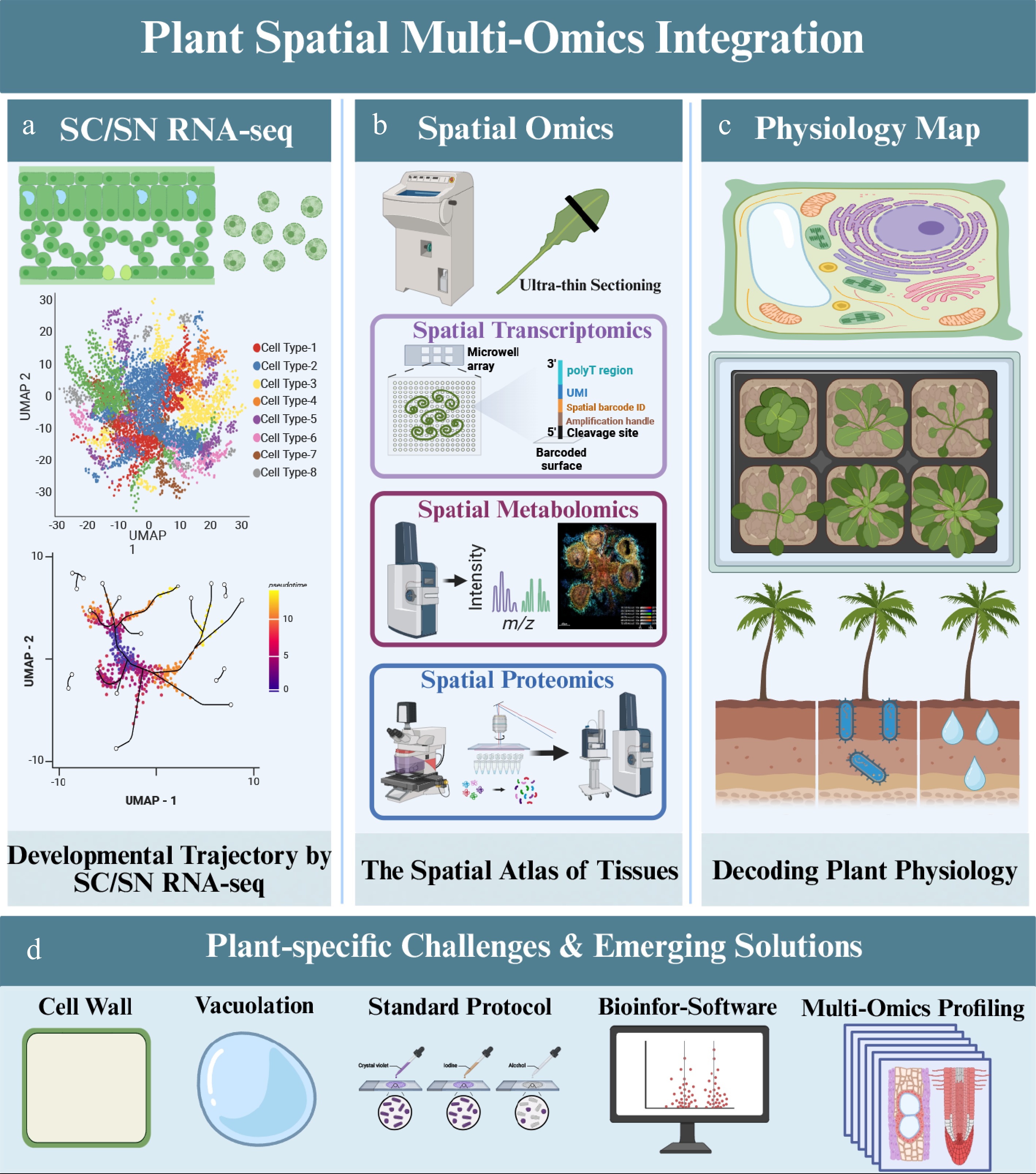
Figure 1.
A strategic roadmap for integrating single-cell and spatial multi-omics to decode plant physiological complexity. This schematic illustrates the workflow for constructing a multi-dimensional 'digital twin' of plant tissues. (a) Single-cell and single-nucleus transcriptomic profiling resolves cellular heterogeneity and developmental trajectories. (b) Spatially resolved transcriptomic, metabolomic, and proteomic methods map molecular states back to tissue coordinates. The capture strategy shown in panel (b) represents one illustrative technical route for spatial transcriptomics and does not encompass all available spatial transcriptomics platforms. (c) Cross-modality integration generates a spatially anchored molecular atlas of plant tissues. (d) Plant-specific constraints—including rigid cell walls, vacuolation, and analyte delocalization—shape experimental design and interpretation. Created with BioRender.
-
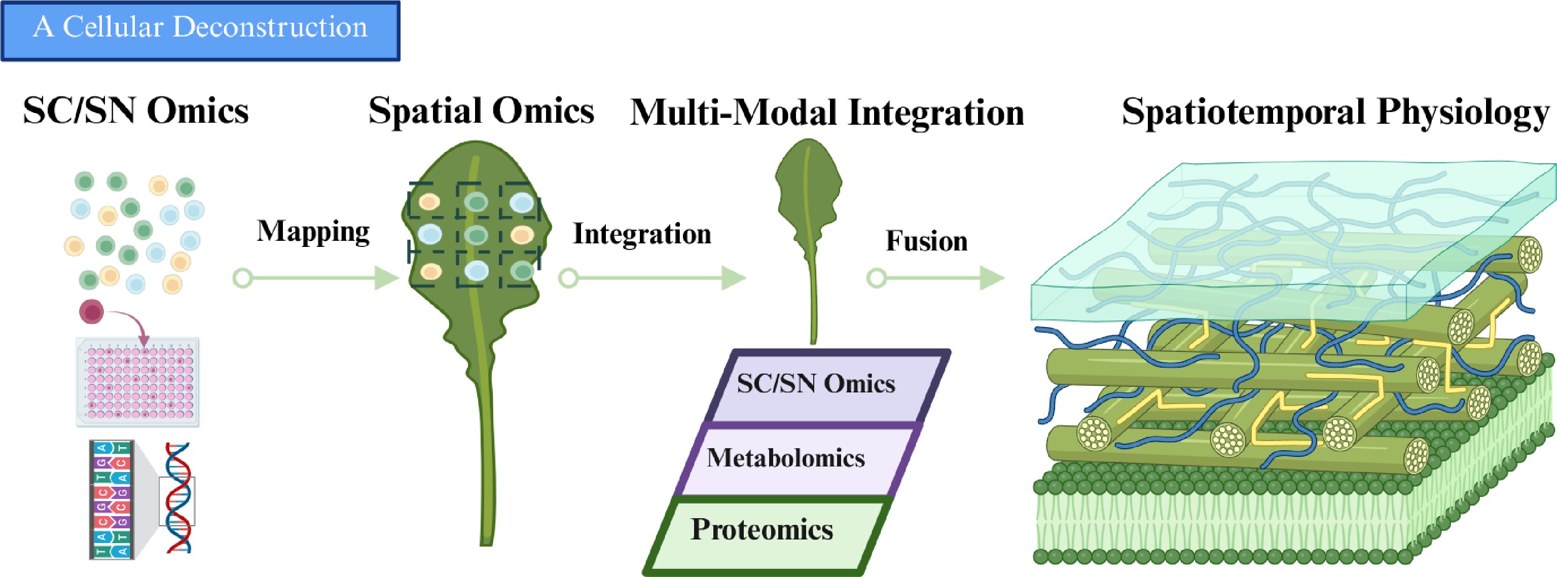
Figure 2.
Minimum operational framework for a plant spatial 'digital twin'. The framework includes spatially registered histology, a matched sc/snRNA-seq reference for improved detection of rare cell populations and spatial deconvolution, same-section co-detection of complementary molecular layers, region-matched proteomics from adjacent sections, explicit cross-section registration with uncertainty control, and orthogonal validation of inferred relationships. Created with BioRender.
-
Modality class Representative platforms/methods Typical output/use Main plant constraints Failure modes/QC Status in plants Protoplast-based scRNA-seq 10x Chromium; Drop-seq-type workflows; DNBelab C Series; Smart-seq2/3 Single-cell transcriptomes; atlases, trajectories, stress states Protoplasting bias; stress artifacts; selective cell loss Low viability; composition bias; organellar RNA; QC: viability, genes/UMIs, doublets Mature, but tissue-dependent snRNA-seq 10x Chromium nuclei workflows; DNBelab C Series; plate- or droplet-based nuclei workflows Nuclear transcriptomes; difficult, lignified, mature, or frozen tissues Nuclear/cytoplasmic differences; debris, starch, metabolites Ambient RNA; low complexity; QC: nuclei integrity, genes/UMIs, marker concordance Highly practical Single-cell chromatin/
multiome10x Single Cell ATAC; 10x Multiome; sci-ATAC-seq-type workflows Accessibility ± RNA; regulatory inference High-quality nuclei required; sparse signal Low fragments; poor TSS; QC: FRiP/TSS, doublets, integration quality Emerging to moderately established Capture-based whole-transcriptome ST 10x Visium/Visium HD; Stereo-seq; DBiT-seq-type workflows; plant-adapted commercial ST services Section-wide spatial transcriptomes; zonation and gradients Section quality; permeabilization; cell walls; vacuolation; cuticles Diffusion; mixed capture; organellar RNA; QC: section integrity, genes/UMIs, spatial concordance Most mature spatial transcriptomic class Imaging-/ hybridization-based targeted ST smFISH; RNAscope; MERFISH; seqFISH-type methods; in situ sequencing-related workflows High-precision localization of selected transcripts Probe penetration; autofluorescence; optical opacity Background; undercounting; QC: signal/background, specificity, reproducibility Powerful, but not routine ROI-/LCM-guided spatial transcriptomics LCM-RNA-seq; Geo-seq-type workflows; histology-guided ROI profiling Region-specific transcriptomes; defined anatomical domains RNA preservation; low input; regional contamination Degradation; low yield; QC: RNA quality, region purity, library complexity Practical for focused questions Spatial proteomics: imaging-based Multiplex IF; cyclic IF; CODEX-type workflows; imaging mass cytometry (IMC) In situ protein localization and tissue-domain validation Limited antibodies; epitope preservation; autofluorescence Weak specificity; background; QC: antibody validation, controls, image quality Emerging and reagent-limited Spatial proteomics:
LCM-guidedLCM-LC–MS/MS; low-input microproteomics; nanoPOTS-type workflows Region-matched proteomes Low input; extraction from wall-rich tissues; peptide loss Shallow depth; variable recovery; QC: peptide/protein counts, reproducibility Promising, but demanding Spatial metabolomics/
MSIMALDI-MSI; DESI-MSI; SIMS/TOF-SIMS; AFADESI-MSI Spatial metabolite maps; defense and stress chemistry Delocalization; ion suppression; wax/cuticle barriers Analyte loss; annotation uncertainty; QC: freezing, matrix consistency, calibration, validation Increasingly important, but method-sensitive Table 1.
Practical comparison of major single-cell, single-nucleus, and spatial omics modalities in plant research.
-
Analytical goal/research scenario Preferred omics layer(s) Recommended modality/representative methods Key sample-preparation priorities Core QC checkpoints Major pitfalls/interpretation caveats Cell-type and cell-state discovery in dissociable tissues Transcriptome Protoplast-based scRNA-seq; 10x Chromium; Drop-seq-type workflows; Smart-seq2/3 Optimize enzymatic digestion, osmotic balance, and handling time; minimize protoplasting stress; process biological replicates independently Cell viability; genes/UMIs per cell; organellar RNA fraction; doublets; recovery of expected marker-defined populations Dissociation bias can distort cell composition; stress-induced transcription may masquerade as biology; fragile or highly vacuolated cells may be underrepresented Profiling difficult, mature, lignified, frozen, or chemically interfering tissues Transcriptome snRNA-seq; nuclei-compatible droplet workflows; plate-based nuclear RNA-seq Preserve nuclei integrity; reduce debris, starch, mucilage, and phenolic carryover; standardize homogenization and filtration Nuclei integrity; genes/UMIs per nucleus; ambient RNA; organellar contamination; agreement with expected markers Nuclear and whole-cell transcriptomes are not directly equivalent; low-complexity nuclei and background contamination can inflate false heterogeneity Organ-wide spatial mapping of tissue domains, zonation, and expression gradients Spatial transcriptome Capture-based whole-transcriptome ST; 10x Visium/Visium HD; Stereo-seq; DBiT-seq-type methods Optimize sectioning, fixation, permeabilization, RNA capture, and histology registration for each tissue type Section integrity; genes/UMIs per spot/bin; organellar fraction; histology concordance; spatial reproducibility across sections Spot/bin signals often reflect mixed-cell capture rather than true single-cell resolution; diffusion and registration errors can distort boundaries High-resolution validation of selected spatial markers Targeted spatial transcriptome smFISH; RNAscope; MERFISH; seqFISH-type methods Optimize fixation, probe penetration, autofluorescence suppression, and imaging depth; preserve anatomy for segmentation Signal-to-background ratio; probe specificity; reproducibility across sections; segmentation quality Limited gene panels restrict discovery; poor penetration or high background may produce false negatives or misleading enrichment patterns Molecular profiling of anatomically defined regions, rare structures, or histologically selected domains Region-resolved transcriptome and/or proteome LCM-RNA-seq; Geo-seq-type workflows; histology-guided ROI profiling; LCM-LC–MS/MS; low-input microproteomics Maintain RNA/protein integrity during sectioning and microdissection; define ROIs precisely; minimize contamination from adjacent regions RNA quality or protein recovery; region purity; library/proteome complexity; concordance with histology Region-level data should not be overinterpreted as true single-cell profiles; low input and contamination are common limiting factors Inference of regulatory programs and chromatin dynamics Chromatin accessibility, optionally paired with transcriptome scATAC-seq; 10x Single Cell ATAC; sci-ATAC-seq-type workflows; single-cell multiome; 10x Multiome Use exceptionally clean nuclei; reduce debris and clumps; optimize accessibility workflow; prepare matched RNA references where possible Fragment counts; TSS enrichment; FRiP or equivalent; doublets; cluster stability; concordance with RNA-based annotation Data sparsity is high; annotation is often indirect; low-quality nuclei can mimic biological variability; inferred regulatory links require orthogonal validation Protein-level spatial profiling and validation of transcript-defined domains Protein spatial layer Multiplex IF; cyclic IF; CODEX-type workflows; imaging mass cytometry (IMC); immunostaining; LCM-guided microproteomics Validate antibodies rigorously; optimize fixation, epitope preservation, autofluorescence reduction, and extraction from wall-rich tissues Antibody specificity; positive/negative controls; reproducibility across sections; peptide/protein identifications for MS-based workflows Antibody availability is a major bottleneck in plants; transcript and protein abundance may diverge substantially; low-input proteomics may have limited depth Mapping metabolites, specialized chemistry, defense compounds, or storage molecules in situ Metabolite spatial layer MALDI-MSI; DESI-MSI; SIMS/TOF-SIMS; AFADESI-MSI Rapid freezing; prevent analyte delocalization; optimize matrix deposition or ionization conditions; handle cuticle/wax-rich tissues carefully Spatial fidelity; matrix consistency where relevant; mass accuracy; calibration; reproducibility; confidence of metabolite annotation Metabolite identification is often uncertain; ion suppression, analyte delocalization, and tissue chemistry strongly affect signals; transcript levels do not necessarily predict metabolite abundance Cross-modal integration, comparative validation, and atlas building in non-model or crop species RNA + chromatin and/or protein and/or metabolite sc/snRNA-seq integrated with scATAC-seq, multiome, ST, imaging proteomics, MSI, or ROI-guided omics Harmonize sample processing and metadata across modalities; use matched or serial sections where relevant; validate marker conservation and species-specific annotations Registration quality; modality concordance; mapping rate; annotation confidence; replicate stability; robustness of integrated conclusions Cross-modal integration can create false confidence if individual layers are weak; cross-species marker transfer may be misleading; apparent novelty may reflect incomplete annotation or protocol bias Table 2.
Cross-omics practical decision guide for plant single-cell, single-nucleus, and spatial profiling.
Figures
(2)
Tables
(2)