











-
For decades, 'bulk' omics technologies have served as the foundation of plant systems biology, providing a global view of molecular responses[1]. However, these population-averaged measurements inherently mask cellular heterogeneity, obscuring the rare cell types and microenvironmental niches that often drive developmental decisions and stress adaptations. The advent of single-cell/nucleus RNA sequencing (sc/snRNA-seq) marked a paradigm shift, allowing researchers to dissect plant tissues at cellular resolution and reconstruct developmental trajectories with unprecedented precision[2]. Yet, standard SC workflows lack a critical dimension: spatial context. Because tissue dissociation is required to isolate cells or nuclei, the native positional context—essential for understanding cell-to-cell communication and morphogen gradients—is lost. To bridge this gap, spatial transcriptomics (ST) has emerged as a transformative technology, enabling the mapping of gene expression within the morphological context of intact tissues. Furthermore, as biological function is ultimately executed by proteins and metabolites rather than transcripts alone, the field is now moving beyond the transcriptome. The integration of spatial proteomics and metabolomics promises to deliver a holistic 'digital twin' of plant tissues, linking gene regulatory networks to their functional end-products[3].
Despite these advances, the application of spatial multi-omics in plants faces challenges that are distinct from those in mammalian systems, primarily because of the rigid cell wall, extensive vacuolization, abundant plastids, and complex secondary metabolism. As a result, methods that offer the greatest molecular breadth are not always the ones most compatible with plant tissues. This trade-off is particularly acute in tropical species, where heat adaptation, pathogen pressure, and specialized metabolism often intersect with anatomically recalcitrant tissues. For this reason, tropical species are not merely an application subset of plant spatial biology; they represent some of the most demanding and informative systems for testing whether SC and spatial multi-omics technologies can capture tissue function under real agronomic and ecological constraints. This review is organized around a progression from single-cell and single-nucleus profiling toward spatially resolved multi-omics, treating the former as the molecular resolution foundation and the latter as the spatial integration framework, with the ultimate goal of outlining a path toward unified spatiotemporal plant tissue models.
-
Plant cell-resolved omics now encompasses a rapidly expanding set of technologies for capturing transcripts, chromatin states, proteins, and metabolites across dissociated cells, nuclei, and intact tissue sections. At the level of modality design, these platforms differ in molecular scope, spatial resolution, throughput, and compatibility with discovery-driven vs targeted analysis. However, in plants, nominal platform capability does not always translate directly into effective performance, because molecular capture is strongly conditioned by tissue anatomy, biochemical background, and sample handling. The following sections, therefore, compare the major platform classes and their analytical strengths, while a later section addresses the plant-specific preparation constraints that often determine whether these capabilities can be realized in practice. A comparative overview of the major platform classes discussed in this review is provided in Table 1.
Table 1. Practical comparison of major single-cell, single-nucleus, and spatial omics modalities in plant research.
Modality class Representative platforms/methods Typical output/use Main plant constraints Failure modes/QC Status in plants Protoplast-based scRNA-seq 10x Chromium; Drop-seq-type workflows; DNBelab C Series; Smart-seq2/3 Single-cell transcriptomes; atlases, trajectories, stress states Protoplasting bias; stress artifacts; selective cell loss Low viability; composition bias; organellar RNA; QC: viability, genes/UMIs, doublets Mature, but tissue-dependent snRNA-seq 10x Chromium nuclei workflows; DNBelab C Series; plate- or droplet-based nuclei workflows Nuclear transcriptomes; difficult, lignified, mature, or frozen tissues Nuclear/cytoplasmic differences; debris, starch, metabolites Ambient RNA; low complexity; QC: nuclei integrity, genes/UMIs, marker concordance Highly practical Single-cell chromatin/
multiome10x Single Cell ATAC; 10x Multiome; sci-ATAC-seq-type workflows Accessibility ± RNA; regulatory inference High-quality nuclei required; sparse signal Low fragments; poor TSS; QC: FRiP/TSS, doublets, integration quality Emerging to moderately established Capture-based whole-transcriptome ST 10x Visium/Visium HD; Stereo-seq; DBiT-seq-type workflows; plant-adapted commercial ST services Section-wide spatial transcriptomes; zonation and gradients Section quality; permeabilization; cell walls; vacuolation; cuticles Diffusion; mixed capture; organellar RNA; QC: section integrity, genes/UMIs, spatial concordance Most mature spatial transcriptomic class Imaging-/ hybridization-based targeted ST smFISH; RNAscope; MERFISH; seqFISH-type methods; in situ sequencing-related workflows High-precision localization of selected transcripts Probe penetration; autofluorescence; optical opacity Background; undercounting; QC: signal/background, specificity, reproducibility Powerful, but not routine ROI-/LCM-guided spatial transcriptomics LCM-RNA-seq; Geo-seq-type workflows; histology-guided ROI profiling Region-specific transcriptomes; defined anatomical domains RNA preservation; low input; regional contamination Degradation; low yield; QC: RNA quality, region purity, library complexity Practical for focused questions Spatial proteomics: imaging-based Multiplex IF; cyclic IF; CODEX-type workflows; imaging mass cytometry (IMC) In situ protein localization and tissue-domain validation Limited antibodies; epitope preservation; autofluorescence Weak specificity; background; QC: antibody validation, controls, image quality Emerging and reagent-limited Spatial proteomics:
LCM-guidedLCM-LC–MS/MS; low-input microproteomics; nanoPOTS-type workflows Region-matched proteomes Low input; extraction from wall-rich tissues; peptide loss Shallow depth; variable recovery; QC: peptide/protein counts, reproducibility Promising, but demanding Spatial metabolomics/
MSIMALDI-MSI; DESI-MSI; SIMS/TOF-SIMS; AFADESI-MSI Spatial metabolite maps; defense and stress chemistry Delocalization; ion suppression; wax/cuticle barriers Analyte loss; annotation uncertainty; QC: freezing, matrix consistency, calibration, validation Increasingly important, but method-sensitive -
Bulk RNA sequencing has been instrumental for studying plant physiology, but it averages signals across cell populations and therefore masks cell-type-specific programs. The transition to scRNA-seq enabled high-resolution dissection of cellular heterogeneity and regulatory programs (Fig. 1a)[4−6]. Early implementations primarily relied on isolating single cells via fluorescence-activated cell sorting (FACS) or laser capture microdissection (LCM) into multi-well plates[4]. While these plate-based strategies offer high sensitivity and full-length transcript coverage, they are inherently constrained by low throughput, high cost per cell, and labor-intensive workflows. From a technology perspective, plant scRNA-seq has been implemented on multiple high-throughput platforms. A fundamental challenge underlying all single-cell sequencing platforms is the need to amplify or otherwise process the extremely limited nucleic acid content of a single cell—typically picogram-scale RNA—to quantities sufficient for library construction and sequencing. Early plate-based methods addressed this through full-length cDNA amplification strategies (e.g., Smart-seq2[7]), which offer high sensitivity but low throughput[8]. Droplet-based platforms instead rely on in-droplet barcoding and 3′-end capture to scale throughput, accepting reduced per-cell sensitivity as a trade-off.
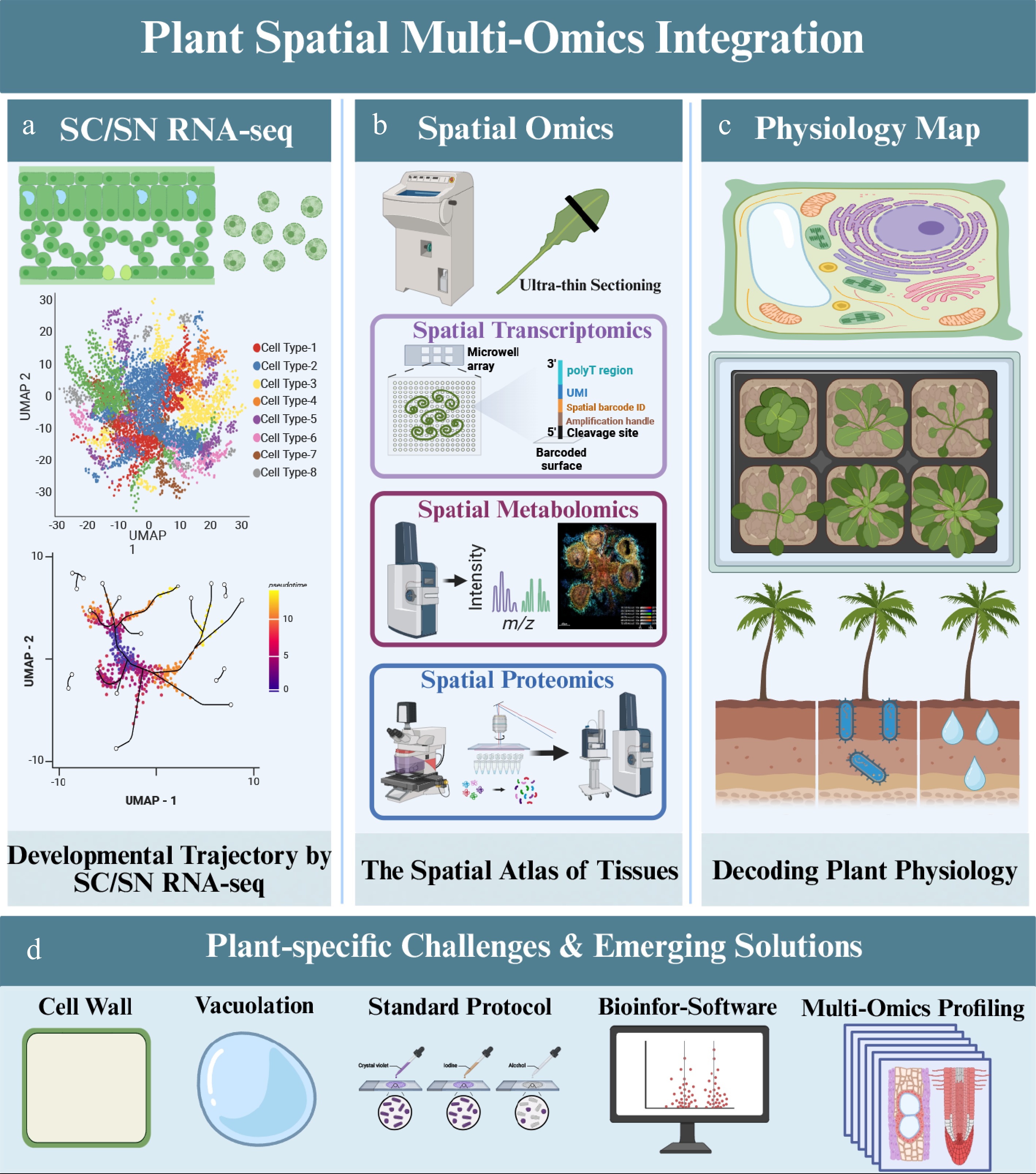
Figure 1.
A strategic roadmap for integrating single-cell and spatial multi-omics to decode plant physiological complexity. This schematic illustrates the workflow for constructing a multi-dimensional 'digital twin' of plant tissues. (a) Single-cell and single-nucleus transcriptomic profiling resolves cellular heterogeneity and developmental trajectories. (b) Spatially resolved transcriptomic, metabolomic, and proteomic methods map molecular states back to tissue coordinates. The capture strategy shown in panel (b) represents one illustrative technical route for spatial transcriptomics and does not encompass all available spatial transcriptomics platforms. (c) Cross-modality integration generates a spatially anchored molecular atlas of plant tissues. (d) Plant-specific constraints—including rigid cell walls, vacuolation, and analyte delocalization—shape experimental design and interpretation. Created with BioRender.
Droplet-based systems, such as the 10x Genomics Chromium and BGI's DNBelab C Series platforms, have enabled scalable profiling of tens of thousands of cells, facilitating multi-field plant studies[9,10]. The first major application of single-cell technologies is the construction of cell atlases, which allow us to understand cellular composition and transcriptomic features across cell clusters[11−14]. Beyond cataloging cell types, these studies have reshaped our understanding of plant tissues as dynamic systems composed of continuous transcriptional states rather than a small set of fixed identities. In Arabidopsis roots and stomatal lineages, sc transcriptomics resolved intermediate states and developmental branching points that are largely inaccessible to bulk profiling[4,15]. Similarly, in pathogen-challenged leaves, cell-resolved analyses revealed that immune activation is not uniformly distributed across tissues but instead involves highly selective and context-dependent responses among neighboring cell populations[16]. In metabolic systems, sc approaches further demonstrated that specialized biosynthetic programs are often confined to restricted cellular niches, providing a more mechanistic framework for linking transcriptional regulation to tissue function[17,18]. In tropical crops, sc profiling has begun to reveal tissue-specific cellular heterogeneity that bulk approaches cannot resolve: in cassava leaves, developmental and physiological specialization is distributed across distinct epidermal, mesophyll, and vascular cell populations[19], while in banana root tips, single-cell transcriptomics has uncovered cell-fate determination programs underlying root development[20].
Spatial transcriptomics (ST) extends transcriptome profiling into intact tissue contexts, making it possible in principle to reconstruct spatial gradients and tissue programs that are lost during dissociation. While sc/snRNA-seq resolves cellular heterogeneity at high resolution, it requires tissue dissociation and thus removes the positional information that is often essential for interpreting cell fate and function in plants[21]. ST addresses this limitation by measuring gene expression in situ, enabling the reconstruction of spatial gradients and tissue programs within their native morphology (Fig. 1b).
Current ST technologies can be broadly organized into three groups: capture-based NGS platforms, imaging-based transcript mapping (e.g., multiplexed FISH), and microdissection-based approaches (e.g., laser capture microdissection, LCM), in which RNA is sequenced from histologically defined regions of interest (ROI)[22]. NGS-based platforms are typically transcriptome-wide and discovery-driven, and in current practice, they are often the most suitable choice for atlas-scale mapping across tissues and conditions; however, limited transcript capture per spatial unit and spot/bin-limited resolution can reduce sensitivity and mix signals from multiple cells, often requiring computational deconvolution with sc/snRNA-seq references. Imaging-based platforms achieve cellular to subcellular localization but are usually lower-throughput and less scalable due to targeted gene panels and intensive imaging workflows. Microdissection-based strategies offer flexible ROI selection and compatibility with standard RNA-seq assays, but their effective spatial resolution and throughput are constrained by ROI size and sampling effort[23−25].
Advances in capture-based NGS ST, including Visium HD (10x Genomics) and Stereo-seq (Spatial enhanced resolution omics-sequencing)-derived high-density workflows, have pushed nominal physical resolution toward the cellular scale. In plants, however, nominal resolution should not be interpreted as equivalent to effective biological resolution. Accurate cell-level inference remains limited by irregular cell geometry, segmentation difficulty, autofluorescence, and lateral analyte diffusion during tissue processing[26]. These constraints are especially relevant in plant tissues because the stronger or longer permeabilization often required to overcome rigid cell walls can improve transcript release at the cost of reduced spatial fidelity.
From a practical standpoint, it is therefore useful to distinguish formal platform specifications from current deployability in plant systems. In current plant research, capture-based NGS workflows remain the most practical options for transcriptome-wide spatial mapping, because they combine broad molecular coverage with comparatively mature experimental workflows. By contrast, imaging-based high-plex methods and true same-section multimodal workflows remain less standardized in plants and often require substantial assay customization. For crop-focused studies, this distinction is important: the most technically advanced platform is not necessarily the most robust choice for a given tissue type or experimental objective.
Despite these intrinsic limitations, capture-based NGS ST remains the near-term workhorse for plant spatial omics because it combines transcriptome-wide coverage with practical deployability[1]. Recent studies illustrate its value in resolving developmental plasticity and tissue compartmentalization: stereo-seq in tomato callus identified chlorenchyma niches associated with shoot regeneration[27], while spatially resolved transcriptomic and metabolomic analyses in cotton revealed localized molecular programs linked to ovule and fiber development[12]. Together, these findings show that ST in plants does more than localize expression patterns; it reveals how tissue architecture constrains biological function. This is especially relevant in tropical crops, where developmental performance and stress resilience often depend on localized microenvironments within anatomically complex tissues.
-
Improvements in plant nuclei isolation have made sc epigenomic profiling increasingly feasible, particularly for chromatin accessibility assays such as scATAC-seq. Unlike scRNA-seq, which captures gene output, scATAC-seq identifies open chromatin regions and maps cis-regulatory elements (CREs) and transcription factor binding sites, thereby adding a mechanistic layer to gene regulation[28]. Landmark studies have generated chromatin accessibility atlases in Arabidopsis and rice, revealing cell-type-specific regulatory signatures, developmental divergence, and accessibility dynamics associated with domestication-related traits[14,29]. Importantly, the value of sc epigenomics lies not simply in adding another omics layer, but in refining how cell identity is interpreted. Whereas transcriptomes reflect current gene output, chromatin accessibility captures regulatory potential—the genes and regulatory programs that a cell is poised to activate or repress. This distinction is especially important in plants, where closely related developmental states may show only subtle transcriptional differences yet possess markedly different capacities for regeneration, differentiation, or stress adaptation. Under such conditions, accessibility maps can help identify cell populations that are permissive to future transitions, improving causal interpretation beyond marker-based state descriptions. This regulatory perspective may be particularly valuable in tropical plants, where heat stress, pathogen pressure, and repeated environmental fluctuation can decouple immediate transcriptional output from underlying chromatin-based competence. Other single-cell epigenomic modalities are beginning to be explored in plant contexts. TAC-seq (Transposase-Accessible Chromatin sequencing with unique molecular identifiers) offers improved sensitivity for low-input chromatin accessibility profiling. Multimodal single-cell approaches such as SHARE-seq and SNARE-seq, which simultaneously capture chromatin accessibility and gene expression from the same nucleus, represent a particularly promising direction for plants, as they can directly link regulatory state to transcriptional output without requiring separate experiments.
However, scATAC-seq in plants faces several technical challenges beyond those shared with scRNA-seq. Tn5 transposase activity can be reduced by high concentrations of polysaccharides and phenolic compounds that co-purify with nuclei, and the relatively sparse signal per cell (compared to RNA) makes clustering and peak calling more sensitive to nuclei quality and sequencing depth.
Beyond scATAC profiling, spatial epigenomics represents a promising but still largely prospective direction for plant systems biology. While currently pioneered in mammalian systems, emerging technologies such as Spatial-CUT&Tag[30]—which combines in situ Tn5 transposition with microfluidic deterministic barcoding—have enabled genome-scale profiling of chromatin accessibility directly in tissue sections[31]. By overlaying epigenetic accessibility with gene expression, this approach has successfully delineated tissue-region-specific regulatory landscapes in the mouse brain and tonsil. Although not yet broadly established in plants, adapting spatial chromatin profiling would allow researchers to move beyond mapping where genes are expressed toward identifying where regulatory competence is established within native tissue architecture.
-
Spatial proteomics in plants is currently best understood in a broad operational sense that includes both multiplexed imaging-based protein mapping and image-guided region-resolved microproteomics. This broader definition is useful because true high-plex sc protein imaging remains technically immature in plants, whereas LCM-coupled and microregional proteomic strategies are becoming increasingly feasible. Conventional bulk proteomics quantifies average protein abundance across large populations of plant cells. Using these approaches, comprehensive plant proteome resources have been generated, including the Arabidopsis proteomics and phosphoproteomics atlas[32] and a proteome map covering ~15,000 proteins across 14 rice tissues[33]. Bulk proteomics has also been widely applied to investigate plant physiology in diverse species, such as barrel clover (Medicago truncatula)[34], wheat (Triticum aestivum)[35], tomato (Solanum lycopersicum)[36], and Chinese white pear (Pyrus bretschneideri)[37].
Within this broad framework, multiplexed imaging-based approaches—including cycIF (Cyclic Immunofluorescence), CODEX (CO-Detection by indEXing), IBEX (Iterative Bleaching Extends multiplexity), MIBI (Multiplexed Ion Beam Imaging), and IMC (Imaging Mass Cytometry)—represent a major branch of spatial proteomics and have recently gained substantial attention, including recognition as Nature Methods' Method of the Year in 2024[38]. In plant systems, however, these antibody- or tag-dependent imaging workflows remain rare because of limited reagent availability, specialized instrumentation requirements, and sample-handling constraints that differ substantially from those in animal tissues. In particular, epitope preservation may be compromised by fixation or embedding protocols optimized for RNA retention, while clearing and permeabilization strategies that improve imaging access can alter protein accessibility or tissue morphology. High intrinsic autofluorescence from chlorophyll, phenolics, and cell wall-associated compounds further complicates multiplexed fluorescence imaging, often necessitating quenching or spectral unmixing strategies. Together, these trade-offs make cross-compatible transcriptomic–proteomic workflows especially challenging in plants.
Nevertheless, spatial proteomics addresses a major blind spot in transcriptomics: RNA abundance does not directly predict protein localization, post-translational regulation, or enzymatic activity. This gap is especially consequential in plants, where developmental outputs and stress responses are often governed by protein turnover, subcellular targeting, and phosphorylation-dependent signaling. For this reason, even lower-throughput or lower-resolution spatial proteomic approaches can yield functional insights that transcript-only maps cannot provide, and at present, most plant applications are better described as region-resolved or image-guided microproteomics rather than true high-plex sc spatial proteomics.
Complementary to multiplexed imaging-based approaches, spatially resolved proteomics can also be achieved through image-guided microproteomics, exemplified by deep visual proteomics (DVP) (Fig. 1b). At present, DVP has not yet been directly established in plant systems, but its logic—image-guided region selection coupled to ultrasensitive proteomics—aligns well with current needs in plant microanatomical analysis. DVP integrates high-content imaging and AI-based cell classification with laser capture microdissection (LCM) and ultra-sensitive mass spectrometry to enable proteome profiling from histologically defined microregions. It has been applied to diverse biomedical specimens, including the tumor microenvironment in tonsil cancer[39] and orthotopically transplanted human colon organoids[40]. In plants, recent advances in sample preparation and mass spectrometry have substantially improved proteome depth from low-input materials, alleviating a key bottleneck of LCM-based workflows. As a result, LCM-enabled spatial proteomics is emerging as a promising strategy to link tissue microanatomy with proteome remodeling during development and under environmental stresses, providing region-resolved insights into protein abundance, post-translational modification, and pathway states. For example, integrating LCM with nanodroplet-based sample preparation enabled proteome profiling of small microregions from tomato pericarp and identified ~1,187–1,449 proteins across distinct tissue types, revealing tissue-specific enzymes and pathways associated with energy transport and source–sink relationships in fruit[41]. In plants, such studies begin to reveal how distinct microanatomical domains within a single organ partition biochemical labor. In fruit tissues, for instance, region-resolved proteomes can distinguish compartments specialized for transport, storage, cell wall remodeling, or metabolic conversion, thereby moving beyond descriptive anatomy toward functional tissue zoning. In addition, an integrative spatial multi-omics study spanning gymnosperms to angiosperms combined spatial transcriptomics, proteomics, and metabolomics to trace axial developmental programs and supported a trajectory of reductive evolution in seed plant vascular development (Fig. 1c)[42]. These considerations are likely to be even more important in tropical plants and fruit crops, where thick cuticles, abundant storage compounds, strong defense chemistry, and heterogeneous tissue maturation increase the difficulty of low-input protein extraction while also making region-specific protein states especially informative.
At the single-cell level, multimodal capture technologies such as CITE-seq enable simultaneous measurement of transcriptomes and surface protein epitopes from the same cell, providing a direct link between gene expression and protein abundance without requiring separate experiments[43]. Although CITE-seq has been primarily developed and validated in mammalian immune cell contexts, its underlying logic—co-barcoding of RNA and antibody-derived tags in the same droplet—is in principle applicable to plant protoplast-based workflows where suitable antibody panels can be established. The development of plant-compatible CITE-seq or analogous co-capture strategies would substantially narrow the RNA-to-protein inference gap at single-cell resolution.
-
Metabolomics provides a direct view of the chemical state of plant tissues, complementing transcriptomic and proteomic measurements by profiling the metabolites that most closely reflect realized biochemical function. In current plant research, metabolomic analysis is increasingly distributed across three analytical scales—bulk, sc[44,45], and spatial[46,47]—each addressing distinct biological questions. Compounds such as carbohydrates, amino acids, lipids, nucleotides, and flavonoids serve not only as metabolic substrates and structural precursors but also as key regulators of photosynthesis, respiration, development, and stress adaptation[48].
Bulk metabolomics remains the discovery workhorse for profiling whole organs or large tissue regions. Depending on the physicochemical properties of the target analytes, targeted metabolomics using LC–MS/MS (e.g., QQQ, QTrap) enables high-confidence, ultra-sensitive, and absolute quantitation of pathway sentinels like phytohormones (ABA, jasmonates, auxin)[49], phenylpropanoids, cyanogenic glycosides, and lipids. Complementing this, GC–MS excels at quantifying volatiles, derivatized sugars, and organic acids. For broader discovery, untargeted metabolomics using high-resolution LC-MS (e.g., QTOF, Orbitrap) paired with reversed-phase or HILIC chromatography can profile thousands of semi-polar or polar metabolites in a single run, facilitating functional analysis and the identification of novel bioactive compounds[50].
Sc metabolomics addresses cellular heterogeneity by capturing the metabolic fingerprints of individual cells[46]. In plants, this is particularly relevant because many adaptive and agronomic traits are determined not only by which genes are expressed, but by where metabolites actually accumulate. Pigments, defense compounds, volatiles, storage carbohydrates, and signaling molecules are often highly localized, and these spatially restricted chemical pools can define tissue function more directly than upstream transcripts. Current approaches utilize microextraction (e.g., micropipette-based sampling)[51] or microfluidic techniques[52,53], followed by gentle ionization and high-sensitivity MS detection. These strategies can reveal cell-type-specific levels of core metabolites, uncovering differences in guard cells, vascular parenchyma, and secretory structures that bulk analysis inherently averages away. However, most current applications focus on animal models. In plants, key challenges include the extreme sensitivity required to detect low-abundance analytes, metabolic turnover during isolation, and the physical barrier of the cell wall[44]. Future method development must prioritize rapid sampling and quenching, native handling protocols, and live-cell-compatible workflows to integrate metabolic states with sc transcriptomic identities.
Spatial metabolomics preserves tissue architecture, mapping native metabolite distributions in situ within plant cross-sections. Mass Spectrometry Imaging (MSI) is the central technology in this domain. MALDI-MSI, depending on matrix selection, offers broad coverage of metabolites, lipids, and peptides at resolutions ranging from 1.2 to 50 µm (Fig. 1b). Alternatively, ambient ionization techniques like DESI, nano-DESI, and LAESI reduce sample preparation requirements and offer a complementary spectral coverage to MALDI[54]. Success in spatial metabolomics hinges on meticulous sample preparation—including fresh-frozen cryosectioning and homogeneous matrix deposition—and analytical refinements such as on-tissue derivatization and ion mobility separation to disentangle isomers[54]. A central technical concern is metabolite delocalization during sectioning, thaw-mounting, and matrix application, which can blur true spatial patterns and generate misleading molecular gradients. To minimize this risk, plant tissues generally require rapid freezing, minimal thaw time, careful control of section thickness, and matrix deposition methods that limit analyte spreading. Although on-tissue derivatization can greatly improve detection of poorly ionizing metabolites, it must be applied cautiously because excessive solvent exposure may distort native localization. These spatial maps have successfully pinpointed defense compounds in epidermal tissues, resolved sugar and organic acid gradients in fruits and stems, and linked metabolic niches to root exudation and rhizosphere assembly[55].
By bridging the gap between gene expression potential and phenotypic reality, spatial metabolomics provides one of the closest readouts of realized biochemical phenotype and is therefore crucial for constructing a multidimensional atlas of plant tissues (Fig. 1c). It can reveal metabolic microenvironments that are difficult to infer from transcriptome data alone, including localized defense chemistry in epidermal layers, sugar gradients associated with sink activity, and secretory hotspots in specialized tissues. This capability is especially important in tropical plants and metabolite-rich crops, where key agronomic and ecological traits often emerge from compartmentalized secondary metabolism, localized defense chemistry, and tissue-specific storage or secretion rather than uniform organ-wide regulation. As discussed later for sectioning-centered workflows, these gains in spatial chemical coverage are highly dependent on controlling freezing, section handling, and matrix application in a tissue-specific manner.
-
Plant cell-resolved omics advances through two experimentally distinct but conceptually linked upstream routes: dissociation-centered workflows for sc and sn profiling, and sectioning-centered workflows for spatial assays. In both cases, plant-specific tissue properties strongly shape data quality before molecular readout begins. Thick cell walls, vacuoles, cuticles, lignified matrices, chloroplast-rich cytoplasm, and abundant secondary metabolites can reduce recovery efficiency, distort representation, compromise analyte integrity, and increase background[56]. As a result, sample preparation is not a routine preliminary step in plants but a major determinant of assay feasibility, effective resolution, and interpretability.
-
In plants, the dissociation step is often the dominant source of bias[57,58]. Initially, progress in plant-adapted scRNA-seq was driven by protoplast-based dissociation[59−61]. However, dissociation efficiency of protoplasts is highly tissue- and cell-type-dependent due to variation in wall composition, cuticle properties, lignification, vacuolation, and cell geometry. As a result, some cell types are preferentially recovered while others are depleted, yielding compositional bias that complicates comparisons across organs, developmental stages, and species[57,62,63]. In addition, mechanical processing and enzymatic digestion of the cell wall can activate stimulus-responsive pathways, introducing artifactual expression signatures. These limitations have accelerated adoption of snRNA-seq as a complementary, and in some tissues preferable, route to cell-resolved transcriptomes.
SnRNA-seq is particularly attractive for tissues where whole-cell enzymatic dissociation is inefficient, highly perturbative, or biased[10,57]. Typical plant nuclei isolation workflows converge on several practical principles: ice-cold handling to slow degradation and aggregation; gentle tissue disruption to release nuclei while minimizing rupture; nuclei-protective buffers to stabilize nuclear membranes and limit RNA leakage; and stepwise filtration to reduce debris[14,64−67]. When higher purity is required, fluorescence staining and fluorescence-activated nuclei sorting (FANS) can enrich intact nuclei and reduce cytoplasmic contamination and ambient RNA. In many plant tissues, this nuclei-first strategy improves feasibility and reproducibility relative to protoplasting, and can mitigate the magnitude of dissociation-associated transcriptional artifacts[68−70]. Nevertheless, snRNA-seq is not fully standardized across plants, and nuclei isolation often requires substantial re-optimization across species, organs, and developmental stages. Nuclear fragility, chromatin state, and the biochemical background of plant tissues vary widely, and metabolites such as polysaccharides and phenolics can promote clumping or co-isolate with nuclei and debris[57,71]. Suboptimal conditions commonly manifest as nuclear aggregation, rupture, and carryover of organellar fragments and other particulate material, all of which can degrade library quality and reduce effective throughput. These issues create recurring trade-offs: more stringent cleanup and sorting can improve purity but add operational complexity and may reduce yield; gentler processing can preserve integrity but may leave unresolved aggregates and higher background[57].
Furthermore, it is crucial to recognize that the nuclear transcriptome is not strictly identical to the whole-cell transcriptome. While recent high-resolution scRNA-seq studies using protoplasts—such as the comprehensive grapevine leaf atlas during Plasmopara viticola infection[72]—successfully capture mature cytoplasmic mRNA and provide a holistic view of cellular responses, snRNA-seq inherently profiles a different RNA pool. Specifically, snRNA-seq captures a higher proportion of unspliced intronic reads and nascent transcripts, while entirely missing transcripts localized to the cytoplasm, chloroplasts, and mitochondria. Although mammalian studies have extensively benchmarked these discrepancies[73], systematic comparisons between sn and whole-cell transcriptomes remain scarce in plants. Future plant multi-omics studies must account for these biological compartmentalization differences when integrating nuclear and whole-cell datasets, especially when investigating rapid stress responses where cytoplasmic mRNA turnover is highly dynamic. This distinction is especially relevant for tropical stress biology, where heat, drought, and pathogen responses often involve rapid transcript turnover and cell-type-specific signaling, making the biological interpretation of scRNA-seq and snRNA-seq datasets non-interchangeable.
Recent methodological advances further illustrate that progress in plant sc omics often depends as much on upstream sample preparation as on downstream sequencing chemistry. A notable example is FX-Cell and its cryopreservation-compatible derivatives, which combine fixation, optimized enzymatic digestion, and RNase control to enable scRNA-seq from difficult-to-digest or cryopreserved plant tissues[56]. By improving cell release efficiency and extending compatibility to field-relevant sampling scenarios, these workflows provide a practical route for anatomically recalcitrant tissues that are poorly served by conventional protoplasting. At the same time, they highlight a broader principle that is equally relevant to spatial omics: in plants, robust molecular profiling frequently depends on controlling pre-analytical tissue handling, degradation, and biochemical interference before platform-specific readout begins.
-
Sectioning-centered workflows introduce a distinct set of pre-analytical constraints in plant spatial omics. These challenges affect not only ST, but also region-resolved proteomics and metabolomics, because tissue preservation, embedding, sectioning, section adhesion, and post-section handling all influence whether molecular information is retained with sufficient spatial fidelity. In plants, these constraints are particularly severe because morphology-preserving conditions do not necessarily preserve analyte accessibility, localization, or assay compatibility.
A major plant-specific barrier is the cell wall, which can impede both section quality and molecular access. In ST, rigid walls, cuticles, and lignified tissues hinder delineation of cell boundaries and restrict the penetration of lysis buffers or hybridization probes (Fig. 1d)[1,27,74]. As a result, permeabilization conditions often require tissue-specific optimization, as demonstrated in Arabidopsis leaves[23], maize ear[28], soybean nodule maturation[75], and cotton ovules[76]. However, stronger or longer permeabilization may improve molecular release at the cost of increased analyte diffusion and reduced spatial fidelity. In addition, large vacuoles dilute mRNA density per unit area, while chloroplast-rich tissues generate abundant plastid-encoded transcripts that, when co-captured alongside nuclear mRNA, dilute effective sequencing depth and complicate cell-type-specific signal interpretation[22].
For spatial metabolomics, sectioning introduces an additional challenge of chemical mobility. Many small metabolites are highly diffusible and may redistribute during freezing, cryosectioning, thawing, matrix deposition, or on-tissue derivatization, thereby blurring true tissue boundaries. Plant-specific chemical complexity can further exacerbate ion suppression and reduce the detectability of low-abundance compounds[44,48,55]. For spatial proteomics, the central trade-off is often between structural preservation and molecular accessibility: fixation and embedding conditions that improve section quality may reduce protein extractability, alter epitope accessibility, or compromise compatibility with antibody-based imaging and low-input mass spectrometry[32,34,36].
These issues are further compounded by the biochemical background of plant tissues. Secondary metabolites, chlorophyll, and cell wall-associated compounds can generate intense autofluorescence that interferes with imaging-based platforms and reduces the fidelity of cell segmentation. At the same time, phenolic oxidation during tissue handling or sectioning can promote cross-linking or degradation of endogenous RNAs and proteins, further reducing signal quality[49,55]. Crucially, these physicochemical barriers are often especially pronounced in tropical and metabolite-rich crops. Thick waxy cuticles, heavily lignified secondary walls, abundant polysaccharides, and phenolic-rich matrices can simultaneously reduce section integrity, impair probe penetration, increase background fluorescence, and complicate analyte retention. In practice, this means that protocol optimization in recalcitrant tissues rarely depends on a single adjustment. Rather, fixation, embedding, section thickness, permeabilization duration, quenching strategy, matrix application, and imaging parameters often need to be tuned in combination. For this reason, tropical plants represent a stringent testbed for both protocol robustness and cross-species transferability in spatial omics.
-
Across both dissociation-centered and sectioning-centered workflows, several recurring technical failure modes shape the effective performance of plant cell-resolved omics. First, tissue-specific physicochemical properties create strong representation bias, either through preferential release of particular cell types in dissociation workflows or through uneven section quality and molecular capture in spatial assays. Second, pre-analytical degradation and analyte delocalization can distort biological interpretation before sequencing or imaging begins. Third, biochemical background—including organellar RNA, polysaccharides, phenolics, autofluorescence, and ion suppression—can substantially reduce effective sensitivity across modalities. These shared constraints have two practical implications. The first is that nominal platform specifications do not necessarily reflect effective performance in plant tissues, where upstream sample compatibility often dominates over downstream chemistry. The second is that broadly transferable workflows remain limited, making optimization at the level of species, organ, developmental stage, and molecular modality difficult to avoid. In this sense, recent advances such as FX-Cell are important not only as technical solutions for sc profiling, but also as reminders that robust plant omics often depends on solving upstream sample handling problems in a tissue-aware manner.
-
Beyond wet-lab considerations, plant sc/snRNA-seq datasets present analysis challenges that directly impact downstream spatial multi-omics integration[6,77]. In parallel, the field still lacks broadly adopted, plant-tailored standards for sc/snRNA-seq analysis pipelines (from QC and ambient RNA correction to integration and annotation), which limits cross-study comparability and the transfer of reference atlases to spatial datasets[6,78−80]. Among the analytical challenges in plant sc/snRNA-seq, cell-type annotation remains the most consequential bottleneck, as it directly determines the biological interpretability of all downstream analyses. Community resources are beginning to narrow this gap; for example, PlantscRNAdb Release 4.0 integrates 107 scRNA-seq datasets across 33 plant species and introduces a plant ontology of cell types (POCT), while the associated deep learning tool PCmaster_anno improves automated annotation using the curated reference compendium[81]. A major challenge remains data preprocessing and cell annotation. Plant sc datasets are especially sensitive to dissociation-induced artifacts, organellar transcript carryover, ambient RNA contamination, and uneven recovery of fragile or wall-rich cell types, all of which can distort clustering and marker detection. In addition, cell annotation remains difficult because marker genes are often developmental-stage-dependent, species-specific, or shared across closely related states. Accordingly, reference-based annotation is powerful, but it should be applied cautiously when labels are transferred across organs, genotypes, or species[81]. More broadly, commonly used frameworks such as Seurat-[82] or Scanpy-[83] based workflows, together with ambient RNA correction tools such as SoupX, provide useful analytical starting points, but their outputs in plants still depend strongly on tissue-specific preprocessing quality and the biological comparability of reference datasets.
For ST, the key computational issue is that physical capture units do not necessarily correspond to biological cells. Spot- or bin-level measurements often mix transcripts from adjacent cells, making deconvolution essential for estimating cellular composition. However, the reliability of deconvolution depends heavily on the quality and experimental compatibility of the sc/snRNA-seq reference atlas. Mismatches in genotype, developmental stage, stress condition, or sample preparation can all reduce mapping accuracy. Several representative tools have been developed for this purpose, including RCTD[84] and Cell2location[85] for reference-based deconvolution, Tangram[86] and SpatialScope[77] for mapping sc/snRNA-seq-defined cell states back into tissue space, and BayesSpace[87] for refining spatial domains and improving effective subspot resolution. Although these methods differ in statistical formulation, they share a common dependence on reference quality, spatial registration, and assumptions about spot composition. In plants, this problem is further compounded by irregular cell geometry and strong autofluorescence, both of which hinder accurate image-based segmentation. In practice, current analytical workflows often combine reference-based label transfer, deconvolution, batch correction, and downstream trajectory or network inference. Anchor-based integration frameworks and deep generative models can assist in mapping sc/snRNA-seq references onto spatial datasets, but their output should be cross-checked against histology, known marker distributions, and tissue anatomy rather than accepted uncritically. In plant systems, benchmarking these tools remains particularly important because irregular cell shapes, lower transcript density, and tissue-specific preparation artifacts can violate assumptions established from mammalian datasets. As a result, segmentation quality can become the hidden bottleneck that limits whether nominally high-resolution data can be interpreted at the true sc level.
These computational issues become even more consequential in multi-omics integration, where datasets must be aligned across different molecular scales, spatial resolutions, and temporal dynamics. Transcriptomic, proteomic, and metabolomic signals may arise from the same tissue region but reflect different biological timescales. Consequently, successful integration requires more than co-localization; it demands explicit modeling of resolution mismatch, batch effects, registration uncertainty, and lagged biological responses. Establishing plant-specific computational benchmarks in these areas will be essential for the field to move from descriptive atlas generation toward predictive systems biology. Taken together, these experimental and computational constraints indicate that modality selection in plant cell-resolved omics must be guided not only by nominal platform capability, but also by the analytical objective and interpretive scope of each molecular layer. A cross-omics analytical framework is summarized in Table 2.
Table 2. Cross-omics practical decision guide for plant single-cell, single-nucleus, and spatial profiling.
Analytical goal/research scenario Preferred omics layer(s) Recommended modality/representative methods Key sample-preparation priorities Core QC checkpoints Major pitfalls/interpretation caveats Cell-type and cell-state discovery in dissociable tissues Transcriptome Protoplast-based scRNA-seq; 10x Chromium; Drop-seq-type workflows; Smart-seq2/3 Optimize enzymatic digestion, osmotic balance, and handling time; minimize protoplasting stress; process biological replicates independently Cell viability; genes/UMIs per cell; organellar RNA fraction; doublets; recovery of expected marker-defined populations Dissociation bias can distort cell composition; stress-induced transcription may masquerade as biology; fragile or highly vacuolated cells may be underrepresented Profiling difficult, mature, lignified, frozen, or chemically interfering tissues Transcriptome snRNA-seq; nuclei-compatible droplet workflows; plate-based nuclear RNA-seq Preserve nuclei integrity; reduce debris, starch, mucilage, and phenolic carryover; standardize homogenization and filtration Nuclei integrity; genes/UMIs per nucleus; ambient RNA; organellar contamination; agreement with expected markers Nuclear and whole-cell transcriptomes are not directly equivalent; low-complexity nuclei and background contamination can inflate false heterogeneity Organ-wide spatial mapping of tissue domains, zonation, and expression gradients Spatial transcriptome Capture-based whole-transcriptome ST; 10x Visium/Visium HD; Stereo-seq; DBiT-seq-type methods Optimize sectioning, fixation, permeabilization, RNA capture, and histology registration for each tissue type Section integrity; genes/UMIs per spot/bin; organellar fraction; histology concordance; spatial reproducibility across sections Spot/bin signals often reflect mixed-cell capture rather than true single-cell resolution; diffusion and registration errors can distort boundaries High-resolution validation of selected spatial markers Targeted spatial transcriptome smFISH; RNAscope; MERFISH; seqFISH-type methods Optimize fixation, probe penetration, autofluorescence suppression, and imaging depth; preserve anatomy for segmentation Signal-to-background ratio; probe specificity; reproducibility across sections; segmentation quality Limited gene panels restrict discovery; poor penetration or high background may produce false negatives or misleading enrichment patterns Molecular profiling of anatomically defined regions, rare structures, or histologically selected domains Region-resolved transcriptome and/or proteome LCM-RNA-seq; Geo-seq-type workflows; histology-guided ROI profiling; LCM-LC–MS/MS; low-input microproteomics Maintain RNA/protein integrity during sectioning and microdissection; define ROIs precisely; minimize contamination from adjacent regions RNA quality or protein recovery; region purity; library/proteome complexity; concordance with histology Region-level data should not be overinterpreted as true single-cell profiles; low input and contamination are common limiting factors Inference of regulatory programs and chromatin dynamics Chromatin accessibility, optionally paired with transcriptome scATAC-seq; 10x Single Cell ATAC; sci-ATAC-seq-type workflows; single-cell multiome; 10x Multiome Use exceptionally clean nuclei; reduce debris and clumps; optimize accessibility workflow; prepare matched RNA references where possible Fragment counts; TSS enrichment; FRiP or equivalent; doublets; cluster stability; concordance with RNA-based annotation Data sparsity is high; annotation is often indirect; low-quality nuclei can mimic biological variability; inferred regulatory links require orthogonal validation Protein-level spatial profiling and validation of transcript-defined domains Protein spatial layer Multiplex IF; cyclic IF; CODEX-type workflows; imaging mass cytometry (IMC); immunostaining; LCM-guided microproteomics Validate antibodies rigorously; optimize fixation, epitope preservation, autofluorescence reduction, and extraction from wall-rich tissues Antibody specificity; positive/negative controls; reproducibility across sections; peptide/protein identifications for MS-based workflows Antibody availability is a major bottleneck in plants; transcript and protein abundance may diverge substantially; low-input proteomics may have limited depth Mapping metabolites, specialized chemistry, defense compounds, or storage molecules in situ Metabolite spatial layer MALDI-MSI; DESI-MSI; SIMS/TOF-SIMS; AFADESI-MSI Rapid freezing; prevent analyte delocalization; optimize matrix deposition or ionization conditions; handle cuticle/wax-rich tissues carefully Spatial fidelity; matrix consistency where relevant; mass accuracy; calibration; reproducibility; confidence of metabolite annotation Metabolite identification is often uncertain; ion suppression, analyte delocalization, and tissue chemistry strongly affect signals; transcript levels do not necessarily predict metabolite abundance Cross-modal integration, comparative validation, and atlas building in non-model or crop species RNA + chromatin and/or protein and/or metabolite sc/snRNA-seq integrated with scATAC-seq, multiome, ST, imaging proteomics, MSI, or ROI-guided omics Harmonize sample processing and metadata across modalities; use matched or serial sections where relevant; validate marker conservation and species-specific annotations Registration quality; modality concordance; mapping rate; annotation confidence; replicate stability; robustness of integrated conclusions Cross-modal integration can create false confidence if individual layers are weak; cross-species marker transfer may be misleading; apparent novelty may reflect incomplete annotation or protocol bias -
It is important to recognize that these temporal discordances are not merely technical artifacts but reflect the inherent biology of information transfer across regulatory layers. Transcriptional bursting, mRNA processing, translational regulation, and post-translational modification each introduce distinct timescales and sources of variation, meaning that low cross-omics correlation at a single time point may represent genuine biological decoupling rather than measurement error. The goal of multi-omics integration is therefore not to force concordance, but to model these layered dynamics jointly and extract mechanistic insights that no single modality can provide alone.
A major long-term goal of spatial biology is to construct a unified 'digital twin' of plant tissues by integrating transcriptomic, proteomic, and metabolomic maps. However, this ambition is constrained by a major resolution gap: while ST can approach cellular resolution, MALDI-MSI typically operates at 5–50 µm, and LCM proteomics often remains limited to multicellular regions (> 50–100 µm). Aligning gene expression with protein or metabolite abundance therefore requires deconvolution, down-sampling, or graph-based integration strategies that can reconcile disparate spatial scales (Fig. 1d). A plant tissue 'digital twin' should thus be understood not as a static overlay of omics layers, but as a predictive representation of tissue organization in which cell identities, regulatory states, protein activities, and metabolite distributions are spatially registered and interpretable across time. Simple juxtaposition is insufficient unless the layers are quantitatively aligned, biologically comparable, and capable of supporting falsifiable predictions across modalities. In this sense, the value of a digital twin lies in its ability to generate testable hypotheses about cell fate transitions, metabolic niche formation, and stress signal propagation across neighboring tissue domains.
A second major hurdle is image registration and the physical compatibility of assays. Traditionally, spatial omics technologies have been viewed as destructive, necessitating the use of consecutive serial sections to capture different molecular layers. This strategy has been successfully employed in recent plant studies; for instance, Sun et al. combined ST with metabolomics to decode cotton fiber development[12], and Zhan et al. integrated scRNA-seq with MALDI-MSI to map taxoid biosynthesis in Taxus leaves[88]. However, relying on serial sections introduces alignment errors due to tissue distortion during cryosectioning—a problem exacerbated in plants by rigid cell walls and variable turgor pressure, which cause non-linear warping. To address this, recent methodological breakthroughs have demonstrated the feasibility of performing multi-omics on the exact same tissue section. Protocols such as those described by Vicari et al. and Godfrey et al. allow for non-destructive mass spectrometry imaging followed by ST on the same slide, enabling precise cell-to-metabolite correlation[89,90]. While currently pioneered in mammalian tissues, adapting these 'same-section' workflows to plant tissues—potentially by overcoming matrix compatibility and cell wall permeabilization issues—represents a critical next step for the field (Fig. 1d).
Furthermore, the temporal lag between regulatory layers complicates biological interpretation. A burst in gene expression may not result in protein accumulation or metabolic output until hours or days later. Consequently, a spatial snapshot taken at a single time point may show low correlation between a transcript and its corresponding enzyme or product, leading to false-negative conclusions about regulatory networks. Bridging these gaps requires not only improvements in wet-lab protocols but also the development of sophisticated multi-modal deep learning frameworks capable of inferring cross-modality relationships—such as reconstructing spatial pseudotime trajectories or predicting metabolic flux—from spatially registered datasets. Achieving this goal will require not only same-section or precisely registered multi-omics measurements, but also perturbation-based validation, temporal sampling, and benchmark datasets that link inferred networks to experimentally observed phenotypes.
In plant systems, we suggest that a spatial 'digital twin' should be defined operationally rather than aspirationally. At a minimum, it should include: (i) a spatially registered histological framework; (ii) a matched sc/snRNA-seq reference to improve sensitivity to rare cell populations, refine cell-state annotation, and support spatial deconvolution; (iii) co-detection of at least two mechanistically complementary molecular layers on the same section, with transcriptomic–metabolomic co-measurement representing a practical current benchmark; (iv) region-matched proteomic information obtained from adjacent sections, for example through LCM-based proteomics; (v) explicit cross-section registration and uncertainty control; and (vi) orthogonal validation of key inferred relationships using independent assays. Under this definition, a digital twin is not a static overlay of omics maps, but a spatially anchored and experimentally testable representation of tissue state (Fig. 2). A more advanced implementation would further incorporate temporal sampling across developmental or stress trajectories, enabling predictive modeling of lagged relationships between chromatin accessibility, transcription, protein abundance, and metabolite accumulation.
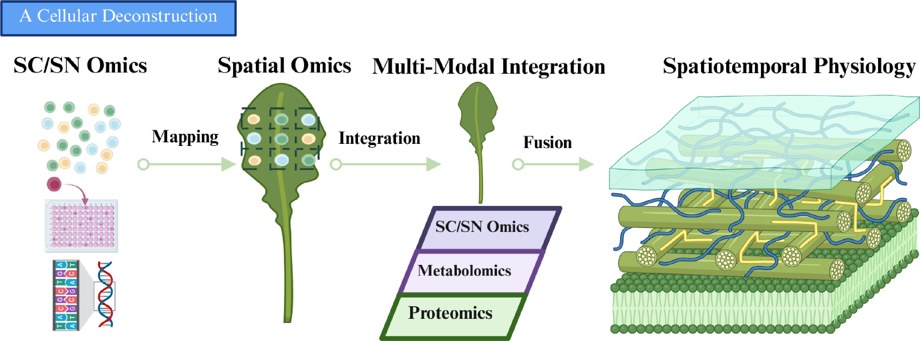
Figure 2.
Minimum operational framework for a plant spatial 'digital twin'. The framework includes spatially registered histology, a matched sc/snRNA-seq reference for improved detection of rare cell populations and spatial deconvolution, same-section co-detection of complementary molecular layers, region-matched proteomics from adjacent sections, explicit cross-section registration with uncertainty control, and orthogonal validation of inferred relationships. Created with BioRender.
-
Plant cell-resolved biology is moving from descriptive atlas generation toward more mechanistic and ultimately predictive models of tissue function. In plants, however, progress is shaped by a constraint that is more severe than in many animal systems: the effective performance of these technologies depends not only on platform design but also on whether plant tissues can preserve analyte accessibility, spatial fidelity, and molecular integrity throughout sample preparation and analysis. As a result, the central challenge is not simply to increase resolution or assay complexity, but to build tissue-aware workflows that remain robust under the anatomical and biochemical conditions characteristic of plant organs.
Three priorities are likely to define the next stage of the field. First, upstream standardization remains essential. Dissociation bias, sectioning artifacts, analyte delocalization, organellar interference, and autofluorescence continue to limit cross-study comparability and effective resolution, especially in non-model and recalcitrant species. Second, the current resolution gap between transcriptomics and functional omics must be narrowed. Spatial proteomics and metabolomics still lag behind transcriptomic profiling in routine cellular-scale applicability, yet they are indispensable for understanding the protein activities and chemical microenvironments that ultimately determine plant phenotype. Third, integration must become both more rigorous and more interpretable. Progress toward plant 'digital twins' will depend not only on same-section or well-registered multimodal measurements, but also on computational frameworks that explicitly model scale mismatch, temporal lag, and uncertainty across modalities.
In this context, the most productive near-term strategy is unlikely to be maximal modality stacking for every experiment. A more effective path is to combine the most informative molecular layers for a defined biological question, using workflows that are compatible with the tissue, species, and analyte class under study. This principle is especially important for tropical crops and metabolite-rich species, which should not be regarded merely as downstream application targets. Rather, they represent stringent benchmark systems for testing whether plant sc and spatial multi-omics methods are truly transferable under real anatomical, chemical, and agronomic constraints.
Looking ahead, the long-term value of plant spatial multi-omics will lie not in producing ever more complex maps, but in generating experimentally testable models of how regulatory states, protein functions, and metabolic outputs are coordinated across tissue space and developmental time. Achieving this will require closer coupling between method development, benchmark datasets, and biological validation. If these challenges can be addressed, plant spatial multi-omics will become not only a descriptive toolkit but a predictive framework for understanding development, stress adaptation, and crop performance.
-
During the preparation of this manuscript, the authors used Google Gemini 3.1 Pro for language polishing, grammar checking, and improving readability. The AI-assisted tool was not used to generate scientific conclusions, create original research data, or replace the authors' critical interpretation of the literature. The authors carefully reviewed, edited, and verified all AI-assisted outputs and take full responsibility for the content of the final manuscript.
-
The authors confirm their contributions to the paper as follows: review topic conceptualization and overall framework design: Xia Y, Zhang H, Cai Z; literature review, manuscript drafting, and visualization: Zhang B, Zhang M, Xu Z, Lam TKY; literature discussion and manuscript revision: Wang J, Zhu L. All authors reviewed the results and approved the final version of the manuscript.
-
This study was supported by the Startup Research Funds from Zhejiang Wanli University to Zhang H and Xia Y; and by the Research Grants Council of Hong Kong (GRF Grant No. 12106224 and AoE Grant No. AoE/M-402/25-N) to Xia Y.
-
The authors declare no conflict of interest.
-
Received 25 January 2026; Accepted 6 May 2026; Published online 13 July 2026
-
# Authors contributed equally: Bingxu Zhang, Mengting Zhang, Zuowei Xu, Thomas Ka Yam Lam
- Copyright: © 2026 by the author(s). Published by Maximum Academic Press on behalf of Hainan University. This article is an open access article distributed under Creative Commons Attribution License (CC BY 4.0), visit https://creativecommons.org/licenses/by/4.0/.
-
About this article
Cite this article
Zhang B, Zhang M, Xu Z, Lam TKY, Wang J, et al. 2026. Single-cell and spatial multi-omics technologies in plants: exploring transcriptomics, proteomics, metabolomics, and emerging integrations. Tropical Plants 5: e025 doi: 10.48130/tp-0026-0024 

Single-cell and spatial multi-omics technologies in plants: exploring transcriptomics, proteomics, metabolomics, and emerging integrations
- Received: 25 January 2026
- Revised: 31 March 2026
- Accepted: 06 May 2026
- Published online: 13 July 2026
Abstract: Plant life depends on the precise spatiotemporal coordination of gene expression, protein abundance, and metabolic activity. Although single-cell sequencing has transformed the resolution of cellular heterogeneity, tissue dissociation inevitably disrupts the positional context required to understand cell–cell communication and local regulatory environments. Spatial omics technologies address this limitation by mapping molecular states directly onto native tissue architecture. Here, we review the evolution from plant single-cell omics to spatially resolved profiling, with emphasis on plant-specific constraints such as rigid cell walls, extensive vacuolization, organellar RNA interference, and metabolite-driven autofluorescence. We further compare routinely deployable approaches with emerging proof-of-concept methods across transcriptomic, proteomic, and metabolomic layers, highlighting key considerations for platform selection in challenging plant tissues. Finally, we discuss the major barriers to multi-omics integration—including resolution mismatch, image registration, and temporal asynchrony—and outline a roadmap toward predictive, spatiotemporally resolved plant 'digital twins', with particular relevance for crop improvement in tropical species.
-
Key words:
- Spatial omics /
- Single-cell /
- Single-nucleus /
- Tropical plants /
- Transcriptomics /
- Metabolomics /
- Proteomics