RESEARCH ARTICLE   Open Access    

Descriptive epidemiology and seroprevalence investigations of Crimean-Congo Hemorrhagic Fever virus in domestic animals of northeast Afghanistan

More Information
  • Received: 08 October 2024
    Revised: 29 October 2024
    Accepted: 11 November 2024
    Published online: 12 December 2024
    Animal Advances  1 Article number: e007 (2024)  |  Cite this article
  • This study investigates CCHF epidemiological cases at a national level from 2007 to 2024, focusing on tick species identification, CCHFV molecular detection, intrinsic, and extrinsic factors associated with the disease's distribution in domestic animals (cattle, sheep, goats, camels, and chickens) in Kunduz and Takhar provinces of Afghanistan. Analyzing national surveillance data for CCHF prevalence from 2007 to 2024, encompassing 1,200 samples (720 ticks and 480 blood) were analyzed. Data concerning intrinsic and extrinsic factors were collected, and seroprevalence was determined using RT-PCR and ELISA. The highest number of confirmed positive cases in humans were reported in 2023 (n = 1,236), and 2022 (n = 389), indicating an annual increase in CCHF cases, with a total case fatality rate of 463, the highest CFR recorded in 2023 (n = 114). Averaging 30.2% over eight years, with a notable death increase until 2018. Among 4,672 collected tick species, Hyalomma predominated, followed by Rhipicephalus, with Dermacentor least found. RT-PCR and ELISA revealed 73 positive cases in Kunduz and 81 in Takhar, with higher seropositivity in the latter. Rustaq (10%) and Dasht-e-Archi (8.2%) showed the highest CCHF prevalence. The present study highlights that early detection plays a crucial role in CCHF mitigation, despite Afghanistan's limited testing capacity and knowledge of CCHF from a one-health perspective.
  • Wheat (Triticum aestivum) is an important global food crop and contains carbohydrates along with other crucial nutrients such as proteins, small amounts of lipids, vitamins, minerals, as well as phytochemicals[1]. Dietary fiber is the carbohydrate oligomers and polymers, which are resistant to digestion and absorption in the human small intestine, leading to partially or complete fermentation in the human large intestine[2]. Whole wheat grains contain between 9% and 20% dietary fiber, and the main components of dietary fiber are cell wall polysaccharides, primarily arabinoxylan and (1,3;1,4)-β-D-glucan (β-glucan), representing approximately 70% and 20% of total dietary fiber, respectively[3]. In addition, dietary fiber in wheat grain also contains resistant starch, which is not digested in the small intestine and is able to reach the large intestine and colon relatively unchanged[4].

    Wheat grains contain a starch-rich endosperm, an embryo (germ) and a fiber-rich outer covering (bran). Wheat-derived foods are made from whole wheat or white flour, which is made from wheat grains that have had the bran and germ removed[5]. Regular consumption of whole grain products has been advocated as a dietary recommendation, but white flour-based products, which are produced through milling to separate the bran and germ from the white flour, continue to dominate in most countries. Improving the fiber content of white flour preparations, and enhancing the general palatability of whole wheat preparations have the potential to improve fiber consumption.

    Consumption of wheat-derived foods with high contents of arabinoxylan, β-glucan, and resistant starch can reduce the negative effects of diseases such as type 2 diabetes, as well as cardiovascular and gastrointestinal diseases[68]. Moreover, dietary intervention is increasingly encouraged as an effective way for preventing obesity and diabetes and is associated with beneficial effects on human health. Clinical analyses have demonstrated that dietary fiber exerts a beneficial impact on blood pressure and serum cholesterol levels, while also having a protective effect against the development of specific types of cancer, particularly colorectal and breast cancers[9].

    In this review, we summarize research on three major components of dietary fiber in wheat: arabinoxylan, β-glucan, and resistant starch. We describe their compositions, biosynthetic pathways, and key enzymes, as well as the effects of these components on the health benefits of wheat fiber. The current advances in QTL/gene mapping and genetic improvement of wheat fiber are summarized and the challenges and perspectives on further research aimed at improving dietary fiber in wheat are discussed.

    Arabinoxylan is a major component in the cell wall of wheat grain. In wheat starchy endosperm, arabinoxylan accounts for 70% of the cell wall polysaccharides and its content based on whole wheat bran ranges from 5% to 27%[10]. Arabinoxylan is one of the major sources of dietary fiber in human diet and is also a crucial factor determining wheat end-use quality and its utilization in animal feed and distilling[11]. The intake of arabinoxylan-rich bread is negatively correlated with postprandial glycemic responses in healthy adult subjects[12]. Indeed, in the gastrointestinal tract, arabinoxylan is intertwined with carbohydrates such as starch, thus delaying the absorption of carbohydrates and reducing post-meal blood sugar levels.

    The classification of arabinoxylan typically involves two fractions: water-extractable (WE-AX) and water-unextractable (WU-AX). WE-AXs constitute ~25%–30% of the total arabinoxylans in wheat grain[13]. The arabinoxylan in white flour consists of a linear (1,4) linked backbone of β-D-xylopyranosyl (xylose) residues[14]. The monomers that constitute the arabinoxylan polysaccharide are xylose and arabinose, which are biosynthesized in the cytoplasm and Golgi because of the existence of many isoforms of enzymes associated with biosynthesis. The arabinoxylan backbone is biosynthesized in the Golgi through the complex interplay of metabolites and the cytosol, that is a process regulated by various membrane channels including uridine diphosphate-sugar transporters.

    Biosynthesis of the final arabinoxylan structure involves participation of two major types of enzymes including the glycosyltransferases (GTs) and glycoside hydrolases (GHs). GTs catalyze the development of glycosidic bonds through transferring the nucleoside diphosphate sugars, which act as a sugar donor containing a nucleoside phosphate, onto a particular receptor. Among the 124 GTs which are related to the biosynthesis of the cell wall in the wheat starchy endosperm, the genes encoding TaGT47_2, TaGT43_2, and TaGT43_1 involved in arabinoxylan backbone biosynthesis and TaGT61_1 in the arabinosylation of xylan are expressed at the highest levels[1517] (Fig. 1). Specifically, all three homoeologs encoding TaGT43_2 and TaGT47_2 are expressed in the wheat endosperm and are regarded as a participant in the arabinoxylan backbone biosynthesis, while two xylan arabinosyl transferases, TaXAT1 and TaXAT2, belonging to the GT61 family, are involved in decorating the arabinoxylan backbone with arabinosyl and xylosyl sidechains[16]. Finally, members of the GH family could be involved in the breakdown of cellulose, allose, mannan, or arabinoxylan[15].

    Figure 1.  The composition, biosynthetic pathways, and key enzymes for the three major classes of dietary fiber in wheat. GT47, GT43, GT61, and GT2 are glycosyltransferases. CslF, CslH, and CslJ are Cellulose-synthase-like (Csl) genes. GBSSI, granule-bound starch synthase I. SSI to SSIV, starch synthase I to IV. SBE, starch-branching enzyme. ISA, isoamylase-type debranching enzyme. LD, limit dextrinase. The blue ellipses represent UDP-D-xylose, and the green diamonds represent UDP- arabinopyranose.

    In recent years, multiple QTLs for arabinoxylan and WE-AX content have been identified using different wheat populations[1820] (Table 1). Genes for arabinoxylan and WE-AX content have also been identified. Four genes were reported to participate in the arabinoxylan biosynthesis pathway; these genes encode a glucosyltransferase, a peroxidase, a glucosidase, and a methyltransferase[21]. Marcotuli et al. found 19 QTLs associated with arabinoxylan content[22], nine of which coincide with annotated genes encoding enzymes involved in arabinoxylan biosynthesis[15,17,23]. These genes include: i) Gal7, encoding the glycosyl hydrolase GH35; ii) a cluster of GT1 genes, including TaUGT1 and cisZog1; iii) CelC, encoding the glycosyl hydrolase GH1; iv) Ugt12887 and TaUGT1, both encoding the glucuronosyltransferase GT1; and v) Gsl12 and Cel8, encoding a (1,3)-β-D-glucan synthase and GH, respectively[22]. Eight candidate genes for arabinoxylan content such as genes encoding F-box domain proteins, the disease-resistance protein RPM1, and the transcription factor bZIP29 were identified through analysis of arabinoxylan content in 562 wheat genotypes combined with genome-wide association study (GWAS)[24]. QTLs with stable effects on total arabinoxylan or WE-AX content might be useful for improving arabinoxylan contents in wheat breeding through molecular marker-assisted selection. For example, QTL on chromosome 1BL of the Chinese wheat cultivar Yumai 34, contributed 24.2% of the phenotypic variation in arabinoxylan levels, leading to the molecular marker development based on allele-specific single nucleotide polymorphism (SNP)[25]. In addition, five SNPs on chromosomes 1BL and 5BS were reported to be associated with total arabinoxylan levels and 13 SNPs on chromosomes 1BL, 2BS, 6BS, 7A, and 7BL were related with WE-AX content[26]. Among which, SNP on 1BL were relevant to both traits and explained 13.29%–17.22% phenotypic variation for total arabinoxylan and 11.56%–19.37% for WE-AX, respectively and competitive allele-specific PCR (KASP) markers were developed for utilization during wheat breeding process.

    Table 1.  QTLs/genes and molecular markers for wheat dietary fiber content.
    TraitQTLChrNearest markerPosition (cM)Candidate genesReference
    Arabinoxylan content
    WE-AXQAxvs.inra-1B1BXcfa214782.7[18]
    QAxfg1.inra-6B6BwPt-221822.2PC1
    TOT-AXQGax.aww-2A.12Awpt-3114-2A85.7[19]
    QGax.aww-4D.14Dgpw-95001-4D46.0
    WE-AXQgWE-AX.caas-1B1BHVM23–Sec1[20]
    QgWE-AX.caas-5A5AXgwm443–Xcwem44
    QgWE-AX.caas-5B5BXbarc142–Xwmc28
    QgWE-AX.caas-7A7AXbarc174–Xbarc108
    Grain dietary fiber contentglucosyl-transferase (GT)
    peroxidase glucosidase methyltransferase
    [21]
    Arabinoxylan contentQGax.mgb-1A.11Awsnp_Ex_c45880_5155017270.1Gal7[22]
    QGax.mgb-2B.12BTdurum_contig45838_263107.39TaUGT1
    QGax.mgb-3A.13AKukri_c17966_634122.68CelC
    QGax.mgb-5A.15AEx_c95453_149926.51Ugt12887
    QGax.mgb-5A.35Atplb0056b09_100063.69TaUGT1
    QGax.mgb-7A.17ATdurum_contig69003_45942.08Gsl12
    QGax.mgb-7A.27Awsnp_Ex_c21854_31021668130.27Cel8
    Arabinoxylan contentAX-950863561BF-box/FBD/LRR-repeat protein;[24]
    AX-944703194BDisease resistance protein RPM1/PIK6-NP-like;
    AX-947130155DbZIP transcription factor 29
    Arabinoxylan contentY34Ukr-RH13-TOTAX;1B[25]
    Y34Cla-JI15-TOTAX
    Arabinoxylan content1B1B_646895451[26]
    1B_653086336
    1B_653681771
    1B_654915479
    5B5B_14665450
    2B2B_184634480
    6B6B_26597224
    7A7A_234827309
    7A_264333614
    7A_458678969
    7A_474572231
    7A_516508921
    7A_700824770
    7B7B_454100716
    β-Glucan content
    QGbg.mgb-1A.11AKukri_rep_c110838_25310.6[35]
    QGbg.mgb-1A.21AKukri_c43410_34881.6Cel9
    QGbg.mgb-2A.12ATdurum_contig10785_81611.2WSs2A
    QGbg.mgb-2A.22AExcalibur_c44834_80197Bamy1
    QGbg.mgb-2B2BBobWhite_c25359_13214.5Wxl1
    QGbg.mgb-3B3BBS00091867_5197.2Xip-II
    QGbg.mgb-5B5BTdurum_contig35470_227I166
    QGbg.mgb-7A.17Atplb0024a09_82949.7
    QGbg.mgb-7A.27ATdurum_contig19352_7690.91-FEH
    QGbg.mgb-2A.12AIWB128035.8–48.0[36]
    QGbg.mgb-2B.12BIWB301150.1–3.9
    QGbg.mgb-2B.22BIWB2378329.9–47.9GLU1a
    QBgn3AXwmc202–Xgwm2[38]
    1BXwmc419–Xwmc134
    5BXwmc149–Xgwm335
    6DXhbe404–Xcfd188
    QTL 14M100022501_F_0glutathione S-transferase 3-like[39]
    QTL 25M100013840_F_1
    QTL 31M100079925_F_0
    Resistant starch content
    A-type starch granulesQga.caas-4AL4AXwmc262 – Xj133[61]
    Qga.caas-1DL1DXcfd48.1 – Xcfd48.2
    Qga.caas-7BL7BXwmc311 – Xbarc50
    B-type starch granules4STR132- TR126[62]
    B-type starch granules4ABGC1-A[63]
    4B
    4D
    BGC1-B
    BGC1-D
    4B
    4D
    BGC1-B
    BGC1-D
    Amylose contentqhams7A.17AC7A.8493170 – C7A.3957649940.0GBSSI[65]
    Resistant starch content2DXbarc59[67]
    5B
    Amylose and resistant starch content2ABDSBEIIa[50,70,71]
    2ABDSBEIIb
    Amylose content7ABDSSIIIa[7274]
    1ABDSSIIIa
     | Show Table
    DownLoad: CSV

    The genes which are responsible for the extension of xylan backbone in wheat are the putative orthologs of the Arabidopsis (Arabidopsis thaliana) GT genes IRREGULAR XYLEM10 (IRX10), IRX14, and IRX9, named TaGT47_2, TaGT43_1, and TaGT43_2, respectively[15]. Downregulating TaGT47_2 or TaGT43_2 expression by RNA interference (RNAi) resulted in as much as a 50% decrease in total arabinoxylan levels with a concomitant increase in arabinosylation levels ranging from 25% to 30%[17]. In these lines, the cell walls of starchy endosperm showed a reduction of xylan and arabinoxylan epitopes (as detected by immunolabeling) and a 50% reduction in cell wall thickness than the wild-type control. Freeman et al. also reported the reduction of WE-AX levels in wheat through downregulating TaGT43_2 and TaGT47_2[27]. A mutant with TaGT43_2 three homoeologs knock-out showed a reduction in arabinoxylan content of approximately 65%[28]. Silencing of the wheat TaXAT1 gene in the GT61 family, which is expressed at the highest levels in starchy endosperm, induced a 70%–80% reduction in α-(1,3)-linked arabinofuranosyl (Araf) residues substitution of β-(1,4)-D-xylopyranosyl (Xylp) residues in arabinoxylan[16]. Overall, these results provide evidence for the relevance of specific genes in controlling the arabinoxylan content in wheat. However, thus far, RNAi has not yet been used to improve arabinoxylan content.

    β-glucan accounts for approximately 20% of total cell wall polysaccharides in wheat grain and is the second most abundant cell wall polysaccharide[10]. β-glucan is effective at reducing postprandial blood glucose levels[29]. β-glucans interact with lipids and biliary salts by forming glucan–biliary salt complexes directly or by creating a viscous layer that hinders absorption, thus decreasing the adsorption of biliary salts in the bowel[30], thereby reducing blood cholesterol levels and mitigating the risk of cardiovascular disease[7]. The β-glucan level in wheat grains is much lower relative to barley or oat. Particularly, the β-glucan content is 2.5%-11.5% in barley and 2%-7.8% in oats, but only 0.4%-1.4% in wheat[31]. The widespread consumption of wheat makes it an ideal target for enhancing β-glucan content.

    β-glucans are long or short-chain polymers of glucose subunits connected by (1,3) and (1,4) linkages. Single (1,3) linkages are isolated by two or three (1,4) linkages, leading to trisaccharide and tetrasaccharide products after digestion by lichenase, which is a specific enzyme[32]. β-glucans are enriched in the subaleurone layer of the wheat seed, with a minor amount in the endosperm[33]. In cereals, β-glucans are biosynthesized by enzymes belonging to the cellulose synthase-like superfamily, which in turn is a component of the large glycosyltransferase GT2 family[34].

    Studies examining genetic variability associated with wheat β-glucan content using different wheat populations have been carried out in recent years (Table 1). Seven QTLs related to wheat grain β-glucan were found in tetraploid durum wheat, leading to the identification of candidate genes encoding GT or GH[35]. These genes include: i) Cel9, encoding the endo-β-(1,4)-glucanase GH9; ii) WSs2A, encoding the starch synthase II; iii) Bamy1, encoding the β-amylase GH14; iv) Wxl1, encoding the (1,4)-beta-xylanase GH10; v) Xip-II, encoding the xylanase inhibitor protein GH18; vi) 1-FEH, encoding the fructan 1-exohydrolase GH32. Three additional QTLs, named QGbg.mgb-2A.1, QGbg.mgb-2B.1, and QGbg.mgb-2B.2, were identified from a durum wheat using recombinant inbred line (RIL) population derived from a cross between two elite wheat cultivars Duilio and Avonlea by the same research group[36]. GLU1a gene encoding β-glucosidase 1a (GLU1a) was identified as candidate gene for QGbg.mgb-2B.2, and this enzyme catalyzes the hydrolysis of the glycosidic linkage in glycosides to form of intramolecular hemiacetal and hemiketal and the corresponding free aglycon[37]. In hexaploidy wheat, the QTLs on chromosomes 3A, 1B, 5B, and 6D were identified to be associated with β-glucan level[38] , among which the QTL located between markers Xwmc149 and Xgwm213 on chromosome 5B explained 15% of the phenotypic variation. Finally, Ivanizs et al. found three QTLs associated with β-glucan content in Aegilops biuncialis[39]: QTLs 2 and 3 were located on chromosomes 1M and 5M, and tetraploid wheat also have QTLs influencing the synthesis of β-glucan on group 1 and 5 chromosomes[40]. Another QTL 1 co-located with GTS3L encoding glutathione S-transferase participated in multiple metabolic pathways. However, most of these QTLs are not stable across different environments[36,38], which limited the utilization of marker-assisted selection during wheat breeding for β-glucan improvement. There are three cellulose-synthase-like (Csl) family members: CslF, CslH, and CslJ[41] (Fig. 1), among which CslF primarily participates in the synthesis of β-glucan in wheat. TaCslF6 has been identified as the crucial candidate for wheat β-glucan biosynthesis[42], with three homoeologs mapped near the centromeres on wheat chromosomes 7AL, 7BL and 7DL[32]. In hexaploid wheat, RNAi was employed to specifically suppress TaCslF6 in grain, leading to a 30% to 52% decrease in β-glucan content[42]. In durum wheat, Marcotuli et al. reported that the expression of CslF6 has a positive correlation with β-glucan content[43].

    The strategies of interspecific hybridization and chromosome engineering can be used to introgress genes of distantly related wild and cultivated relatives to increase β-glucan content in wheat. Barley (Hordeum vulgare) has been successfully utilized in interspecific hybridization, since barley grain contained the highest β-glucan content[44]. It has been reported that HvCslF4, HvCslF6 and HvCslF9 located on chromosome 2HS, 7HL and 1HS greatly regulate β-glucan content in barley grain[45]. Several genes from barley were introgressed into wheat, which led to increased β-glucan content in wheat grain. Türkösi et al. developed a wheat/barley Robertsonian translocation (RobT) line 7BS.7HL, which confers increased grain β-glucan content (0.9%) than the control (0.7%)[46]. Danilova et al. developed a set of six compensating RobT chromosomes containing barley chromosome 7H (carrying the HvCslF6 gene related to β-glucan synthesis) and three group-7 chromosomes of wheat, among which the average β-glucan content of 7AS·7HL (1.01%), 7BS·7HL (0.82%), and 7DS·7HL (0.85%) were increased compared to the control (0.72%), although they were still much lower than the barley parent (5.81%)[32]. Subsequently, the same group used induced wheat–barley homologous recombination to shorten the barley chromosome 7HL arm in these RobTs to small segments[44]. They showed that the β-glucan level was increased in wheat lines containing two or four copies of the barley Cellulose synthase-like F6 gene (HvCslF6). Hence, by increasing the copies of the HvCslF6 gene through combining different recombinant chromosomes in a single line, the β-glucan content in wheat grain could be further increased. In addition, a wheat addition line containing the HvCslF9 gene showed a much higher β-glucan content[47]. Taken together, it is helpful to introgress barley HvCslF6 to wheat for β-glucan content improvement in wheat grains, but the β-glucan content in barley was still much more than that in translocation lines. Thus, other key genes from barley could be the candidates for increasing β-glucan biosynthesis in wheat.

    For the purpose of identification of genotypes with high β-glucan content that can be utilized as interspecific gene resource transferring to cultivated wheat, Marcotuli et al. screened a set of cultivated and wild Triticum and Aegilops species for β-glucan content. They found two Aegilops species including Ae. umbellulata, UU genome and Ae. markgrafii, CC genome exhibiting superior β-glucan content than Triticum species and they were the putative candidates for wheat β-glucan content improvement[48]. Specifically, species with the U genome seemed to have a greatly higher β-glucan level (about 5.3%) compared to cultivated hexaploid bread wheat (about 0.83%). Another candidate gene is ISA1, encoding Isoamylase 1 (ISA1), which is an isoamylase-type debranching enzyme playing an important role in amylopectin biosynthesis. Sestili et al. knocked down Isa1 in durum wheat using RNAi: the resulting transgenic lines had reduced starch content, as well as moderately enhanced phytoglycogen and β-glucan contents[49]. These findings demonstrate that transgenic strategy can be utilized to regulate the content of β-glucan in wheat grains.

    Starch is the main polysaccharide in wheat, which can be classified into amylose and amylopectin based on the polymerization form of glucose[50]. Amylose, a linear structure connected by α1, 4 bonds, is densely packed within the starch granule in the wheat grain endosperm[50]. Amylopectin, a branched molecule containing both α1, 4 and α1, 6 bonds, generates the complex structure that occupies most of space within the starch granule of wheat grain endosperm. In wheat grain, amylopectin typically constitutes over 70% of the total content, while amylose accounts for less than 30%. According to its digestibility, starch is generally categorized as rapidly and slowly digestible starch, or resistant starch, depending on its post-consumption behavior[51]. Resistant starch refers to dietary starch that cannot be digested in the small intestine and undergoes fermentation exclusively in the colon[52]. The consumption of resistant starch has been shown to directly attenuate postprandial blood glucose levels, serum cholesterol levels, the glycemic index, and insulin levels, thereby contributing to the prevention or reduction of obesity, diabetes, cardiovascular disease, and chronic kidney disease[8]. There are five types of resistant starch (type 1−5), the augmentation of amylose content can decelerate the rate of starch digestion and increase the content of resistant starch type 2[53]. By focusing on biochemical pathways that enhance the amylose content within a starch granule allows for the synthesis of high-amylose grains with higher levels of resistant starch type 2, and consequently an increased amount of dietary fiber in refined flour[53].

    Starch biosynthesis is a complicated and finely-regulated process that requires several functional enzymes, including sucrose synthase (SUS), ADP-glucose pyrophosphorylase (AGPase), granule-bound starch synthase I (GBSSI), starch synthase (SS), starch-branching enzyme (SBE) and de-branching enzyme (DBE)[54,55] (Fig. 1). In this process, the sucrose biosynthesized in leaves is transported into grains, and then it is decomposed into fructose and UDP-glucose by SUS. Following this, the products are converted into ADP-glucose by AGPase. Subsequently, soluble ADP-glucose is transported into the amyloplasts as a direct substrate, where the starch biosynthesis is initiated[8]. GBSSI controls amylose biosynthesis independently, while SBE, DBE and SS are responsible for amylopectin biosynthesis[5456]. The amount of resistant starch is positively related to the proportion of amylose[57]. Therefore, increasing GBSSI or decreasing SS and SBE gene expression is the most common means to increase the resistant starch content in cereal endosperm.

    In recent years, multiple QTLs and genes related to resistant starch content in wheat have been reported (Table 1). The quantity and proportion of A- or B-type starch granules, as well as the quantity of amylose, are related to resistant starch content. A-type granules contain higher amounts of amylose than B-type starch granules[58]. The proportion of B-type granules is positively related to amylose and resistant starch content[59,60]. Using 240 RILs derived from a cross between wheat lines PH82-2 and Neixiang 188, three QTLs associated with the contents of A-type starch granules, named Qga.caas-1DL, Qga.caas-7BL, and Qga.caas-4AL, were identified, explaining 5.6%, 5.2%, and 3.8% of the phenotypic variation, respectively[61]. Howard et al. developed an F2 population of Aegilops with variation in B-type starch granule content and used this population to identify one major QTL accounting for 44% of the phenotypic variation[62]. Similarly, the progeny derived from Ae. peregrina × KU37 were crossed, revealing a link between B-GRANULE CONTENT 1 (BGC1) and resistant starch content[63]. BGC1 inhibits the formation of A-granules during early grain development but promotes the formation of B-granules during middle grain development.

    In general, starch with a greater proportion of amylose has a higher level of resistant starch[64]. Thus, QTLs and genes regulating amylose content might also affect resistant starch content. Analysis of an F2 population developed by crossing the high-amylose donor 'TAC 75' and high-yielding variety 'WH 1105' identified qhams7A.1, a stable QTL associated with amylose content. This QTL includes the GBSSI gene[65]. A high-amylose line carrying the missense mutation g.35767184 T > C in GBSSI produces the mutant protein GBSSI_L539P, which is associated with high-amylose starch. The L539P mutation improves GBSSI activity by affecting its starch-binding ability, suggesting that this mutant allele could be a molecular target for enhancing amylose and resistant starch content in wheat grain[66]. Various additional molecular markers related to resistant starch content have also been developed. For example, bulk segregation analysis identified the SSR marker Xbarc59 located on chromosomes 2D and 5B of spring wheat[67]. In the F2 generation obtained from a cross between Wuchun 4 and M344, genotypes with high, medium, and low resistant starch contents, were efficiently screened using the Xbarc59 marker.

    Ethyl methanesulfonate (EMS)-targeted mutagenesis has been successfully used to develop wheat lines with high amylose and high resistant starch contents. In common wheat grains, the amylose content varies from 20%–30%. Mishra et al. produced a bread wheat population comprising 101 EMS-induced mutant lines, with amylose content ranging from ~3%–76% and resistant starch content ranging from 1–41%[68]. Irshad et al. identified stable mutants with significant changes in resistant starch content using an EMS-induced population of wheat variety J411[69].

    Four types of enzymes are required for starch biosynthesis: GBSSI is required for amylose biosynthesis, while amylopectin is produced by the concerted action of several classes of starch synthase (SSI, SSII, SSIII, and SSIV), branching (SBEI, SBEIIa, and SBEIIb), and debranching enzymes[54,55]. By suppressing the expression of SBE or SS genes or enhancing the expression of the GBSSI gene in the amyloplast, starch biosynthesis can be modified to increase the amylose content, thus leading to an increased proportion of resistant starch.

    The waxy (Wx) gene encodes GBSSI. A mutation in Wx-B1 reduced GBSSI activity by affecting its starch-binding ability, indicating that modifying GBSSI activity can modulate amylose content. Accordingly, screening an EMS-mutagenized population of bread wheat variety J411 revealed two mutants with high levels of resistant starch and two mutants with low levels of resistant starch, with the mutants with high levels of resistant starch showing higher GBSSI expression and the mutants with low levels of resistant starch showing higher SBE expression[69].

    Durum wheat with a SBEIIa/b-AB double mutation exhibited a 22% and 115% increase in amylose and resistant starch content, respectively[70]. Introducing a series of deletions and SNPs into SBEIIa and SBEIIb also increased the amylose content to an unprecedented level of ~85%, with a concomitant increase in resistant starch content[50]. Schönhofen et al. detected an increase in amylose content of up to 60% and a 10-fold increase in resistant starch content in SBEII mutants[71]. The combination of Targeting Induced Local Lesions in Genomes (TILLING) technology and breeding led to the successful introduction of SBEIIa mutations in bread wheat and durum wheat[49]. These mutants showed increases in amylose and resistant starch contents ranging from 47%–52.7% and 4.7%–6.8%, respectively.

    Whole meal flour from a SSIIIa triple mutant had more resistant starch and higher levels of arabinoxylans and β-glucans than the wild type[72]. Schoen et al. identified SSIIa knockout mutations in all three genomes of the wheat variety 'Jagger', generating a triple knockout mutant affecting the activities of all three homoeologs[73]. The triple knockout mutant had increased amylose and resistant starch levels without the shriveled grain phenotype observed in other SSIIa knockout mutants[7476]. Importantly, since all the mutants were obtained using an elite wheat cultivar, they are immediately available for practical application.

    Plant breeding using crop varieties with high levels of resistant starch and marker-assisted selection are effective methods for improving resistant starch content. Combined mutations in SBEIIa and SBEIIb paralogs by EMS were obtained in hexaploid wheat cultivar Lassik including: SBEIIa/b-AB; SBEIIa/b-A, SBEIIa-D; SBEIIa/b-B, SBEIIa-D; and SBEIIa/b-AB, SBEIIa-D. The quintuple mutant line (SBEIIa/b-AB, SBEIIa-D) exhibited a significant increase in amylose and resistant starch contents[77]. Hogg et al. also observed an increase in total fiber content in pasta made from a SSIIa null durum wheat line[75]. In addition, Botticella et al. developed a breeding strategy to produce a set of durum wheat SSIIa null mutants[78]. The authors introgressed SSIIa null mutations into the Italian elite durum wheat cultivar Svevo and showed that the semolina flour produced from two SSIIa null mutant lines had increased arabinoxylans, β-glucans, and resistant starch contents. The same authors also used mutagenesis followed by conventional marker-assisted breeding to generate three mutant lines that produced starch with an amylose content of 0%, 46%, and 79%[76].

    RNAi has also been used to generate transgenic wheat lines with increased amylose content. Downregulating SBEIIb alone had no effect on amylose content, whereas the concomitant downregulation of SBEIIa and SBEIIb resulted in starch comprising more than 70% amylose[79]. When rats were fed whole meal flour from the RNAi double downregulated lines, improvements in several indices of large-bowel function were observed. Similarly, the RNAi-mediated downregulation of SBEIIa in durum wheat induced an up to 75.05% increase in amylose content[80]. Finally, suppressing SSI by RNAi dramatically enhanced amylose content (31.4%)[81].

    Clustered regularly interspaced short palindromic repeats (CRISPR)/CRISPR-associated nuclease 9 (Cas9) is widely used to introduce specific DNA modifications into targeted genes[82]. This method has been successfully used to create transgene-free lines with various amylose contents in crops including rice and maize[8385]. Using the modern winter wheat cv. Zhengmai 7698 and the spring wheat cv. Bobwhite, Li et al. employed CRISPR/Cas9-targeted mutagenesis of TaSBEIIa to successfully generate transgene-free high-amylose plants[86]. The flours of lines affected in the functions of all three homoeologs showed increased amylose, resistant starch, protein, and soluble pentosan contents, which benefit human health.

    The physicochemical properties of arabinoxylan, β-glucan, and resistant starch are closely related to dietary fiber content in wheat. Further optimization of arabinoxylan, β-glucan, and resistant starch biosynthesis in wheat endosperm represents an important future direction in wheat breeding for improving the nutritional quality of grain. Nonetheless, there are still many hurdles to overcome.

    The increased amylose and resistant starch contents in SBEII, SSII, and SSIII mutants are related to changes in grain characteristics that may limit their usefulness in breeding programs. Indeed, these mutants show reduced starch contents and grain weight, leading to yield penalties[7076]. For example, Hazard et al. reported that mutations in the durum wheat genes SBEIIa/b-AB were associated with an average reduction in kernel weight of 5.2% and a 15% reduction in grain yield[70]. Schönhofen et al. found that SBEII mutants exhibited reductions in kernel weight and grain yield of 2.8% and 5.8%, respectively[71]. Schoen et al. reported that SSIIa triple mutants had a 21.29% reduction in thousand-grain weight, which was also significantly reduced in SSIIa single mutants compared to the control[73]. Similarly, targeting of SBEIIa by CRISPR/Cas9 was associated with a slight reduction in grain length and width, resulting in a decrease in the thousand-grain weight in different mutant lines, with the highest effect observed in aabbdd triple-null lines[86]. Thus, the effects of partial- or triple-null alleles are dosage dependent, with triple-null lines exhibiting the most profound impacts on yield penalty. Accordingly, Fahy et al. reported that the SSIIIa triple mutants exhibited shrunken grains with a weight reduction of ~11%, whereas the grains of SSIIIa single and double mutants were not significantly different from those of the control[72]. The same research group also revealed a possible dosage effect on starch content. Marcotuli et al. reported QTLs associated with wheat grain β-glucan, while the associated genes include starch synthesis II, supporting a link between the biosynthetic pathways of starch and β-glucan[35], and the negative correlation between grain weight and β-glucan content were also observed in wheat[48].

    High-amylose wheat varieties have been successfully developed during the past decades through modification of the SBEII, SSII, or SSIII genes, which lead to a reduced glycemic index and are beneficial for human health[70,72,75]. Unfortunately, the grain- or flour-processing quality of these varieties was negatively affected. Given the crucial role of a continuous gluten network, the dilution effect, mechanical shear effect, competitive water absorption, and steric hindrance effect of dietary fiber may potentially disrupt structure, thereby resulting in suboptimal rheological properties of dough[53]. For example, Schönhofen et al. reported that SBEII mutations exert notable influences on bread-making quality. The traits related to end-use quality such as enhanced grain hardness, and increased starch damage, water absorption, and protein content in flour; with diminished flour extraction, farinograph development, stability times, starch viscosity, and loaf volume[71]. Li et al. found that increasing amylose content drastically reduced cooking quality and that bread made from high-amylose wheat flour had a lower loaf volume, a more dense crumb structure, and higher hardness than wild-type wheat bread[64]. Li et al. reported that bread and biscuits made of high-amylose flours from TasbeIIa triple-null lines had decreased volume, brighter color, higher hardness, and lower sensory scores compared to the wild type[86].

    Durum wheat SBEIIa/b-AB mutant lines showed favorable performance on pasta firmness but negative effects on pasta color and semolina extraction[70]. These mutants showed an average adverse in firmness of 12.4% relative to the wild-type lines. However, the average amount of semolina extracted from grains after milling was 4.6% lower in the SBEIIa/b-AB mutants vs the wild type. The SBEIIa/b-AB mutants also displayed an average increase in cooking loss of 20.4%, a 6.1% reduction in cooked weight, and a 10.4% decrease in color score. Furthermore, Hogg et al. reported that durum wheat accessions containing SSIIa null mutation showed high-amylose content and lead to increased grain protein content with lower semolina yield[75]. Pasta made from semolina of the SSIIa null mutant exhibited reduced water absorption, higher cooking loss, shorter cooking time, and significantly firmer texture even when overcooked, as compared to the wild-type line. The brightness of cooked and uncooked pasta from the SSIIa null mutant was diminished compared to the wild type. Therefore, efforts aimed at improving dietary fiber content must monitor effects on processing quality and select for traits with minimal impacts on quality traits. Consequently, the balance of grain protein, starch and dietary fiber is more important, enhancing the dietary fiber content of wheat while preserving yield and end-use quality.

    The wheat endosperm is composed of an outer layer called bran (14%−15%), an inner starchy endosperm (81%−83%) and embryo (2%−3%). The bran, composed of the aleurone, endocarp, mesocarp, and epicarp, forms the outer layer of the grain. It contains trace minerals and indigestible cellulose materials, including the β-glucans. In contrast to barley, the distribution of β-glucans in the wheat endosperm is less consistent and at lower levels. The aleurone layer was found to have high concentrations of β-glucans, accounting for approximately 29% of its dry weight[42]. As a dietary fiber supplement incorporated into food, wheat bran exhibits remarkable efficacy in promoting cardiovascular and gastrointestinal health[87]. Therefore, increasing bran or aleurone layer thickness is an effective way to improve dietary fiber in wheat. Actually, in rice, Li et al. & Liu et al. has proved that increasing the thickness of aleurone layer is an effective way to improve the nutrition properties. They screened the ta1 and ta2 mutants displaying increase in the number of aleurone cell layers compared to the wild type, resulting in elevated levels of all examined nutritional factors, including lipids, proteins, vitamins, minerals, and dietary fibers[88,89]. The ta1 aleurone thickened phenotype is caused by mutations in the OsmtSSB1, which interacts with RECA3 and TWINKLE to inhibit abnormal recombination of mitochondrial genomic DNA in rice aleurone cells, ensuring optimal mitochondrial energy supply[88].The phenotype of ta2 is attributed to a dominant negative mutation in the DNA demethylase gene OsROS1, which holds a pivotal role in epigenetics based on molecular genetic evidence. In wild-type rice cells, this mutation leads to a notable increase in the aleurone layer from 1−2 to 4−10 layers, consequently enhancing the nutritional quality of rice caryopsis significantly[89]. In wheat, Chen et al. reported a lgp1 mutant with low gluten protein exhibiting significantly increased bran content and dietary fiber content compared to the wild type[90], which could be used for wheat dietary fiber improvement.

    As discussed above, partial-null and triple-null alleles of starch biosynthesis-related genes, especially SSIIa, SSIIIa, and SBEII triple-null lines, incur yield penalties and affect wheat processing quality. In particular, triple-null lines demonstrated more profound impacts on yield penalty and end-use quality than other mutants. This observation suggests that fine-tuning the expression of genes or the activities of enzymes involved in amylose content could be an effective strategy to increase fiber content while minimizing impacts on grain weight and end-use quality. For example, it might be possible to use unique combinations of mutant ssIIa, ssIIIa, or sbeIIa homoeologs in wheat breeding programs. In addition, moderate changes in the expression of starch biosynthesis-related genes that are highly expressed in the endosperm could improve dietary fiber content without severely affect yield and quality traits in rice[91,92]. Specifically, Xu et al. generated a series of Wxb mutants using cytidine base editors (CBEs)[92], leading to 1.4%–11.9% increases in amylose content without affecting the quality or appearance of milled rice. Another approach could be to target starch biosynthesis-related genes with relatively low expression levels in endosperm, since mutations in these genes tend to cause moderate changes in starch biosynthesis, thus limiting undesired effects[93].

    Several other studies successfully fine-tuned gene expression using CRISPR/Cas9 to target gene regulatory sequences[85,94]. Huang et al. generated six novel Wx alleles by editing the region near the TATA box of the Wxb promoter[91]. This resulted in downregulated Wx expression and increased grain amylose content. Liu et al. engineered quantitative variation for yield-related traits in maize by generating weak promoter alleles of CLAVATA3/embryo surrounding region-related (CLE) genes and a null allele of a newly identified and partially redundant compensating CLE[95]. While CLE knockout alleles had a deleterious effect on yield, fine-tuning CLE expression by editing its cis-regulatory elements boosted yield. Finally, CRISPR-dCas9 can be utilized to target the addition or removal of DNA methylation to silence or activate genes in Arabidopsis[96]. Therefore, modulating DNA methylation at cis-regulatory elements represents an additional strategy to fine-tune the expression of genes.

    For β-glucan improvement, the identification of the key functional genes such as TaCslF6 had been contributed to increased β-glucan content[43]. However, the lack of major and stable QTL still limited and prevented the utilization of marker-assisted selection for β-glucan improvement in wheat breeding. Thus, enhancing our understanding on the genetic regulation of β-glucan biosynthesis and identifying more superior genetic resources for enhancing β-glucan content is essential. The integration of high-throughput markers with genome sequencing techniques holds promise for genetic identification, elucidating genetic control, and developing molecular markers linked to key genes and QTL associated with this trait[8789].

    As discussed above, different (and non-mutually exclusive) strategies can be adopted to fine-tune gene expression and/or enzymatic activity. We provided several examples in which these strategies were efficiently employed in rice, maize, and Arabidopsis. In Fig. 2, we propose several strategies for improving dietary fiber content in wheat grain. However, it remains to be assessed whether and to what extent these approaches can be used to increase dietary fiber content in wheat without affecting yield and end-use quality. This is one of the major challenges that should be addressed in the future.

    Figure 2.  Strategies for improving dietary fiber content in wheat grain.
  • The authors confirm contribution to the paper as follows: study conception and design: Yao Y, Yang C, Chen Q; data collection: Yang C, Chen Q, Zhang X, Zhang J; draft manuscript preparation: Yang C, Chen Q; manuscript revision: Rossi V, Du J, Xin M, Ni Z, Sun Q, Yao Y. All authors have reviewed and approved the final version of the manuscript.

  • Data sharing not applicable to this article as no datasets were generated or analyzed during the current study.

  • This work was supported by the National Natural Science Foundation of China (Grant No. 32125030), the National Key Research and Development Program of China (2022YFD1200203), and Frontiers Science Center for Molecular Design Breeding (Grant No. 2022TC149).

  • The authors declare that they have no conflict of interest.

  • [1]

    Leblebicioglu H, Sunbul M, Memish ZA, Al-Tawfiq JA, Bodur H, et al. 2015. Consensus report: preventive measures for Crimean-Congo hemorrhagic fever during Eid-al-Adha festival. International Journal of Infectious Diseases 38:9−15

    doi: 10.1016/j.ijid.2015.06.029

    CrossRef   Google Scholar

    [2]

    Sabir DK, Mohammad SM, Khwarahm NR, Arif SK, Tawfeeq BA. 2024. Epidemiological study of the 2023 Crimean-Congo hemorrhagic fever outbreak in Iraq. IJID One Health 2:100017

    doi: 10.1016/j.ijidoh.2024.100017

    CrossRef   Google Scholar

    [3]

    Karim AM, Hussain I, Lee JH, Park KS, Lee SH. 2017. Surveillance of Crimean-Congo haemorrhagic fever in Pakistan. The Lancet Infectious Diseases 17:367−68

    doi: 10.1016/S1473-3099(17)30119-6

    CrossRef   Google Scholar

    [4]

    Zia A, Khalil AT, Alam N, Khan AQ, Khan MA, et al. 2024. Prevalence of Crimean Congo hemorrhagic fever in Khyber Pakhtunkhwa, Pakistan. Travel Medicine and Infectious Disease 59:102722

    doi: 10.1016/j.tmaid.2024.102722

    CrossRef   Google Scholar

    [5]

    Sahak MN, Arifi F, Saeedzai SA. 2019. Descriptive epidemiology of Crimean-Congo hemorrhagic fever (CCHF) in Afghanistan: reported cases to National Surveillance System, 2016–2018. International Journal of Infectious Diseases 88:135−40

    doi: 10.1016/j.ijid.2019.08.016

    CrossRef   Google Scholar

    [6]

    Gunes T, Poyraz O, Vatansever Z. 2011. Crimean-Congo hemorrhagic fever virus in ticks collected from humans, livestock, and picnic sites in the hyperendemic region of Turkey. Vector-Borne and Zoonotic Diseases 11:1411−16

    doi: 10.1089/vbz.2011.0651

    CrossRef   Google Scholar

    [7]

    Ince Y, Yasa C, Metin M, Sonmez M, Meram E, et al. 2014. Crimean-Congo hemorrhagic fever infections reported by ProMED. International Journal of Infectious Diseases 26:44−46

    doi: 10.1016/j.ijid.2014.04.005

    CrossRef   Google Scholar

    [8]

    Butt MH, Ahmad A, Misbah S, Mallhi TH, Khan YH. 2021. Crimean‐Congo hemorrhagic fever and Eid‐ul‐Adha: A potential threat during the COVID‐19 pandemic. Journal of Medical Virology 93:618−19

    doi: 10.1002/jmv.26388

    CrossRef   Google Scholar

    [9]

    Mallhi TH, Khan YH, Alotaibi NH, Alzarea AI, Tanveer N, et al. 2020. Celebrating Eid-ul-Adha in the era of the COVID-19 pandemic in Pakistan: potential threats and precautionary measures. Clinical Microbiology and Infection 26:1714−15

    doi: 10.1016/j.cmi.2020.07.019

    CrossRef   Google Scholar

    [10]

    Qaderi S, Mardani M, Shah A, Shah J, Bazgir N, et al. 2021. Crimean-Congo hemorrhagic fever (CCHF) in Afghanistan: A retrospective single center study. International Journal of Infectious Diseases 103:323−28

    doi: 10.1016/j.ijid.2020.11.208

    CrossRef   Google Scholar

    [11]

    National Statistics and Information Authority (NSIA). 2024. Population of Kunduz and Takhar provinces. https://nsia.gov.af/home

    [12]

    Guglielmone AA, Robbins RG, Apanaskevich DA, Petney TN, Estrada-Peña A, et al. 2014. The hard ticks of the world. Dordrecht: Springer. doi: 10.1007/978-94-007-7497-1

    [13]

    Schuster I, Mertens M, Mrenoshki S, Staubach C, Mertens C, et al. 2016. Sheep and goats as indicator animals for the circulation of CCHFV in the environment. Experimental and Applied Acarology 68:337−46

    doi: 10.1007/s10493-015-9996-y

    CrossRef   Google Scholar

    [14]

    Remington JS, Wilson CB, Nizet V, Klein JO, Maldonado YA. 2011. Infectious diseases of the fetus and newborn. Amsterdam: Elsevier Health Sciences. doi: 10.1016/C2009-0-50442-4

    [15]

    National Disease Surveillance System. 2024. Infectious Diseases Outbreak Situation Reports. www.emro.who.int/afg/information-resources/infectious-disease-outbreak-situation-reports.html

    [16]

    Mostafavi E, Ghasemian A, Abdinasir A, Nematollahi Mahani SA, Rawaf S, et al. 2022. Emerging and re-emerging infectious diseases in the WHO Eastern Mediterranean region, 2001−2018. International journal of health policy and management 11:1286−300

    doi: 10.34172/ijhpm.2021.13

    CrossRef   Google Scholar

    [17]

    Hamdard E, Zahir A, Karwand B, Nazari ZU, Shi F. 2025. Silent public threat: Crimean-Congohemorrhagic fever outbreak spikes during Eid-al-Adha in Afghanistan (Reported cases to National Surveillance System, 2015−2024). Journal of Infection and Public Health 18:102591

    doi: 10.1016/j.jiph.2024.102591

    CrossRef   Google Scholar

    [18]

    Raheemi H, Abbas H, Afsheen Z, Rizwan HM, Sajid MS. 2024. Epizootiology and seroprevalence of Crimean-Congo hemorrhagic fever virus in ruminant population of East Afghanistan. Kuwait Journal of Science 51:100131

    doi: 10.1016/j.kjs.2023.09.005

    CrossRef   Google Scholar

    [19]

    Akuffo R, Brandful JAM, Zayed A, Adjei A, Watany N, et al. 2016. Crimean-Congo hemorrhagic fever virus in livestock ticks and animal handler seroprevalence at an abattoir in Ghana. BMC Infectious Diseases 16:324

    doi: 10.1186/s12879-016-1660-6

    CrossRef   Google Scholar

    [20]

    Matthews J, Secka A, McVey DS, Dodd KA, Faburay B. 2023. Serological prevalence of Crimean–Congo hemorrhagic fever virus infection in small ruminants and cattle in the Gambia. Pathogens 12:749

    doi: 10.3390/pathogens12060749

    CrossRef   Google Scholar

    [21]

    Schulz A, Barry Y, Stoek F, Ba A, Schulz J, et al. 2021. Crimean-Congo hemorrhagic fever virus antibody prevalence in Mauritanian livestock (cattle, goats, sheep and camels) is stratified by the animal’s age. PLoS Neglected Tropical Diseases 15:e0009228

    doi: 10.1371/journal.pntd.0009228

    CrossRef   Google Scholar

    [22]

    Grech-Angelini S, Lancelot R, Ferraris O, Peyrefitte CN, Vachiery N, et al. 2020. Crimean-Congo hemorrhagic fever virus antibodies among livestock on Corsica, France, 2014–2016. Emerging Infectious Diseases 26:1041

    doi: 10.3201/10.3201/eid2605.191465

    CrossRef   Google Scholar

    [23]

    Taraku A, Sas MA, Lugaj A, Bizhga B, Berxholi K, Martin H. 2018. Crimean-Congo hemorrhagic fever virus infections in cattle in Kosovo. Journal of Veterinary Medicine and Research 5:1119

    Google Scholar

    [24]

    Keesing F, Ostfeld RS, Okanga S, Huckett S, Bayles BR, et al. 2018. Consequences of integrating livestock and wildlife in an African savanna. Nature Sustainability 1:566−73

    doi: 10.1038/s41893-018-0149-2

    CrossRef   Google Scholar

    [25]

    Rehman A, Nijhof AM, Sauter-Louis C, Schauer B, Staubach C, et al. 2017. Distribution of ticks infesting ruminants and risk factors associated with high tick prevalence in livestock farms in the semi-arid and arid agro-ecological zones of Pakistan. Parasites & Vectors 10:190

    doi: 10.1186/s13071-017-2138-0

    CrossRef   Google Scholar

    [26]

    Rocha JF, Martínez R, López-Villalobos N, Morris ST. 2019. Tick burden in Bos taurus cattle and its relationship with heat stress in three agroecological zones in the tropics of Colombia. Parasites & Vectors 12:73

    doi: 10.1186/s13071-019-3319-9

    CrossRef   Google Scholar

    [27]

    Obanda V, Agwanda B, Blanco-Penedo I, Mwangi IA, King'ori E, et al. 2021. Livestock presence influences the seroprevalence of Crimean Congo hemorrhagic fever virus on sympatric wildlife in Kenya. Vector-Borne and Zoonotic Diseases 21:809−16

    doi: 10.1089/vbz.2021.0024

    CrossRef   Google Scholar

    [28]

    Msimang V, Weyer J, Le Roux C, Kemp A, Burt FJ, et al. 2021. Risk factors associated with exposure to Crimean-Congo haemorrhagic fever virus in animal workers and cattle, and molecular detection in ticks, South Africa. PLoS Neglected Tropical Diseases 15:e0009384

    doi: 10.1371/journal.pntd.0009384

    CrossRef   Google Scholar

    [29]

    Spengler JR, Estrada-Peña A, Garrison AR, Schmaljohn C, Spiropoulou CF, et al. 2016. A chronological review of experimental infection studies of the role of wild animals and livestock in the maintenance and transmission of Crimean-Congo hemorrhagic fever virus. Antiviral research 135:31−47

    doi: 10.1016/j.antiviral.2016.09.013

    CrossRef   Google Scholar

    [30]

    Balinandi S, von Brömssen C, Tumusiime A, Kyondo J, Kwon H, et al. 2021. Serological and molecular study of Crimean-Congo hemorrhagic fever virus in cattle from selected districts in Uganda. Journal of Virological Methods 290:114075

    doi: 10.1016/j.jviromet.2021.114075

    CrossRef   Google Scholar

    [31]

    Altaliby MAS, Esmaeel SA, Hussain KHJ. 2023. Seroprevalence of Crimean-Congo haemorrhagic fever in sheep and goats in Iraq. Bulgarian Journal of Veterinary Medicine 26:202−7

    doi: 10.15547/bjvm.2021-0019

    CrossRef   Google Scholar

    [32]

    Al-Abri SS, Al Abaidani I, Fazlalipour M, Mostafavi E, Leblebicioglu H, et al. 2017. Current status of Crimean-Congo haemorrhagic fever in the World Health Organization Eastern Mediterranean Region: issues, challenges, and future directions. International Journal of Infectious Diseases 58:82−89

    doi: 10.1016/j.ijid.2017.02.018

    CrossRef   Google Scholar

  • Cite this article

    Hamdard E, Zahir A, Mosawi SH, Din Muhammad S, Karwand B, et al. 2024. Descriptive epidemiology and seroprevalence investigations of Crimean-Congo Hemorrhagic Fever virus in domestic animals of northeast Afghanistan. Animal Advances 1: e007 doi: 10.48130/animadv-0024-0007
    Hamdard E, Zahir A, Mosawi SH, Din Muhammad S, Karwand B, et al. 2024. Descriptive epidemiology and seroprevalence investigations of Crimean-Congo Hemorrhagic Fever virus in domestic animals of northeast Afghanistan. Animal Advances 1: e007 doi: 10.48130/animadv-0024-0007

Figures(5)  /  Tables(9)

Article Metrics

Article views(998) PDF downloads(138)

RESEARCH ARTICLE   Open Access    

Descriptive epidemiology and seroprevalence investigations of Crimean-Congo Hemorrhagic Fever virus in domestic animals of northeast Afghanistan

Animal Advances  1 Article number: e007  (2024)  |  Cite this article

Abstract: This study investigates CCHF epidemiological cases at a national level from 2007 to 2024, focusing on tick species identification, CCHFV molecular detection, intrinsic, and extrinsic factors associated with the disease's distribution in domestic animals (cattle, sheep, goats, camels, and chickens) in Kunduz and Takhar provinces of Afghanistan. Analyzing national surveillance data for CCHF prevalence from 2007 to 2024, encompassing 1,200 samples (720 ticks and 480 blood) were analyzed. Data concerning intrinsic and extrinsic factors were collected, and seroprevalence was determined using RT-PCR and ELISA. The highest number of confirmed positive cases in humans were reported in 2023 (n = 1,236), and 2022 (n = 389), indicating an annual increase in CCHF cases, with a total case fatality rate of 463, the highest CFR recorded in 2023 (n = 114). Averaging 30.2% over eight years, with a notable death increase until 2018. Among 4,672 collected tick species, Hyalomma predominated, followed by Rhipicephalus, with Dermacentor least found. RT-PCR and ELISA revealed 73 positive cases in Kunduz and 81 in Takhar, with higher seropositivity in the latter. Rustaq (10%) and Dasht-e-Archi (8.2%) showed the highest CCHF prevalence. The present study highlights that early detection plays a crucial role in CCHF mitigation, despite Afghanistan's limited testing capacity and knowledge of CCHF from a one-health perspective.

    • Crimean-Congo Hemorrhagic Fever (CCHF) is a tick-borne disease prevalent in Afghanistan, with a Case Fatality Ratio (CFR) of 10% to 50%. Its incidence is rising in northeast Afghanistan, with animals as the primary source of infection. First identified in the 1940s in the Crimean Peninsula, it is caused by an enveloped negative-sense single-stranded RNA virus belonging to the Bunyaviridae family, Nairovirus genus. The main mode of transmission is through ticks, especially the Hyalomma species[1].

      Wild animals, such as rabbits, hedgehogs, and certain rat species, serve as reservoirs for CCHF in different regions. Domestic animals like cattle, sheep, goats, camels, horses, dogs, donkeys, and poultry also act as reservoirs and amplifying hosts[1]. They can be asymptomatically infected or harbor infected ticks, with cattle being particularly important for CCHFV transmission[2]. Detecting CCHF viral RNA in clinical samples is crucial during the acute phase, especially before symptoms appear when antibody detection isn't feasible. Rapid and reliable diagnostic methods are essential due to high fatality rates, pathogenicity, and potential human-to-human transmission[3,4]. Early, accurate detection and monitoring of viral load are crucial for managing cases and ensuring biosafety, given the absence of specific treatment or approved vaccines[5]. CCHF is a major public health concern in Eastern Europe, Africa, the Middle East, and Asia, where the Hyalomma tick is common. People involved in animal husbandry and slaughtering, especially in rural areas of Afghanistan, face significant risk[6]. Transmission of CCHFV happens through tick bites, contact with crushed infected ticks, animal secretions or blood on injured skin or mucosa, and exposure to contaminated surgical instruments[1,7].

      In Afghanistan, CCHF is mainly reported among livestock workers, but cases have also been documented among healthcare personnel, veterinarians, meat inspectors, butchers, livestock traders, hunters, farmers, ranchers, and the general population[1]. Occupational exposure to infected animals and humans increases the risk of contracting CCHF. The first case was reported in 1998 in Takhar province, northeast Afghanistan. The WHO noted a substantial rise in cases, with 30 reported in 2018 and 947 from all 34 provinces in 2023, leading to 100 fatalities. Afghanistan is an endemic area for CCHF, facilitated by the Hyalomma tick's ecological range.

      CCHF prevalence rises throughout the year, particularly during Eid-Al-Adha, a religious holiday characterized by widespread animal sacrifices and unprofessional slaughtering in rural/urban areas[5]. Eid-ul-Adha is an annual religious festival during which millions of farm animals, including goats, cows, sheep, and camels, are slaughtered. This period, typically falling between June and September, is considered the most susceptible time for disease contraction, particularly Crimean-Congo Hemorrhagic Fever (CCHF). The preference for self-slaughter due to the unavailability of butchers and the convenience of house slaughtering by professional butchers contributes to animal-to-human disease transmission. Notably, CCHF is primarily confined to rural areas of Afghanistan[8,9].

      In 2022, Afghanistan was among the countries with the highest number of CCHF cases reported by the WHO. The number of confirmed cases has been on the rise in Afghanistan recently, but the capacity for laboratory testing and case management remains limited[5,10]. Various lab tests diagnose CCHFV, such as ELISA, serum neutralization, antigen detection, virus isolation, and RT-PCR. RT-PCR is preferred for its simplicity, specificity, and sensitivity[4].

      Therefore, this study aimed to investigate the CCHF virus in Afghanistan, focusing on identifying its primary reservoirs and transmission factors. We conducted molecular and seroprevalence analyses, examined tick morphology, and reviewed national surveillance data from 2007 to 2024. We assessed seroprevalence and molecular detection in blood and tick samples from domestic animals in Kunduz and Takhar provinces. Our findings could guide future surveillance efforts to address this public health threat.

    • Kunduz province, strategically located at a border intersection with Takhar, Baghlan, Balkh, and Tajikistan, is a pivotal crossing point. Kunduz has a population of 1,308,389 residents, comprising both rural and urban dwellers[11].

      Takhar, situated in the Northeastern Region of Afghanistan, is one of 34 provinces. The province has a population of 1,109,573 inhabitants, including rural and urban populations. The main occupations in these provinces encompass agriculture, animal husbandry, clothing production, labor, carpet weaving, and business[11].

    • A cross-sectional study was conducted from January to March 2024 in the Kunduz and Takhar provinces, Afghanistan. With an expected prevalence of 50%, a sample size of 427 livestock per province was calculated at a 95% confidence level and 5% precision.

      Districts, farms, and villages were chosen based on WHO-identified high outbreak areas. Animal species (cows, sheep, camels, goats, and chickens) were randomly selected, regardless of age, sex, or breed, without tagging animals on farms. A systematic sampling method ensured each animal had an equal chance of selection, with owners consenting before sampling.

    • Four hundred and eighty blood samples were collected equally from four districts each in Kunduz and Takhar provinces. Trained veterinarians assisted in drawing 5 ml blood samples from cattle, sheep, camels, goats, and chickens via the jugular vein using BD Vacutainer 10 ml Hematology (K₃EDTA) tubes, regardless of age. Samples were promptly transported on dry ice to the Central Veterinary Diagnostic and Research Laboratory (CVDRL) to maintain cold chain integrity. Upon arrival, serum was obtained through centrifugation, transferred to labeled 5 ml cryogenic vials, and stored at −20°C until further serological and molecular testing.

    • CCHF-suspected samples were meticulously investigated for CCHF RNA presence. Total RNA extraction from serum samples utilized the Viral Nucleic Acid Isolation Kit from BioPerfectus Technologies. Extracted RNA was reverse transcribed to cDNA, and amplification was conducted using the one-step RealStar® 1.0 RT-PCR kit from Altona Diagnostics (Germany) on an AriaMx real-time PCR machine.

    • Ticks were systematically collected from livestock farms, including cattle, sheep, camels, goats, and chickens, with tick collectors wearing full-body protective clothing. Animals underwent thorough examinations to locate ticks in specific areas. Ticks were carefully removed using blunt forceps and transferred into labeled safety-lock Eppendorf tubes®. Live ticks were transported to the CVDRL in Kabul for morphological examinations, then stored at −80 °C for mRNA extraction and further analysis.

    • Ticks were identified based on their geomorphological features under a light stereomicroscope using a multiple electronic entomology key[12]. The ticks were identified up to the species level based on morphological characteristics of the ticks for species identification and recorded respectively.

    • The sera were serologically tested as described by Schuster et al.[13]. All samples were first tested in an adapted commercial species-specific indirect CCHFV-IgG ELISA. In the adapted commercial species-specific indirect CCHFV-IgG ELISA, the samples with an OD value > 0.7 were considered positive. In a second step, samples with divergent results were run in a commercial species-adapted indirect CCHFV-IgG immunofluorescence assay (IFA) to obtain the result.

      Samples collected from the field were transferred through a cold chain system and stored in a −80 °C freezer until RNA extraction. Total RNA for RT-PCR and real-time PCR was subsequently extracted and purified from frozen tissues using the Viral Nucleic Acid Isolation Kit (Silica-Based Spin Column) from Jiangsu Bioperfectus Technologies Co Ltd. (Jiangsu), following the manufacturer's protocols. This process aimed to eliminate genomic (g) DNA.

    • Serum samples and ticks were individually washed twice with PBS and crushed with a pestle in 200–300 µl of liquid nitrogen in 2 ml cryogenic vials to detect CCHFV RNA. RNA extraction was performed using the QIAamp Viral RNA Mini Kit according to the manufacturer's instructions, and total RNA was stored at −70 °C until use. Gel electrophoresis assessed RNA quality, where the presence of two distinct bands indicated high-quality RNA: the top band represented 28S ribosomal RNA (rRNA) at 4.8 kb, and the lower band represented 18S rRNA at 2.0 kb. Additionally, an in-house molecular method was used alongside a commercial kit for CCHF virus detection.

    • A comprehensive questionnaire gathered socio-demographic data and assessed CCHF risk factors. Before administering the structured questionnaire, community engagement activities identified potential additional risk factors for CCHF exposure. Data collected from livestock owners included animal types and numbers, sample collection details, location, weather conditions, and individual animal specifics. Intrinsic factors (species, sex, age, and breed) and extrinsic factors (husbandry practices, body condition score, and tick infestation count) were recorded. Questions on CCHF awareness and public health aspects included closed, multiple-choice, and open-ended questions. Moderated interviews were conducted with farm owners in the local language.

    • All data underwent statistical analysis using SPSS Statistics 23.0. Proportions were calculated for qualitative variables, while mean with standard deviation (SD) and median with interquartile range (IQR) were calculated for quantitative variables. The chi-square test of independence and the Fisher exact test were utilized to determine associations among various independent factors (species, sex, breed, housing, hygiene, tick infestation, body condition score, and feeding systems) with CCHF seropositivity rates in cattle, sheep, camels, goats, and chickens. Minitab® 18 software was employed, with statistical significance set at p < 0.05[14].

    • Data extracted from Afghanistan's national surveillance system for 2007−2024 revealed 4,667 suspected cases, with 2651 laboratory-confirmed positives and 463 reported deaths. Additional cases were reported annually: 163 in 2016, 245 in 2017, 483 in 2018, 412 in 2022, 1,442 in 2023, and 113 as of March 2024, with the highest in 2023 (Fig. 1). Notably, confirmed positive cases peaked in 2023 (1,236), followed by 2022 (389), 2018 (139), and 2017 (104). This indicates an annual increase in CCHF cases, posing a significant public health threat, with a total case fatality rate of 463, highest in 2023 (114) (Fig. 1).
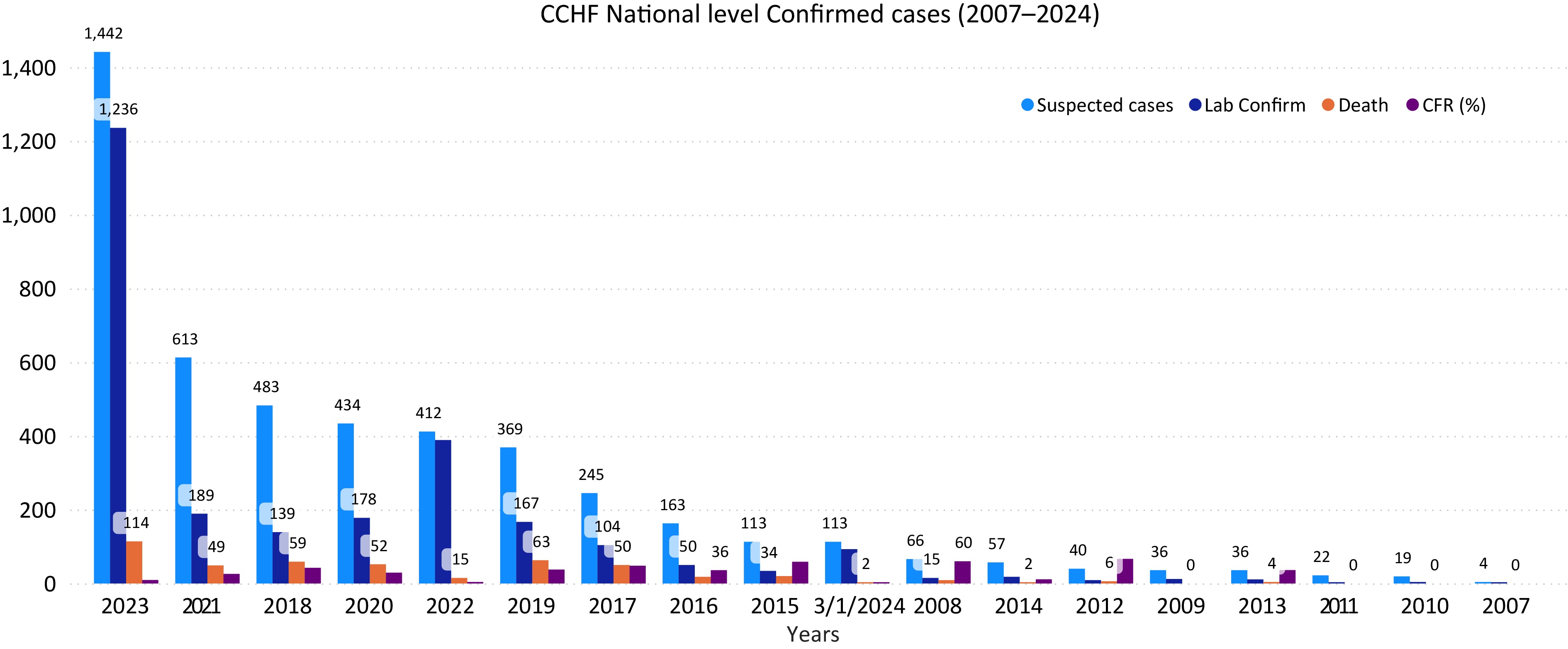
      Figure 1. 

      Number of suspected and confirmed CCHF cases and death in Afghanistan, 2007–2024. The horizontal axis year (from 2007 to 2024) and vertical axis shows the number of CCHF cases.

      From 2007 to 2024, the average case fatality ratio (CFR) of confirmed CCHF cases in Afghanistan was 30.2%. The CFR varied annually: 36% in 2016, 48% in 2017, 42.2% in 2018, 32% in 2019, 29% in 2020, 25% in 2021, 17% in 2022, 11% in 2023, and 2.1% as of March 2024. While CCHF cases increased until 2018, deaths subsequently declined (Fig. 1). Possible reasons for reduced CFR include improved public knowledge leading to prompt action, rapid blood donation supply, and increased preventive measures. Comparing January−March incidence from 2022−2024, no cases were reported in January−March 2022−2023, but 26 cases in January 2024, 47 in February, and 64 in March, indicating an anticipated increase in 2024 prevalence. Occupationally, most reported cases were in the 'others' category (23%), followed by unemployed (17%), housewives (14.5%), health staff (12.8%), shepherds (11%), butchers (7%), animal dealers and farmers (7.6%), and students (6.7%) (Fig. 2).

      Figure 3. 

      National surveillance data due to CCHF outbreak on an annual basis for the northeast region provinces (Kunduz, Takhar, Badakhshan, and Baghlan) during the period of 2007−2024. The horizontal axis shows CCHF prevalence on year basis and vertical axis shows the number of CCHF confirmed cases by the national surveillance system for the northeast region provinces.

    • Data from the national surveillance system for northeast region provinces (Kunduz, Takhar, Badakhshan, and Baghlan) showed higher CCHF prevalence in Kunduz (29.6%), followed by Takhar (25.4%), Badakhshan (24%), and Baghlan (20.8%) (Fig. 3). These findings suggest a higher likelihood of future prevalence in Kunduz and Takhar provinces. Hence, early mitigation is crucial, necessitating intensified biosecurity and tick prevention measures on animal farms.
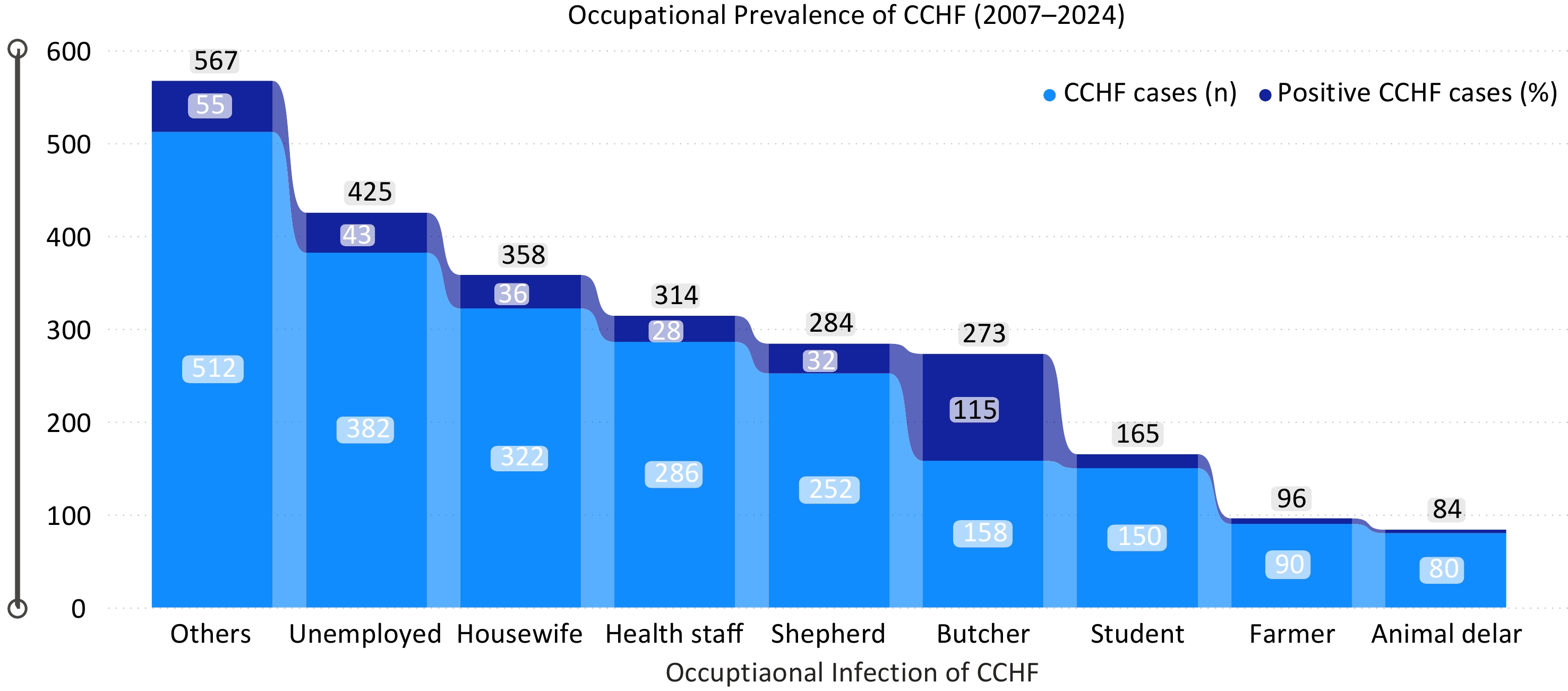
      Figure 2. 

      Occupational prevalence due to CCHF for the period of 2007−2024. The blue color indicates number of recorded cases while the dark blue color indicates the number of death cases respectively. The horizontal axis indicates the occupation of persons, and the vertical axis indicates the total number of CCHF cases due to occupational incidences of CCHF for the period of 2007−2024.

    • A total of 720 tick samples were collected, each containing an average of 27 ticks, totaling 4,672 ticks from Kunduz and Takhar provinces (Fig. 4). Tick species were identified based on morphological characteristics, revealing Hyalomma (H. asiaticum and H. marginatum), Rhipicephalus, Argas, Ornithodorus, Dermacentor, and Linognathus ticks. Hyalomma species were the most prevalent, followed by Rhipicephalus, while Dermacentor was least found. This indicates a significant presence of Hyalomma ticks, the primary vectors of CCHF, suggesting a high risk of transmission from infected animals to humans in the region and nationally (Fig. 4).
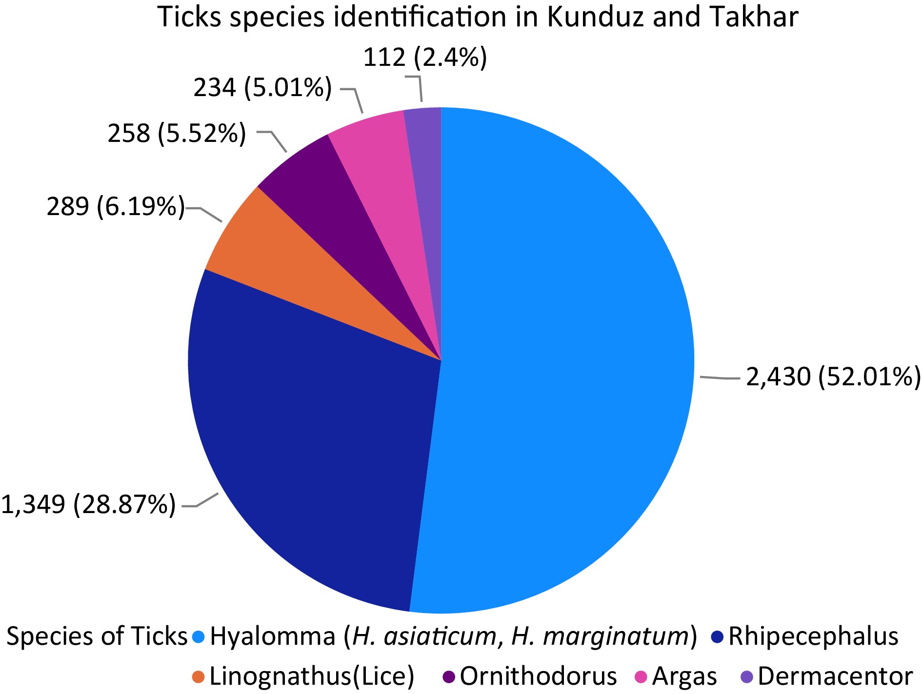
      Figure 4. 

      Different species of ticks presents in Kunduz and Takhar provinces with their percentage. Each species of the ticks found are exhibited in the figure with number and percentage.

    • A total of 720 ticks and 480 blood samples were collected, covering eight districts in two provinces equally. Among the samples tested by RT-PCR and IgG ELISA, 73 ticks in Kunduz and 81 ticks in Takhar were confirmed positive. In blood samples, 29 in Kunduz and 36 in Takhar tested positive (Fig. 5). Seropositivity was higher in Takhar than in Kunduz, with Rustaq in Takhar showing the highest prevalence. In Kunduz, Dasht-e-Archi had the highest prevalence. Overall, 102 ticks (17%) and 117 blood samples (19.5%) out of 720 and 480, respectively, were presumed positive for CCHF (Fig. 5).
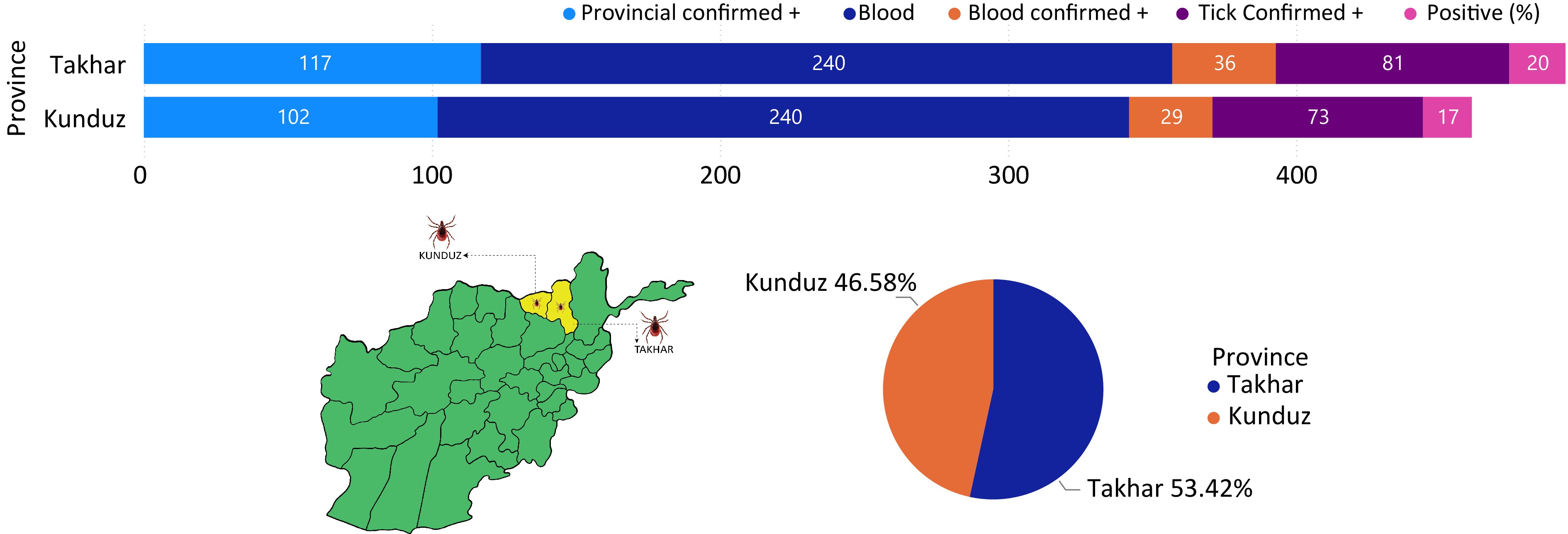
      Figure 5. 

      Prevalence of CCHF in Kunduz and Takhar provinces.

    • Intrinsic factors, notably animal species, demonstrated a significant association with CCHF prevalence, with cattle showing the highest prevalence, followed by sheep, goats, and camels. Seroprevalence was notably higher in females than males (Table 1). Animals older than 2 years were more susceptible than younger ones, although differences between indigenous and exotic breeds were non-significant, despite higher prevalence in indigenous animals (Tables 2, 3). Extrinsic factors such as housing system, feeding, hygiene practices, body condition score, and tick infestation were also explored (Tables 48). Free-ranging animals had a higher prevalence than tethered ones, with significant associations observed between housing systems and seroprevalence (Table 4). Pasture-grazing animals exhibited higher seroprevalence than stall-fed ones, while animals receiving good hygienic practices had lower prevalence compared to those with poor hygiene (Tables 5, 6). Obese animals demonstrated a higher prevalence than emaciated and average-weight animals, with significant differences based on body condition scores (Table 7). Significant associations were found within districts and between provinces (Kunduz and Takhar) regarding tick infestation, with tick-infested animals showing higher seroprevalence (Tables 8, 9).

      Table 1.  Association of sera-molecular prevalence of CCHF within animal species in the Kunduz and Takhar provinces of Afghanistan.

      Study province Study district Variables Examined Positive Seroprevalence (%) χ2 value p-value
      Kunduz Kunduz-Center Cattle 60 15 30 0.1816 0.0609
      Sheep 40 9 32.5
      Goat 30 3 26.66666667
      Camel 15 0 13.33333333
      Chicken 5 0 0
      Dasht-e-Archi Cattle 60 18 25 0.6031 0.088
      Sheep 40 13 22.5
      Goat 30 8 10
      Camel 15 2 0
      Chicken 5 0 0
      Imam Sahib Cattle 60 9 15 0.5714 0.061
      Sheep 40 11 27.5
      Goat 30 5 16.66666667
      Camel 15 2 13.33333333
      Chicken 5 0 0
      Char Dara Cattle 60 5 8.333333333 0.0742 0.045
      Sheep 40 4 10
      Goat 30 3 10
      Camel 15 0 0
      Chicken 5 0 0
      Takhar Taloqan Cattle 60 14 23.33333333 0.4867 0.067
      Sheep 40 11 27.5
      Goat 30 5 16.66666667
      Camel 15 1 6.666666667
      Chicken 5 0 0
      Rustaq Cattle 60 22 36.66666667 0.5683 0.109
      Sheep 40 16 40
      Goat 30 9 30
      Camel 15 3 20
      Chicken 5 0 0
      Khwaja Bahawodeen Cattle 60 9 15 0.4477 0.053
      Sheep 40 8 20
      Goat 30 4 13.33333333
      Camel 15 0 0
      Chicken 5 0 0
      Khwaja Ghar Cattle 60 3 5 0.4953 0.043
      Sheep 40 5 12.5
      Goat 30 2 6.666666667
      Camel 15 0 0
      Chicken 5 0 0

      Table 2.  Association of sera-molecular prevalence of CCHF within sex of animals in the Kunduz and Takhar provinces of Afghanistan.

      Study province Study district Variables Examined Positive Seroprevalence (%) χ2 value p-value
      Kunduz Kunduz-Center Male 77 12 15.58441558 0.5098 0.0361
      Female 73 15 20.54794521
      Dasht-e-Archi Male 77 11 14.28571429 0.0052 0.0302
      Female 73 30 41.09589041
      Imam Sahib Male 77 10 12.98701299 0.1713 0.0103
      Female 73 17 23.28767123
      Char Dara Male 77 4 5.194805195 0.2301 0.0016
      Female 73 8 10.95890411
      Takhar Taloqan Male 77 11 14.28571429 0.1079 0.0067
      Female 73 20 27.39726027
      Rustaq Male 77 19 24.67532468 8.125 0.015
      Female 73 31 42.46575342
      Khwaja Bahawodeen Male 77 7 9.090909091 0.1222 0.003
      Female 73 14 19.17808219
      Khwaja Ghar Male 77 3 3.896103896 0.1914 0.001
      Female 73 7 9.589041096

      Table 3.  Association of sera-molecular prevalence of CCHF within age of animals in the Kunduz and Takhar provinces of Afghanistan.

      Study province Study district Variables Examined Positive Seroprevalence (%) χ2 value p-value
      Kunduz Kunduz-Center < 6 months 20 3 15 1 0.0137
      1 > Year 30 3 23.33333333
      > 2 Year 40 7 25
      2 > Year 60 14 35
      Dasht-e-Archi < 6 months 20 3 15 1 0.027
      1 > Year 30 7 10
      > 2 Year 40 10 17.5
      2 > Year 60 21 23.33333333
      Imam Sahib < 6 months 20 2 10 1 0.012
      1 > Year 30 5 16.66666667
      > 2 Year 40 9 22.5
      2 > Year 60 11 18.33333333
      Char Dara < 6 months 20 1 5 1 0.007
      1 > Year 30 3 10
      > 2 Year 40 2 5
      2 > Year 60 6 10
      Takhar Taloqan < 6 months 20 2 10 1 0.016
      1 > Year 30 5 16.66666667
      > 2 Year 40 9 22.5
      2 > Year 60 15 25
      Rustaq < 6 months 20 5 25 1 0.035
      1 > Year 30 10 33.33333333
      > 2 Year 40 13 32.5
      2 > Year 60 22 36.66666667
      Khwaja Bahawodeen < 6 months 20 2 10 1 0.0103
      1 > Year 30 3 10
      > 2 Year 40 5 12.5
      2 > Year 60 11 18.33333333
      Khwaja Ghar < 6 months 20 1 5 1 0.006
      1 > Year 30 2 6.666666667
      > 2 Year 40 2 5
      2 > Year 60 5 8.333333333

      Table 4.  Association of sera-molecular prevalence of CCHF within breed of animals in the Kunduz and Takhar provinces of Afghanistan.

      Study province Study district Variables Examined Positive Seroprevalence (%) χ2 value p-value
      Kunduz Kunduz-Center Indigenous 140 26 18.57142857 0.5571 0.45
      Exotic 10 1 10
      Dasht-e-Archi Indigenous 140 39 27.85714286 0.6757 0.504
      Exotic 10 2 20
      Imam Sahib Indigenous 140 26 18.57142857 0.5571 0.45
      Exotic 10 1 10
      Char Dara Indigenous 140 12 8.571428571 0.3558 0.401
      Exotic 10 0 0
      Takhar Taloqan Indigenous 140 30 21.42857143 0.4654 0.465
      Exotic 10 1 10
      Rustaq Indigenous 140 47 33.57142857 0.8684 0.541
      Exotic 10 3 30
      Khwaja Bahawodeen Indigenous 140 20 14.28571429 0.7389 0.429
      Exotic 10 1 10
      Khwaja Ghar Indigenous 140 10 7.142857143 0.399 0.395
      Exotic 10 0 0

      Table 5.  Association of sera-molecular prevalence of CCHF with housing system of animals in the Kunduz and Takhar provinces of Afghanistan.

      Study province Study district Variables Examined Positive Seroprevalence (%) χ2 value p-value
      Kunduz Kunduz-Center Extensive 75 19 25.33333333 0.5086 0.007
      Intensive 75 8 10.66666667
      Dasht-e-Archi Extensive 75 29 38.66666667 0.0181 0.023
      Intensive 75 12 16
      Imam Sahib Extensive 75 22 29.33333333 0.0031 0.018
      Intensive 75 5 6.666666667
      Char Dara Extensive 75 10 13.33333333 0.026 0.003
      Intensive 75 2 2.666666667
      Takhar Taloqan Extensive 75 26 34.66666667 0.0005 0.029
      Intensive 75 5 6.666666667
      Rustaq Extensive 75 39 52 0.0005 0.07
      Intensive 75 11 14.66666667
      Khwaja Bahawodeen Extensive 75 17 22.66666667 0.0007 0.01
      Intensive 75 4 5.333333333
      Khwaja Ghar Extensive 75 9 12 0.0141 0.003
      Intensive 75 1 1.333333333

      Table 6.  Association of sera-molecular prevalence of CCHF with feeding system of animals in the Kunduz and Takhar provinces of Afghanistan.

      Study province Study district Variables Examined Positive Seroprevalence (%) χ2 value p-value
      Kunduz Kunduz-Center Stall feeding 75 6 8 0.0076 0.0145
      Pasture grazing 75 21 28
      Dasht-e-Archi Stall feeding 75 6 8 4.8928 0.064
      Pasture grazing 75 35 46.66666667
      Imam Sahib Stall feeding 75 3 4 0.0001 0.027
      Pasture grazing 75 24 32
      Char Dara Stall feeding 75 1 1.333333333 0.0053 0.005
      Pasture grazing 75 11 14.66666667
      Takhar Taloqan Stall feeding 75 5 6.666666667 0.0004 0.029
      Pasture grazing 75 26 34.66666667
      Rustaq Stall feeding 75 9 12 0.002 0.088
      Pasture grazing 75 41 54.66666667
      Khwaja Bahawodeen Stall feeding 75 3 4 0.002 0.013
      Pasture grazing 75 18 24
      Khwaja Ghar Stall feeding 75 2 2.666666667 0.066 0.001
      Pasture grazing 75 8 10.66666667

      Table 7.  Association of sera-molecular prevalence of CCHF with hygenenic measures for animals in the Kunduz and Takhar provinces of Afghanistan.

      Study province Study district Variables Examined Positive Seroprevalence (%) χ2 value p-value
      Kunduz Kunduz-Center Good 75 5 6.666666667 0.0024 0.018
      Poor 75 22 29.33333333
      Dasht-e-Archi Good 75 7 9.333333333 0.0001 0.056
      Poor 75 34 45.33333333
      Imam Sahib Good 75 4 5.333333333 0.0007 0.023
      Poor 75 23 30.66666667
      Char Dara Good 75 2 2.666666667 0.026 0.003
      Poor 75 10 13.33333333
      Takhar Taloqan Good 75 7 9.333333333 0.0052 0.019
      Poor 75 24 32
      Rustaq Good 75 12 16 0.0013 0.061
      Poor 75 38 50.66666667
      Khwaja Bahawodeen Good 75 4 5.333333333 0.0077 0.01
      Poor 75 17 22.66666667
      Khwaja Ghar Good 75 3 4 0.2205 0.0008
      Poor 75 7 9.333333333

      Table 8.  Association of sera-molecular prevalence of CCHF with body condition score of animals in the Kunduz and Takhar provinces of Afghanistan.

      Study province Study district Variables Examined Positive Seroprevalence (%) χ2 value p-value
      Kunduz Kunduz-Center Obese 50 15 44 5.9152 0.0002
      Average 50 4 14
      Emaciated 50 8 24
      Dasht-e-Archi Obese 50 22 30 1.0036 0.001
      Average 50 7 8
      Emaciated 50 12 16
      Imam Sahib Obese 50 16 32 1.3038 0.0004
      Average 50 3 6
      Emaciated 50 8 16
      Char Dara Obese 50 7 14 7.6074 1.194
      Average 50 1 2
      Emaciated 50 4 8
      Takhar Taloqan Obese 50 16 32 2.748 0.0003
      Average 50 4 8
      Emaciated 50 11 22
      Rustaq Obese 50 17 34 2.386 0.0053
      Average 50 6 12
      Emaciated 50 27 54
      Khwaja Bahawodeen Obese 50 11 22 2.4731 0.0001
      Average 50 1 2
      Emaciated 50 9 18
      Khwaja Ghar Obese 50 4 8 0.0001 2.627
      Average 50 1 2
      Emaciated 50 5 10

      Table 9.  Association of sera-molecular prevalence of CCHF with tick infestation in animals in the Kunduz and Takhar provinces of Afghanistan.

      Study province Study district Variables Examined Positive Seroprevalence (%) χ2 value p-value
      Kunduz Kunduz-Center Indigenous 75 19 25.33333333 0.0508 0.007
      Exotic 75 8 10.66666667
      Dasht-e-Archi Indigenous 75 32 42.66666667 0.0013 0.041
      Exotic 75 9 12
      Imam Sahib Indigenous 75 18 24 0.1103 0.005
      Exotic 75 9 12
      Char Dara Indigenous 75 8 10.66666667 0.2663 0.0008
      Exotic 75 4 5.333333333
      Takhar Taloqan Indigenous 75 24 32 0.0052 0.019
      Exotic 75 7 9.333333333
      Rustaq Indigenous 75 39 52 0.0005 0.0702
      Exotic 75 11 14.66666667
      Khwaja Bahawodeen Indigenous 75 18 24 0.002 0.013
      Exotic 75 3 4
      Khwaja Ghar Indigenous 75 8 10.66666667 0.0666 0.0018
      Exotic 75 2 2.666666667
    • Afghanistan is currently facing an intensified surge of Crimean-Congo Hemorrhagic Fever (CCHF) nationwide. Domestic ruminants, including cattle, sheep, goats, camels, and chickens, can act as reservoir hosts for CCHFV, aiding virus transmission through tick bites or direct contact with infected tissues. This situation raises substantial public health concerns. From 2007 to 2024, Afghanistan has seen an annual rise in confirmed CCHF cases and associated deaths. Public surveillance data indicates 4,667 suspected cases during this period, with 2,651 confirmed positive cases and 463 deaths. Specific numbers for certain years include: 163 cases in 2016, 245 in 2017, 483 in 2018, 412 in 2022, 1,442 in 2023, and 113 as of March 2024. The highest confirmed cases were in 2023 (1,236), followed by 2022, 2018, and 2017. Despite a rise until 2018, there has been a decline in deaths since then[5].

      The present investigation compared CCHF incidences nationally from January to March in 2022 to 2024. Surprisingly, no cases were reported in January to March in 2022 and 2023. However, in January 2024, 26 cases were confirmed, followed by 47 in February and 64 in March, totaling 137 cases with a CFR of 1%. These findings indicate a higher tendency for increased CCHF cases in 2024 compared to previous years[15].

      The present findings on occupational transmission of CCHF from 2007 to 2024 aligns with previous studies[16]. Most cases were from individuals categorized as 'others' (23%), followed by the unemployed (17%), housewives (14.5%), health staff (12.8%), shepherds (11%), butchers (7%), animal dealers, and farmers (7.6%), and students (6.7%). These patterns correspond with studies by Ahmad et al., Sahak, and research in Pakistan, which reported CFR rates ranging from 10% to 40%[5,17].

      Program experts suggest that the increase in CCHF cases may be linked to environmental factors. Drought and a lack of fodder in the West and North regions have led to dry pastures, prompting the migration of livestock and people to areas with better grazing conditions. This movement increases the potential for infected tick exposure as migrating herds mix with others[2,4,10].

      Data from the national surveillance system for the northeast region provinces (Kunduz, Takhar, Badakhshan, and Baghlan) showed Kunduz had the highest prevalence (29.6%), followed by Takhar (25.4%), Badakhshan (24%), and Baghlan (20.8%)[18]. This suggests a higher likelihood of prevalence in Kunduz and Takhar in the future. Domestic ruminants like cattle, goats, and sheep can act as reservoir hosts for CCHFV, making tick-borne diseases a significant concern due to their veterinary and public health implications. Hyalomma species are major vectors for CCHFV transmission to both animal and human hosts through bites[18,19]. Across eight districts in the Kunduz and Takhar provinces, a total of 720 ticks and 480 blood samples were collected. Of the 360 ticks sampled in each province, 73 in Kunduz and 81 in Takhar tested positive for CCHFV using RT-PCR and IgG ELISA (Fig. 5). Regarding blood samples, 29 out of 240 were positive in Kunduz, while 36 out of 240 were positive in Takhar. Seropositivity was higher in Takhar province than in Kunduz. In Takhar, Rustaq had the highest prevalence, followed by Taloqan, Khwaja Bahawodeen, and Khwaja Ghar, ranging from 10% to 2%. In Kunduz, Dasht-e-Archi had the highest prevalence, followed by Kunduz Center, Imam Sahib, and Char Dara, ranging from 8.2% to 2.4% (Fig. 5).

      Remarkably, among the eight districts of both provinces, Rustaq showed the highest prevalence of CCHF at 10%, followed by Dasht-e-Archi at 8.2%. Across both provinces, 102 (17%) tick samples were presumed positive and 117 (19.5%) blood samples out of 720 and 480, respectively (Fig. 5).

      These findings are in line with parallel studies conducted in various countries. For instance, in Gambia[20], a higher prevalence was reported in cattle compared to small ruminants (sheep and goats), which aligns with the present results. Similarly, studies[18,21] in different locations also revealed higher seropositivity of CCHFV in cattle than in goats and sheep, consistent with the present findings. Additionally, research in Pakistan reported the highest seroprevalence of CCHFV antibodies in cattle, followed by sheep and goats. Studies in Corsica, France[22] and Kosovo, Germany, also found higher seropositivity in cattle compared to sheep and goats[23].

      The present findings reveal higher seroprevalence in cattle compared to sheep, goats, and camels, suggesting they could serve as a source of CCHFV transmission to these animals during grazing interactions. This possibility is supported by previous research[24]. The elevated seroprevalence in cattle may be attributed to Hyalomma ticks, the primary carriers of CCHFV, which prefer feeding on larger animals like cattle. Ticks readily attach to cattle for feeding, facilitating efficient viral transfer between infected ticks and cattle. CCHFV replicates to higher levels in cattle compared to sheep, goats, and camels, leading to a higher viral load in the bloodstream. This increases the likelihood of ticks acquiring CCHFV when feeding on infected cattle.

      In the present study, a significantly higher seroprevalence of CCHF was found in female domestic animals compared to males (p > 0.05). This aligns with previous research[18,20,25,26]. The elevated seroprevalence in female domestic animals could be attributed to factors such as pregnancy stress, lactation stress, and limited access to balanced nutrition, which may reduce immunity and decrease their resistance to tick infestations.

      The present study supports previous findings that local breeds exhibit higher seropositivity compared to exotic breeds, as reported in previous studies[18,25]. Similarly, previous research[18,25] indicates that indigenous cattle breeds experience more tick infestations and external parasites compared to exotic breeds, potentially leading to higher seroprevalence. This similarity could be attributed to factors such as poor hygiene, limited access to quality feed, and inferior husbandry practices observed in Indigenous breeds compared to exotic breeds found in the study areas. The current study highlights higher seroprevalence in animals raised extensively or on communal grazing systems, while those in intensive housing systems exhibit lower seroprevalence. These findings align with previous research[18,24,27] . The increased seroprevalence in extensively raised animals may be attributed to their closer proximity to tick vectors, lack of acaricide use, and poor hygiene management practices on the farm. Conversely, the lower seroprevalence in animals kept in intensive housing systems may result from effective tick control measures, such as regular acaricide application and good hygiene practices, which reduce tick populations[27].

      Early studies support the present findings that higher seroprevalence in older and tick-infested cattle is age-dependent[28]. Seroprevalence in cattle increases with age and the presence of tick infestation, as documented in previous research[29]. Studies conducted in Kenya, northwestern Senegal, Afghanistan, and Uganda[30] also support this association between seroprevalence and age in cattle. Additionally, research suggests that the seroprevalence of CCHFV antibodies in domestic ruminants is dependent on age, with older animals exhibiting higher seroprevalence rates than younger ones[21]. This higher seroprevalence with age may be attributed to increased production of IgG antibodies in response to continuous exposure to CCHFV-infected ticks in older animals in endemic areas, compared to younger animals with maternal immunity.

      The present investigations have identified a correlation between the body condition of domestic animals and CCHFV antibody seroprevalence. Previous research[18] found a high seroprevalence in overweight ruminants, correlating with weight. They observed that obese animals were more susceptible to CCHF compared to emaciated animals due to weakened immunity. The present study supports this, revealing that obese domestic animals exhibited the highest seroprevalence, followed by those of average weight, and then emaciated animals, respectively.

      Furthermore, heavily infested ruminants play a crucial role in CCHFV transmission and can become sources of infection for healthy animals compared to tick-free animals, as reported previously[18,31]. Similarly, it was reported that tick-infested cattle have a higher seroprevalence compared to tick-free animals[28], which is fully consistent with the current study.

      Afghanistan, situated in the ecological range of the Hyalomma tick, experiences an annual increase in CCHF incidence[10,32]. The variation in seropositivity observed in the present study may be attributed to the endemicity of CCHF in the region, the significant abundance of ticks, and host behavior patterns influenced by climate changes and drought. Additionally, differences in laboratory examinations for molecular and serological detection of CCHFV antibodies, including specificity and sensitivity could contribute to these variations. These insights call for further investigation into the associated factors contributing to the rising number of CCHF cases within the country.

    • The higher seroprevalence underscores a significant healthcare concern, given the recent rise in CCHF cases and fatalities in Afghanistan. The initial report highlights a notably elevated prevalence of CCHFV nationally and regionally, urging urgent attention to mitigate further spread, particularly in livestock. Extrinsic risk factors (husbandry practices, animal condition, and tick infestation) and intrinsic factors (species, sex, age, and breed) show significant associations with CCHFV seroprevalence, detected through IgG antibodies and RT-PCR analysis. Collaborating with Afghan molecular experts, an in-house molecular method has been developed for CCHF virus detection in ticks and blood samples, facilitating deeper genome studies. Early detection and understanding of risk factors in animal hosts aid in mapping endemic areas. Given CCHF's impact on human health, especially those in direct animal contact, control strategies are imperative. Livestock plays a vital role in rural Afghans' livelihoods and can transmit diseases. Raising local awareness, collaborating with health and veterinary departments, promoting animal health practices, and intensifying livestock husbandry alongside establishing active disease surveillance are essential for enhancing one-health approaches.

      • All procedures were reviewed and approved by the Animal Care and Research Committee of Ministry of Agriculture Irrigation and Livestock (MAIL), identification number: (KBL-2023–MAIL-03), approval date: 2023-12-09, and implemented based on the standard of Experimental Animal Care and Use Guidelines of Animals. The research followed the "Replacement, Reduction, and Refinement" principles to minimize harm to animals. This article provides details on the housing conditions, care, and pain management for the animals, ensuring that the impact on the animals is minimized during the experiment.

      • The authors confirm contribution to the paper as follows: writing – draft manuscript preparation, investigation, conceptualization: Hamdard E; formal analysis, data curation: Karwand B, Din Muhammad S; data collection – review & editing, methodology: Zahir A, Din Muhammad S, Mosavi SH. writing – final draft and editing: Sayedpoor S. All authors reviewed the results and approved the final version of the manuscript.

      • The original contributions presented in this study are included in the article/supplementary material. Further inquiries can be directed to the corresponding author.

      • The authors declare that they have no conflict of interest.

      • Copyright: © 2024 by the author(s). Published by Maximum Academic Press on behalf of Nanjing Agricultural University. This article is an open access article distributed under Creative Commons Attribution License (CC BY 4.0), visit https://creativecommons.org/licenses/by/4.0/.
    Figure (5)  Table (9) References (32)
  • About this article
    Cite this article
    Hamdard E, Zahir A, Mosawi SH, Din Muhammad S, Karwand B, et al. 2024. Descriptive epidemiology and seroprevalence investigations of Crimean-Congo Hemorrhagic Fever virus in domestic animals of northeast Afghanistan. Animal Advances 1: e007 doi: 10.48130/animadv-0024-0007
    Hamdard E, Zahir A, Mosawi SH, Din Muhammad S, Karwand B, et al. 2024. Descriptive epidemiology and seroprevalence investigations of Crimean-Congo Hemorrhagic Fever virus in domestic animals of northeast Afghanistan. Animal Advances 1: e007 doi: 10.48130/animadv-0024-0007

Catalog

  • About this article

/

DownLoad:  Full-Size Img  PowerPoint
Return
Return