ARTICLE   Open Access    

A clinical retrospective study of anlotinib in combination with gemcitabine and albumin-bound paclitaxel for the first-line treatment of advanced pancreatic cancer

  • # Authors contributed equally: Yawen Zheng, Yuhan Zhou

More Information
  • Anlotinib has demonstrated efficacy and tolerability in treating various tumors. The purpose of this retrospective study is to assess the efficacy and safety of anlotinib combined with gemcitabine and albumin-bound paclitaxel as a first-line treatment for advanced pancreatic cancer. Patients with advanced pancreatic cancer who are not candidates for surgery were divided into two groups: anlotinib plus gemcitabine/nab-paclitaxel (A + AG) (n = 32) and gemcitabine/nab-paclitaxel alone (AG) (n = 39). Data was analyzed for clinical characteristics, survival benefits, and side effects. The median progression-free survival (mPFS) (7.4 m vs 5.1 m, p = 0.003) and the median overall survival (mOS) (15 m vs 9.6 m, p = 0.001) were both prolonged in the A + AG group compared to AG group. The objective response rate (ORR) (12.5% vs 5.1%, p = 0.399) and the disease control rate (DCR) (87.5% vs 74.4%, p = 0.233) only disclosed increasing trends in the A + AG group compared to the AG group. Univariate analysis identified that pathology type (p = 0.002) affects PFS, while pathology type (p = 0.003), and CA199 level (p = 0.020) affect OS in the AG groups. In the A + AG group, the Thymidine kinase (TR) level affected PFS (p = 0.014) and OS (p = 0.035). Multivariate analysis showed no clinical factors correlated with PFS and OS in both the A + AG and AG groups. The most common adverse effects were leukopenia (91.5%), fatigue/poor appetite (85.9%), nausea/vomiting (80.3%), and thrombocytopenia (77.5%). The incidence of grade III/IV adverse reactions was similar in both groups. No grade V adverse reactions occurred. This study demonstrates that anlotinib combined with the AG chemotherapy regimen provides a new first-line therapeutic choice for patients with advanced pancreatic cancer.
  • Pancreatic cancer (PC) is among the most lethal malignancies, and its mortality closely parallels its morbidity. PC is estimated to be the second leading cause of cancer deaths in the next decade, with incidence increasing rapidly[1,2]. Surgical pancreatectomy remains the only curative approach for PC. Unfortunately, the majority of patients are unresectable at the time of diagnosis due to extensive local spread and metastasis[3,4]. PC is an atypical disease whose symptoms are easily confused with other noncancerous gastrointestinal symptoms, including nausea, abdominal pain, weight loss, and jaundice, leading to later medical consultations. In addition, the pancreas is an easily overlooked organ (16 cm × 6 cm × 2 cm) hidden behind the stomach and duodenum, and pancreatic cancer holds few blood vessels compared to other gastrointestinal cancers, making PC insensitive to imaging detection, such as computed tomography (CT) and magnetic resonance imaging (MRI)[5]. Large-scale screening programs can effectively reduce the disease burden in various gastrointestinal cancers, including esophageal cancer and colorectal cancer. However, there are no noninvasive diagnostic methods that facilitate early diagnosis of population-based PC[69]. This fact sheds light on the necessity for discovering a novel powerful diagnostic approach with higher compliance for PC in the early stages of development.

    The human body encompasses trillions of indigenous microbes, including eukaryota, bacteria, and archaea. Microbiota are a collection of microorganisms that live within/on the human body, and the human microbiome refers to the collective genome encoded by the microbiota[10,11]. Body sites such as the skin, oral cavity, stomach, bladder, vaginal cavity, nasal cavity, and lungs host rich microbial communities; among them, the gastrointestinal tract harbors the greatest density of microbiota, which means the gut microbiota receive the most attention. Bacterial analysis is the main focus when mentioning the microbiota unless otherwise specified. Recently, facilitated by the advancement of high-throughput 16S rRNA sequencing and whole-genome shotgun metagenomic technology, we can gain a glimpse into microbial diversity and abundance[12,13]. Accumulating studies have suggested that the human microbiota plays a vital role in human health, both in physiological and pathological conditions. Given that the microbiota is associated with a variety of diseases, its analysis may help in disease diagnosis, prognosis, prevention, and therapy[1419]. Among several sample sources that can reflect the fluctuation of the human microbiota, such as intestinal mucosa and secretions, stool, and oral saliva are undoubtedly the most noninvasive and accessible means. It has been reported that diagnostic models based on discriminating bacterial species from oral or fecal samples show high sensitivity and specificity in various malignancies, including hepatocellular carcinoma and colorectal cancer[2022]. Further studies reveal that dysbiosis of the gut microbiota participates in carcinogenesis via abnormal metabolites or immune modulation. There are also articles mentioning the relationship between gut microbiota and PC, which provides a basis for the diagnostic value of gut microbiota in PC. Farrell et al. compared oral microbial composition between PC patients and matched healthy controls via Human Oral Microbe Identification Microarray (HOMIM). They identified 16 species/clusters, which showed a significantly different frequency between the two groups[23]. Similarly, Michaud et al. compared antibodies against oral bacteria between 405 PC patients and 416 matched controls and showed that individuals expressing higher levels of antibodies against Porphyromonas gingivalis (P. gingivalis) have a twofold higher risk of PC than those with lower levels of antibodies[24]. In addition to HOMIM and antibodies, there are various methods to investigate the microbial composition, including the long-lasting gold standard, 16S rRNA sequencing[25]. Based on next-generation sequencing technology, several studies aimed to reveal the entirety of the genetic information contained in a sample from PC patients and looked to discriminate bacteria as a diagnostic biomarker for PC[2640].

    Considering the possibility of human microbiota serving as a noninvasive diagnostic tool in the screening of PC, we sought to systematically review the studies revealing different microbial compositions between PC patients and controls and the possible use of the models established on discriminated taxa for the early diagnosis of PC. Given the significant variations between different detection methods for the microbial community, this systematic review focuses only on articles using 16S rRNA sequencing.

    The aim of this systematic review was to collect all original research articles that evaluated the possibility of gut microbiota, measured by 16S rRNA sequencing, as a noninvasive prognostic tool for PC. The research was performed in accordance with procedures recommended by the Cochrane Collaboration.

    A comprehensive literature search for eligible articles was performed in PubMed, Cochrane Library, Embase, and Web of Science up to 20 November, 2024. The search approach followed the combinations of the terms 'pancreatic cancer', 'pancreatic ductal adenocarcinoma', 'pancreatic adenocarcinoma', 'gut', 'fecal', 'stool', 'oral', 'salivary', 'microbial', 'microbiome', and 'microbiota'. Relevant reviews were screened to seek missing studies. Titles and abstracts were screened to remove duplicates. This systematic review was limited to English-language articles only.

    The articles that met all following terms were included in this review: assessed the gut microbial community in oral or fecal samples from PC patients compared with healthy controls, used 16S rRNA sequencing, and found several discriminated bacteria as outcomes. Studies were removed if they were published as letters or case reports, as they failed to provide enough data for the present review. Studies examining fungi or viruses in PC patients were excluded because they did not share the same sequencing method with bacteria. Articles focused on specific bacterial species, such as Helicobacter pylori, were also removed because several meta-analyses related to this topic have been published. The reviewers screened the studies based on the inclusion criteria independently, and disagreements were discussed to resolve the differences.

    Two reviewers independently retrieved the articles. The basic information extracted from the articles was summarized as: study characteristics, population characteristics, and methodologic characteristics. The study characteristics were as follows: first author, publication year, country, publication type, and study type. The population and methodologic characteristics were displayed in terms of sample size, age, sex, body mass index, smoking status, sample source, temperature for storage, measurement method, diversity assessment, and administration of antibiotics or probiotics. To minimize biases, a quality assessment was conducted following the Newcastle–Ottawa Scale (NOS)[41], which is based on three components: the selection of study groups, comparability, and ascertainment of exposure/outcome. Two independent reviewers scored the NOS on major aspects of risk and applicability assessment.

    The search process is displayed in a PRISMA flow diagram, as shown in Fig. 1. A total of 80 papers were retrieved from PubMed, the Cochrane Library, Embase, and the Web of Science. Following a thorough screening of their full titles and abstracts, 18 reviews or meta-analyses were excluded. The full-text articles were further examined and those that did not meet all the inclusion criteria were excluded. Fourty-seven papers were excluded due to the following reasons: use of invasive methods to obtain samples (n = 5), and not being human studies (n = 10); lack of controls (3); not pancreatic cancer (26); other detection methods (3). Finally, 15 further studies were ultimately included.

    Figure 1.  Flowchart.

    Table 1 displays the characteristics of the 15 studies included in our review published between 2013 and 2024. Among the 15 studies, five originated from China, four were from the USA, another two were from Israel and Japan, and one was from Iran and Germany. The majority of studies were published as journal articles, and only two were conference proceedings. Only one of the studies was designed as a prospective study, while the remaining studies were case–control studies. The NOS score ranged from 6 to 8 in the included studies.

    Table 1.  Study characteristics.
    Study Country Publication type Study type NOS score
    Fan et al., 2018[26] USA Journal article Prospective cohort 8
    Torres et al., 2015[31] USA Journal article Controlled 8
    Olson et al., 2017[29] USA Journal article Pilot 7
    Lin et al., 2013[34] USA Conference proceedings Pilot 7
    Lu et al., 2019[28] China Journal article Controlled 8
    Vogtmann et al., 2019[32] Iran Journal article Controlled 8
    Ren et al., 2017[30] China Journal article Controlled 8
    Half et al., 2019[27] Israel Journal article Controlled 8
    Half et al., 2015[33] Israel Conference proceedings Pilot 6
    Kartal et al., 2022[35] Germany Journal article Case–control 6
    Chen et al., 2023[36] China Journal article Controlled 7
    Hashimoto et al., 2022[38] Japan Journal article Controlled 8
    Sono et al., 2024[39] Japan Journal article Controlled 7
    Zhao et al., 2024[37] China Journal article Controlled 8
    Yang et al., 2023[40] China Journal article Controlled 6
    NOS, Newcastle-Ottawa Scale. USA, United States of America.
     | Show Table
    DownLoad: CSV

    The population characteristics are described in Table 2. The sample size of the included studies ranged between 28 (13 PC) in the smallest study by Lin et al., and 732 (361 PC) in the largest study by Fan et al. To increase the specificity of the diagnostic taxa, four reviews included other pathological conditions, such as pancreatitis and nonalcoholic fatty liver disease, in the control group rather than healthy individuals only. The majority of journal articles were able to provide necessary demographic information such as age, gender, and body mass index. The sample type included an oral wash, saliva, tongue coat, and stool, which were all obtained by noninvasive methods. Half of the studies emphasized that samples were stored at −80 °C before analysis. It is recognized that the administration of antibiotics, probiotics, and prebiotics may affect gut microbiota. All the case–controlled journal articles limited antibiotic usage when enrolled. Smoking status, a well-known risk factor for PC, also plays a role in gut microbiota analysis, as mentioned in only four articles. Although all these articles adopted 16S rRNA sequencing, hypervariable regions vary with different papers including V3–V4, V4–V5, and V3–V5, while several articles did not mention the hypervariable regions they chose for analysis. Six articles assessed both α-diversity and β-diversity in the analysis, the Simpson index and the Shannon index were the most used as an outcome of α-diversity, and β-diversity is mainly represented by the principal coordinate analysis of weighted and unweighted UniFrac distances. The above description is shown in Table 3.

    Table 2.  Population characteristics.
    Study Sample size Age % male BMI % smoking
    PC Control PC Control PC Control PC Control PC Control
    Fan et al., 2018[26] 361 371 68.5 68.3 57.1 57.1 57.3 49.9
    Torres et al., 2015[31] 8 100
    (other disease 78 HC 22)
    71.1 60.7
    (other cancers)
    75.0 50.0
    Olson et al., 2017[29] 40 97
    (IPMN 39; HC 58)
    < 70, 64.0%; ≥ 70, 35.0% < 70, 42.0%; ≥ 70, 59.0% (IPMN); < 70, 81.0%;
    ≥ 70, 19.0% (HC)
    53.0 56.0 (IPMN); 40.0 (HC) Normal 38.0%; abnormal 61.0% Normal 36.0%;
    abnormal 64.0% (IPMN); normal 43.0%;
    abnormal 57.0% (HC)
    44.0 46.0 (IPMN); 31.0 (HC)
    Lin et al., 2013[34] 13 15
    (pancreatitis 3 HC 12)
    Lu et al., 2019[28] 30 25 50.8 ± 5.3 48.2 ± 6.0 70.0 80.0 22.5 ± 1.2 22.6 ± 1.6
    Vogtmann et al., 2019[32] 273 285 < 70, 63.7%;
    ≥ 70, 36.3%
    < 70, 67.4%; ≥ 70, 32.6% 60.4 46.0 Normal 57.5%; abnormal 42.5% Normal 46.3%;
    abnormal 53.7%
    30.5 25.6
    Ren et al., 2017[30] 85 57 56.0 (33.0–78.0) 52.0 (43.0–67.0) 55.3 63.2 22.7 (19.5–26.0) 23.2 (18.5–27.1)
    Half et al., 2019[27] 30 35
    (NAFLD 16; PCL 6 HC 13)
    68.9 ± 6.2 51.0 ± 10.8 (NAFLD);
    66.0 ± 15.3 (PCL);
    59.0 ± 8.7 (HC)
    53.3 75.0 (NAFLD);
    83.3 (PCL); 46.2 (HC)
    Half et al., 2015[33] 15 15
    Kartal et al., 2022[35] 57 79 (HC 50 CP 29)
    Chen et al., 2023[36] 40 54 (HC 50 CP 15)
    Hashimoto et al., 2022[38] 5 68 70.0−89.0 54.0 40.0 42.6
    Sono et al., 2024[39] 30 18 63.7 63.0 53.3 66.7 22.0 24.5 36.7 88.9
    Zhao et al., 2024[37] 29 9 67.6 ± 10.8 30.5 ± 6.8 58.6 33.3 22.4 ± 2.9 20.8 ± 1.5
    Yang et al., 2023[40] 44 50 47.7 22.4 ± 2.8
    /, no related information; BMI, body-mass index; HC, healthy control; IPMN, intraductal papillary mucinous neoplasms; NAFLD, non-alcoholic fatty liver disease; PCL, pre-cancerous lesions. Notes: * It was a prospective study including two large population-based cohorts whose BMI was described in Median or mean.
     | Show Table
    DownLoad: CSV
    Table 3.  Methodologic characteristics.
    Study Sample Temperature
    for storage
    Measurement
    method
    Diversity assessment Antibiotics Probiotic or
    prebiotic
    α-diversity β-diversity
    Fan et al., 2018[26] Oral wash −80 °C 16S V3–V4 Shannon, Simpson PCoA
    Torres et al., 2015[31] Saliva −80 °C 16S Chao1 ANOSIM Not in 2 wk
    Olson et al., 2017[29] Saliva 16S V4–V5 NP Shannon, Inverse Simpson NA Not in 30 d
    Lin et al., 2013[34] Oral wash 16S
    Lu et al., 2019[28] Tongue coat 16S V3–V4 Shannon, Simpson, inverse Simpson, Obs, Chao 1, ACE PCoA Not in 8 wk Not in 8 wk
    Vogtmann et al., 2019[32] Saliva −70 °C 16S V4 Observed SVs, Shannon, Faith's PD PCoA
    Ren et al., 2017[30] Stool −80 °C 16S V3–V5 Shannon, Simpson, Chao 1 PCoA Not in 8 wk Not in 8 wk
    Half et al., 2019[27] Stool −80 °C 16S Shannon PCoA Not in 8 wk
    Half et al., 2015[33] Stool 16S ANOSIM
    Kartal et al., 2022[35] Stool and saliva −80 °C 16S V4 Shannon, Simpson Unweighted TINA index
    Chen et al., 2023[36] Fecal and saliva −80 °C 16S V3–V4 Chao1, Shannon observed species, and PD whole tree PCoA Not in 4 wk Not in 4 wk
    Hashimoto et al., 2022[38] Stool and saliva −80 °C 16S Shannon PCoA Not in 6 months Not in 6 months
    Sono et al., 2024[39] Stool and saliva 16S V3–V4 Observed features, Shannon BrayeCurtis dissimilarity QIIME 2
    Zhao et al., 2024[37] Stool −80 °C 16S V3–V4 Chao 1 Acex, Shannon Simpson Sobs i Coverage Mothur software Qiime Not in 8 wk Not in 8 wk
    Yang et al., 2023[40] Stool −80 °C 16S Chao 1 Shannon PCoA
    −, no related information. d, day; wk, week.
     | Show Table
    DownLoad: CSV

    Bacterial taxonomic levels are described in Table 4. Combining studies that investigated samples from the oral cavity, six phyla were significantly different between PC patients and healthy controls, including Fusobacteria, Bacteroidetes, Firmicutes, Actinobacteria, Verrucomicrobia, and Proteobacteria. In addition, the abundance of Firmicutes and Proteobacteria was found to differ significantly in at least two studies, and they shared the same changing directions in different articles. However, there were some contradictory findings in the phyla Bacteroidetes and Fusobacteria. Fan et al. found that the phylum Fusobacteria was associated with decreased PC risk (OR = 0.94), while the phylum Bacteroidetes was associated with a higher risk of PC (OR = 1.01)[26]. Their findings were inconsistent with the results from Lu et al., whose findings suggested that PC patients presented a higher abundance of Fusobacteria and a lower abundance of Bacteroidetes than healthy controls[28]. However, the results from Fan et al. were in agreement with the findings from a European cohort containing 405 PC patients and 416 matched controls[24], in which higher antibody levels from Fusobacteria in prediagnosis blood were associated with reduced PC risk. At the genus level, 19 taxa were found to be significantly different between PC patients and healthy controls in five studies: Leptotrichia, Alloprevotella, Bacteroides, Porphyromonas, Aggregatibacter, Neisseria, Streptococcus, Haemophilus, Corynebacterium, Fusobacterium, Actinomyces, Rothia, Solobacterium, Oribacterium, Campylobacter, Atopobium, Parvimonas, Paraprevotella, and Lachnospiraceae G7. Among these identified genera, Bacteroides was found to be higher in PC, while Porphyromonas, Aggregatibacter, Neisseria, and Haemophilus were lower in PC when compared with controls in at least two articles. All five genera were found to change in the same direction between different studies. Nevertheless, there was an inconsistency between articles, such as the genus Leptotrichia that was found by Torres et al.[31] and by Lu et al.[28] to be more abundant in PC patients, while Fan et al.[26] found the genus Leptotrichia to be associated with decreased PC risk (OR = 0.87). This inconsistency may be explained by different experimental designs and various types of published articles.

    Table 4.  Discriminating taxa.
    Study Bacteria taxonomic level
    Phylum Class Order Family Genus
    Fan et al., 2018[26] Bacteroidetes (↑) SR1[C-1] (↑) Fusobacteriales (↓) Leptotrichiaceae (↓) Alloprevotella, Porphyromonas gingivalis, and Aggregatibacter actinomycetemcomitans (↑)
    Fusobacteria (↓) Fusobacteria (↓) Leptotrichia (↓)
    Torres et al., 2015[31] Firmicutes (↑) Leptotrichia Bacteroides (↑)
    Proteobacteria (↓) Porphyromonas Aggregatibacter Neisseria (↓)
    Olson et al., 2017[29] Firmicutes (↑) Bacilli (↑) Lactobacillales (↑) Streptococcaceae (↑) Streptococcus (↑)
    Proteobacteria (↓) Gammaproteobacteria; Betaproteobacteria (↓) Pasteurellales Neisseriales (↓) Pasteurellaceae Neisseriaceae (↓) Haemophilus Neisseria (↓)
    Lin et al., 2013[34] Bacteroides (↑)
    Corynebacterium Aggregatibacter (↓)
    Lu et al., 2019[28] Firmicutes, Fusobacteria and Actinobacteria (↑) Leptotrichiaceae, Fusobacteriaceae, Actinomycetaceae, Lachnospiraceae, Micrococcaceae, Erysipelotrichaceae, and Campylobacteraceae (↑) Leptotrichia, Fusobacterium, Actinomyces, Rothia, Solobacterium, Oribacterium, Campylobacter, Atopobium, and Parvimonas (↑)
    Bacteroidetes (↓) Prevotellaceae, Pasteurellaceae, and Porphyromonadaceae (↓) Porphyromonas, Haemophilus, and Paraprevotella (↓)
    Vogtmann et al., 2019[32] Enterobacteriales (↑) Enterobacteriaceae, Bacteroidaceae, Staphylococcaceae (↑) Lachnospiraceae G7 (↑)
    Haemophilus (↓)
    Ren et al., 2017[30] Bacteroidetes (↑) Prevotella, Veillonella, Klebsiella, Selenomonas, Hallella, Enterobacter, and Cronobacter (↑)
    Firmicutes and Proteobacteria (↓) Gemmiger, Bifidobacterium, Coprococcus, Clostridium IV, Blautia, Flavonifractor, Anaerostipes, Butyricicoccus, and Dorea (↓)
    Half et al., 2019[27] Bacteroidetes (↑) Bacteroidia; Verrucomicrobiae; Clostridia Bacteroidales; Verrucomicrobiales; Clostridiales Porphyromonadaceae; Verrucomicrobiaceae; Clostridiaceae1 Odoribacter, Akkermansia (↑)
    Firmicutes (↓) Clostridiumsensustricto1 (↓)
    Half et al., 2015[33] Bacteroidetes Verrucomicrobia (↑) Sutterella, Veillonella, Bacteroides, Odoribacter, and Akkermansia (↑)
    Firmicutes and Actinobacteria (↓)
    Kartal et al., 2022[35] Veillonella atypica, Fusobacterium, nucleatum/hwasookii, Alloscardovia, omnicolens (↑) Romboutsia timonensis, Faecalibacterium, rausnitzii, Bacteroides, coprocola, Bifidobacterium, and bifidum (↓)
    Chen et al., 2023[36] Bacteroidetes (↑) Veillonella, Peptostreptococcus, Akkermansia, Parvimonas, Solobacterium, Olsenella, and Escherichia-Shigella (↑)
    Firmicutes (↑)
    Proteobacteria (↑)
    Verrucomicrobia (↑)
    Hashimoto et al., 2022[38] Actinomyces, Lactobacillus, Streptococcus, and Veillonella (↑)
    Anaerostipes (↓)
    Sono et al., 2024[39] Firmicutes (↑) Streptococcus (↑)
    Proteobacteria (↓) Neisseria (↓)
    Zhao et al., 2024[37] Moraella, Sphingomonas Oxalobacteriae
    Yang et al., 2023[40] Firmicutes, Bacteroidetes, Proteobacteria, and Actinobacteria (↑) Streptococcus, Lactobacillus, and Bifidobacterium (↑)
     | Show Table
    DownLoad: CSV

    Based on nine studies focusing on fecal samples, six phyla differed between PC patients and controls. It seems that these nine articles came to some consensus at the phylum level, which all found that Bacteroidetes was significantly more abundant in PC patients than in healthy controls, while Firmicutes was less abundant in PC patients. Two of these articles were conducted by Half et al.[27], whose results were somewhat consistent in some genera, such as Odoribacter and Akkermansia. The comparison is mainly performed between articles from Ren et al.[30] and Half et al.[27] Unlike at the phylum level, there were extensive differences that distinguished PC patients from controls at the genus level between the papers from China and Israel, with almost no overlap. Ren et al. reported that PC patients had significantly higher levels of Prevotella, Veillonella, Klebsiella, Selenomonas, Hallella, Enterobacter, and Cronobacter, as well as lower levels of Gemmiger, Bifidobacterium, Coprococcus, Clostridium IV, Blautia, Flavonifractor, Anaerostipes, Butyricicoccus, and Dorea. The inconsistency is probably driven by a difference in methodology as well as population features, including host genetics and lifestyle.

    Five articles established diagnostic models based on the discriminated bacteria derived from their analysis[27,28,30,36,40]. The area values under the receiver operator characteristic curve (AUC) in these five studies were very similar to each other, ranging from 0.802 to 0.927. The details are displayed in Table 5. Based on a sample from the tongue coat, Lu et al. developed a model based on the combination of Fusobacterium, Leptotrichia, and Porphyromonas, with high sensitivity (0.771) and specificity (0.786) to discriminate patients with PC from controls[28]. Ren et al.[30] and Half et al.[27] focused on fecal analysis and established a diagnostic model with values of 0.859 and 0.769 for sensitivity and 0.667 and 0.8 for specificity, respectively. Based on fecal flora, Chen et al.[36] and Yang et al.[40] developed diagnostic models with values of 0.916 and 0.927, respectively, but did not provide sensitivity and specificity.

    Table 5.  Diagnostic models.
    Study Models Sensitivity Specificity AUC
    Lu et al., 2019[28] Fusobacterium, Leptotrichia, and Porphyromonas 0.771 0.786 0.802
    Ren et al., 2017[30] Based on the 40 genera 0.859 0.667 0.842
    Half et al., 2019[27] Based on discriminating taxa 0.769 0.8 0.825
    Chen et al., 2023[36] Random forest 0.916
    Yang et al., 2023[40] Random forest 0.927
    AUC, area under the receiver operator characteristic curve.
     | Show Table
    DownLoad: CSV

    Fifteen studies examining the difference in the microbial community between PC patients and controls were included in the present systematic review. Although very limited comparisons were allowed due to the great heterogeneity between these studies, there existed an optimistic conclusion that microbial analysis holds the potential to provide a novel diagnostic tool for the early detection of PC.

    It has been reported that the majority of humans may share a 'core microbiota'[42], which has evolved with humans for thousands of years and participates in physiological activities such as nutrient absorption and immune regulation. It is also undoubted that human microbiota may be affected by various external factors, including geographic location, ethnicity, lifestyle, and medications[11,12]. Differences in host location have been reported to be the strongest phenotypic determinant with microbiota variations in humans with similar ancestry[43]. Therefore, the geographical differences of the included articles contribute to inevitable heterogeneity in this systematic review. In addition, there are mutual regulations between microbiota and smoking status or diabetes[14,44], which are also recognized as risk factors for PC. There were limited included articles that described the conditions of smoking or diabetes in detail. Finally, the administration of antibiotics, probiotics, and prebiotics has a crucial impact on the microbiota. Although five journal articles excluded individuals who had taken antibiotics within an indicated period, the majority of the articles failed to provide information about probiotic or prebiotic issues. The sample size, a highly relevant factor in statistical strength, varies from 28 to 732 people in the papers included in this report. Compared with small sample sizes, studies with large sample sizes potentially represent better credibility in the results. Among the nine studies, there was only one prospective evaluation based on two large cohorts. The variations in study design and sample size also led to heterogeneity between articles.

    The present research focused on studies that investigated the microbial community based on 16S rRNA sequencing, whose protocols are divergent between articles. The variations in sample handling, variable regions, sequencing depth, and sequencing platform resulted in significant methodological heterogeneity in the present review.

    In addition to 16S rRNA sequencing, detection methods for microbes include plasma antibody analysis, enzyme-linked immunosorbent assay, quantitative polymerase chain reaction, and microbe identification microarray[4547]. Combining the results of different techniques and populations, there are two promising microbes involved in PC development, P. gingivalis and Leptotrichia. Periodontal disease is an acknowledged risk factor for PC[48]. Furthermore, P. gingivalis is a major pathogen responsible for periodontal disease[49], and it was found to be increased in PC by multiple articles. Torres et al.[31] and Fan et al.[26] found that P. gingivalis differed significantly between PC and controls. Moreover, Michaud et al. found that a higher level of antibodies against P. gingivalis was associated with an increased risk of PC. Further studies have revealed that P. gingivalis promotes PC carcinogenesis by producing PAD enzymes, which induce mutations in P53 or KRAS or initiate Toll-like receptor (TLR) signaling pathways, which are critical in PC development[47,50]. Fan et al.[26], Torres et al.[31], and Lu et al.[28] found that Leptotrichia differed between PC and controls despite inconsistent directions. Leptotrichia, known as an opportunistic pathogen involved in periodontal disease, is associated with a decreased risk of PC by Fan et al.[26], and Torres et al.[31] are regarded as a protective bacterium for PC[51,52]. In addition, Torres et al. suggested a relatively high ratio of Leptotrichia to Porphyromonas in PC patients. However, further investigation is needed to reveal their role in pancreatic cancer development[31].

    P. gingivalis and Leptotrichia, along with their metabolic products, contribute to the development of pancreatic cancer through multiple mechanisms. Here are the possible mechanisms: P. gingivalis can induce inflammation by releasing lipopolysaccharides (LPS) and other virulence factors, thereby triggering an immune response. This includes the activation of Toll-like receptors (TLRs) on pancreatic cells and immune cells, leading to the secretion of pro-inflammatory cytokines (such as IL-1β, IL-6, TNF-α)[53]. P. gingivalis and Leptotrichia produce short-chain fatty acids (SCFAs) and other metabolites during carbohydrate fermentation. While some SCFAs can be protective, elevated levels may contribute to the activation of pro-inflammatory pathways[54]. Enzymes produced by P. gingivalis (such as gingipains) and Leptotrichia can degrade extracellular matrix components and epithelial cells. Damage inflicted on pancreatic tissues can stimulate further inflammation and repair processes[55]. Both bacteria can modulate the adaptive immune response, leading to reduced efficacy in tumor cell recognition. For example, P. gingivalis can induce the polarization of macrophages toward an M2 phenotype, which is immunosuppressive, and stimulate regulatory T cells (Tregs)[56].

    A single bacterium fails to show powerful diagnostic ability in early PC detection, and obvious diagnostic models based on multiple bacteria have better performance. As reported in the study by Zheng et al.[20], the multibacterial model based on 30 optimal microbial markers identified from East China can also discriminate hepatocellular carcinoma independently in Northwest China and Central China. The AUC was 80.64% (95% CI 74.47% to 86.80%) for early HCC diagnosis in the discovery phase, which was relatively stable in two validation phases, with values of 76.80% (95% CI 67.90% to 85.70%) and 79.20% (95% CI 67.40% to 90.90%) between early HCC and controls[20]. Yu et al. established a diagnostic model from fecal metagenomes, which can distinguish colorectal cancer from controls with an AUC of 84% in China. Moreover, the AUC values are 72% and 77% when validated in France and Australia, respectively[57]. These studies suggest the possibility of universal bacterial models in discerning gastrointestinal cancers from healthy controls even with microbial variations from geographical and population differences. The analysis by Torres et al. of salivary microbial profiles supports prior work suggesting that salivary microbial communities of patients diagnosed with pancreatic cancer are distinguishable from salivary microbial communities of healthy patients or patients with other diseases, including non-pancreatic cancers[31]. At the phylum level, pancreatic cancer patients tended to have higher proportions of Firmicutes and lower proportions of Proteobacteria. The most striking difference between the microbial profiles of pancreatic cancer patients and other patient groups was in the ratio of the bacterial genera Leptotrichia and Porphyromonas. The LP ratio had been identified as a potential biomarker from a preliminary analysis and an analysis of the full dataset found significantly higher LP ratio in pancreatic cancer patient saliva than in other patients. Based on a sample from the tongue coat, Lu et al.[26] developed a model based on the combination of Fusobacterium, Leptotrichia, and Porphyromonas, with high sensitivity (0.771) and specificity (0.786) to discriminate patients with PC from controls. Zhao's study indicated that pancreatic cancer can significantly increase the species richness and diversity of gut microbes in patients[37]. The dominant species of gut microbes in pancreatic cancer and healthy people are Bacteroides and Lachnospira. The study by Yang et al. demonstrated significant differences in intestinal microbiome composition between PC patients and healthy people, and found that the intestinal microbial richness of PC patients was higher, and the Streptococcus content was significantly increased[40]. Through LEfSe, RF analysis and verified by ROC curve, it was found that it had important discrimination ability in the PC group.

    At the same time, none of them performed independent validation in different geographic regions, which limits the reliability of the multibacterial model. Nevertheless, these studies indicate that a noninvasive strategy for the early diagnosis of PC may be achieved by microbiota-targeted biomarkers, which need further investigation in a larger population and cross-regional validation.

    The association between the microbiota and PC is widely recognized. However, whether it is a causal relationship and the mechanisms contributing to these observations remain elusive. Emerging studies have moved into the field of elucidating mechanisms by which the microbiota influences the initiation and progression of PC.

    According to the present studies, the microbiota may impact PC development through local and/or remote means. The pancreas, which used to be considered a sterile organ, harbors its own microbial environment, at least under pathological conditions[58]. Considering that the pancreas gland is directly connected to the gastrointestinal tract via a pancreatic duct in the duodenum, it appears possible that some microorganisms directly translocate to the pancreatic parenchyma through the pancreatic duct. The relatively higher prevalence of PCH (pancreatic head carcinoma) than PCB (pancreatic body and tail carcinoma) may provide feasible evidence for this hypothesis due to the closer anatomical location of the pancreatic head to the duodenum[59,60]. In addition, oral gavage of Bifidobacterium pseudolongum leads to the presence of the bacterium in the pancreas in a mouse model of PC[61]. Another alternative hypothesis is explaining how microbiota gain access to the pancreas; it is based on mesenteric lymph and/or mesenteric venous drainage[62]. The gut microbiota is an important component of the gut barrier, and disturbance of the microbiota may induce dysfunction of the gut barrier. The microbes may migrate into pancreatic parenchyma as a consequence of intestinal barrier damage and lymphatic dysfunction, which is caused by dysbiosis. The pancreatic microbiota, especially the intratumoral microbiota, may affect the prognosis and therapeutic efficacy of pancreatic cancer. Riquelme et al. compared tissue microbiota between long-term survival and short-term survival PC patients by 16S rRNA sequencing and found a significant difference in diversity and dominant taxa between them. Patients with a higher abundance of Pseudoxanthomonas-, Streptomyces-, Saccharopolyspora-, Bacillus clausii in tumor tissues presented with long survival, suggesting the importance of pancreatic microbiota in prognosis prediction[63]. Geller et al. found the presence of γ-proteobacteria in pancreatic tumors, probably induced via gemcitabine resistance, owing to gemcitabine degradation by γ-proteobacteria products, such as cytidine deaminase[64]. In addition, Pushalkar et al. also suggested that the intratumoral microbiota influences the response to immunotherapy[61]. These findings indicate the potential of microbial modulation as adjuvant therapy in PC treatment.

    It is well-established that chronic inflammation is paramount for PC initiation and progression[65]. Chronic pancreatitis, a typical condition of pancreatic inflammation, as well as diabetes and obesity, which may cause systematic inflammation, are risk factors for PC. The microbiota plays a critical role in the development of chronic pancreatitis[66], diabetes, and obesity, by which microbes indirectly impact PC tumorigenesis. Furthermore, an increased abundance of LPS-producing bacteria has been observed in a variety of gastrointestinal malignancies, including pancreatic cancer. Ren et al. found that LPS-generating bacteria, such as Prevotella, Hallella, and Enterobacter, were enriched in fecal samples from PC patients[30]. Lipopolysaccharide (LPS), a major microbe-specific molecular compound localized in the outer membrane of gram-negative bacteria may be recognized by Toll-like receptors (TLRs), which are expressed in immune cells. The combination of LPS and TLR activates multiple downstream proinflammatory pathways, including the NF-κB/MAPK signaling pathway, which plays an important role in inflammation[6769]. The microbiota exerts a carcinogenic impact by maintaining an inflammatory environment.

    The immunosuppressive microenvironment is an essential feature of pancreatic cancer. Recently, immune therapy of PC has attracted much attention despite the limited response thus far[61]. The microbes have a dual impact on malignancies. On the one hand, bacterial ablation improves the efficacy of checkpoint-targeted immunotherapy, which suggests the immune-suppressive effect of microbiota; on the other hand, antibiotic administration limits the effectiveness of PD-1 therapy, which indicates that microbes may provoke cancer immunity[7073]. Although an intact gut microbiota is essential for the maturation of the immune system, the absence of microbiota may lead to hypoplastic lymphoid organs and immune cells. Some microbes are responsible for innate and adaptive immune suppression in tumorigenesis by the interaction between LPS and TLRs. The binding and activation of TLRs, including TLR7, TLR9, and TLR5[61,74,75] , via bacterial products maintain an immune-suppressive microenvironment by T cells and another lymphocyte inactivation as a consequence of the macrophage M2-like phenotype. Riquelme et al. found that the gut microbiome can affect immune cell infiltration in pancreatic tumors, and microbiota from long-term surviving patients may induce a strong antitumor immune response[63].

    Diabetes and obesity, as the systemic inflammatory status mentioned above, are also the two most common metabolic diseases worldwide. Considering the essential role of gut microbiota in the digestion and metabolism of nutrients, the gut microbiota is regarded as an intermediate key linking metabolic disorders and pancreatic cancer. Ren et al. found that the abundance of butyrate-producing bacteria, including Coprococcus, Clostridium IV, Blautia, and Flavonifractor, decreased in the stool of PC patients, suggesting that gut microbiota may affect PC development through metabolites[30]. Butyrate is a short-chain fatty acid (SCFA) derived from the fermentation of fiber by gut microbiota and has shown an antitumor effect in various malignancies. Butyrate can help maintain the integrity of the gut barrier by providing an energy source for intestinal epithelial cells and inhibiting microbe translocation by gut leakage[76,77]. In addition, butyrate plays an essential role in multiple cancerous activities by regulating epigenetic processes, including proliferation, differentiation, apoptosis, and invasion.

    It is estimated that approximately 10-20% of malignancies are attributed to infectious factors. Several specific microbes have been identified as carcinogenic pathogens, such as Helicobacter pylori in gastric cancer and HPV in cervical cancer. However, no specific pathogen has been found to be the causative agent for PC[78,79]. Accumulating epidemiological studies suggest a close relationship between H. pylori, hepatitis virus, and PC despite still controversy between studies[8083]. In addition, some articles highlight the importance of fungi in the carcinogenesis of PC. Aykut et al. found that pancreatic cancer tissue harbors 3,000-fold increased fungi compared with healthy tissue, especially Malassezia spp. Administration of amphotericin B to eliminate fungi may delay the progression and invasion of PC in mouse models[84]. Further research suggested that glycans on the fungal wall may bind and activate mannose-binding lectin (MBL) and then drive the complement system cascade, which is necessary for oncogenic progression.

    The development of pancreatic cancer is a long-term process that develops through several histopathological facets, including the following: chronic inflammation, pancreatic intraepithelial neoplasia, and finally pancreatic cancer[85]. The microbiota may play a key role in the entire process from chronic pancreatitis to precancerous conditions, even to pancreatic cancer. A qualified diagnostic model can distinguish PC patients from confusing diseases such as other gastrointestinal malignancies rather than healthy individuals only. Four studies included in this review have considered this and recruited pancreatitis, NAFLD, and IPMN patients as controls[27,29,31]. Although independent articles explain the relationship between chronic pancreatitis or IPMN and microbiota, a comparative model from a homogeneous region and population is still needed to establish a more qualified diagnostic model for pancreatic cancer with higher specificity.

    The present study had several limitations. First some of the sample sizes of the articles included were small, with only 13 individuals in some disease groups, which is mainly due to the low rate of pancreatic cancer incidence. Secondly, not all medical records had randomized controls, furthermore, the specimen sites studied in the individual articles differed, which may have led to a bias in the measured flora.

    This systematic-based review on limited existing human studies, supporting the possibility of microbiota analysis, searched for a useful noninvasive diagnostic tool in PC detection. The variations in study design and population, methodological characteristics between studies, as well as the influence of region, drugs, and diet on the microbiota led to heterogeneity in this systematic review. Therefore, future studies with larger sample sizes and controlled covariates are needed for a better understanding of the relationship between microbiota and PC, as well as several different diseases.

  • Not applicable.

  • The authors confirm contribution to the paper as follows: draft manuscript preparation: Zhang Y, Du H; manuscript reviewed and editing: Chen H. All authors reviewed the results and approved the final version of the manuscript.

  • The datasets generated during and/or analyzed during the current study are available from the corresponding author on reasonable request.

  • The authors declare that they have no conflict of interest.

  • [1]

    Siegel RL, Miller KD, Fuchs HE, Jemal A. 2022. Cancer statistics, 2022. CA: A Cancer Journal for Clinicians 72:7−33

    doi: 10.3322/caac.21708

    CrossRef   Google Scholar

    [2]

    Zhang Y, Liu X, Wang Y, Lai S, Wang Z, et al. 2022. The m6A demethylase ALKBH5-mediated upregulation of DDIT4-AS1 maintains pancreatic cancer stemness and suppresses chemosensitivity by activating the mTOR pathway. Molecular Cancer 21:174

    doi: 10.1186/s12943-022-01647-0

    CrossRef   Google Scholar

    [3]

    Ansari D, Tingstedt B, Andersson B, Holmquist F, Sturesson C, et al. 2016. Pancreatic cancer: yesterday, today and tomorrow. Future Oncology 12:1929−46

    doi: 10.2217/fon-2016-0010

    CrossRef   Google Scholar

    [4]

    Chen W, Zheng R, Baade PD, Zhang S, Zeng H, et al. 2016. Cancer statistics in China, 2015. CA: A Cancer Journal for Clinicians 66:115−32

    doi: 10.3322/caac.21338

    CrossRef   Google Scholar

    [5]

    Stolzenberg-Solomon RZ, Schairer C, Moore S, Hollenbeck A, Silverman DT. 2013. Lifetime adiposity and risk of pancreatic cancer in the NIH-AARP diet and health study cohort. American Journal of Clinical Nutrition 98:1057−65

    doi: 10.3945/ajcn.113.058123

    CrossRef   Google Scholar

    [6]

    Blackford A, Parmigiani G, Kensler TW, Wolfgang C, Jones S, et al. 2009. Genetic mutations associated with cigarette smoking in pancreatic cancer. Cancer Research 69:3681−88

    doi: 10.1158/0008-5472.CAN-09-0015

    CrossRef   Google Scholar

    [7]

    Spanknebel K, Conlon KC. 2001. Advances in the surgical management of pancreatic cancer. Cancer Journal 7:312−23

    Google Scholar

    [8]

    Park W, Chawla A, O'Reilly EM. 2021. Pancreatic cancer: a review. JAMA 326:851−62

    doi: 10.1001/jama.2021.13027

    CrossRef   Google Scholar

    [9]

    Von Hoff DD, Ervin T, Arena FP, Chiorean EG, Infante J, et al. 2013. Increased survival in pancreatic cancer with nab-paclitaxel plus gemcitabine. New England Journal of Medicine 369:1691−703

    doi: 10.1056/NEJMoa1304369

    CrossRef   Google Scholar

    [10]

    Conroy T, Hammel P, Hebbar M, Ben Abdelghani M, Wei AC, et al. 2018. FOLFIRINOX or gemcitabine as adjuvant therapy for pancreatic cancer. The New England Journal of Medicine 379:2395−406

    doi: 10.1056/NEJMoa1809775

    CrossRef   Google Scholar

    [11]

    Wilke H, Muro K, Van Cutsem E, Oh SC, Bodoky G, et al. 2014. Ramucirumab plus paclitaxel versus placebo plus paclitaxel in patients with previously treated advanced gastric or gastro-oesophageal junction adenocarcinoma (RAINBOW): a double-blind, randomised phase 3 trial. The Lancet Oncology 15:1224−35

    doi: 10.1016/S1470-2045(14)70420-6

    CrossRef   Google Scholar

    [12]

    Garon EB, Ciuleanu TE, Arrieta O, Prabhash K, Syrigos KN, et al. 2014. Ramucirumab plus docetaxel versus placebo plus docetaxel for second-line treatment of stage IV non-small-cell lung cancer after disease progression on platinum-based therapy (REVEL): a multicentre, double-blind, randomised phase 3 trial. Lancet 384:665−73

    doi: 10.1016/S0140-6736(14)60845-X

    CrossRef   Google Scholar

    [13]

    Liu ZL, Chen HH, Zheng LL, Sun LP, Shi L. 2023. Angiogenic signaling pathways and anti-angiogenic therapy for cancer. Signal Transduction and Targeted Therapy 8:198

    doi: 10.1038/s41392-023-01460-1

    CrossRef   Google Scholar

    [14]

    Hoff PM, Machado KK. 2012. Role of angiogenesis in the pathogenesis of cancer. Cancer Treatment Reviews 38:825−33

    doi: 10.1016/j.ctrv.2012.04.006

    CrossRef   Google Scholar

    [15]

    Shen G, Zheng F, Ren D, Du F, Dong Q, et al. 2018. Anlotinib: a novel multi-targeting tyrosine kinase inhibitor in clinical development. Journal of Hematology & Oncology 11:120

    doi: 10.1186/s13045-018-0664-7

    CrossRef   Google Scholar

    [16]

    Li S, Niu M, Deng W, Li N, Wei C, Luo S. 2022. Anlotinib is effective in the treatment of advanced pancreatic cancer: a case report. Anti-Cancer Drugs 33:e558−e561

    doi: 10.1097/CAD.0000000000001173

    CrossRef   Google Scholar

    [17]

    Luo D, Liao S, Li Q, Lin Y, Wei J, et al. 2022. Case report: A case of locally advanced pancreatic cancer which achieved progression free for over 12 months by subsequent therapy with anlotinib hydrochloride plus Tegafur-Gimeracil-Oteracil Potassium (TS-1). Frontiers in Oncology 12:862600

    doi: 10.3389/fonc.2022.862600

    CrossRef   Google Scholar

    [18]

    Wang Y, Wang B, Xiang L, Deng J, Xu B, et al. 2022. Case Report: Anlotinib combined with PD-1 inhibitor and sequential GA regimen or FOLFIRINOX Chemotherapy in treatment of KRAS G12V mutated pancreatic ductal adenocarcinoma with liver metastasis: a case and literature review. Frontiers in Immunology 13:1016647

    doi: 10.3389/fimmu.2022.1016647

    CrossRef   Google Scholar

    [19]

    Han B, Li K, Zhao Y, Li B, Cheng Y, et al. 2018. Anlotinib as a third-line therapy in patients with refractory advanced non-small-cell lung cancer: a multicentre, randomised phase II trial (ALTER0302). British Journal of Cancer 118:654−61

    doi: 10.1038/bjc.2017.478

    CrossRef   Google Scholar

    [20]

    Xu Q, Wang J, Sun Y, Lin Y, Liu J, et al. 2022. Efficacy and safety of sintilimab plus anlotinib for PD-L1-positive recurrent or metastatic cervical cancer: a multicenter, single-arm, prospective phase II trial. ournal of Clinical Oncology 40:1795−805

    doi: 10.1200/JCO.21.02091

    CrossRef   Google Scholar

    [21]

    Sun Y, Zhou A, Zhang W, Jiang Z, Chen B, et al. 2021. Anlotinib in the treatment of advanced hepatocellular carcinoma: an open-label phase II study (ALTER-0802 study). Hepatol Int 15:621−29

    doi: 10.1007/s12072-021-10171-0

    CrossRef   Google Scholar

    [22]

    Zhou AP, Bai Y, Song Y, Luo H, Ren XB, et al. 2019. Anlotinib versus sunitinib as first-line treatment for metastatic renal cell carcinoma: a randomized phase II clinical trial. The Oncologist 24:e702−e08

    doi: 10.1634/theoncologist.2018-0839

    CrossRef   Google Scholar

    [23]

    Ma J, Song Y, Shou J, Bai Y, Li H, et al. 2020. Anlotinib for patients with metastatic renal cell carcinoma previously treated with one vascular endothelial growth factor receptor-tyrosine kinase inhibitor: a phase 2 trial. Frontiers in Oncology 10:664

    doi: 10.3389/fonc.2020.00664

    CrossRef   Google Scholar

    [24]

    Huang J, Xiao J, Fang W, Lu P, Fan Q, et al. 2021. Anlotinib for previously treated advanced or metastatic esophageal squamous cell carcinoma: a double-blind randomized phase 2 trial. Investigational New Drugs 10:1681−89

    doi: 10.1002/cam4.3771

    CrossRef   Google Scholar

    [25]

    Li J, Liang H, He J, Sui X, Qin Y. 2021. Anlotinib combined with chemotherapy for recurrence of pulmonary sarcomatoid cancer previously surgically treated: a case report and literature review. Frontiers in Oncology 11:639168

    doi: 10.3389/fonc.2021.639168

    CrossRef   Google Scholar

    [26]

    Liu S, Zhi W, Zhang L. 2024. Efficacy of anlotinib in Chinese patients with metastatic breast cancer: a retrospective observational study. Journal of Cellular and Molecular Medicine 28:e18008

    doi: 10.1111/jcmm.18008

    CrossRef   Google Scholar

    [27]

    Teraoka H, Sawada T, Nishihara T, Yashiro M, Ohira M, et al. 2001. Enhanced VEGF production and decreased immunogenicity induced by TGF-β1 promote liver metastasis of pancreatic cancer. British Journal of Cancer 85:612−17

    doi: 10.1054/bjoc.2001.1941

    CrossRef   Google Scholar

    [28]

    Zhan G, Hu J, Da S, Weng J, Zhou C, et al. 2023. A real-world study of anlotinib combined with GS regimen as first-line treatment for advanced pancreatic cancer. Frontiers in Endocrinology 14:1110624

    doi: 10.3389/fendo.2023.1110624

    CrossRef   Google Scholar

    [29]

    Sha H, Tong F, Ni J, Sun Y, Zhu Y, et al. 2024. First-line penpulimab (an anti-PD1 antibody) and anlotinib (an angiogenesis inhibitor) with nab-paclitaxel/gemcitabine (PAAG) in metastatic pancreatic cancer: a prospective, multicentre, biomolecular exploratory, phase II trial. Signal Transduction and Targeted Therapy 9:143

    doi: 10.1038/s41392-024-01857-6

    CrossRef   Google Scholar

    [30]

    Bhargavan R, Philip FA, Km JK, Augustine P, Thomas S. 2024. Comparison of modified frailty index, clinical frailty scale, ECOG score, and ASA PS score in predicting postoperative outcomes in cancer surgery: a prospective study. Indian Journal of Surgical Oncology 15(4):938−45

    doi: 10.1007/s13193-024-01995-x

    CrossRef   Google Scholar

    [31]

    Li S, Wang H. 2023. Research progress on mechanism and management of adverse drug reactions of anlotinib. Drug Design, Development and Therapy 17:3429−37

    doi: 10.2147/DDDT.S426898

    CrossRef   Google Scholar

    [32]

    Burris HA, 3rd, Moore MJ, Andersen J, Green MR, Rothenberg ML, et al. 1997. Improvements in survival and clinical benefit with gemcitabine as first-line therapy for patients with advanced pancreas cancer: a randomized trial. Journal of Clinical Oncology 15:2403−13

    doi: 10.1200/JCO.1997.15.6.2403

    CrossRef   Google Scholar

    [33]

    National Health Commission Of The People's Republic Of China. 2019. Chinese guidelines for diagnosis and treatment of pancreatic cancer 2018 (English version). Chinese Journal of Cancer Research 31:278−94

    doi: 10.21147/j.issn.1000-9604.2019.02.03

    CrossRef   Google Scholar

    [34]

    Tempero MA, Malafa MP, Al-Hawary M, Behrman SW, Benson AB, et al. 2021. Pancreatic Adenocarcinoma, Version 2.2021, NCCN Clinical Practice Guidelines in Oncology. Journal of the National Comprehensive Cancer Network 19:439−57

    doi: 10.6004/jnccn.2021.0017

    CrossRef   Google Scholar

    [35]

    Dallas NA, Gray MJ, Xia L, Fan F, van Buren G, 2nd, et al. 2008. Neuropilin-2-mediated tumor growth and angiogenesis in pancreatic adenocarcinoma. Clinical Cancer Research 14:8052−60

    doi: 10.1158/1078-0432.CCR-08-1520

    CrossRef   Google Scholar

    [36]

    Pang N, Yang Z, Zhang W, Du Y, Zhang L, et al. 2024. Cancer-associated fibroblasts barrier breaking via TGF-β blockade paved way for docetaxel micelles delivery to treat pancreatic cancer. International Journal of Pharmaceutics 665:124706

    doi: 10.1016/j.ijpharm.2024.124706

    CrossRef   Google Scholar

    [37]

    Lee J, Lee J, Yun JH, Choi C, Cho S, et al. 2017. Autocrine DUSP28 signaling mediates pancreatic cancer malignancy via regulation of PDGF-A. Scientific Reports 7:12760

    doi: 10.1038/s41598-017-13023-w

    CrossRef   Google Scholar

    [38]

    Kindler HL, Wroblewski K, Wallace JA, Hall MJ, Locker G, et al. 2012. Gemcitabine plus sorafenib in patients with advanced pancreatic cancer: a phase II trial of the University of Chicago Phase II Consortium. Investigational New Drugs 30:382−6

    doi: 10.1007/s10637-010-9526-z

    CrossRef   Google Scholar

    [39]

    Scott LJ. 2018. Apatinib: a review in advanced gastric cancer and other advanced cancers. Drugs 78:747−58

    doi: 10.1007/s40265-018-0903-9

    CrossRef   Google Scholar

    [40]

    Lin B, Song X, Yang D, Bai D, Yao Y, et al. 2018. Anlotinib inhibits angiogenesis via suppressing the activation of VEGFR2, PDGFRβ and FGFR1. Gene 654:77−86

    doi: 10.1016/j.gene.2018.02.026

    CrossRef   Google Scholar

    [41]

    Abbaspour Babaei M, Kamalidehghan B, Saleem M, Huri HZ, Ahmadipour F. 2016. Receptor tyrosine kinase (c-Kit) inhibitors: a potential therapeutic target in cancer cells. Drug Design, Development and Therapy 10:2443−59

    doi: 10.2147/DDDT.S89114

    CrossRef   Google Scholar

    [42]

    Levina V, Marrangoni A, Wang T, Parikh S, Su Y, et al. 2010. Elimination of human lung cancer stem cells through targeting of the stem cell factor-c-kit autocrine signaling loop. Cancer Research 70:338−46

    Google Scholar

    [43]

    Mulligan LM. 2014. RET revisited: expanding the oncogenic portfolio. Nature Reviews 14:173−86

    doi: 10.1038/nrc3680

    CrossRef   Google Scholar

    [44]

    Touat M, Ileana E, Postel-Vinay S, André F, Soria JC. 2015. Targeting FGFR signaling in cancer. Clinical Cancer Research 21:2684−94

    doi: 10.1158/1078-0432.CCR-14-2329

    CrossRef   Google Scholar

    [45]

    Babina IS, Turner NC. 2017. Advances and challenges in targeting FGFR signalling in cancer. Nature Reviews Cancer 17:318−32

    doi: 10.1038/nrc.2017.8

    CrossRef   Google Scholar

    [46]

    Szturz P, Raymond E, Abitbol C, Albert S, de Gramont A, et al. 2017. Understanding c-MET signalling in squamous cell carcinoma of the head & neck. Critical Reviews in Oncology/Hematology 111:39−51

    doi: 10.1016/j.critrevonc.2017.01.004

    CrossRef   Google Scholar

    [47]

    Yang L, Zhou X, Sun J, Lei Q, Wang Q, et al. 2020. Reactive oxygen species mediate anlotinib-induced apoptosis via activation of endoplasmic reticulum stress in pancreatic cancer. Frontiers in Cell and Developmental Biology 11:766

    doi: 10.1038/s41419-020-02938-4

    CrossRef   Google Scholar

    [48]

    Zhang X, Liu Y, Zhang Z, Tan J, Zhang J, et al. 2021. Multi-omics analysis of anlotinib in pancreatic cancer and development of an anlotinib-related prognostic signature. Frontiers in Cell and Developmental Biology 9:649265

    doi: 10.3389/fcell.2021.649265

    CrossRef   Google Scholar

    [49]

    Su Y, Luo B, Lu Y, Wang D, Yan J, et al. 2022. Anlotinib induces a T cell-inflamed tumor microenvironment by facilitating vessel normalization and enhances the efficacy of PD-1 checkpoint blockade in neuroblastoma. Clinical Cancer Research 28:793−809

    doi: 10.1158/1078-0432.CCR-21-2241

    CrossRef   Google Scholar

    [50]

    Qiu X, Lu C, Sha H, Zhu Y, Kong W, et al. 2024. Efficacy and safety of second-line therapy by S-1 combined with sintilimab and anlotinib in pancreatic cancer patients with liver metastasis: a single-arm, phase II clinical trial. Frontiers in Immunology 15:1210859

    doi: 10.3389/fimmu.2024.1210859

    CrossRef   Google Scholar

    [51]

    Nan X, Xie C, Zhu Q, Zhang J, Fu S, et al. 2020. Hand-foot syndrome and survival in patients with advanced non-small-cell lung cancer receiving anlotinib: a subgroup analysis of data from the ALTER 0303 study. International Journal of Clinical Oncology 25:1492−98

    doi: 10.1007/s10147-020-01683-0

    CrossRef   Google Scholar

  • Cite this article

    Zheng Y, Zhou Y, Xia H, Liang W, Wei X, et al. 2025. A clinical retrospective study of anlotinib in combination with gemcitabine and albumin-bound paclitaxel for the first-line treatment of advanced pancreatic cancer. Gastrointestinal Tumors 12: e001 doi: 10.48130/git-0025-0001
    Zheng Y, Zhou Y, Xia H, Liang W, Wei X, et al. 2025. A clinical retrospective study of anlotinib in combination with gemcitabine and albumin-bound paclitaxel for the first-line treatment of advanced pancreatic cancer. Gastrointestinal Tumors 12: e001 doi: 10.48130/git-0025-0001

Figures(1)  /  Tables(7)

Article Metrics

Article views(410) PDF downloads(94)

ARTICLE   Open Access    

A clinical retrospective study of anlotinib in combination with gemcitabine and albumin-bound paclitaxel for the first-line treatment of advanced pancreatic cancer

Gastrointestinal Tumors  12 Article number: e001  (2025)  |  Cite this article

Abstract: Anlotinib has demonstrated efficacy and tolerability in treating various tumors. The purpose of this retrospective study is to assess the efficacy and safety of anlotinib combined with gemcitabine and albumin-bound paclitaxel as a first-line treatment for advanced pancreatic cancer. Patients with advanced pancreatic cancer who are not candidates for surgery were divided into two groups: anlotinib plus gemcitabine/nab-paclitaxel (A + AG) (n = 32) and gemcitabine/nab-paclitaxel alone (AG) (n = 39). Data was analyzed for clinical characteristics, survival benefits, and side effects. The median progression-free survival (mPFS) (7.4 m vs 5.1 m, p = 0.003) and the median overall survival (mOS) (15 m vs 9.6 m, p = 0.001) were both prolonged in the A + AG group compared to AG group. The objective response rate (ORR) (12.5% vs 5.1%, p = 0.399) and the disease control rate (DCR) (87.5% vs 74.4%, p = 0.233) only disclosed increasing trends in the A + AG group compared to the AG group. Univariate analysis identified that pathology type (p = 0.002) affects PFS, while pathology type (p = 0.003), and CA199 level (p = 0.020) affect OS in the AG groups. In the A + AG group, the Thymidine kinase (TR) level affected PFS (p = 0.014) and OS (p = 0.035). Multivariate analysis showed no clinical factors correlated with PFS and OS in both the A + AG and AG groups. The most common adverse effects were leukopenia (91.5%), fatigue/poor appetite (85.9%), nausea/vomiting (80.3%), and thrombocytopenia (77.5%). The incidence of grade III/IV adverse reactions was similar in both groups. No grade V adverse reactions occurred. This study demonstrates that anlotinib combined with the AG chemotherapy regimen provides a new first-line therapeutic choice for patients with advanced pancreatic cancer.

    • Pancreatic cancer (PC) is an extremely fatal disease of the digestive system, with a five-year survival rate of merely 10%[13]. PC-related deaths accounted for 88.12% of new cases, second only to liver cancer, and became one of the six leading causes of death in China in 2015[4]. Several risk factors are associated with PC, including family history, obesity[5], type 2 diabetes[5], and smoking[6]. Surgical operation remains the only potential cure, though fewer than 20% of patients qualify for surgical treatment[7]. At diagnosis, the majority of PC patients have either locally advanced stage (30%−35%) or distant metastasis (50%−55%)[8]. Systemic chemotherapy is the preferred treatment for advanced PC, focusing on enhancing quality of life and extending survival time. Currently, first-line chemotherapy with the gemcitabine plus albumin-coated paclitaxel (AG) regimen and fluorouracil, leucovorin, irinotecan, and oxaliplatin (FOLFIRINOX) regimen have shown clinical benefits in advanced PC[9,10].

      The synergistic effect of anti-angiogenic drugs in conjunction with chemotherapy has been demonstrated in several cancer types[11,12]. Anti-angiogenic drugs can inhibit abnormal tumor angiogenesis, improve the distribution and penetration of drugs in tumor tissues, and enhance the efficacy and tolerance of chemotherapy[13]. A variety of anti-angiogenic drugs, such as bevacizumab combined with chemotherapy, have been used in many clinical studies in pancreatic cancer. Although improvement of PFS has been observed in some clinical trials[14], the results do not show significant survival advantages. Anlotinib is a novel small molecule multi-target receptor tyrosine kinase (RTK) inhibitor that effectively inhibits vascular endothelial growth factor receptor (VEGFR), platelet-derived growth factor receptor (PDGFR), fibroblast growth factor receptor (FGFR), and stem cell factor receptor (c-Kit) and other kinases, exhibiting significant anti-angiogenesis and tumor proliferation inhibition[15]. Clinical research has demonstrated the effectiveness of anlotinib in treating multiple types of tumors, including pancreatic cancer[1618], non-small-cell lung cancer[19], soft tissue sarcoma[20], advanced medullary thyroid carcinoma[21], metastatic renal cell carcinoma[22,23], esophageal carcinoma[24], and hepatocellular carcinoma[21].

      Several clinical studies have revealed encouraging outcomes from the combination of anlotinib and chemotherapy. In non-small-cell lung cancer, this combination demonstrated strong tumor-suppressing activity with manageable side effects[25]. For metastatic breast cancer, the combination of anlotinib and chemotherapy achieved a disease control rate of 79.3%[26]. Given the hypoxia and nutrient-deprived microenvironment of pancreatic cancer, which is linked to elevated levels of vascular endothelial growth factor (VEGF) and the occurrence of distant metastasis[11,27], exploring the value of anlotinib combined with chemotherapy is clinically significant. At present, the clinical impact of anlotinib in treating pancreatic cancer remains uncertain. Some retrospective studies have reported that anlotinib alone or in combination with gemcitabine plus S-1 could prolong the survival of unresectable or metastatic PC patients[14,28]. A recent phase II clinical study showed that anlotinib in combination with immunotherapy and chemotherapy achieved OS data of 13.7 months in the first-line treatment of advanced PC[29]. More clinical data are still needed to prove the value of anlotinib in PC.

      The aim of this study was to evaluate the potential efficacy and safety of anlotinib combined with the AG regimen as a first-line treatment for advanced pancreatic cancer, expanding anlotinib's indications, providing clinical treatment references, and exploring more effective strategies for pancreatic cancer treatment.

    • We retrospectively collected data from patients with advanced pancreatic cancer receiving first-line treatment at the Department of Oncology, Nanjing First Hospital (Nanjing, China) from May 2018 to September 2023. Patients were assigned to two groups based on the treatment protocol: 39 patients in the chemotherapy group (AG regimen) and 32 patients in the combination therapy group (anlotinib plus AG regimen). The criteria for inclusion were as follows: (1) patients diagnosed with pancreatic cancer through histopathology or cytology with at least one measurable lesion as defined by the Response Evaluation Criteria in Solid Tumors version 1.1 (RECIST 1.1); (2) defined as stage IV pancreatic cancer according to the Union for International Cancer Control (UICC)/American Joint Committee on Cancer (AJCC) 8th edition staging criteria; (3) pre-treatment ECOG score of 0−2; (4) no prior anti-tumor therapy, or only one neoadjuvant or adjuvant chemotherapy with recurrence more than six months post-chemotherapy; (5) expected survival of at least three months; (6) aged 18−80 years; (7) had received at least four cycles of treatment. The criteria for exclusion were as follows: patients with severe dysfunction of the heart, brain, liver, or kidneys, as well as those with significant coagulation abnormalities, those who were diagnosed with two or more systemic malignant primary tumor types, those who were unable to conduct follow-up and those who were in special populations such as pregnant women, postpartum women, and individuals with mental illness. This study protocol was reviewed and approved by the Nanjing First Hospital, approval number KY20240520-KS-05.

    • (1) Chemotherapy group: Gemcitabine 1,000 mg/m2 via intravenous infusion on days 1 and 8, followed by albumin-bound paclitaxel 125 mg/m2 via intravenous infusion on days 1 and 8, 21 days per cycle.

      (2) Combined treatment group: Anlotinib hydrochloride capsules 12 mg orally in the morning for 14 consecutive days, then discontinued for one week, gemcitabine 1,000 mg/m2 via intravenous infusion on days 1 and 8, and albumin-bound paclitaxel 125 mg/m2 via intravenous infusion on days 1 and 8, 21 days per cycle.

    • Patients were followed up monthly during treatment and every three months post-treatment. The deadline for follow-up was March 2024. Medical history, CA199 level (detected by electrochemiluminescence immunoassay), and imaging examinations were collected during follow-up. Primary endpoints were OS and PFS, with OS defined as the duration from the initiation of treatment to either death or the most recent last follow-up, and PFS from treatment start to tumor progression or death. The secondary endpoints were as follows: ORR, DCR, and time to treatment failure.

      Tumor treatment response was assessed using RESECT 1.1, with outcomes categorized as complete response (CR), partial response (PR), stable disease (SD), and progressive disease (PD). ORR was determined by dividing the total of CR and PR by the total number of cases. DCR was calculated by dividing the sum of CR, PR, and SD by the total number of cases.

      Adverse events were assessed and recorded according to CACTE version 4.0, focusing on grade III/IV events and the patient's tolerance.

    • Data were analyzed using SPSS 25.0 and GraphPad Prism 9.0. Continuous variables conforming to normal distribution were expressed as mean ± standard deviation and qualitative variables as rates (%). Independent samples t-test, non-parametric test, and χ2 or Fisher's exact tests were used to compare baseline characteristics, survival, efficacy, and safety. Kaplan-Meier curves were generated to estimate OS and PFS. Independent risk factors affecting survival were identified through univariate and multivariate Cox regression. In this study, p < 0.05 was considered statistically significant.

    • This study examined the clinical data of 71 patients diagnosed with pancreatic cancer between May 2018 and September 2023, including 32 patients in the A + AG regimen group and 39 patients receiving AG regimen chemotherapy. Patients' gender, age, past history, body mass index (BMI) (calculated as weight in kilograms divided by the square of height in meters), Eastern Cooperative Oncology Group (ECOG) score[30], pathological type, metastasis site, carbohydrate antigen 19–9 (CA19-9) level, and thioredoxin reductase (TR) are presented in Table 1. A comparative analysis revealed no statistically significant differences in the baseline characteristics between the two groups (p > 0.05).

      Table 1.  Patient characteristics.

      Clinical characteristics A + AG group
      (n = 32)
      AG group
      (n = 39)
      p-value
      Gender 0.551
      Male 20 (62.5%) 27 (69.2%)
      Female 12 (37.5%) 12 (30.8%)
      Age (years, ) 64.0 ± 10.0 66.9 ± 8.7 0.199
      Smoking history 10 (31.3%) 10 (25.6%) 0.601
      Drinking history 11 (34.4%) 15 (38.5%) 0.722
      Past medical history
      Hypertension 14 (43.8%) 15 (38.5%) 0.652
      Diabetes 10 (31.3%) 13 (33.3%) 0.852
      BMI (kg/m2), ) 22.6 ± 2.5 21.4 ± 2.8 0.089
      ECOG score 0.801
      0−1 19 (59.4%) 22 (56.4%)
      2 13 (40.6%) 17 (43.6%)
      Pathological type 0.782
      Adenocarcinoma 28 (87.5%) 36 (92.3%)
      Other types 4 (12.5%) 3 (7.7%)
      Metastatic site
      Liver 25 (78.1%) 23 (59.0%) 0.086
      Lung 10 (31.3%) 15 (38.5%) 0.527
      Peritoneum 4 (12.5%) 9 (23.1%) 0.252
      Lymph node 17 (53.1%) 21 (53.8%) 0.952
      CA19-9 (U/ml) 0.307
      ≤ 27 9 (28.1%) 7 (21.9%)
      > 27 23 (71.9%) 32 (78.1%)
      TR (u/ml) 0.185
      ≤ 4 12 (37.5%) 9 (23.1%)
      > 4 20 (62.5%) 30 (76.9%)
      The figures in parentheses indicate the proportion of patients as percentages.
    • All patients had measurable lesions, and the clinical efficacy was evaluated following RECIST version 1.1 in both groups. No significant differences in ORR and DCR were observed between the A + AG group and the AG group (ORR: 12.5% vs 5.1 %, p = 0.399; DCR: 87.5% vs 74.4%, p = 0.233) (See Table 2).

      Table 2.  Comparison of tumor response status between the two groups.

      Tumor response A + AG group (n = 32) AG group (n = 39) p-value
      CR 0 0
      PR 4 2
      SD 24 27
      PD 4 10
      ORR 4 (12.5%) 2 (5.1%) 0.399
      DCR 28 (87.5%) 29 (74.4%) 0.233
    • With follow-up up to March 2024, PFS was observed in 71 patients and OS in 65 patients, while the remaining six patients were lost to further analysis. The overall mPFS was 5.8 months (95 % confidence interval (CI) = 4.883−6.717) (Fig. 1a) and mOS was 11.4 months (95% CI = 9.355−13.505) (Fig. 1b). In the AG regimen group, mPFS was 5.1 months (95% CI = 3.632−6.568) and mOS was 9.6 months (95% CI = 8.703−10.557). In the A + AG group, mPFS was 7.4 months (95% CI = 4.212−10.588), and mOS was 15.0 months (95% CI = 9.496−12.564). Survival outcomes in the A + AG group significantly exceeded those of the AG group (PFS: p = 0.003; OS: p = 0.001), indicating meaningful clinical benefits (Fig. 1c, d). However, no significant differences in OS were observed within clinical subgroups, such as ECOG score or metastasis location (p > 0.05) (Fig. 1e & f).
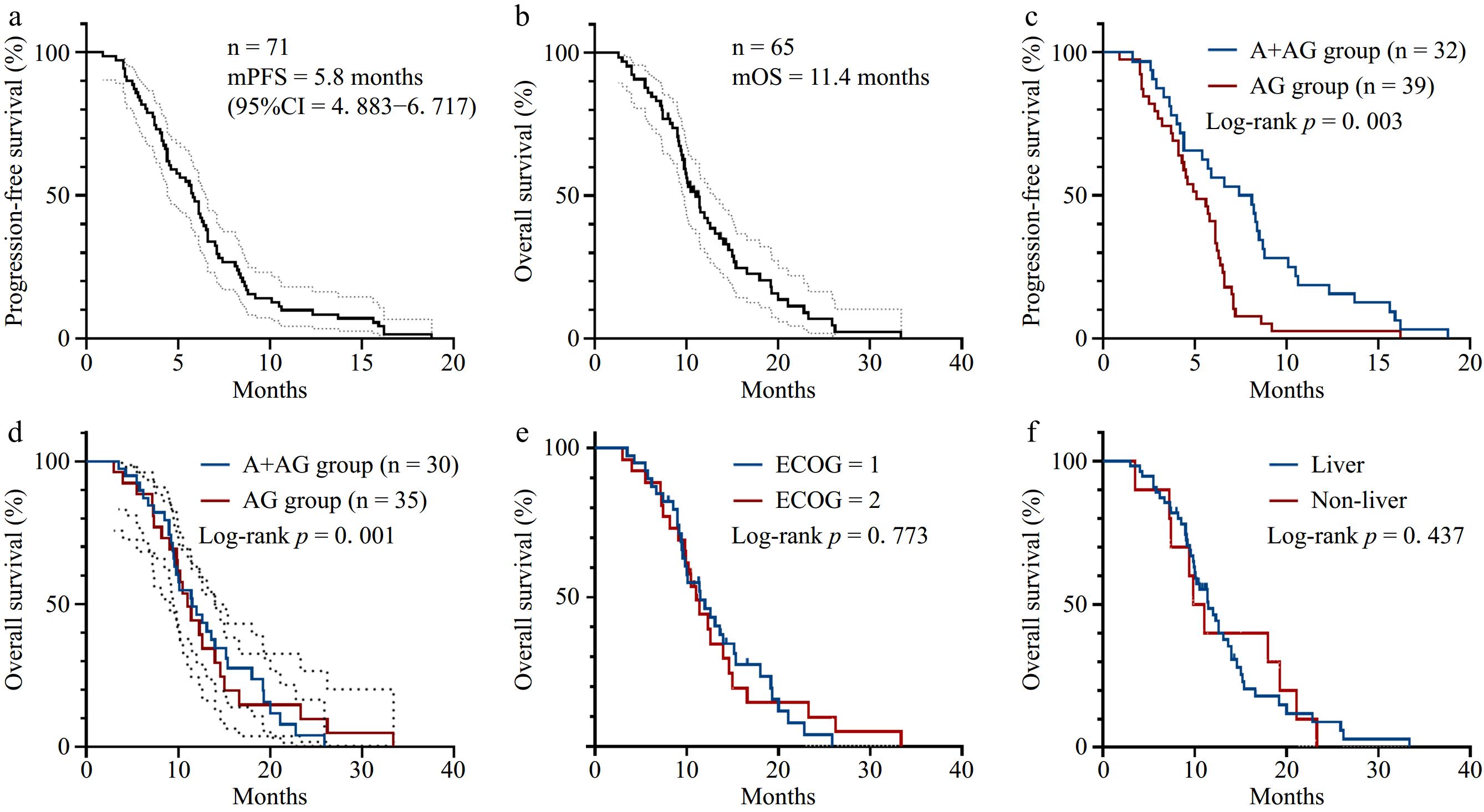
      Figure 1. 

      Survival curves of patients in this study. (a) PFS survival curve of the overall population with mPFS of 5.8 months (95% CI = 4.883−6.717). (b) OS survival curve of the overall population with mOS of 11 months (95% CI = 9.355−13.505). (c) PFS survival curves of the A + AG regimen group and the AG regimen group (p = 0.003). (d) OS survival curves of the A + AG regimen group and AG regimen group (p = 0.001). (e) OS survival curves according to the ECOG score. (f) OS survival curves according to the metastasis site.

    • A COX analysis based on baseline characteristics was performed on the PFS of patients in the AG and A + AG groups. Univariate analysis identified pathology type as a relevant clinical factor affecting PFS (Table 3). while pathology type and CA199 level were the relevant clinical factors affecting OS (Table 4). In the A + AG group, univariate analysis identified TR as the relevant clinical factor affecting PFS (Table 5), and TR was the relevant clinical factor affecting OS (Table 6). Multivariate analysis showed no clinical factors correlated with PFS and OS in both groups.

      Table 3.  Univariate and multivariate analysis of factors influencing PFS in patients in the AG regimen group.

      Clinical features Univariate analysis Multivariate analysis
      HR 95% CI p HR 95% CI p
      Gender
      Male 1.00
      Female 0.70 0.34−1.44 0.331
      Age 1.03 0.99−1.07 0.141
      BMI (kg/m2 ) 1.09 0.96−1.24 0.205
      Hypertension
      True 1.000
      False 0.909 0.467−1.769 0.778
      ECOG score 1.24 0.64−2.38 0.526
      Pathological type
      Adenocarcinoma 1.00 1.00
      Other types 10.20 2.24−46.53 0.003 1.07 0.18−6.52 0.943
      Lymph node metastasis
      True 1.00
      False 0.72 0.37−1.42 0.342
      Liver metastasis
      True 1.00
      False 1.28 0.67−2.46 0.461
      Pulmonary metastasis
      True 1.00
      False 0.79 0.40−1.55 0.492
      Peritoneal metastasis
      True 1.00
      False 0.70 0.32−1.53 0.372
      CA199 level
      Negative 1.00
      Positive 1.97 0.85−4.58 0.113
      TR 0.96 0.85−1.07 0.457

      Table 4.  Univariate and multivariate analysis of factors influencing OS in patients in the AG regimen group.

      Clinical features Univariate analysis Multivariate analysis
      HR 95% CI p HR 95% CI p
      Gender
      Male 1.00
      Female 1.30 0.62−2.73 0.487
      Age 1.00 0.96−1.05 0.981
      BMI (kg/m2 ) 1.01 0.90−1.14 0.858
      Hypertension
      True 1.00
      False 0.59 0.28−1.22 0.156
      ECOG score 1.13 0.56−2.26 0.737
      Pathological type
      Adenocarcinoma 1.00 1.00
      Other types 11.28 2.45−51.98 0.002 3.34 0.51−21.82 0.208
      Lymph node metastasis
      True 1.00
      False 0.88 0.44−1.77 0.719
      Liver metastasis
      True 1.00
      False 0.64 0.30−1.37 0.253
      Pulmonary metastasis
      True 1.00
      False 0.88 0.44−1.78 0.725
      Peritoneal metastasis
      True 1.00
      False 1.35 0.58−3.14 0.482
      CA199 level
      Negative 1.00 1.00
      Positive 2.95 1.18−7.35 0.020 3.34 1.12−10.22 0.031
      TR 1.05 0.93−1.18 0.461

      Table 5.  Univariate and multivariate analysis of factors influencing PFS in patients in the A + AG regimen group.

      Clinical features Univariate analysis Multivariate analysis
      HR 95% CI p HR 95% CI p
      Gender
      Male 1.00
      Female 0.72 0.35−1.50 0.383
      Age 1.00 0.96−1.04 0.941
      BMI (kg/m2 ) 0.98 0.85−1.14 0.789
      Hypertension
      True 1.00
      False 0.79 0.38−1.64 0.521
      ECOG score 0.97 0.47−2.00 0.934
      Pathological type
      Adenocarcinoma 1.00
      Other types 1.27 0.43−3.70 0.664
      Lymph node metastasis
      True 1.00
      False 0.98 0.48−1.99 0.950
      Liver metastasis
      True 1.00
      False 1.92 0.78−4.72 0.155
      Pulmonary metastasis
      True 1.00
      False 1.40 0.62−3.18 0.424
      Peritoneal metastasis
      True 1.00
      False 0.78 0.27−2.26 0.649
      CA199 level
      Negative 1.00 1.00
      Positive 2.31 0.93−5.77 0.073 2.39 0.94−6.06 0.067
      TR 1.17 1.03−1.33 0.014 1.11 0.97−1.28 0.128

      Table 6.  Univariate and multivariate analysis of factors influencing OS in patients in the A + AG regimen group.

      Clinical features Univariate analysis Multivariate analysis
      HR 95% CI p HR 95% CI p
      Gender
      Male 1.00
      Female 1.34 0.55−3.25 0.521
      Age 1.06 0.96−1.05 0.056 1.06 0.98−1.15 0.170
      BMI (kg/m2 ) 1.07 0.88−1.31 0.475
      Hypertension
      True 1.00 1.00
      False 0.37 0.14−1.00 0.050 0.84 0.24−2.92 0.789
      ECOG score 0.78 0.31−1.95 0.588
      Pathological type
      Adenocarcinoma 1.00
      Other types 1.38 0.39−4.89 0.614
      Lymph node metastasis
      True 1.00
      False 1.18 0.49−2.87 0.709
      Liver metastasis
      True 1.00
      False 0.56 0.18−1.75 0.321
      Pulmonary metastasis
      True 1.00
      False 0.86 0.35−2.10 0.735
      Peritoneal metastasis
      True 1.00
      False 1.48 0.34−6.47 0.605
      CA199 level
      Negative 1.00
      Positive 1.83 0.67−5.05 0.242
      TR 1.18 1.01−1.38 0.035 1.07 0.88−1.31 0.496
    • Adverse reactions were evaluated concerning CACTE version 4.0. The typical side effects related to anlotinib treatment included hypertension, fatigue, loss of appetite, hyperlipidemia, hand-foot syndrome, proteinuria, and hepatic dysfunction[31]. Notably, the A + AG group showed a markedly higher incidence of hypertension compared to the AG group (p = 0.020). However, the incidence of grade III/IV hypertension did not differ statistically between the two groups. The incidence of other adverse reactions did not differ significantly between the two groups, as shown in Table 7. During the treatment period, no patients died due to grade V adverse reactions, and all observed adverse reactions remained manageable.

      Table 7.  Comparison of adverse reactions between the two groups.

      Grade III adverse reactions Grade III/IV adverse reactions
      Group A + AG Group AG p value Group A + AG Group AG p value
      Leucopenia 17 (53.2%) 23 (58.9%) 0.800 13 (40.6%) 12 (30.8%) 0.387
      Decreased hemoglobin 17 (53.1%) 16 (41.1%) 0.437 4 (12.5%) 7 (17.9%) 0.763
      Thrombocytopenia 15 (46.9%) 21 (53.9%) 0.729 11 (34.4%) 8 (20.5%) 0.189
      Hypoalbuminemia 13 (40.6%) 20 (51.3%) 0.511 8 (25.0%) 7 (17.9%) 0.469
      Diarrhea 19 (59.3%) 18 (46.1%) 0.384 2 (6.3%) 1 (2.6%) 0.585
      Pleural and abdominal fluid 8 (25.0%) 3 (7.7%) 0.094 3 (9.4%) 5 (12.8%) 0.936
      Nausea and vomiting 18 (56.2%) 26 (66.7%) 0.513 7 (21.9%) 6 (15.4%) 0.482
      Fatigue and poor appetite 24 (75.0%) 29 (74.4%) 1.000 5 (15.6%) 3 (7.7%) 0.500
      Hepatic dysfunction 11 (34.4%) 13 (33.3%) 1.000 9 (28.1%) 4 (10.3%) 0.053
      Hemorrhage 3 (9.4%) 1 (2.6%) 0.471 1 (3.1%) 0 0.921
      Hypertension 8 (25.0%) 5 (12.8%) 0.312 14 (43.8%) 11 (28.2%) 0.172
      Hand-foot syndrome 6 (18.7%) 5 (12.8%) 0.153 2 (6.3%) 0 0.200
      Hyperlipidemia 10 (31.2%) 10 (25.6%) 0.797 6 (18.8%) 3 (7.7%) 0.301
      Proteinuria 16 (50.0%) 17 (43.6%) 0.764 4 (12.5%) 2 (5.1%) 0.399
    • Currently, chemotherapy is the sole effective option for treating unresectable pancreatic adenocarcinoma. Since 1997, gemcitabine has been the primary chemotherapy medication for individuals with locally advanced or metastatic pancreatic cancer, while the survival data is merely 5.7 months[32]. The current National Comprehensive Cancer Network (NCCN) and Chinese Society of Clinical Oncology (CSCO) guidelines recommend regimens including the AG regimen, gemcitabine plus capecitabine (GX regimen), and FOLFIRINOX regimen for patients with unresectable locally advanced or metastatic PC[33,34]. Compared to the FOLFIRINOX regimen, the AG regimen is more widely used in clinical practice due to relatively mild adverse effects. However, regardless of the clinical choice of the FOLFIRINOX or the AG regimen, the overall survival (OS) of advanced PC patients still does not exceed 1 year[9].

      It is well recognized today that anticancer drugs often are most effective when used in combination. Targeted drug combination chemotherapy is an important research direction for pancreatic cancer. A multicenter, randomized phase IIb trial assessing the efficacy of gemcitabine combined with nimotuzumab in KRAS wild-type pancreatic cancer revealed improved OS in the combination group in comparison with the placebo group. This suggests that optimizing systemic chemotherapy and exploring broad-spectrum targeted therapies remain important directions for future research in pancreatic cancer.

      Angiogenesis plays a crucial role in the progression and spread of tumors. It has been revealed that several proangiogenic factors, including VEGF, transforming growth factor (TGF)-β, and platelet-derived growth factor (PDGF)-A overexpressed in PC and involved in disease progression[3537]. However, the therapeutic effectiveness of traditional drugs targeting angiogenesis, such as bevacizumab, in treating PC patients is far from expected. Sorafenib, a drug that inhibits VEGFR-2 and PDGFR-β, has demonstrated effectiveness against pancreatic cancer in preclinical studies. Unfortunately, the combination of sorafenib and gemcitabine did not improve advanced PC patients' prognosis in a multicenter phase II clinical study[38]. Other studies demonstrated that apatinib, a novel VEGFR-2 tyrosine kinase inhibitor can promote radiosensitivity in the treatment of advanced PC with tolerable side effects[39]. Nonetheless, anti-angiogenic therapies remain clinically promising by normalizing tumor vasculature, improving drug delivery, enhancing the local immune response, and reducing distant metastasis. Targeting angiogenesis remains a viable strategy for treating advanced pancreatic cancer.

      Anlotinib, a multi-targeted anti-tumor therapy, specifically targets critical components of the angiogenic pathway, including VEGFR-2, VEGFR-3, PDGFR-α, PDGFR-β, and FGFR1-4, exhibiting a lower inhibitory concentration 50 (IC50). Due to its target concentration, anlotinib has strong efficacy in anti-angiogenic therapy with a low risk of adverse reactions[40]. Furthermore, anlotinib effectively inhibits c-Kit[41,42], the rearranged during transfection proto-oncogene (Ret)[43], FGFR[44,45], and hepatocyte growth factor receptor (c-Met)[46], which collectively suppress the growth and movement of tumor cells. A study indicates that anlotinib activates the endoplasmic reticulum stress pathway( PERK/peIF2α/ATF4 ), inducing reactive oxygen species production, which blocks cell proliferation and causes cell cycle arrest at the G2/M phase thereby inducing apoptosis of PC cells[47]. Multi-omics analysis has shown that anlotinib significantly inhibits ribosomal activity, further curtailing the proliferation of PC cells by regulating cell cycle dynamics, RNA metabolism, and lysosomal function[48]. Anlotinib also can promote tumor vascular normalization, transforming the immunosuppressive tumor microenvironment into one that stimulates immune responses. This action significantly reduces tumor growth and helps prevent systemic immune suppression. Furthermore, when combined with PD-1 checkpoint inhibitors, anlotinib can reverse the immunosuppression caused by PD-L1 upregulation after monotherapy, prolong the period of vascular normalization, and ultimately lead to tumor regression[49]. A recent clinical study reported the benefit of combining S-1, sintilimab, and anlotinib as second-line therapy for pancreatic cancer (PC) patients with liver metastasis, extending overall survival (OS)[50]. These findings suggest that anlotinib-related combination therapy could offer a novel treatment approach for PC patients. Additionally, recent studies have shown that combining penpulimab, anlotinib, nab-paclitaxel, and gemcitabine yields promising clinical outcomes, particularly in enhancing ORR and DCR in metastatic PC[29]. Our study focuses on exploring the combination of anlotinib with gemcitabine and nab-paclitaxel, aiming to provide clinicians with more treatment options.

      In this study, we evaluated the effectiveness and safety of combining anlotinib with albumin-bound paclitaxel and gemcitabine as a first-line treatment for PC. We observed that ORR reached 12.5% and DCR was 87.5% in this regimen. Both mPFS and mOS of the A + AG group were superior to the AG group. Moreover, the inclusion of anlotinib did not raise the risk of severe adverse reactions when compared to chemotherapy alone. While patients receiving anlotinib showed a notably higher risk of developing hypertension, there was no significant difference in the occurrence of grade III/IV hypertension, which could be tolerated by most patients, and this adverse reaction can be managed with dose regulation and symptomatic treatment. Intriguingly, some adverse reaction is also correlated with the efficacy of anlotinib, such as hand-foot syndrome is positively correlated with prolonged survival in NSCLC treated with anlotinib[51].

      It is necessary to state that this retrospective study has many limitations, including retrospective data, which has selection bias and information bias that is difficult to avoid. It is necessary to conduct prospective or randomized controlled trials in the future. Secondly, the sample size of this study is limited and it is a single-center study, which may lead to data bias and be limited by treatment conditions, making it difficult to comprehensively reflect the effectiveness and safety of the treatment. Future research should involve multi-center studies with larger sample sizes. In addition, the management strategy for adverse reactions is imperfect, which may affect the accurate evaluation of drug safety.

    • In conclusion, our findings suggest that combining anlotinib with gemcitabine and albumin-bound paclitaxel may be an effective first-line treatment option for advanced pancreatic cancer with significant survival advantages and tolerable adverse reactions, which is of great clinical significance. To further confirm these findings and improve the therapeutic regimen of anlotinib in PC, large-scale, prospective, multi-center randomized controlled trials, along with long-term follow-up data, are required.

      • We would like to extend our sincere thanks to all the colleagues and patients who contributed to this study. This study was supported by the Natural Science Foundation of Jiangsu Province (Grant No. BK20231127) to Xiaowei Wei.

      • This study protocol was reviewed and approved by the Nanjing First Hospital, approval number KY20240520-KS-05.

      • The authors confirm contribution to the paper as follows: analysis design, clinical data collection, statistical analysis performed: Zheng Y; draft manuscript preparation: Zhou Y; treated patients, manuscript revision: Zhou J, Wei X, Xia H, Liang W. All authors reviewed the results and approved the final version of the manuscript.

      • The datasets produced and/or analyzed in this study are available upon reasonable request from the corresponding author.

      • The authors declare that they have no conflict of interest.

      • # Authors contributed equally: Yawen Zheng, Yuhan Zhou

      • Copyright: © 2025 by the author(s). Published by Maximum Academic Press, Fayetteville, GA. This article is an open access article distributed under Creative Commons Attribution License (CC BY 4.0), visit https://creativecommons.org/licenses/by/4.0/.
    Figure (1)  Table (7) References (51)
  • About this article
    Cite this article
    Zheng Y, Zhou Y, Xia H, Liang W, Wei X, et al. 2025. A clinical retrospective study of anlotinib in combination with gemcitabine and albumin-bound paclitaxel for the first-line treatment of advanced pancreatic cancer. Gastrointestinal Tumors 12: e001 doi: 10.48130/git-0025-0001
    Zheng Y, Zhou Y, Xia H, Liang W, Wei X, et al. 2025. A clinical retrospective study of anlotinib in combination with gemcitabine and albumin-bound paclitaxel for the first-line treatment of advanced pancreatic cancer. Gastrointestinal Tumors 12: e001 doi: 10.48130/git-0025-0001

Catalog

  • About this article

/

DownLoad:  Full-Size Img  PowerPoint
Return
Return