











-
Hyphomycetous fungi constitute one of the most diverse groups of asexual Ascomycota. To date, more than 2,265 genera and approximately 13,800 species have been described worldwide[1−3]. Hyphomycetes occupy a wide range of ecological niches and are commonly encountered in terrestrial habitats such as forest litter, decaying wood, soil, and plant surfaces[4−6]. Many taxa are also distributed in freshwater and marine environments, where they play important roles in the decomposition of organic matter[7−9]. As saprobes, hyphomycetous fungi contribute substantially to nutrient cycling through the degradation of plant-derived substrates[10−12]. In addition, some species occur as endophytes, colonizing healthy plant tissues without causing visible symptoms and potentially enhancing host tolerance to abiotic stress or resistance to plant pathogens[13,14].
China encompasses a wide range of climatic zones and ecosystems and is recognized as a global hotspot for fungal diversity. In recent years, intensive taxonomic studies have substantially increased the number of documented fungal taxa from the country, particularly within Ascomycota[15−17]. Despite this progress, many regions remain insufficiently explored, and numerous fungal taxa are still known only from limited collections. Guizhou, Yunnan, and Guangxi are located in southwestern China and are characterized by complex topography, high habitat heterogeneity, and diverse vegetation types, providing a wide range of ecological niches for fungal communities. Recent studies have reported an increasing number of hyphomycetous fungi from these provinces, suggesting that the diversity of this fungal group in the region remains substantially underestimated[18−21].
Traditional classifications of hyphomycetous fungi were largely based on conidiophore and conidial morphology, which frequently resulted in artificial groupings and unstable taxonomic frameworks[12,22]. Morphological convergence and phenotypic plasticity have further complicated the delimitation of genera and species within this group. The application of molecular phylogenetic approaches has profoundly reshaped the taxonomy of asexual fungi by enabling accurate phylogenetic placement of morphologically ambiguous taxa. Multigene sequence data derived from ribosomal DNA regions and protein-coding genes have revealed extensive polyphyly in many traditionally circumscribed genera and families of hyphomycetous fungi[16,23,24]. Consequently, integrative taxonomy that combines detailed morphological observations with multigene phylogenetic analyses has become essential for resolving species boundaries, clarifying generic concepts, and establishing natural classifications within hyphomycetous fungi[25,26].
Although integrative taxonomic studies have substantially improved the classification of hyphomycetous fungi, many taxa from understudied regions remain poorly characterized, and their phylogenetic relationships are still unclear. In particular, saprobic hyphomycetous fungi associated with decaying plant substrates in southwestern China have received comparatively limited attention, despite the ecological complexity and high habitat diversity of this region. A comprehensive assessment of these fungi is therefore necessary to clarify their taxonomic placement and to improve the current understanding of hyphomycetous fungal diversity and evolution.
In the present study, hyphomycetous fungi collected from saprobic substrates in Guizhou, Yunnan, and Guangxi were investigated using an integrative taxonomic framework. Detailed morphological examinations were combined with multigene phylogenetic analyses to assess species boundaries, clarify phylogenetic relationships, and evaluate the diversity of hyphomycetous fungi in southwestern China. This study aims to contribute to a more robust taxonomic framework for hyphomycetous fungi and to provide a foundation for future ecological and evolutionary studies of this diverse fungal group.
-
Samples of submerged leaves, twigs, decaying wood, and dead herbaceous materials were collected from freshwater habitats, including rivers, lakes, and ponds, in Guangxi Zhuang Autonomous Region, Guizhou Province, and Yunnan Province, southwestern China, between July 2024 and August 2025. Samples were placed in sterile containers and incubated in sealed moist chambers at 20–24 °C. Moist chambers consisted of sterilized plastic boxes lined with moistened sterile tissue paper, with samples placed on sterilized plastic straws to avoid direct contact with the moist surface. The boxes were incubated at room temperature for approximately one week and examined periodically for the development of fungal structures. Morphological characteristics were recorded from fresh specimens, with macromorphological features examined using an Olympus SZ61 stereomicroscope (Olympus, Tokyo, Japan) and documented with a Canon 700D digital camera (Canon, Tokyo, Japan). Micromorphological observations were conducted on material mounted in water, and measurements were obtained using Tarosoft® Image Framework version 0.9.7 (Tarosoft, Bangkok, Thailand). Images were processed and arranged using Adobe Photoshop CS6 (Adobe Systems, San Jose, USA).
Pure cultures were obtained by single-spore isolation following Liu et al.[15]. Conidia were suspended in sterile distilled water, spread onto water agar (WA), and incubated at 25 °C for 12–36 h. Individual germinated conidia were aseptically transferred to fresh potato dextrose agar (PDA; Difco, Detroit, USA) and incubated at 24–28 °C, depending on the growth response of individual isolates, to obtain optimal colony development. Additional germinated conidia remaining on the agar surface were excised together with small agar blocks and mounted in water on glass slides to document germ tube emergence and orientation. The dried specimens are preserved in two herbaria: the Herbarium of Guizhou Medical University (GMB) and the Cryptogam Herbarium at the Kunming Institute of Botany, Chinese Academy of Sciences (KUN-HKAS). Living cultures are maintained in the Guizhou Medical University Culture Collection (GMBC). Fungal nomenclature follows MycoBank.
DNA extraction, PCR amplification, and sequencing
-
Colonies were grown on potato dextrose agar plates for two to four weeks until fully developed. Mycelium was harvested using a sterile scalpel, and genomic DNA was isolated with the BIOMIGA Fungus Genomic DNA Extraction Kit (Solarbio, Beijing, China) following the manufacturer's protocol. Six loci, namely ITS, LSU, SSU, tef1-α, rpb2, and tub2, were then amplified by PCR with the following primer pairs: ITS (ITS1 and ITS4)[27], LSU (LR0R and LR5)[27], SSU (NS1 and NS4)[27], tef1-α (TEF1-983F and TEF1-2218R)[28], rpb2 (RPB2-5F and RPB2-7cR)[29], and tub2 (T1 and Bt2b)[29,30]. Polymerase chain reactions were performed in 25 µL volumes consisting of 9.5 µL ddH2O, 1 µL genomic DNA template at 5 to 10 ng, 12.5 µL 2 × Taq PCR Master Mix (Sangon Biotech, Shanghai, China), and 1 µL of each primer at 10 µM. The thermal cycling conditions followed those described by Samarakoon et al.[31]. Successful amplification was confirmed by agarose gel electrophoresis, and the products were sequenced by Sangon Biotech (Shanghai, China). The newly obtained sequences have been deposited in GenBank (
https://www.ncbi.nlm.nih.gov/ ).Sequence alignments and phylogenetic analyses
-
All newly obtained sequences were compared with each other and against reference sequences in GenBank via BLASTn to assess sequence similarity and preliminary identification[32]. Sequence alignments were performed on the MAFFT v. 7.110 online platform using default parameters[33], and alignments were manually refined in BioEdit version 7.0.5.3 where required[34].
Phylogenetic relationships were inferred using the Maximum Likelihood method implemented in RAxML (v8.2.12) under the GTRGAMMA model, with support estimated from 1,000 bootstrap replicates[35]. Bayesian inference (BI) analyses were carried out in MrBayes (v3.2.2)[36], employing six Markov chains run for 1,000,000 generations and sampling trees at intervals of 1,000 generations. The initial 25% of trees were excluded as burn-in, and posterior probabilities were calculated from the remaining dataset. All phylogenetic analyses were executed on the CIPRES Science Gateway v.3.3 platform[37]. Phylogenetic trees were visualized using FigTree version 1.4.3[38]. All newly generated sequences were submitted to GenBank, and their accession numbers are provided in Supplementary Table S1. The selection of loci and outgroup taxa followed previously published phylogenetic frameworks relevant to each family and genus.
Phylogenetic support assessment
-
Statistical support for phylogenetic relationships was assessed using bootstrap percentages from maximum likelihood analyses and posterior probability values from Bayesian inference. Nodes with ML bootstrap values ≥ 70% and BI posterior probabilities ≥ 0.90 were considered strongly supported. No additional statistical analyses were conducted, as this study is based on qualitative morphological comparisons and phylogenetic inference commonly applied in fungal taxonomy.
-
Ascomycota Caval. Sm., Biol. Rev. Cambridge Philos. Soc. 73: 247 (1998).
Dothideomycetes O.E. Erikss. & Winka, Myconet 1: 5 (1997).
Pleosporomycetidae C.L. Schoch, Spatafora, Crous & Shoemaker, Mycologia 98 (6): 1,048 (2007).
Kirschsteiniotheliales Hern.-Restr., R.F. Castañeda, Gené & Crous, Stud. Mycol. 86: 72 (2017).
Kirschsteiniotheliaceae Boonmee & K.D. Hyde, Mycologia 104(3): 705 (2012).
Kirschsteiniothelia D. Hawksw., Bot. J. Linn. Soc. 91: 182 (1985).
Notes – Kirschsteiniothelia was originally introduced in Pleosporaceae with K. aethiops as the type species[39]. Later, based on morphological observations, Barr (1987) transferred it to Pleomassariaceae[39,40]. However, phylogenetic work by Schoch et al. showed that K. aethiops is not a member of either family but instead represents a distinct lineage[41]. As a result, Boonmee et al. proposed a new family, Kirschsteiniotheliaceae, to accommodate this genus[42].
The teleomorph of Kirschsteiniothelia is characterized by dark brown to black, superficial ascomata, textura angularis peridium cells, cylindrical to clavate asci with a small ocular chamber, and ellipsoidal, brown, 1–2-septate ascospores with or without a mucilaginous sheath[43]. The anamorph has septate, dark brown conidiophores and septate, obovoid to fusiform conidia[39,44,45]. Kirschsteiniothelia species are mostly saprobic, occurring on decomposing wood or plant debris in both terrestrial and freshwater habitats, mainly in tropical to subtropical regions. According to Species Fungorum (accessed 3 January 2026), the genus currently comprises 74 accepted species, and one novel species is described in this study.
Kirschsteiniothelia chinensis L.L. Liu, Q.F. Zhang, & Q.R. Li, sp. nov. (Fig. 1)
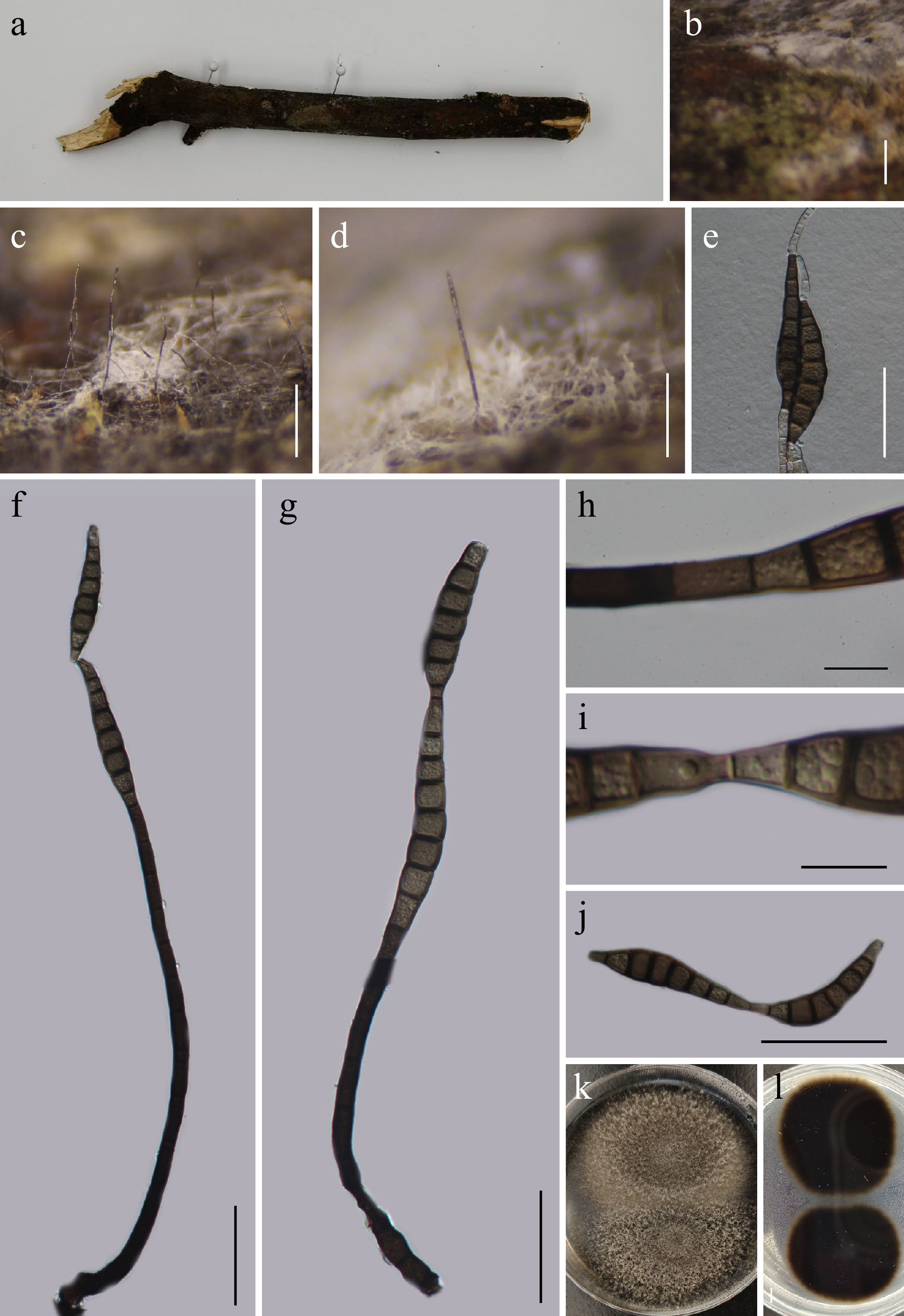
Figure 1.
Kirschsteiniothelia chinensis (GMB5168 holotype). (a) Host. (b)–(d) Colonies on natural substrate. (e) Germinating conidia. (f), (g) Conidiophores and conidia. (h), (i) Conidiogenous cells with conidia. (j) Conidia. (k), (l) Surface and reverse view of culture on PDA. Scale bars: b = 1 mm, c = 0.5 mm, d = 0.25 mm, e–g, j = 50 μm, h, i = 10 μm.
MycoBank number: MB862338
Etymology – The species name refers to China, the country of origin of the holotype specimen.
Saprobic on submerged dead branches in a pond. Sexual morph: not observed. Asexual morph: hyphomycetous. Colonies superficial, hairy, black, glistening, effuse, gregarious. Mycelium partly immersed, consisting of septate, smooth-walled hyphae, pale brown to dark brown. Conidiophores 93–276 × 7.2–10 µm (x = 146.6 × 8.8 µm, n = 15), mononematous, macronematous, straight to slightly flexuous, solitary, cylindrical, unbranched, septate, smooth, brown to dark brown, wider at the base, tapering toward the apex. Conidiogenous cells 3.2–12 × 1.8–7.6 µm (x = 9.1 × 4.6 µm, n = 16), holoblastic, monoblastic, terminal, determinate, cylindrical, smooth, pale brown to brown. Conidia 55.4–122 × 10–16 µm (x = 84.4 × 12.7 µm, n = 25), cylindrical to obclavate, solitary, rostrate, acrogenous, brown to dark brown, straight or slightly curved, smooth, 6–16 euseptate, slightly constricted at septa, apex attenuated, base truncate.
Culture characteristics – Conidia germinate on PDA within 24 h. After 14 d at 25 °C, colonies on PDA reach 30–40 mm in diameter, circular, raised, aerial mycelium sparse to moderate, floccose to velvety, surface white; reverse dark brown to black.
Material examined – CHINA, Yunnan Province, Xishuangbanna Dai Autonomous Prefecture, 24°02'24.48" N, 100°48'38.25" E, elevation 1,450 m, on submerged dead branches in pond, 18 August 2025, Qin-Fang Zhang, 2025XSBN13 (GMB5168, holotype; GMBC5168, ex-type); ibid., KUN-HKAS 152889, isotype.
Additional specimens examined – CHINA, Yunnan Province, Xishuangbanna Dai Autonomous Prefecture, 24°02'24.61" N, 100°48'38.30" E, elevation 1,190 m, on submerged dead branches in pond, 18 August 2025, Qin-Fang Zhang, 2025XSBN80 (GMB5169, GMBC5169).
Notes – In the phylogenetic analysis (Fig. 2), Kirschsteiniothelia chinensis forms a closely related lineage with K. spatiosa (MFLU 21-0128) and K. agumbensis (NFCCI 5714). Comparison of ITS sequences revealed that K. chinensis (GMBC5168) differs from the type strain of K. spatiosa (MFLU 21-0128) by 11.5% (50/434 bp) and from K. agumbensis (NFCCI 5714) by 7.8% (36/459 bp).
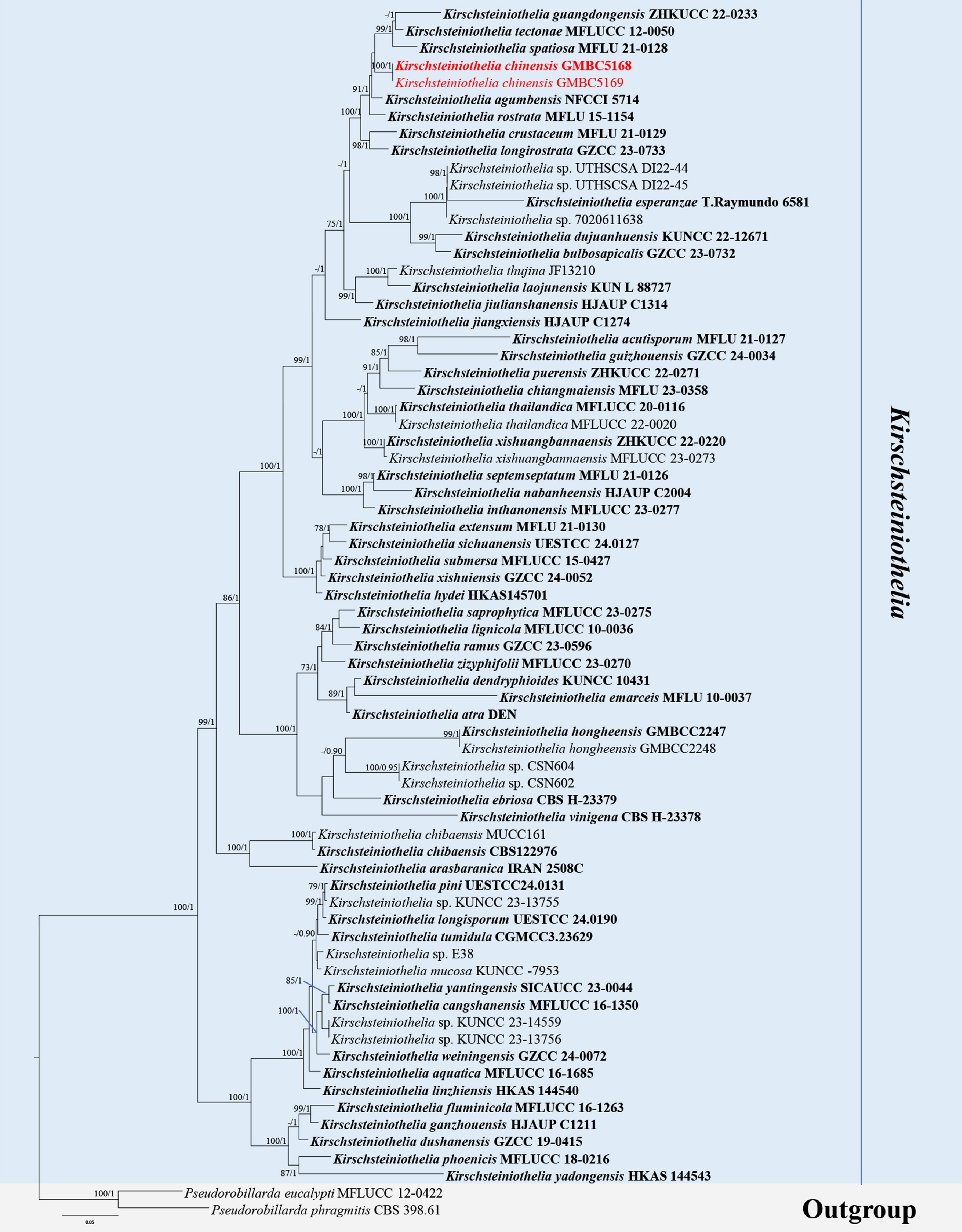
Figure 2.
ML tree of selected Kirschsteiniothelia species based on the ITS-LSU-SSU dataset. The combined dataset comprised 71 taxa and 2,429 characters (ITS: 1–520 bp; LSU: 521–1,405 bp; SSU: 1,406–2,429 bp), including indel regions. Maximum likelihood (lnL = –22,055.122970) and Bayesian analyses produced consistent tree topologies. Pseudorobillarda phragmitis (CBS 398.61) and P. eucalypti (MFLUCC 12-0422) were selected as the outgroup taxa. Bayesian posterior probabilities (PP) ≥ 0.90 and RAxML bootstrap support values ≥ 70 % are shown on the branches. The newly described species is marked in red. Ex-type/type strains are indicated in bold.
Morphologically, K. chinensis is comparable to K. spatiosa in producing hyphomycetous asexual morphs characterized by macronematous, mononematous, cylindrical conidiophores and solitary, acrogenous, rostrate, multiseptate conidia. However, K. chinensis can be distinguished by its shorter conidia (55.4–122 μm vs 90–139 μm) with fewer septa (6–16 vs 8–23) and lacking a mucilaginous sheath, as well as by its longer conidiophores (93–276 μm vs 70–128 μm)[46].
Based on these distinct morphological and molecular differences and its independent phylogenetic placement, K. chinensis is proposed herein as a new species.
Nigrogranaceae Jaklitsch & Voglmayr, Stud. Mycol. 85: 54 (2016).
Nigrograna Gruyter, Verkley & Crous, Stud. Mycol. 75: 31 (2012) [2013].
Notes – Nigrograna was established by de Gruyter et al. with N. mackinnonii (formerly Pyrenochaeta mackinnonii) as the type species[47]. A significant taxonomic conflict exists between Nigrograna and Biatriospora. Earlier phylogenetic studies[48] treated Nigrograna as a synonym of Biatriospora based on sequence data derived from an isolate identified as Biatriospora marina (strain CY1228). However, this placement was challenged by Jaklitsch & Voglmayr[49], who identified taxa (N. mackinnonii) forming sexual morphs that were phylogenetically related to B. marina (CY1228) but differed markedly in ascospore morphology from authentic B. marina. They therefore questioned the identity of the strain (CY1228) used by Ahmed et al.[48] and emphasized the need for fresh collections and molecular data to clarify its taxonomic placement. They also introduced Nigrogranaceae to accommodate Nigrograna and recommended discontinuing the use of Biatriosporaceae due to these uncertainties.
Hongsanan et al. introduced a new species, Biatriospora borsei, characterized by a hyphomycetous asexual morph, that clustered phylogenetically with the unverified strain B. marina CY1228 (known only from a sexual morph)[50]. At that time, no hyphomycetous asexual morph had been reported in Nigrograna, which was therefore considered to possess exclusively pycnidial asexual morphs. Hongsanan et al., therefore, placed B. borsei in Biatriospora and retained the genus within Biatriosporaceae based on both its hyphomycetous morphology and its apparent phylogenetic association with CY1228[50]. Hongsanan et al. assigned their isolate to B. borsei primarily based on close DNA sequence similarity to B. marina strain CY1228. However, no published literature provides valid information regarding the original source or morphological documentation of strain CY1228.
In the present study, we report a new taxon represented by a hyphomycetous asexual morph that clusters with other Nigrograna species and is phylogenetically distant from B. borsei. Importantly, both B. borsei and the unverified B. marina strain CY1228 are nested within the strongly supported Nigrograna s. str. clade, which includes the type species N. mackinnonii. This placement indicates that the hyphomycetous morph is not diagnostic for Biatriospora but instead represents previously undocumented morphological variation within Nigrograna. This finding further demonstrates that B. borsei was originally assigned to an incorrect genus. Accordingly, we propose the new combination Nigrograna borsei comb. nov.
We also emend the generic circumscription of Nigrograna to include both pycnidial and hyphomycetous conidiation. Because the type strain CY1228 is unauthenticated, as reported by Jaklitsch & Voglmayr[49], and the phylogenetic position of authentic B. marina cannot be resolved with the currently available data. Consequently, we recommend discontinuing the use of Biatriosporaceae and placing Biatriospora in Pleosporales, incertae sedis.
Nigrograna borsei (Devadatha & V.V. Sarma) L. Liu & Q.R. Li, comb. nov.
MycoBank No: MB862958
Basionym: Biatriospora borsei Devadatha & V.V. Sarma, Mycosphere 11 (1): 1,768 (2020).
Description – See Hongsanan et al. (2020)[50]
Nigrograna chinensis L.L. Liu, W.M. Li, & Q.R. Li, sp. nov. (Fig. 3)
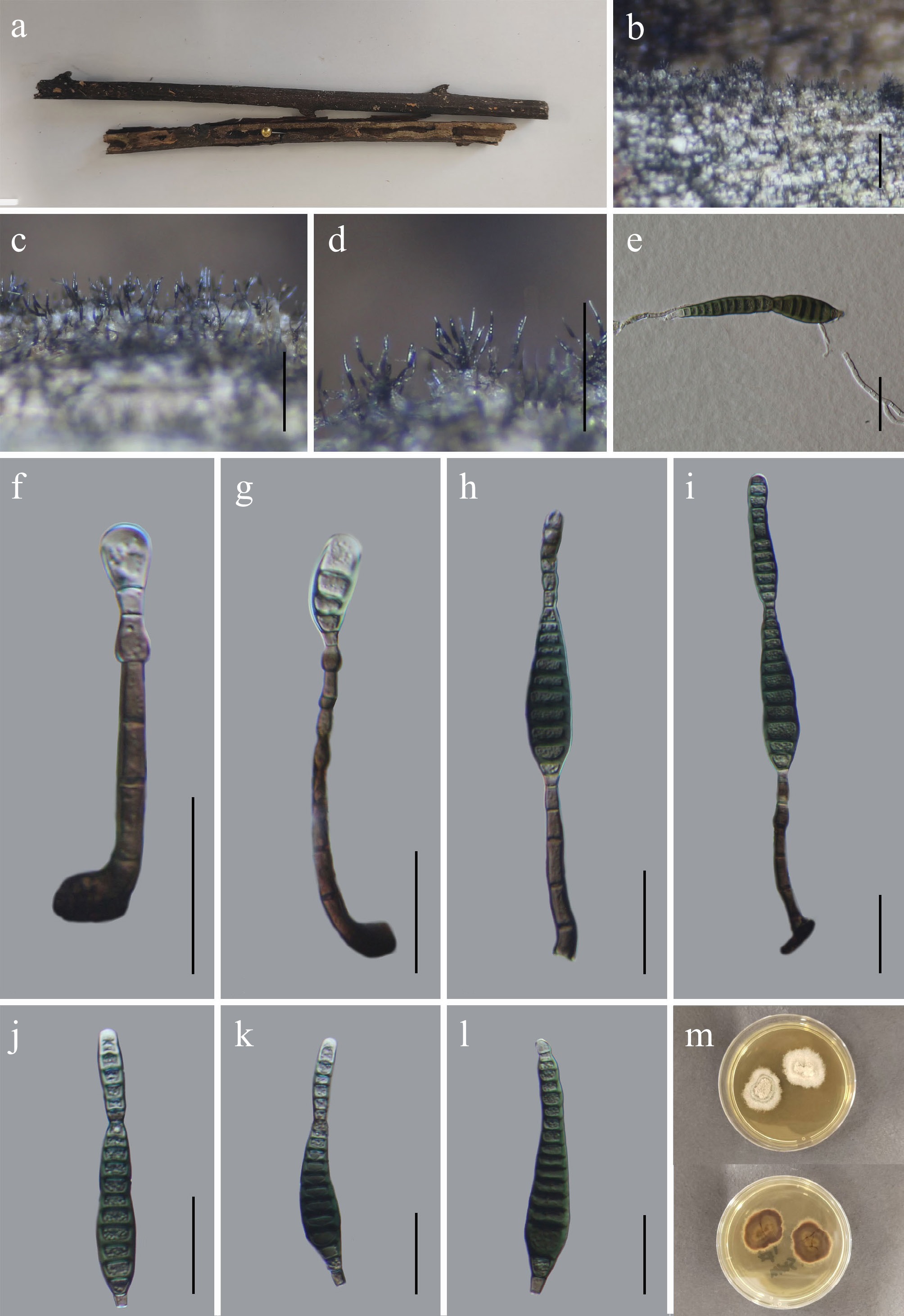
Figure 3.
Nigrograna chinensis (GMB6935 holotype). (a) Host. (b)–(d) Colonies on natural substrate. (e) Germinating conidium. (f)–(i) Conidiophores and conidia. (j)–(l) Conidia. (m) Surface and reverse view of culture on PDA. Scale bars: b = 1 mm, c, d = 0.5 mm, e–l = 30 μm.
MycoBank number: MB862921
Etymology – Referring to China, the country of origin of the holotype specimen.
Saprobic on submerged dead branches along the riverbank. Sexual morph: not observed. Asexual morph: hyphomycetous. Colonies velvety to olivaceous brown. Mycelium mostly superficial, comprising smooth or verruculose, septate, pale to medium brown, extensively ramifying hyphae. Conidiophores 27.2–118.5 × 3.2–6.7 µm (x = 56.2 × 5.5 µm, n = 30), mononematous, macronematous, solitary, unbranched, septate, brown to dark brown, erect to slightly flexuous, smooth-walled, 3–6 septate. Conidiogenous cells 3.0–10.3 × 3.1–5.6 μm (x = 6.5 × 4.0 μm, n = 30) integrated, terminal, monoblastic, holoblastic, pale brown to subhyaline, smooth-walled, cylindrical to slightly swollen; conidial secession schizolytic, often leaving a short basal remnant on the conidiogenous cell. Conidia 25–104.5 × 5–13.5 µm (x = 65.1 × 10.2 µm, n = 30), solitary, acrogenous, narrowly fusiform to clavate, straight to slightly curved, nine to 19 pseudoseptate, sometimes slightly asymmetrical, dark brown to blackish brown at maturity, paler when young, thick-walled, smooth, apex rounded to slightly attenuated, base truncate.
Culture characteristics – Conidia germinate on PDA within 24 h. After 14 d at 25 °C, colonies on PDA reach 20–35 mm in diameter; surface white to cream, filamentous, circular, raised, aerial mycelium sparse to moderate, with dense central tufts, margin even; reverse dark brown to reddish-brown.
Material examined – CHINA, Yunnan Province, Zhaotong City, Wumengshan National Nature Reserve, 25°20′30.05″ N, 103°10′05.90″ E, elevation 2,400 m, on submerged dead branches along the riverbank, 25 October 2024, Wen-Mei Li, 2024WMS157 (GMB6935, holotype; GMBC6935, ex-type); ibid., KUN-HKAS 152890, isotype.
Additional examined specimens – CHINA, Yunnan Province, Zhaotong City, Wumengshan National Nature Reserve, 25°10′30.13″ N, 103°10′05.94″ E, elevation: 2,198 m, on submerged dead branches along the riverbank, 25 October 2024, Wen-Mei Li, 2024WMS142 (GMB6936, GMBC6936).
Notes – In the phylogenetic analyses (Fig. 4), Nigrograna chinensis forms a well-supported sister clade with N. asexualis (ZHKUCC 22-0214). Despite this close phylogenetic relationship, the two species are morphologically unrelated. Nigrograna asexualis has a coelomycetous asexual morph, whereas N. chinensis produces a hyphomycetous morph. Comparative nucleotide sequence analysis showed that N. chinensis (GMBC6935) differs from the type strain of N. asexualis (ZHKUCC 22-0214) by 2.5% (9/354 bp) in the ITS locus, 0.2% (2/858 bp) in the LSU locus, 0.2% (2/1024 bp) in the SSU locus, 3.7% (35/953 bp) in the tef1-α gene, and 8.6% (88/1027 bp) in the rpb2 gene.
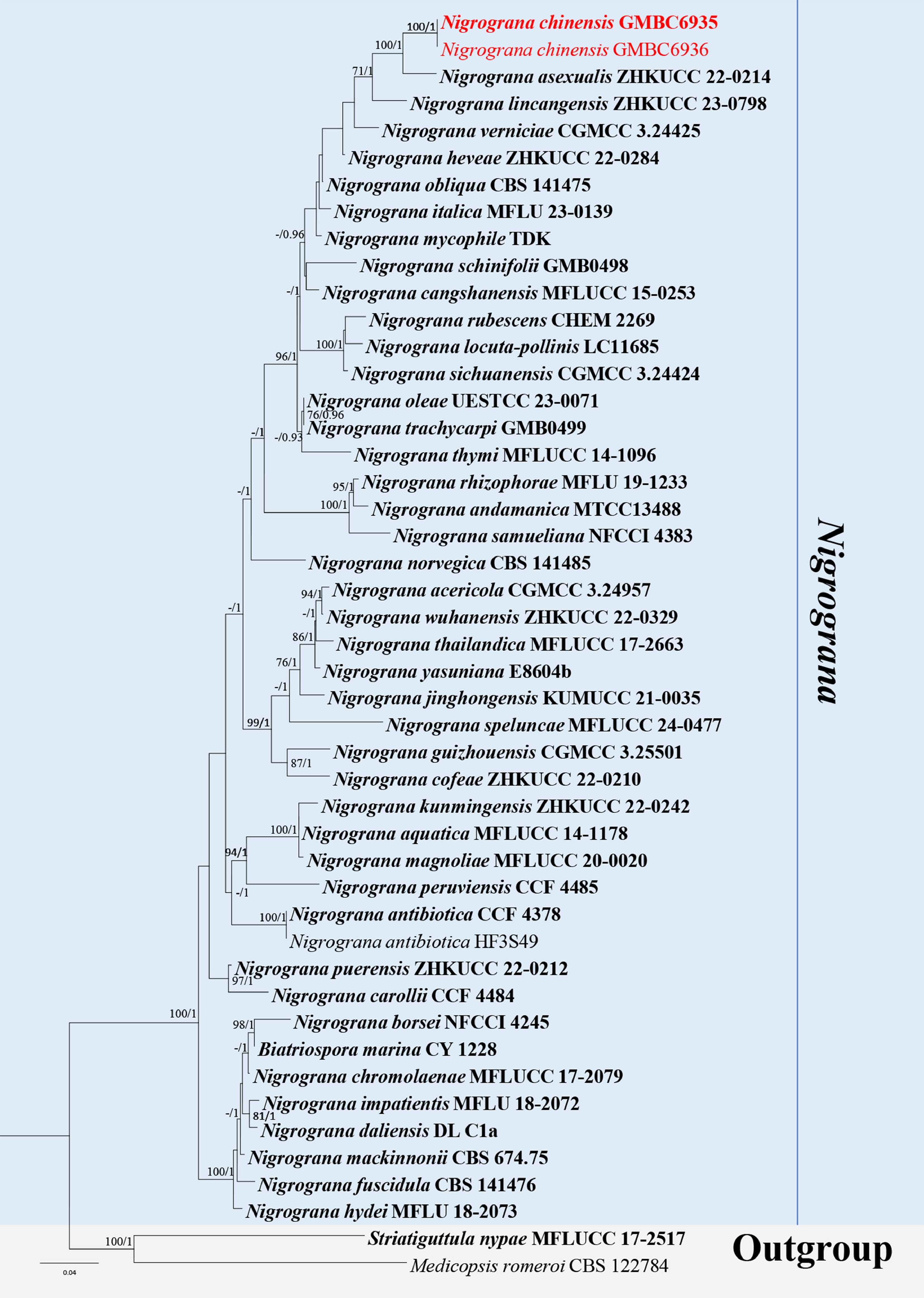
Figure 4.
ML tree of selected Nigrograna species based on the ITS-LSU-SSU-tef1-α-rpb2 dataset. The combined dataset comprised 47 taxa and 4,464 characters (ITS: 1–529 bp; LSU: 530–1,402 bp; SSU: 1,403–2,425 bp; tef1-α: 2,426–3,405 bp; rpb2: 3,406–4,464 bp), including indel regions. Maximum likelihood (lnL = −22,113.458225) and Bayesian analyses produced consistent tree topologies. Striatiguttula nypae (MFLUCC 17-2517) and Medicopsis romeroi (CBS122784) were selected as the outgroup taxa. Bayesian posterior probabilities (PP) ≥ 0.90 and RAxML bootstrap support values ≥ 70 % are shown on the branches. The newly described species is marked in red. Ex-type/type strains are indicated in bold.
Morphologically, Nigrograna chinensis resembles N. borsei in having a hyphomycetous asexual morph, characterized by cylindrical septate conidiophores and brown, sub-clavate, multi-pseudoseptate conidia. However, N. chinensis can be differentiated by having 9 to 19 pseudoseptate, longer conidia (25–104.5 × 5–13.5 µm), whereas N. borsei usually has six (rarely seven) pseudoseptate, smaller conidia (30–45 × 7–9 µm)[50].
The combination of a distinct phylogenetic placement and differences in both the molecular and morphological characters supports the recognition of N. chinensis as a new species.
Pleosporales Luttr. ex M.E. Barr, Prodromus to class Loculoascomycetes: 67 (1987).
Corynesporascaceae Sivan., Mycol. Res. 100(7): 786 (1996).
Corynespora Güssow, J. Royal Agric. Soc. England 65: 272 (1905) [1904].
Notes – Corynespora was established by Güssow, with C. mazei as the type species[51]. Species of Corynespora are widely distributed and are primarily known as pathogens of economically important crops such as pepper, roselle, cotton, papaya, tomato, and rubber, causing leaf and fruit spot diseases[52−55]. In addition to their pathogenic lifestyle, species of the genus have also been reported as endophytes and saprobes[56,57]. The genus includes species widely distributed in tropical and subtropical regions and has been isolated from diverse substrates and environments, including air, soil, lower plants, and lichens. They have been recorded from both aquatic and terrestrial habitats, indicating broad ecological adaptability[53]. According to Species Fungorum (3 March 2026), the genus currently comprises 200 accepted species.
Corynespora guizhouensis L.L. Liu, W.M. Li, & Q.R. Li sp. nov. (Fig. 5)
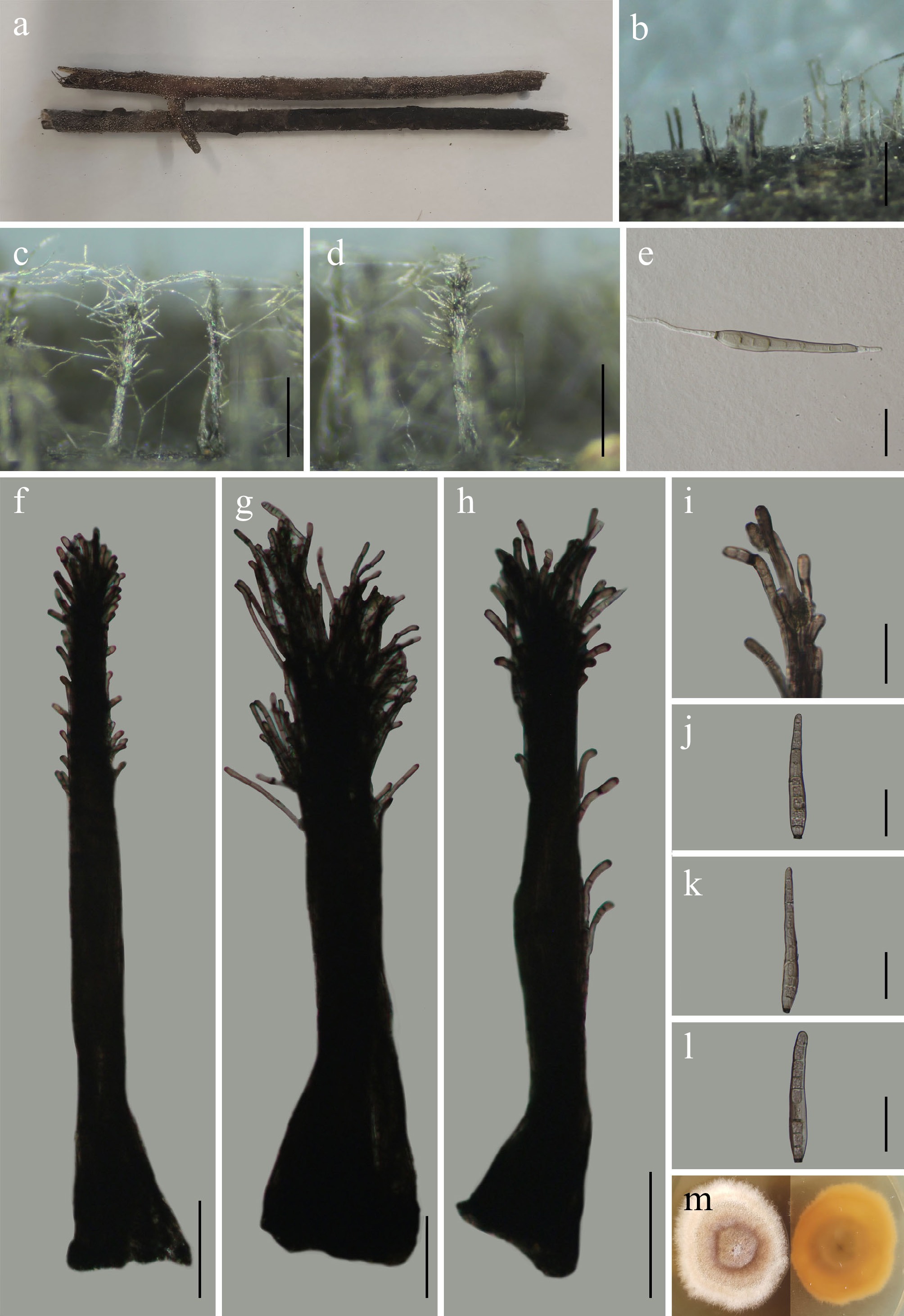
Figure 5.
Corynespora guizhouensis (GMB6927 holotype). (a) Host. (b)–(d) Colonies on natural substrate. (e) Germinating conidium. (f)–(h) Conidiophores and conidia. (i) Conidiogenous cells. (j)–(l) Conidia. (m) Surface and reverse view of culture on PDA. Scale bars: b = 1 mm, c–d = 0.5 mm, f–h = 100 μm, e, i–l = 30 μm.
MycoBank number: MB862927
Etymology – The epithet refers to Guizhou Province, China, where the holotype specimen was collected.
Saprobic on dead branches of an unknown plant. Sexual morph: not observed. Asexual morph: hyphomycetous. Colonies are hairy, effuse, dark brown to black. Mycelium mostly immersed, partly superficial, comprised of septate, branched, pale to dark brown, smooth-walled hyphae. Synnemata 461–897 × 25.3–89.7 µm (x = 663.5 × 49.7 µm, n = 30), formed by aggregated conidiophores, cylindrical to slightly clavate, straight to slightly flexuous, brown to dark brown, thick-walled, often swollen towards the apex; constituent conidiophores macronematous, mononematous, septate, unbranched, and closely compacted. Conidiogenous cells 3.3–7.4 µm wide (x = 5.9 µm, n = 30), terminal, integrated, determinate, monotretic, brown to dark brown, cylindrical, occasionally showing percurrent proliferation. Conidia 46.5–102 × 6–11 µm (x = 80.4 × 8.4 µm, n = 30), solitary, simple, acrogenous, obclavate to cylindrical, subhyaline to pale brown, straight to slightly curved, 4–13 distoseptate, smooth and thick-walled, base truncate with a distinct, darkened basal scar, gradually tapering towards the apex.
Culture characteristics – Conidia germinate on PDA within 24 h. After 21 d at 25 °C, colonies on PDA reach 40–50 mm in diameter; surface pale grey to off-white with a darker central zone, filamentous, circular, raised, velvety to floccose, margin even; reverse yellowish to orange-brown, darker at center.
Material examined – CHINA, Guizhou Province, Guiyang City, Panlong Mountain Park, 26°43′53″ N, 106°49′53.19″ E, elevation 1,198 m, on dead branches of an unknown plant, 24 May 2025, Wen-Mei Li, 2025GY4 (GMB6927, holotype; GMBC6927, ex-type); ibid., KUN-HKAS 152891, isotype.
Additional specimens examined – CHINA, Guizhou Province, Guiyang City, Panlong Mountain Park, 26°43′54″ N, 106°49′54.26″ E, elevation 1,286 m, on dead branches of an unknown plant, 24 May 2025, Wen-Mei Li, 2025GY53 (GMB6928, GMBC6928).
Notes – In the phylogenetic analyses (Fig. 6), Corynespora guizhouensis forms a well-supported (BS = 95% / PP = 1.00) sister clade with C. aquilariae (ZHKUCC 23-0071). Morphologically, C. guizhouensis also resembles C. aquilariae in sharing similar conidial characteristics. However, these two species can be readily distinguished based on conidiophore morphology. In C. guizhouensis, conidiophores are synnematous and significantly larger (461–897 × 25.3–89.7 µm), whereas C. aquilariae possesses mononematous and comparatively smaller conidiophores (145–255 × 6.5–10.5 µm). In addition, conidia of C. guizhouensis are smaller (46.5–102 × 6–11 µm) with fewer distosepta (4–13), while those of C. aquilariae are larger [(73–)85–110(–130) × 13–15.5(–17.5) µm] and have a higher number of distosepta (10–16)[58].
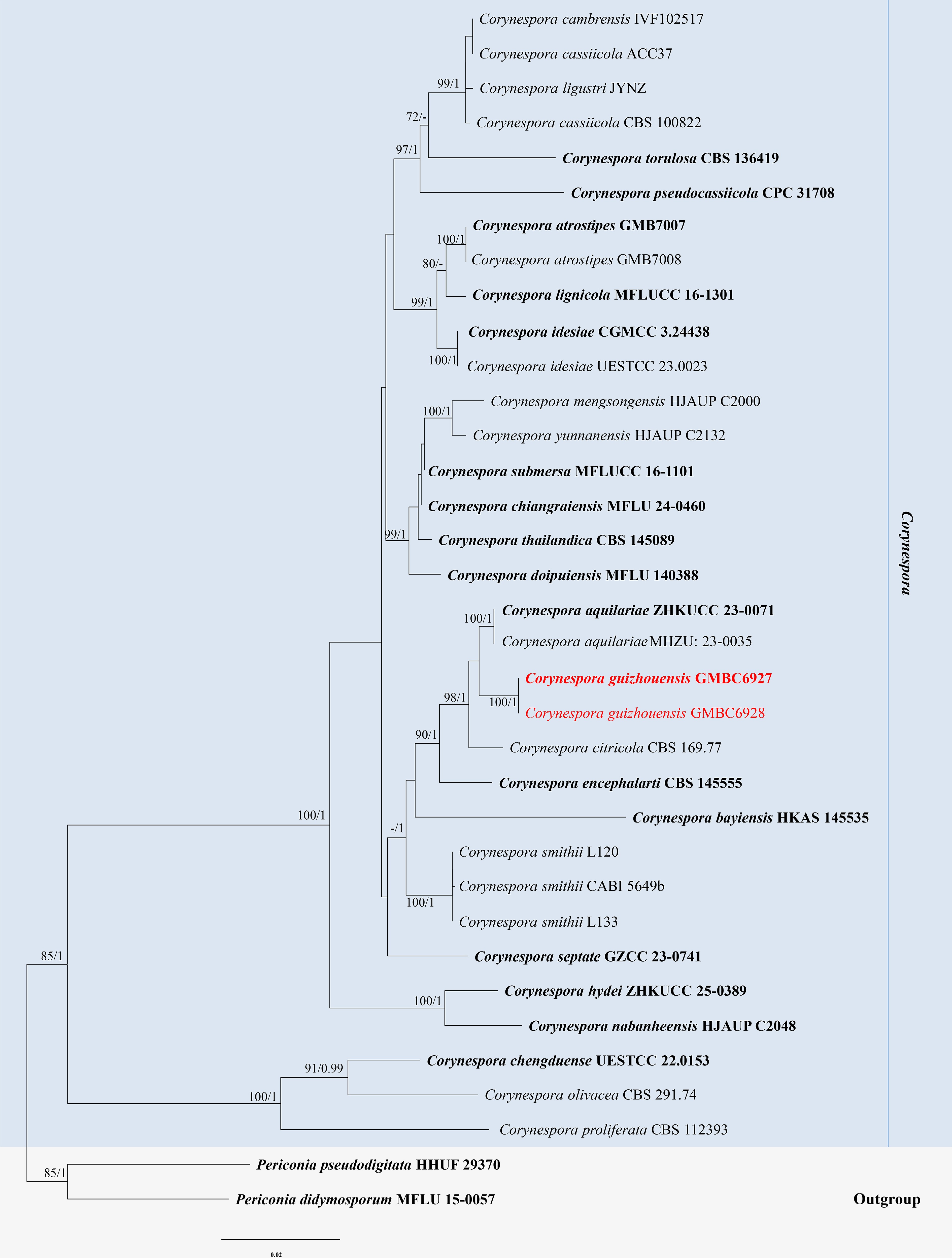
Figure 6.
ML tree of selected Corynespora species based on the ITS-LSU-SSU-tef1-α-rpb2 dataset. The combined dataset comprised 27 taxa and 3,207 characters (ITS: 1–437, LSU: 438–1,297, SSU: 1,298–2,307, tef1-α: 2,304–3,207). Maximum likelihood (lnL = −12,287.196510) and Bayesian analyses produced consistent tree topologies. Periconia digitata (CBS 510.77) and P. igniaria (CBS 845.96) were selected as the outgroup taxa. Bayesian posterior probabilities (PP) ≥ 0.90 and RAxML bootstrap support values ≥ 70 % are shown on the branches. The newly described species is marked in red. Ex-type/type strains are indicated in bold.
Based on these clear differences in conidiophore morphology and its distinct phylogenetic placement, C. guizhouensis is herein introduced as a novel species.
Hermatomycetaceae Locq., Mycol. gén. struct. (Paris): 202 (1984).
Hermatomyces Speg., Anal. Mus. nac. B. Aires, Ser. 3 13: 445 (1910) [1911].
Notes – Hermatomyces was established by Spegazzini with H. tucumanensis as the type species[59]. Species of Hermatomyces are distinguished by sporodochial conidiomata, micronematous conidiophores, and the production of two distinct conidial types, namely cylindrical and lenticular conidia[60]. Cylindrical conidia are hyaline to irregularly pigmented, composed of several columns and cells, whereas lenticular conidia are characteristically pigmented with paler peripheral cells and dark brown central cells, exhibiting different shapes in frontal and lateral views[61]. The sexual morph, only recently discovered, is characterized by ostiolate, dark brown to black ascomata, bitunicate, eight-spored asci, and broadly fusiform, 1-septate ascospores[62].
Species of Hermatomyces are saprobic, occurring mainly on dead plant material, particularly decomposing leaves and wood in terrestrial habitats. The genus shows a pantropical distribution, with numerous records from Southeast Asia, while earlier collections from Africa were reported by Hughes and later revised by Koukol et al.[63,64]. Recent surveys have demonstrated that the genus is likely underexplored, as targeted sampling in tropical regions has resulted in the discovery of several novel taxa[16,62,64,65]. According to Species Fungorum (accessed 16 March 2026), the genus currently comprises 40 accepted species. In this study, a novel species of Hermatomyces is introduced.
Hermatomyces hyalodimorphus L.L. Liu, Q.F. Zhang, & Q.R. Li, sp. nov. (Fig. 7)
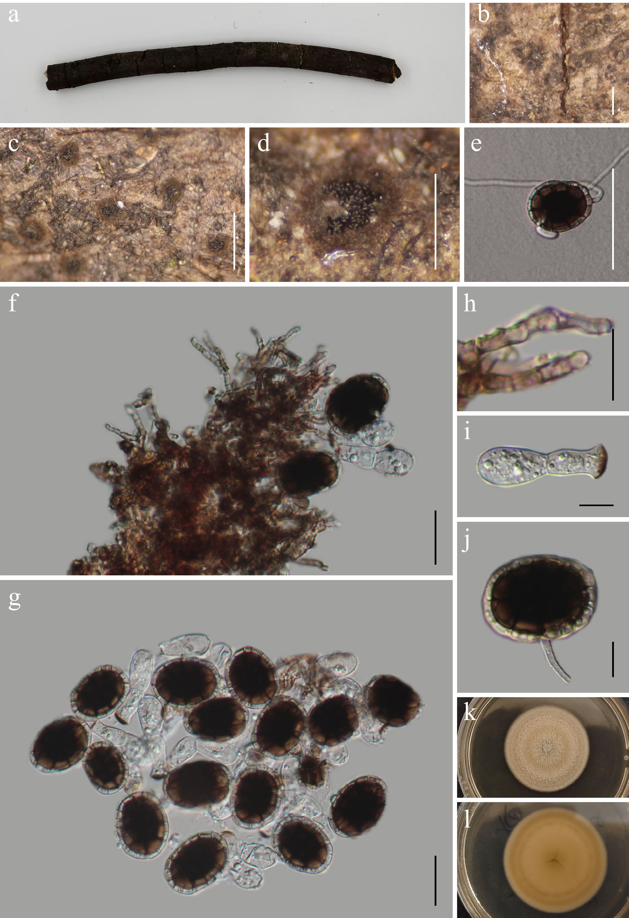
Figure 7.
Hermatomyces hyalodimorphus (GMB5172 holotype). (a) Host. (b)–(d) Colonies on natural substrate. (e) Germinating conidium. (f) Subicular hyphae and conidia. (g) Conidia. (h) Peripheral hyphae. (i) Hyaline conidium. (j) Lenticular conidium. (k), (l) Surface and reverse view of culture on PDA. Scale bars: b, c = 1 mm, d = 0.5 mm, e = 40 μm, f–h = 15 μm, i = 10 μm, j = 5 μm
MycoBank number: MB862928
Etymology – The epithet refers to the presence of conspicuously dimorphic conidia, one of which is hyaline.
Saprobic on dead branches of an unknown plant. Sexual morph: not observed. Asexual morph: hyphomycetous. Colonies superficial, sporodochial, subiculate conidiomata, dark brown to black, circular to irregular in outline, scattered to gregarious, with a dense, velvety, sterile marginal zone surrounding a flattened, dark, abundantly sporulating central disc; conidia easily detached when disturbed. Mycelium is mostly superficial, consisting of a compact network of repent, branched, septate hyphae, pale brown to brown, smooth to finely verruculose; subicular hyphae septate, branched, flexuous to undulate, forming a dense basal stroma. Conidiophores reduced to conidiogenous cells. Conidia holoblastic, produced singly or occasionally in short chains, distinctly dimorphic, dry. Lenticular conidia 24.9–38.1 × 18.5–26.7 µm (x = 32.4 × 23.5 µm, n = 30), thick-walled and muriform, ellipsoidal to oblong in lateral view, circular to broadly ellipsoidal in frontal view, slightly constricted at the septa; central cells dark brown to blackish brown; peripheral cells 13–17, pale brown, forming a conspicuous marginal ring; surface smooth to finely verruculose, occasionally observed with a short, hyaline pedicel-like remnant at the base. Hyaline conidia 32.3–44.5 (x = 37.6 µm, n = 25) long, 8.2–14.1 µm (x =11.4 µm, n = 25) wide, 1-septate, hyaline, smooth-walled, cylindrical to slightly clavate, produced singly or in short chains, occasionally with a truncate base.
Culture characteristics – Conidia germinate on PDA within 24 h. After 28 d at 25 °C, colonies on PDA reach 25–35 mm in diameter; surface grayish-white, with a tree-ring-like pattern, filamentous, circular, raised, velvety to floccose, margin even; reverse light yellow-brown in the center, whitish at the margin.
Material examined – CHINA, Yunnan Province, Xishuangbanna Dai Autonomous Prefecture, 24°02'24.48" N, 100°48'38.25" E, elevation 1,450 m, on dead branches of an unknown plant, 8 August 2025, Qin-Fang Zhang, 2025XSBN34-2 (GMB5172, holotype; GMBC5172, ex-type); ibid., KUN-HKAS 152892, isotype.
Additional examined specimens – CHINA, Yunnan Province, Xishuangbanna Dai Autonomous Prefecture, 24°02'24.61" N, 100°48'38.30" E, elevation 1,190 m, on dead branches of an unknown plant, 18 August 2025, Qin-Fang Zhang, 2025XSBN135 (GMB5173, GMBC5173).
Notes – In the phylogram (Fig. 8), Hermatomyces hyalodimorphus forms a well-supported, distinct lineage within Hermatomyces, close to H. yunnanensis. Comparative analysis of nucleotide base pairs showed that H. hyalodimorphus (GMBC5172) differs from the type strain of H. yunnanensis (HKAS 144361) by 1.1% (3/298 bp) in the ITS locus, 0.8% (6/794 bp) in the LSU locus, and 7.2% (66/917 bp) in the rpb2 gene.
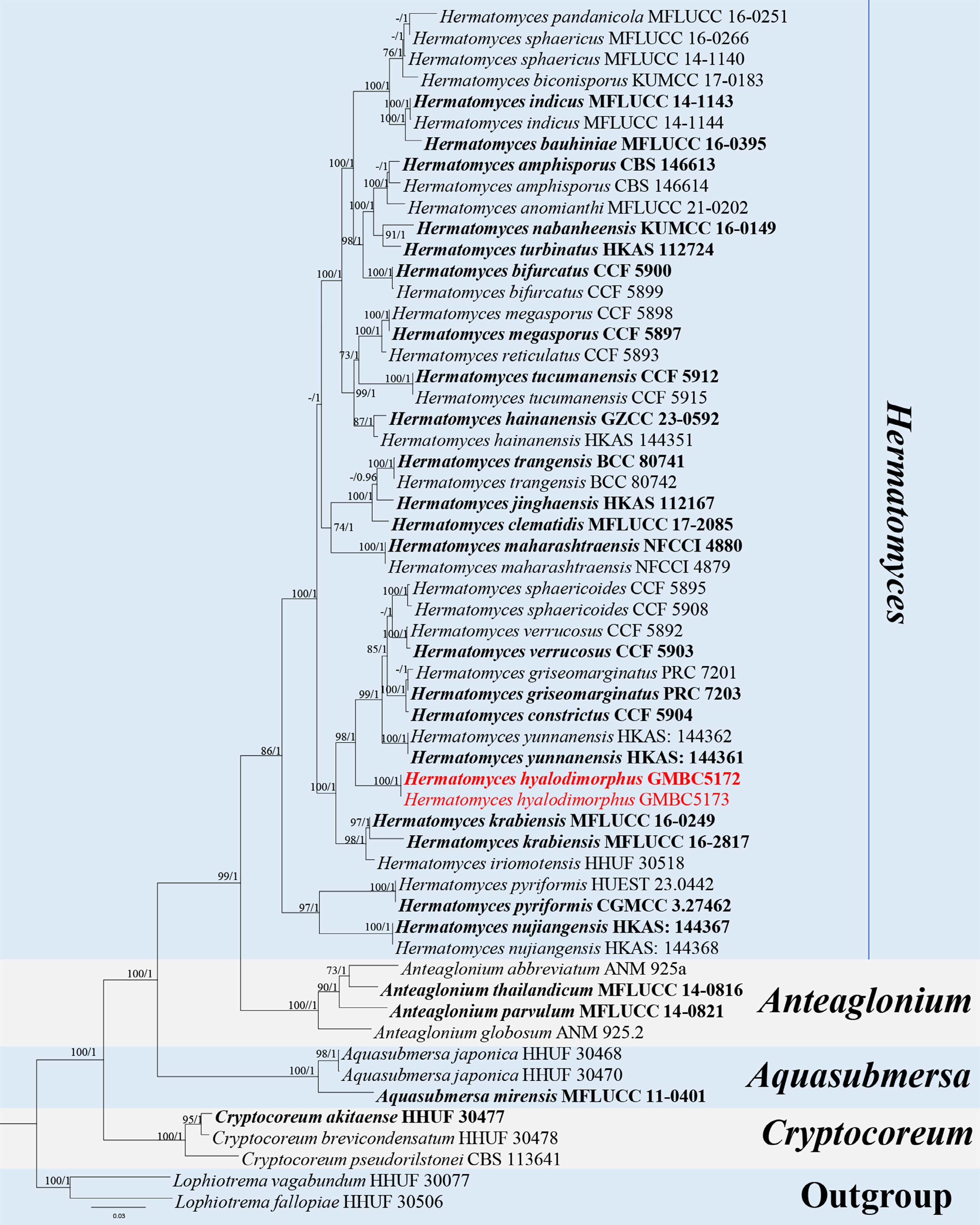
Figure 8.
ML tree of selected species of Hermatomyces and related genera inferred from a combined ITS-LSU- tef1-α-rpb2 dataset. The combined dataset comprised 57 taxa and 3,331 characters (ITS: 1–510 bp; LSU: 511–1,376 bp; tef1-α: 1,377–2,316 bp; rpb2: 2,317–3,331 bp), including indel regions. Maximum likelihood (lnL = −18,361.071594) and Bayesian analyses produced consistent tree topologies. Lophiotrema fallopiae (HHUF 30506) and L. vagabundum (HHUF 30077) were selected as the outgroup taxa. Bayesian posterior probabilities (PP) ≥ 0.90 and RAxML bootstrap support values ≥ 70 % are shown on the branches. The newly described species is marked in red. Ex-type/type strains are indicated in bold.
Morphologically, H. hyalodimorphus resembles H. yunnanensis in the formation of sporodochial, subiculate conidiomata and the presence of lenticular, muriform conidia with dark central cells and pale peripheral cells. However, H. hyalodimorphus differs from H. yunnanensis by producing distinctly dimorphic conidia, including hyaline, 1-septate conidia formed singly or in short chains (absent in H. yunnanensis), and by having thinner lenticular conidia (24.9–38.1 × 18.5–26.7 µm) compared to those of H. yunnanensis (30–39 × 21–36 µm)[66].
Based on its independent phylogenetic placement and distinct morphological characteristics, H. hyalodimorphus is described here as a novel species.
Phaeoseptaceae Boonmee, Thambug. & K.D. Hyde, Mycosphere 9(2): 323 (2018).
Xenopleopunctum J.Y. Zhang, Y.Z. Lu & K.D. Hyde, IMA Fungus 16, e167717 (2025).
Notes – Xenopleopunctum was established by Zhang et al., with X. guizhouense designated as the type species[67]. Species of Xenopleopunctum characterized by effuse, superficial, brown to black colonies, scattered to aggregated; conidiophores brown, cylindrical, sometimes reduced to conidiogenous cells; conidiogenous cells terminal, monoblastic, brown, thick-walled; conidia ellipsoidal to muriform, acrogenous, brown to dark brown, basal region paler with a distinct hyaline basal cell, darker at septa[67,68]. According to Species Fungorum (accessed 16 March 2026), Xenopleopunctum currently contains three accepted species. In this study, two additional species, X. yunnanense and X. dictyosporum, are described based on distinct morphological characters and phylogenetic analyses.
Xenopleopunctum dictyosporum L.L. Liu, W.M. Li, & Q.R. Li, sp. nov. (Fig. 9)
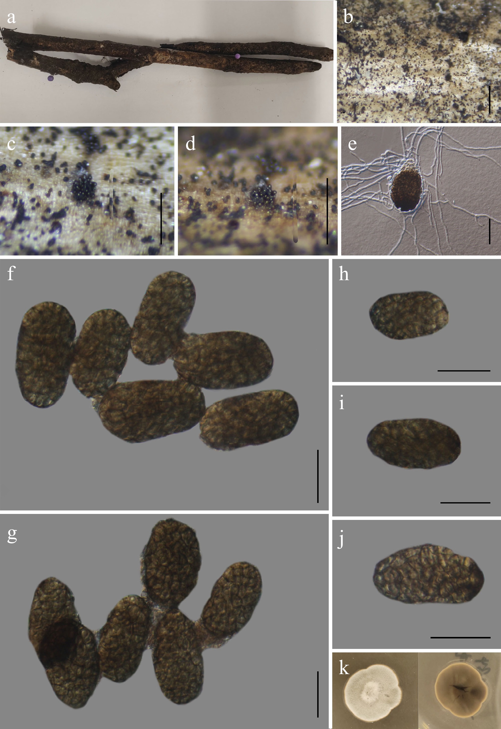
Figure 9.
Xenopleopunctum dictyosporum (GMB6953 holotype). (a) Host. (b)–(d) Colonies on natural substrate. (e) Germinating conidium. (f)–(j) Conidia. (k) Surface and reverse view of culture on PDA. Scale bars: b = 1 mm, c, d = 0.5 mm, f–j = 30μm.
MycoBank number: MB862931
Etymology – The specific epithet dictyosporum refers to the dictyosporous conidia.
Saprobic on dead branches of an unknown plant. Sexual morph: not observed. Asexual morph: hyphomycetous. Colonies superficial, sporodochial, scattered or aggregated in small groups, with irregular margins, appearing as dark, beady to velvety masses composed of densely packed conidiophores and conidia. Mycelium mostly immersed, consisting of smooth-walled, septate, hyaline, branched hyphae. Conidiophores micronematous, hyaline, reduced to conidiogenous cells. Conidiogenous cells integrated, terminal, monoblastic, holoblastic, cylindrical, hyaline, smooth-walled. Conidia 28–46 × 18.5–23.5 µm (x = 40.7 × 21.1 µm, n = 30), solitary, dry, dictyosporous, broadly ellipsoidal to ovate or obovoid, apex broadly rounded and gradually tapering towards the base, septate both longitudinally and transversely, forming a muriform pattern. darker at the septa and paler towards the basal region, usually with a conidiogenous cell attached 5.2–10 × 9.4–14.7 μm (x = 7.0 × 9.2 μm, n = 30).
Culture characteristics – Conidia germinate on PDA within 24 h. After five weeks at 25 °C, colonies on PDA reach 10–20 mm in diameter; circular, raised, aerial mycelium moderately developed, fluffy, concentric zonation absent, margins entire, surface white to pale, reverse yellowish brown to dark brown, pigmentation concentrated in the central zone.
Material examined – CHINA, Yunnan Province, Zhaotong City, Wumengshan National Nature Reserve, 25°21′19.26″ N, 103°15′09.37″ E, elevation 2,519 m, on dead branches, 26 October 2024, Wen-Mei Li, 2024WMS339-2 (GMB6953, holotype; GMBC6953, ex-type); ibid., KUN-HKAS 152894, isotype.
Additional examined specimens – CHINA, Yunnan Province, Zhaotong City, Wumengshan National Nature Reserve, 25°21′17.13″ N, 103°16′11.23″ E, elevation 1,846 m, on dead branches, 26 October 2024, Wen-Mei Li, 2024WMS392 (GMB6954, GMBC6954).
Notes – In the BLASTn search of ITS and LSU sequences, Xenopleopunctum dictyosporum showed the closest similarity to X. sporodochiale. Phylogenetically, X. dictyosporum formed an independent clade, which confirmed that they are separate species (Fig. 10). Pairwise comparison of nucleotide sequences revealed that X. dictyosporum (GMBC6953) differs from the type strain of X. sporodochiale (GZCC 23-0742) by 6.1% (30/488 bp) in the ITS 1.2% (10/853 bp) in the LSU locus. Morphologically, X. dictyosporum can be differentiated from X. sporodochiale by its smaller basel condial cell 5.2–10 × 9.4–14.7 μm compared to 8.5–14 × 6–15 µm in X. sporodochiale[67].
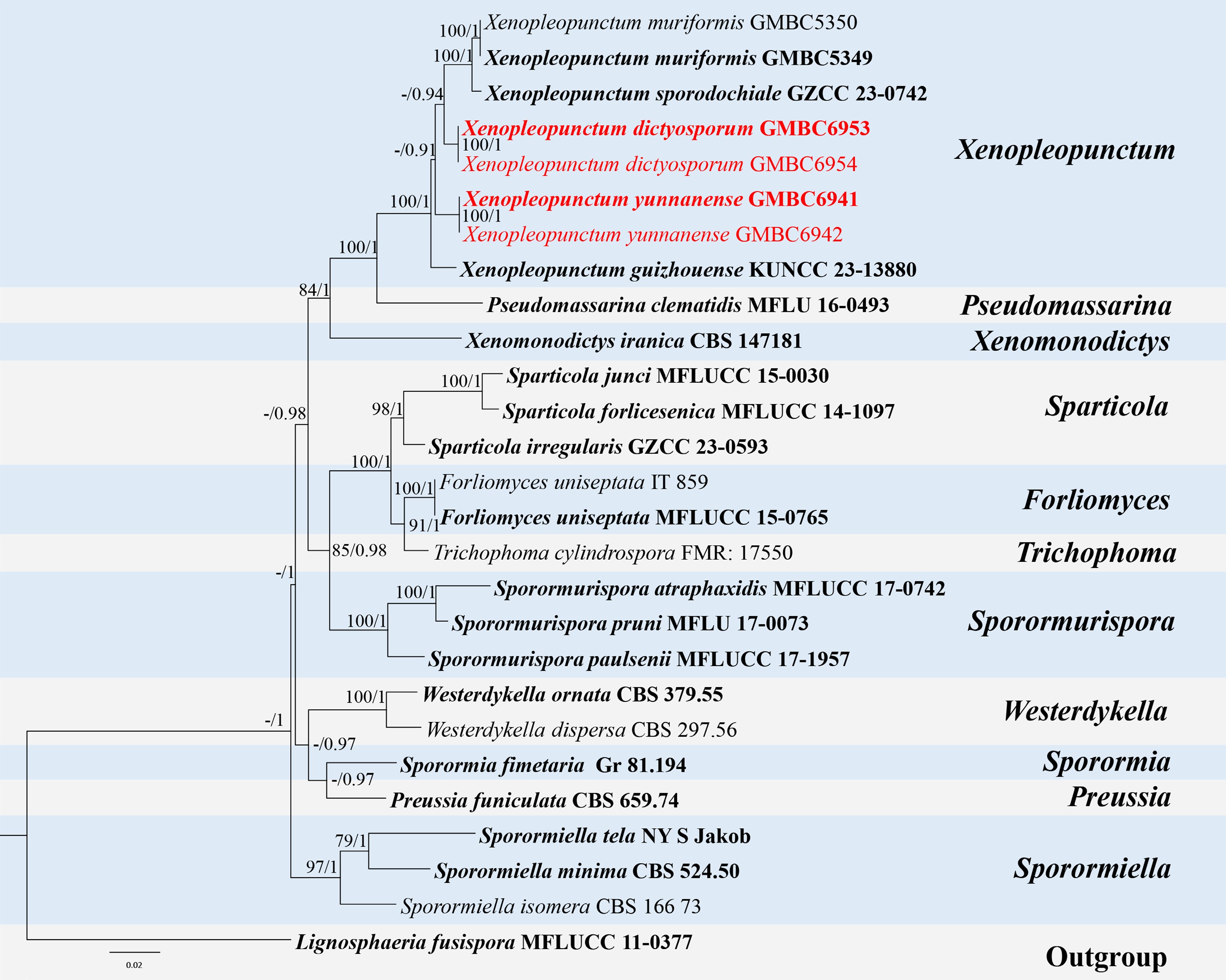
Figure 10.
ML tree of selected species of Xenopleopunctum and related genera inferred from a combined ITS-LSU-SSU dataset. The combined dataset comprised 27 taxa, 2,425 characters (ITS:1–514 bp; LSU: 515–1,418 bp; SSU: 1,419–2,425 bp), including indel regions. Maximum likelihood (lnL = −9,247.818154) and Bayesian analyses produced consistent tree topologies. Lignosphaeria fusispora (MFLUCC 11-0377) was selected as the outgroup taxon. Bayesian posterior probabilities (PP) ≥ 0.90 and RAxML bootstrap support values ≥ 70 % are shown on the branches. The newly described species are marked in red. Ex-type/type strains are indicated in bold.
Based on its distinct phylogenetic position and molecular divergence, X. dictyosporum is proposed here as a novel species.
Xenopleopunctum yunnanense L.L. Liu, W.M. Li, & Q.R. Li, sp. nov. (Fig. 11)
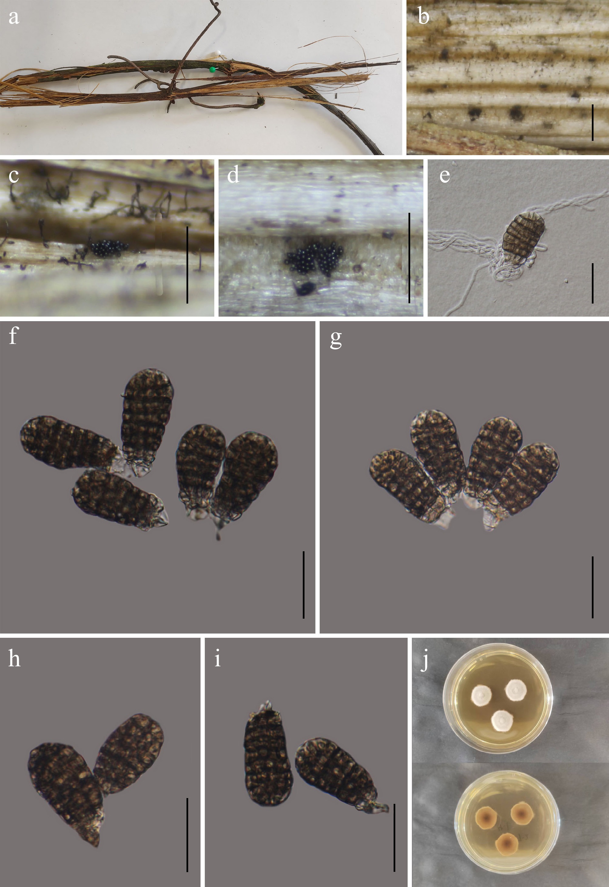
Figure 11.
Xenopleopunctum yunnanense (GMB6941 holotype). (a) Host. (b)–(d) Colonies on natural substrate. (e) Germinating conidium. (f)–(i) Conidia. (j) Surface and reverse view of colonies on PDA. Scale bars: b = 1 mm, c, d = 0.5 mm, e–i = 30 μm.
MycoBank number: MB862929
Etymology – The specific epithet yunnanense refers to Yunnan Province, China, where the holotype was collected.
Saprobic on dead branches of an unknown plant. Sexual morph: not observed. Asexual morph: hyphomycetous. Colonies superficial, sporodochial, black, glistening, gregarious, punctiform. Mycelium immersed, consisting of subhyaline to hyaline, branched, septate hyphae. Conidiogenous cells not observed. Conidia acrogenous, 47–64.5 × 20–29 µm (x = 54.3 × 24.5 µm, n = 30), brown to dark brown, oval to ellipsoidal, broadly obtuse at the apex, solitary, muriform, basal cell absent.
Culture characteristics – Conidia germinating on PDA within 24 h. Colonies on PDA reaching 15–25 mm after 28 d (day–night cycle) at 25 °C, circular, raised, aerial mycelium sparse to moderate, fluffy, concentric zonation indistinct, margins entire to slightly lobate, surface white to pale cream initially, beige to light brown with age, reverse pale brown to dark brown, blackish at center.
Material examined – CHINA, Yunnan Province, Zhaotong City, Wumengshan National Nature Reserve, 25°20′18.90″ N, 103°13′05.90″ E, elevation 2,350 m, on dead branches of an unknown plant, 25 October 2024, Wen-Mei Li, 2024WMS184 (GMB6941, holotype; GMBC6941, ex-type); ibid., KUN-HKAS 152893, isotype.
Additional examined specimens – CHINA, Yunnan Province, Zhaotong City, Wumengshan National Nature Reserve, 25°21′30.13″ N, 103°11′05.94″ E, elevation 2,700 m, on dead branches of an unknown plant, 25 October 2024, Wen-Mei Li, 2024WMS186 (GMB6942, GMBC6942).
Notes – Results from BLASTn searches of ITS and LSU regions showed that Xenopleopunctum yunnanense shares the highest similarity with X. guizhouense and is also closely related phylogenetically (Fig. 10). Pairwise comparison of nucleotide sequences showed that X. yunnanense (GMBC6941) differs from the type strain of X. guizhouense (KUNCC 23-13880) by 4.0% (19/478 bp) in the ITS locus and 1.6% (14/860 bp) in the LSU locus.
Morphologically, the two species share muriform, brown to dark brown conidia. However, X. yunnanense differs from X. guizhouense in lacking a hyaline basal cell, which is present in X. guizhouense as a cylindrical to subglobose, hyaline to subhyaline or pale brown basal cell measuring 8.5–14 × 6–15 µm[67].
Given its clear molecular and morphological differences and its distinct phylogenetic position, X. yunnanense is established here as a new species.
Tubeufiales Boonmee & K.D. Hyde, Fungal Diversity 68 (1): 245 (2014).
Tubeufiaceae M.E. Barr, Mycologia 71: 948 (1979).
Helicoma Corda, Icones fungorum hucusque cognitorum 1: 15 (1837).
Notes – Helicoma was established by Corda, based on the type species H. muelleri[69]. Species of the genus are characterized by intercalary, cylindrical conidiogenous cells bearing denticles, which arise laterally from the lower regions of the conidiophores as tooth-like projections; pleurogenous, helicoid, hygroscopic conidia, tapering towards the apex and rounded at the tip, coiled 1½ to 5 times, becoming loosely coiled in water. According to Species Fungorum (accessed 8 January 2026), the genus Helicoma currently comprises 77 accepted species. Members of Helicoma have a cosmopolitan distribution, reported from both terrestrial and freshwater habitats[70−73]. In this study, Helicoma wuliangshanense is introduced as a novel species isolated from submerged wood in a freshwater habitat.
Helicoma wuliangshanense L.L. Liu, Q.F. Zhang, & Q.R. Li, sp. nov. (Fig. 12)
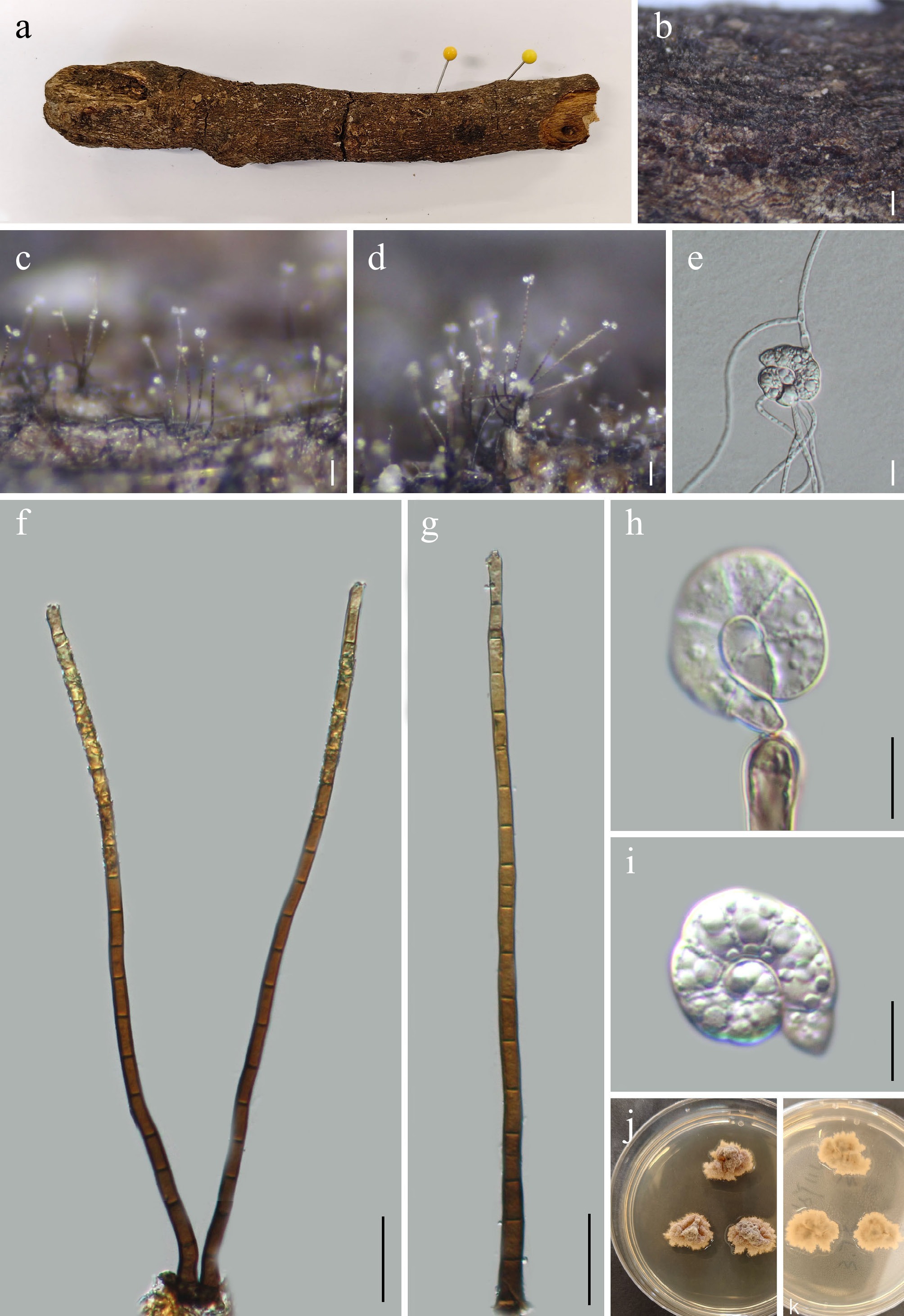
Figure 12.
Helicoma wuliangshanense (GMB5146 holotype). (a) Host. (b)–(d) Colonies on natural substrate. (e) Germinating conidium. (f), (g) Conidiophores. (h) Conidiogenous cells. (i) Conidium. (j), (k) Surface and reverse view of culture on PDA. Scale bars: b = 1 mm, c, d = 0.5 mm, f, g = 50 μm, e, h, i = 10 μm.
MycoBank number: MB862949
Etymology –The epithet refers to Wuliangshan National Nature Reserve, the locality where the holotype was collected.
Saprobic on submerged decaying wood in freshwater habitats. Sexual morph: not observed. Asexual morph: hyphomycetous, helicosporous. Colonies superficial, hairy, effuse, pale brown to dark brown, glistening, scattered to gregarious. Mycelium, partly superficial, partly immersed, consisted of smooth-walled, branched, septate hyaline to pale brown hyphae. Conidiophores 118.8–314.5 × 5.2–7.4 μm (x = 227.5 × 6.3 μm, n = 30), mononematous, macronematous, straight to slightly flexuous, erect, septate, smooth-walled, simple or occasionally branched, sometimes slightly swollen at the base, pale brown to brown, paler towards the apex, darker towards the base. Conidiogenous cells 12.5–31.8 × 2.2–5.5 μm (x = 20.2 × 3.3 μm, n = 30), mono- or polyblastic, holoblastic, determinate, integrated, terminal, cylindrical, pale brown, smooth-walled, denticulate, truncate at the apex after conidial secession. Conidia 40.0–57.5 μm (x = 49.2 μm, n = 30) long and 6.8–9.2 μm (x = 8.3 μm, n = 30) wide, solitary, acrogenous to acropleurogenous, helicoid, tightly coiled 1 to 1.5 times, conidial filament, overall conidial diameter 15.7–21 μm (x = 19.3 μm, n = 30), multi-septate with 6–10 septa, not loosening in water, smooth-walled, hyaline to subhyaline, rounded at the apex, occasionally guttulate.
Culture characteristics – Conidia germinate on PDA within 24 h. After four weeks at 25 °C, colonies on PDA reach 30–40 mm in diameter; circular to irregular, raised, woolly at center, fluffy, concentric zonation indistinct, margins entire to lobate, surface white to yellow-brown; reverse pale brown to dark brown.
Material examined – CHINA, Yunnan Province, Wuliangshan National Nature Reserve, 24°25'33.66" N, 101°23'55.67" E, elevation 1,455 m, on submerged decaying wood in freshwater habitats, 2 August 2024, Qin-Fang Zhang, 2024WLSY114 (GMB5146, holotype; GMBC5146, ex-type); ibid., KUN-HKAS 152895, isotype
Additional specimens examined – CHINA, Yunnan Province, Wuliangshan National Nature Reserve, 24°35'56.42" N, 100°25'55.57" E, elevation 2,180 m, on submerged decaying wood in freshwater habitats, 2 August 2024, Qin-Fang Zhang, 2024WLSY154 (GMB5147, GMBC5147).
Notes – In the phylogram (Fig. 13), Helicoma wuliangshanense is closely related to H. brunneum. Pairwise comparison of nucleotide sequences revealed that H. wuliangshanense (GMBC5146) differs from the type strain of H. brunneum (CGMCC 3.25573) by 3.2% (15/468 bp) in the ITS locus and 1.4% (20/1400 bp) in the LSU locus. Morphologically, H. wuliangshanense shares several characters with H. brunneum and H. dennisii, including effuse, hairy colonies, macronematous and mononematous conidiophores, denticulate conidiogenous cells, and helicoid conidia tightly coiled 1 to 1.5 times. However, H. wuliangshanense can be distinguished from H. brunneum in having a smaller conidial diameter (15.7–21.3 μm vs 18–26 μm), and relatively narrower conidiogenous cells (2–5 μm vs 5–8 μm). Ecologically, H. wuliangshanense is saprobic on submerged decaying wood in freshwater habitats, whereas H. brunneum was described from terrestrial decaying wood[8].
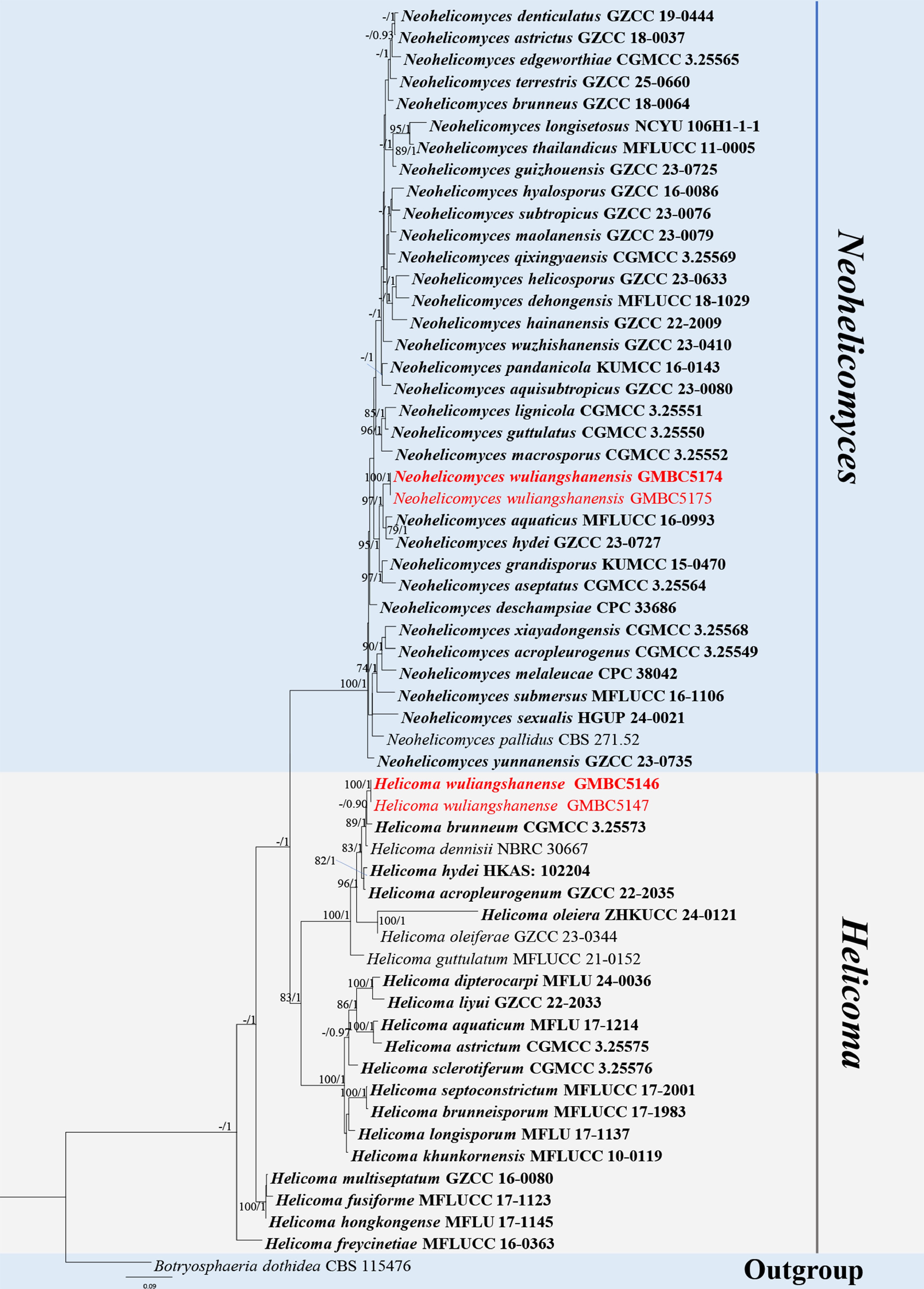
Figure 13.
ML tree of Neohelicomyces and closely related genera (including Helicoma) inferred from a combined ITS-LSU-SSU-tef1-α-rpb2 dataset. The combined dataset comprised 58 taxa, 3,446 characters (ITS: 1–571 bp; LSU: 572–1,431 bp; tef1-α: 1,432–2,345 bp; rpb2: 2,346–3,446 bp), including indel regions. Maximum likelihood (lnL = −23,540.830905) and Bayesian analyses produced consistent tree topologies. Botryosphaeria dothidea (CBS 115476) was selected as the outgroup taxon. Bayesian posterior probabilities (PP) ≥ 0.90 and RAxML bootstrap support values ≥ 70 % are shown on the branches. The newly described species are marked in red. Ex-type/type strains are indicated in bold.
Helicoma wuliangshanense differs from H. dennisii by its shorter conidiophores (118–314 μm vs up to 470 μm), and a greater number of conidial septa (6–10 vs 7–8 septa)[74].
Molecular phylogenetic analyses, coupled with the morphological and ecological distinctions, support the recognition of H. wuliangshanense as a novel species.
Neohelicomyces Z.L. Luo, Bhat & K.D. Hyde, Cryptog. Mycol. 38(1): 39 (2017).
Notes – Neohelicomyces was introduced by Luo et al., typified by N. aquaticus[26]. The genus belongs to Tubeufiaceae and comprises helicosporous hyphomycetes characterized by coiled to helically arranged conidia[26,75−77]. Species of Neohelicomyces are saprobic on dead plant litter in terrestrial and freshwater habitats, with most records reported from Asia, particularly in China (Guizhou, Hainan, and Yunnan Provinces)[16,78]. According to Species Fungorum (accessed 5 January 2026), the genus currently comprises 38 accepted species. The new species introduced herein was isolated from decaying wood in Yunnan Province, China.
Neohelicomyces wuliangshanensis L.L. Liu, Q.F. Zhang, & Q.R. Li, sp. nov. (Fig. 14)
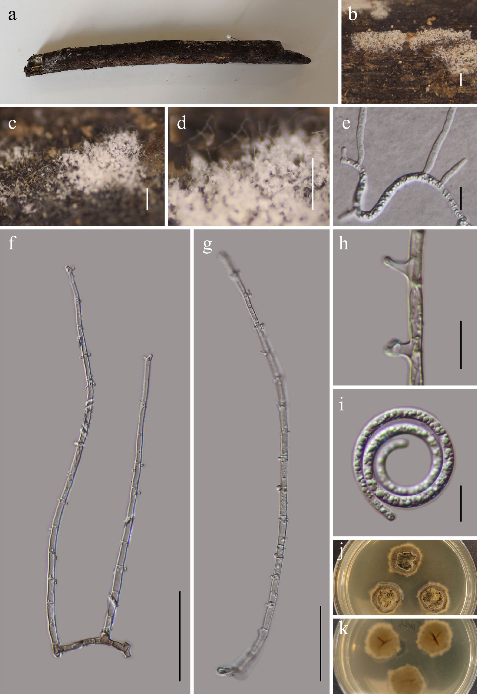
Figure 14.
Neohelicomyces wuliangshanensis (GMB5174 holotype). (a) Host. (b)–(d) Colonies on natural substrate. (e) Germinating conidium. (f), (g) Conidiophores and conidia. (h) Conidiogenous cells. (i) Conidia. (j), (k) Surface and reverse view of culture on PDA. Scale bars: b =1 mm, c, d = 0.5 mm, f, g = 50 μm, e, h, i = 10 μm.
MycoBank number: MB862950
Etymology – The epithet refers to Wuliangshan National Nature Reserve, where the type specimen was collected.
Saprobic on submerged decaying wood in a wetland. Sexual morph: not observed. Asexual morph: hyphomycetous, helicosporous. Colonies effuse, superficial, white to pale brown, gregarious, forming conspicuous glistening conidial masses. Mycelium partly superficial, partly immersed, formed by branched, smooth-walled septate, hyaline to pale brown hyphae. Conidiophores 95.4–380 × 3.2–5.2 μm (x = 226.0 × 4.3 μm, n = 20), simple or sparingly branched, mononematous, macronematous, straight to flexuous, erect, cylindrical, hyaline to pale brown, 7–24-septate, smooth and thick-walled. Conidiogenous cells 1.2–5.9 × 1.0–1.8 μm (x = 2.7 × 1.5 μm, n = 20), integrated, monoblastic, holoblastic, intercalary or terminal, bearing minute denticles, cylindrical to slightly lageniform, truncate at the apex after conidial secession, smooth-walled, hyaline to pale brown. Conidia 137.5–209.2 μm long and 2.5–3.1 μm wide (x = 162.4 × 3.15 μm, n = 20), solitary, acropleurogenous, helicoid, conidial filament coiled two to four times, overall conidial diameter 14–33 μm (x = 28 μm, n = 20), multiseptate, smooth-walled, hyaline, guttulate, becoming loosely coiled in water, rounded at both ends.
Culture characteristics – Conidia germinate on PDA within 24 h. After five weeks at 25 °C, colonies on PDA reach 20–30 mm in diameter; circular, raised, fluffy, concentric zonation distinct, margins entire, surface white to yellow-brown; reverse pale brown to dark brown
Material examined – CHINA, Yunnan Province, Wuliangshan National Nature Reserve, 24°25'33.66" N, 101°23'55.67" E, elevation 1,455 m, on submerged decaying wood in freshwater habitats, 18 August 2025, Qin-Fang Zhang, 2025WLS21 (GMB5174, holotype; GMBC5174, ex-type); ibid., KUN-HKAS 152896, isotype.
Additional examined specimens – CHINA, Yunnan Province, Wuliangshan National Nature Reserve, 24°35'56.42" N, 100°25'55.53" E, elevation 2,180 m, on submerged decaying wood in freshwater habitats, 18 August 2025, Qin-Fang Zhang, 2025WLS84 (GMB5175, GMBC5175).
Notes – In the phylogenetic analysis (Fig. 13), Neohelicomyces wuliangshanensis is closely related to N. aquaticus (MFLUCC 16-0993) and N. hydei (GZCC 23-0727), forming a well-supported clade within Neohelicomyces (BS = 97; PP = 1). Comparative analysis of nucleotide base pairs showed that N. wuliangshanensis (GMBC5174) differs from the type strain of N. aquaticus (MFLUCC 16-0993) by 2.5% (11/440 bp) in the ITS locus, 2.6% (23/868 bp) in the tef1-α gene, and 2.2% (21/945 bp) in the rpb2 gene.
Morphologically, N. wuliangshanensis shares several characteristics with N. aquaticus and N. hydei, including macronematous and mononematous conidiophores, holoblastic conidiogenous cells with denticulate protrusions, and multiseptated, helicoid conidia developing acropleurogenously. However, N. wuliangshanensis differs from N. aquaticus by having longer conidiophores (95.4–380 μm vs 240.5–335.5 μm), a greater range of conidiophore septation (7–24 septa vs fewer septa), a larger conidial diameter (14–33 μm vs not exceeding 18 μm), and conidia coiled more times (2–4 vs 2–2.5 turns)[26].
Neohelicomyces wuliangshanensis differs from N. hydei in having significantly smaller conidiogenous cells (1.2–5.9 × 1.0–1.8 μm vs 7.5–19.5 × 3.5–6 μm) and a larger conidial diameter (14–32.8 μm vs up to 18.5 μm)[8].
Based on strong molecular phylogenetic support and its distinct morphology, N. wuliangshanensis is introduced herein as a new species.
Eurotiomycetes O.E. Erikss. & Winka, Myconet 1(1): 6 (1997).
Sclerococcales Réblová, Unter. & W. Gams, Mycol. Prog. 16(1): 34 (2016) [2017].
Dactylosporaceae Bellem. & Hafellner, Cryptog. Mycol. 3(1): 79 (1982).
Gamsomyces Hern.-Restr. & Réblová, Stud. Mycol. 95: 448 (2020).
Notes – Gamsomyces was established by Réblová et al., with Gamsomyces longisporus (≡ Bactrodesmium longisporum)[22] as the type species[79]. Species of Gamsomyces are characterized by synnematous or sporodochial conidiomata; fasciculate, subhyaline to brown, simple or penicillately branched, macronematous to semi-macronematous conidiophores; integrated, monoblastic conidiogenous cells elongating percurrently; and subcylindrical to subulate, transversely euseptate, brown, fusiform conidia, usually bearing a mucilaginous apical cap[5,79,80]. According to Species Fungorum (accessed 5 January 2026), the genus currently comprises six accepted species. Herein, a new species of Gamsomyces is introduced.
Gamsomyces guangxiensis L.L. Liu, Q.F. Zhang, & Q.R. Li, sp. nov. (Fig. 15)
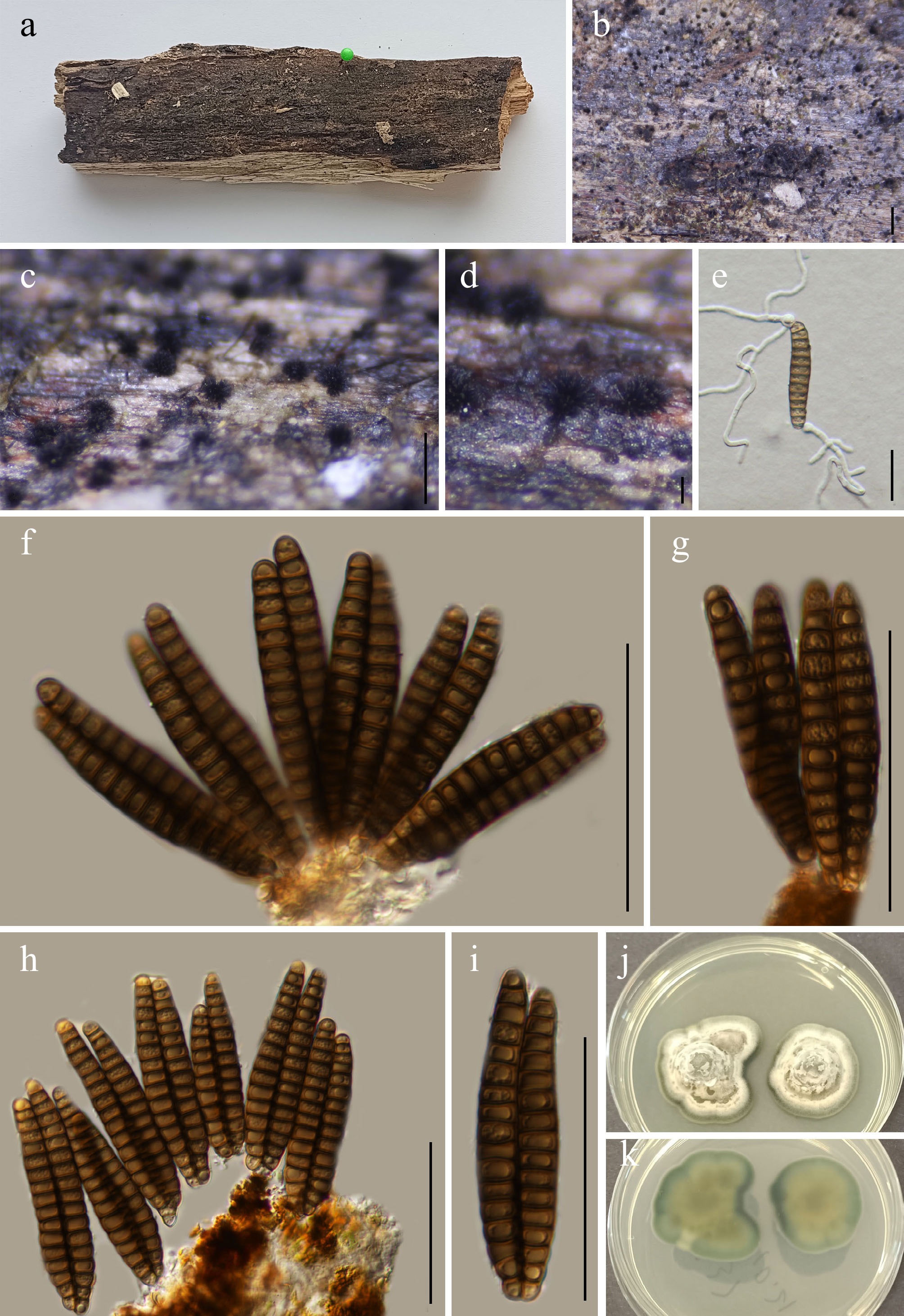
Figure 15.
Gamsomyces guangxiensis (GMB5152 holotype). (a) Host. (b)–(d) Colonies on natural substrate. (e) Germinating conidium. (f)–(i) Conidiophores and conidia. (j), (k) Surface and reverse view of culture on PDA. Scale bars: b = 1 mm, c = 0.5 mm, d = 0.1 mm, e–i = 50 μm.
MycoBank number: MB862951
Etymology – The specific epithet guangxiensis refers to Guangxi, China, where the holotype was collected.
Saprobic on submerged decaying wood in a wetland. Sexual morph: not observed. Asexual morph: hyphomycetous. Colonies sporodochial to synnematous, superficial, scattered to gregarious, dark brown to black, minute, pulvinate to tufted structures on the surface of submerged wood. Mycelium mostly immersed, formed by branched, septate, smooth-walled, hyaline to pale brown hyphae. Conidiophores reduced to conidiogenous cells. Conidiogenous cells monoblastic, terminal, integrated, brown. Conidia 43.2–72.5 × 5.7–8.5 μm (x = 61.6 × 7.3 μm, n = 30), acrogenous, solitary, straight to distinctly curved, narrowly fusiform to fusiform, ends tapering, apex rounded, base truncate to slightly attenuated, pale brown to dark brown, smooth-walled, transversely euseptate, 12–19-septate, not constricted at septa, lack mucilaginous cap at the apex; conidia often aggregated in fascicles at maturity.
Culture characteristics – Conidia germinate on PDA within 24 h. After four weeks at 25 °C, colonies on PDA reach 20–30 mm in diameter; circular, raised in the middle, with dense mycelium, fluffy, concentric zonation distinct, margins entire, surface white to light brown, margins bluish green; reverse pale yellow, bluish green at edges.
Material examined – CHINA, Guangxi Zhuang Autonomous Region, Nonggang National Nature Reserve, 22°20'28.47" N, 106°25'31.35" E, elevation 633 m, on moist submerged decaying wood in a wetland, 2 August 2024, Qin-Fang Zhang, 2024NG37 (GMB5152, holotype; GMBC5152, ex-type); ibid., KUN-HKAS 152897, isotype.
Additional examined specimens – CHINA, Guangxi Zhuang Autonomous Region, Nonggang National Nature Reserve, 22°17'04.45" N, 106°34'25.87" E, elevation 535 m, on moist submerged decaying wood in a wetland, 15 August 2024, Qin-Fang Zhang, 2024NG72 (GMB5153, GMBC5153).
Notes – In the phylogram (Fig. 16), Gamsomyces guangxiensis forms a well-supported sister clade with G. brevis (MFLUCC 24-0107). This clade is clearly separated from other Gamsomyces species, supporting the recognition of G. guangxiensis as a distinct species. Pairwise comparison of nucleotide sequences revealed that G. guangxiensis (GMBC5152) differs from the type strain of G. brevis (MFLUCC 24-0107) by 1.9% (8/432 bp) in the ITS region and 0.1% (1/746 bp) in the LSU region.
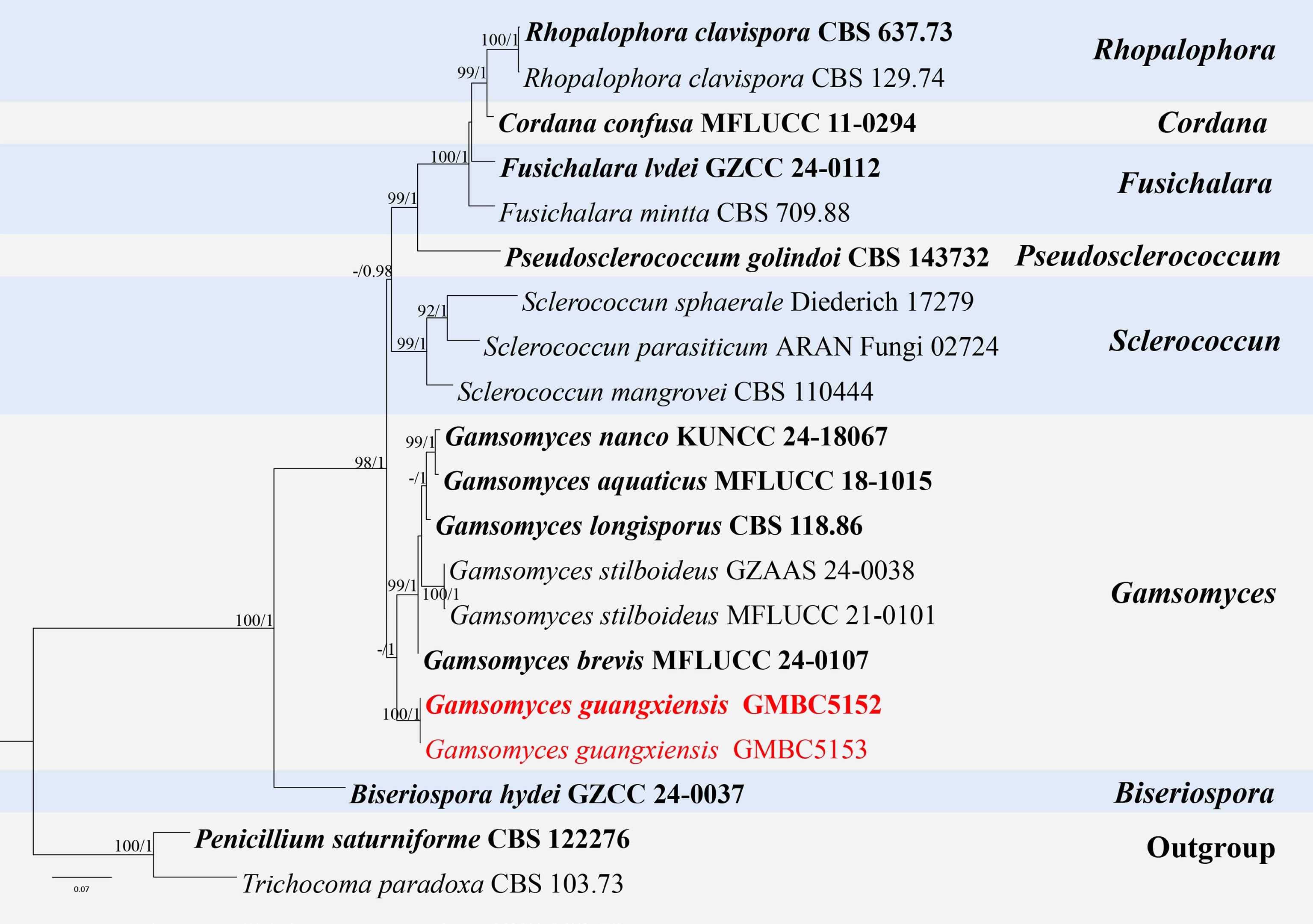
Figure 16.
ML tree of selected species of Gamsomyces and related genera inferred from a combined ITS-LSU dataset. The combined dataset comprised 20 taxa and 1,355 characters (ITS: 1–492 bp; LSU: 493–1,355 bp), including indel regions. Maximum likelihood (lnL = −6,073.633839) and Bayesian analyses produced consistent tree topologies. Penicillium saturniforme (CBS 122276) and Trichocoma paradoxa (CBS 103.73) were selected as the outgroup taxa. Bayesian posterior probabilities (PP) ≥ 0.90 and RAxML bootstrap support values ≥ 70 % are shown on the branches. The newly described species is marked in red. Ex-type/type strains are indicated in bold.
Morphologically, G. guangxiensis is similar to G. brevis in having a hyphomycetous asexual morph with septate, brown to dark brown conidia. However, G. brevis can be distinguished from G. guangxiensis by its larger conidia (57–92 × 4.5–7.5 μm), having a mucilaginous cap at the apex and up to 18 septa. In contrast, G. guangxiensis has smaller conidia (43.2–72.5 × 5.7–8.5 μm), lacks a mucilaginous apical cap, and possesses 12–19 septa[80].
Based on its distinct phylogenetic placement within Gamsomyces s. str., together with its morphological and molecular differences, G. guangxiensis is introduced as a new species.
Leotiomycetes O.E. Erikss. & Winka, Myconet 1(1): 7 (1997).
Helotiales Nannf., Nova Acta R. Soc. Scient. upsal., Ser. 4 8(no. 2): 68 (1932).
Hyaloscyphaceae Nannf., Nova Acta R. Soc. Scient. upsal., Ser. 4 8(no. 2): 258 (1932).
Dematioscypha Svrcek, Ceská Mykologie 31 (4): 193 (1977)
= Haplographium Berk. & Broome, Annals and Magazine of Natural History 3: 361 (1859)
Notes – Dematioscypha was introduced by Svrcek, with D. dematiicola as the type species. Johnston et al. treated Haplographium as a synonym of Dematioscypha, based on the recognition that Haplographium delicatum, the type species of Haplographium, represents the anamorph of Dematioscypha dematicola, the type species of Dematioscypha. Consequently, both genera were considered conspecific. The authors also noted that Haplographium has become taxonomically inconsistent, with many of its species reassigned to other genera, whereas Dematioscypha remains well-defined and widely accepted. Due to the widespread use of the well-defined genus Dematioscypha, they recommended that Dematioscypha be protected over both Schizocephalum and Haplographium[81]. However, fungal databases MycoBank, Species Fungorum, and Index Fungorum still list these genera separately, reflecting differing taxonomic opinions and the lack of universal consensus.
The genus is characterized by brown, penicillate conidiophores with hyaline to subhyaline, sympodial conidiogenous cells that produce hyaline, aseptate conidia aggregated in slimy masses[2]. Species of Dematioscypha are typically saprobic on decaying plant material, especially wood, and occur in both terrestrial and freshwater-associated habitats. According to Species Fungorum (accessed 20 March 2026), the genus currently comprises six accepted species. Herein, a new species of Dematioscypha is introduced based on distinct morphological characteristics and phylogenetic evidence.
Dematioscypha aquatica L.L. Liu, Q.F. Zhang & Q.R. Li, sp. nov. (Fig. 17)
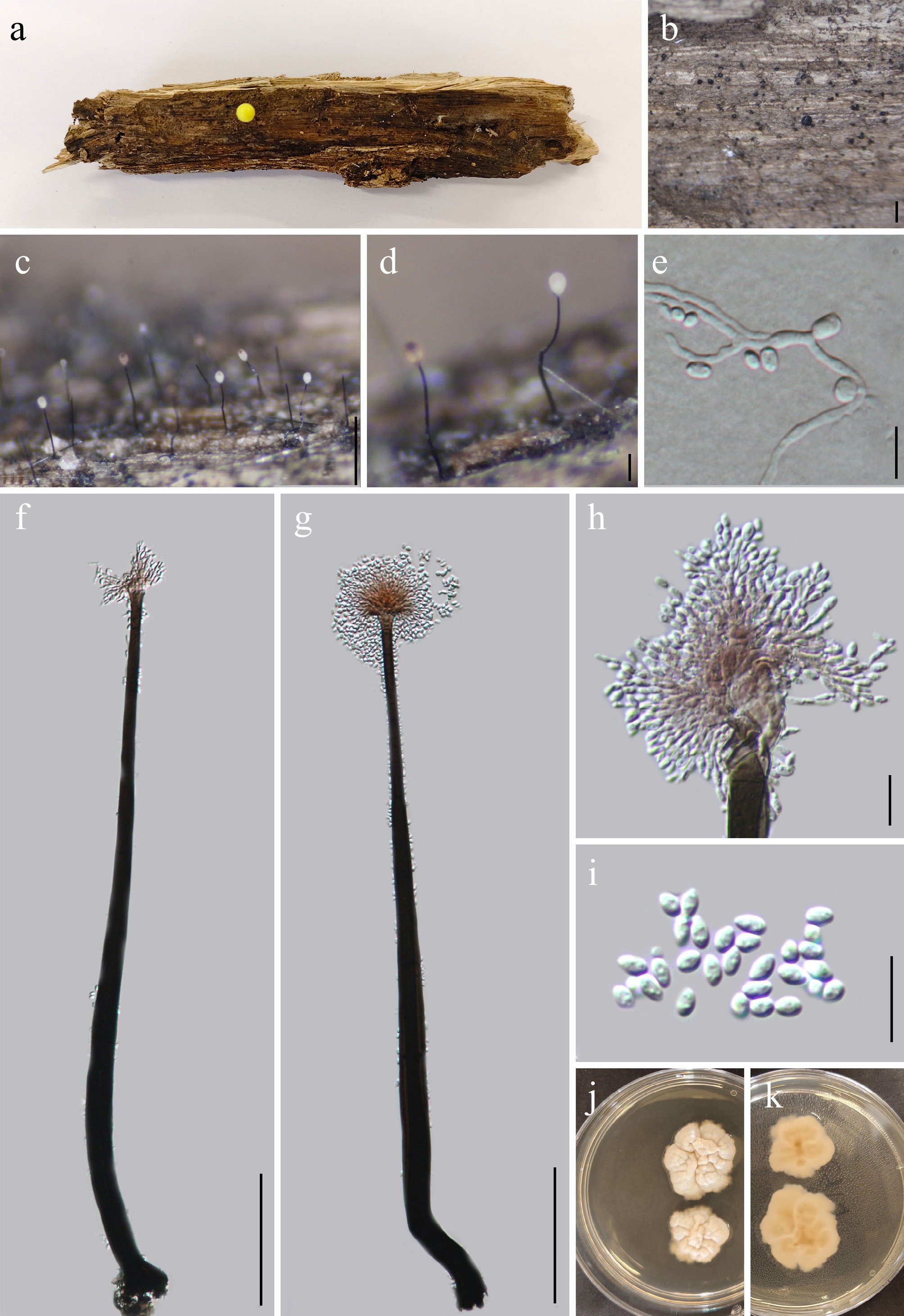
Figure 17.
Dematioscypha aquaticum (GMB5150 holotype). (a) Host. (b)–(d) Colonies on natural substrate. (e) Germinating conidium. (f), (g) Conidiophores and conidia. (h) Conidiogenous cells. (i) Conidia. (j), (k) Surface and reverse view of culture on PDA. Scale bars: b = 1 mm, c = 0.5 mm, d = 0.1 mm, f, g = 100 μm, e, h, i = 10 μm.
MycoBank number: MB862952
Etymology – The specific epithet aquatica refers to its occurrence in a freshwater habitat.
Saprobic on decaying wood in a freshwater swamp. Sexual morph: not observed. Asexual morph: hyphomycetous. Colonies effuse, dark brown to black on the host surface, forming scattered to gregarious conidiophores, each terminating in a conspicuous, spherical to subglobose, glistening, slimy white conidial head. Mycelium immersed in the substrate. Conidiophores 328.8–477.2 × 8.9–14.6 µm (x = 404.6 × 11.2 µm, n = 15), macronematous, mononematous, erect, unbranched, subcylindrical, straight to slightly flexuous, dark brown at the base, gradually becoming pale brown to subhyaline towards the apex, thick-walled, smooth. Conidiogenous cells 20.1–31.8 × 9.3–24.2 µm (x = 25.1 × 19.6 µm, n = 15), integrated, sympodial, terminal, pale brown to subhyaline, smooth-walled, subcylindrical to slightly inflated, straight to slightly curved. Conidia 2.9–4.6 × 1.8–2.7 µm (x = 3.8 × 2.3 µm, n = 20), aggregated in a mucilaginous mass at the apex of conidiophores, smooth, guttulate, hyaline, subcylindrical to ellipsoidal, straight, aseptate.
Culture characteristics – Conidia germinate on PDA within 24 h. After 28 d at 25 °C, colonies on PDA reach 20–30 mm in diameter; circular to irregular, raised, cracked, with dense mycelium, fluffy, margins slightly lobate, surface white to pale; reverse light brown.
Material examined – CHINA, Yunnan Province, Yuxi City, Ailaoshan National Nature Reserve 24°5'7.01" N, 101°31'30.44" E, elevation 1,169 m, on decaying wood in a freshwater swamp, 15 September 2024, Qin-Fang Zhang, 2024ALS204-2 (GMB5150, holotype; GMBC5150, ex-type); ibid., KUN-HKAS 152898, isotype.
Additional examined specimens – CHINA, Yunnan Province, Yuxi City, Ailaoshan National Nature Reserve 24°5'7.25" N, 101°31'30.18 " E, elevation 1,795 m, on decaying wood in a freshwater swamp, 15 September 2024, Qin-Fang Zhang, 2024ALS310 (GMB5151, GMBC5151).
Notes – In the phylogram (Fig. 18), Dematioscypha aquatica forms a well-supported (BS = 99; PP = 1) independent lineage, closely related to D. catenate (CBS 482.67). However, D. catenate is a sexual morph species and lacks a hyphomycetous anamorph, which precludes direct morphological comparison. Morphologically, D. aquatica resembles Haplographium hyalosporum, sharing hyphomycetous asexual morphs, mononematous and macronematous conidiophores, sympodial conidiogenous cells, and hyaline, smooth conidia aggregated in conspicuous slimy heads. However, D. aquatica can be readily distinguished from H. hyalosporum by its much longer conidiophores (328.8–477.2 μm vs 109–249 μm) and smaller aseptate conidia (2.9–4.6 × 1.8–2.7 μm vs 5–8 × 2–4 μm), whereas H. hyalosporum produces aseptate to occasionally 1-septate conidia[3]. Pairwise comparison of nucleotide sequences revealed that D. aquatica (GMBC5150) differs from the type strain of H. hyalosporum (GZCC 18-0031) by 5.8% (21/364 bp) in the ITS locus, 0.4% (3/808 bp) in the LSU locus, and 3.8% (35/921 bp) in the tef1-α gene.
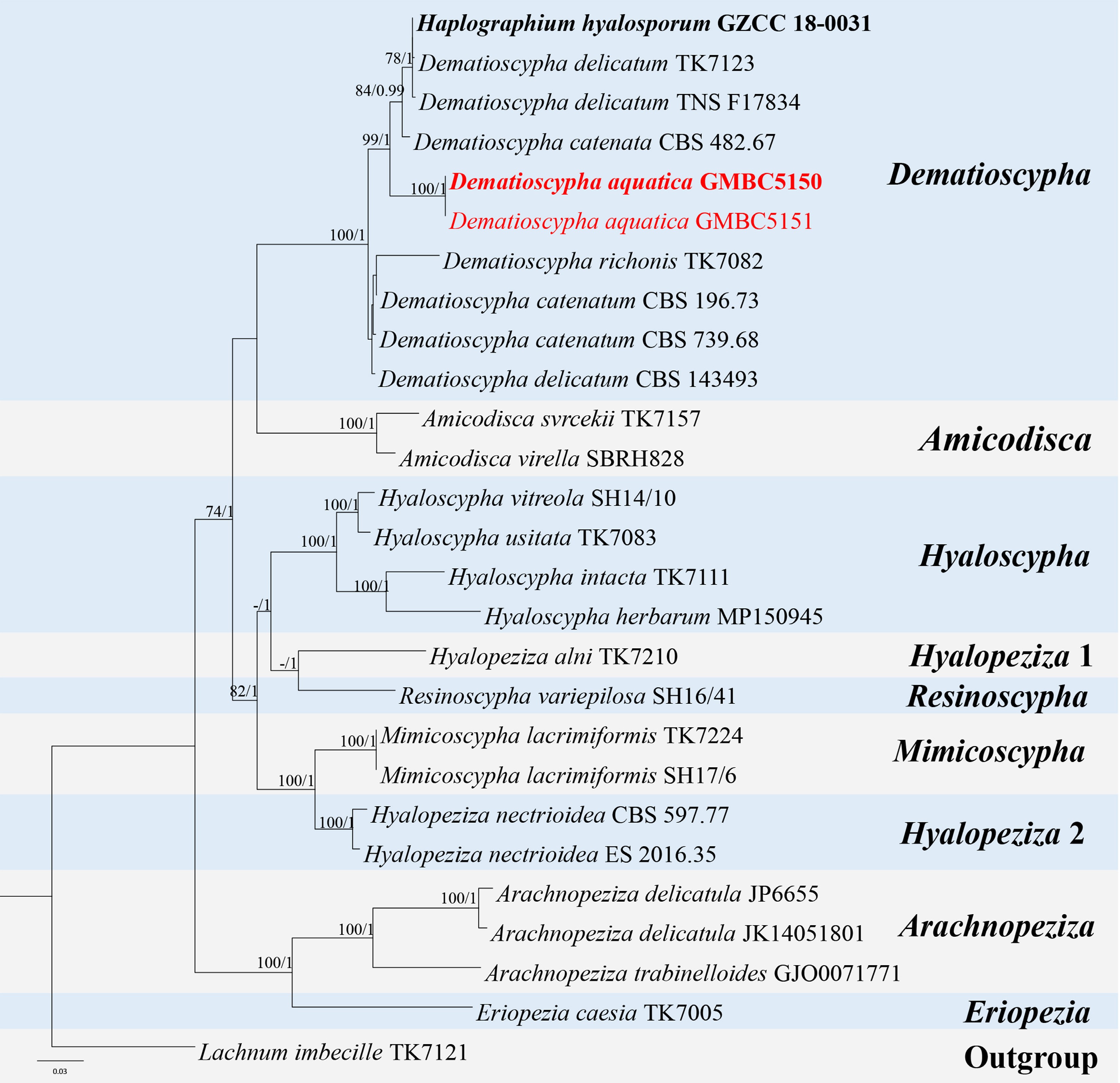
Figure 18.
ML tree of Dematioscypha and related genera inferred from a combined ITS-LSU-tef1-α-rpb2 dataset. The combined dataset comprised 27 taxa, 3,365 characters (ITS: 1–547 bp; LSU: 548–1,493 bp; tef1-α: 1,394–2,313 bp; rpb2: 2,314–3,365 bp) including indel regions. Maximum likelihood (lnL= −16,013.685762) and Bayesian analyses produced consistent tree topologies. Lachnum imbecille (TK7121) was selected as the outgroup taxon. Bayesian posterior probabilities (PP) ≥ 0.90 and RAxML bootstrap support values ≥ 70 % are shown on the branches. The newly described species is marked in red. Ex-type/type strains are indicated in bold.
Based on these distinct morphological and molecular differences, together with its placement in a separate clade in the phylogenetic analysis, Dematioscypha aquatica is introduced as a new species.
Mollisiaceae Rehm [as 'Mollisieae'], Rabenh. Krypt.-Fl., Edn 2 (Leipzig) 1.3(lief. 35): 503 (1891) [1896].
Notes – Mollisiaceae comprises saprobic fungi that occur on decaying wood, bark, leaves, and herbaceous debris, with some taxa reported as endophytes or weak pathogens, and is widely distributed in terrestrial ecosystems[82,83]. Members of the family are characterized by small, discoid to cupulate apothecia, unitunicate, inoperculate asci with an amyloid or hemiamyloid apical apparatus, hyaline, aseptate to septate ascospores, and filiform paraphyses; asexual morphs, when present, are coelomycetous or hyphomycetous[83]. Although traditionally delimited based on morphology, multigene phylogenetic studies have revealed that Mollisiaceae is heterogeneous, highlighting the need for taxonomic revisions and a more refined circumscription of the family within Helotiales[1,81]. In the present study, a new genus, Aquaclavispora, is placed in Mollisiaceae based on both phylogenetic evidence and morphological characteristics, expanding the ecological and morphological diversity currently recognized within the family.
Aquaclavispora L.L. Liu, W.M. Li, & Q.R. Li, gen. nov.
MycoBank number: MB863031
Etymology – Derived from Latin aqua (= water), referring to its aquatic habitat, and clavispora (from clavis = club, spora = spore), referring to the club-shaped conidia.
Saprobic on submerged decaying wood in a freshwater habitat. Sexual morph unknown. Asexual morph hyphomycetous. Colonies effuse, dark brown to black. Conidiophores mononematous, macronematous, solitary, erect to slightly flexuous, cylindrical, dark brown to black, septate. Conidiogenous cells integrated, terminal or intercalary, monoblastic, determinate, loci inconspicuous. Conidia acrogenous, dry, initially in threes, apically slightly connate, becoming divergent at maturity; straight to curved, smooth, thick-walled, obclavate to cylindrical, septate, dark brown to olivaceous brown; cells differentiated, apical cells narrower, basal cell truncate; apex rounded to obtuse, occasionally with a cup-shaped apical sheath, upper cell hyaline.
Type species – Aquaclavispora lignicola L.L. Liu, W.M. Li, & Q.R. Li
Notes – Phylogenetic analyses resolve Aquaclavispora in a well-supported, distinct lineage within the Mollisiaceae, sister to a clade containing Trimmatostroma, Cystodendron, and Mollisia (Fig. 19). However, it can be clearly differentiated based on conidiophore and conidial morphology. Unlike Trimmatostroma, which possesses micronematous conidiophores and produces catenate conidia, Aquaclavispora has macronematous, septate conidiophores and solitary, acrogenous conidia. with apical sheath[69,84]. Aquaclavispora differs from Cystodendron in lacking stromatic tissues and chains of conidia, and in producing larger, obclavate to cylindrical, multiseptate conidia[83]. The genus Mollisia is primarily defined by its sexual morph, and its asexual states are rarely observed in nature, typically developing only after prolonged incubation in culture and characterized by phialocephala-like conidiophores, with phialides producing dimorphic, catenate, hyaline conidia[83].
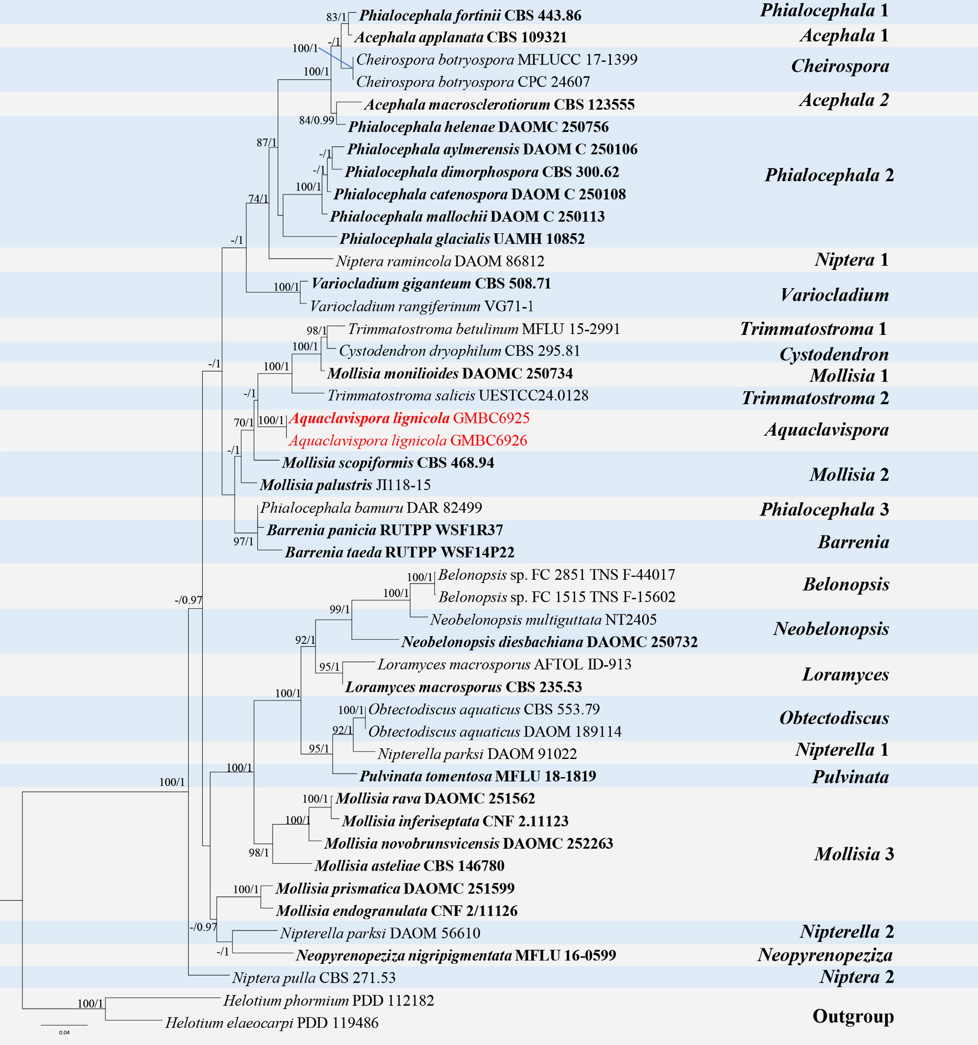
Figure 19.
ML tree of selected Mollisiaceae species based on the ITS-LSU dataset. The combined dataset comprised 43 taxa, 1,398 characters (ITS: 1–551 bp; LSU: 552–1,398 bp), including indel regions. Maximum likelihood (lnL = −8,494.851348) and Bayesian analyses produced consistent tree topologies. Helotium elaeocarpi (PDD 119486) and H. phormium (PDD 112182) were selected as the outgroup taxa. Bayesian posterior probabilities (PP) ≥ 0.90 and RAxML bootstrap support values ≥ 70 % are shown on the branches. Novel species are indicated in red and ex-type/type strains in bold.
Based on these distinct morphological differences from closely related genera, together with its phylogenetic placement, Aquaclavispora is herein introduced as a new genus within Mollisiaceae.
Aquaclavispora lignicola L.L. Liu, W.M. Li, & Q.R. Li, sp. nov. (Fig. 20)
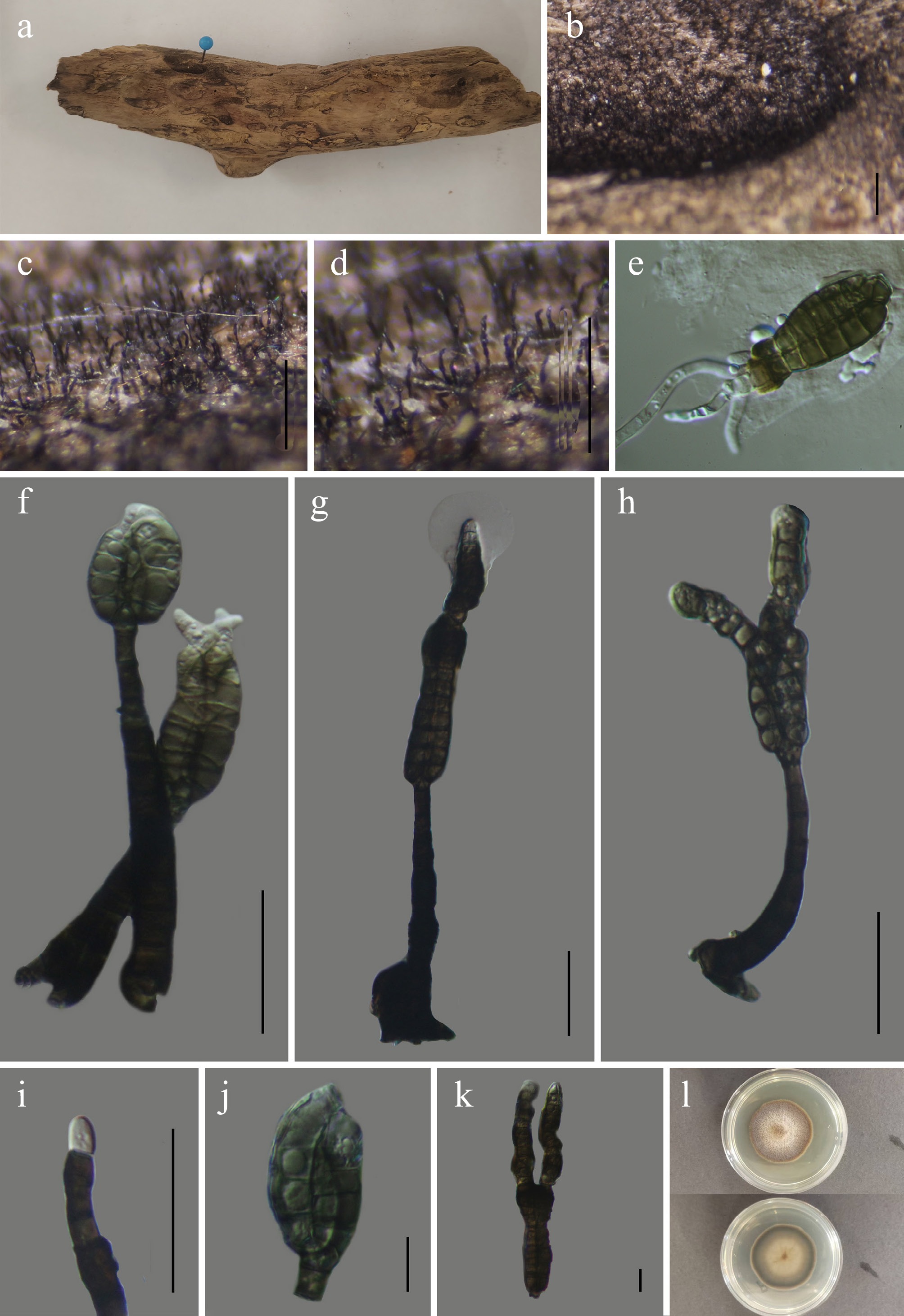
Figure 20.
Aquaclavispora lignicola (GMB6925 holotype). (a) Host. (b)–(d) Colonies on natural substrate. (e) Germinating conidium. (f)–(h) Conidiophores and conidia. (i) Conidiogenous cells with conidia. (j) immature conidia. (k) Mature conidia. (l) Surface and reverse view of culture on PDA. Scale bars: b = 1 mm, c, d = 0.5 mm, f–i = 30 μm, e, j–k = 10 μm.
MycoBank number: MB863032
Etymology – The epithet refers to its occurrence on submerged decaying wood.
Saprobic on submerged decaying wood in a freshwater swamp. Sexual morph: not observed. Asexual morph: hyphomycetous. Colonies on natural substrate, effuse, dark brown to black, velvety. Mycelium partly immersed, partly superficial; hyphae dark brown, thick-walled, septate, branched, smooth to slightly roughened. Conidiophores 44.3–89.5 × 5.9–9.4 µm (x = 66.3 × 7.7 µm, n = 30), mononematous, macronematous, erect to slightly flexuous, solitary, unbranched or rarely branched, cylindrical, thick-walled, dark brown to black, smooth, 5–9-septate, basal swollen, apex slightly attenuated. Conidiogenous cells 4–8.7 × 3.3–6.5 µm (x = 6.5 × 4.7 µm, n = 30) integrated, terminal or intercalary, monoblastic, determinate, cylindrical to doliiform, dark brown, smooth, conidiogenous loci inconspicuous. Conidia aggregated, three slightly connate at the apex, borne on conidiogenous cells, occasionally diverging from the base at maturity; initially three young conidia aggregated subglobosely; at maturity, conidia apically elongate and become divergent apically, forming curved, robustly elongate structures; acrogenous, dry, obclavate to cylindrical, straight to curved, apex rounded to obtuse, base truncate to slightly obconic, smooth, thick-walled, 4–12-septate, 30–150.3 × 9.7–12.4 µm (x = 93.8 × 12.4 µm, n = 30) dark brown to olivaceous brown, cells clearly differentiated, apical cells narrower, basal cell truncate, with cup shaped apical sheath.
Culture characteristics – Conidia germinate within 24 h on PDA. After 40 d at 25 °C, colonies on PDA reach 20–30 mm in diameter; circular, entire; surface flat to slightly raised, velvety to felty; pale brown to dark brown; reverse pale brown to dark brown.
Material examined – CHINA, Guizhou Province, Tongren City, Fodingshan National Nature Reserve, 27°23′0.56″ N, 108°8′30.31″ E, elevation 1,179 m, on submerged decaying wood in a freshwater swamp, 10 August 2024, Wen-Mei Li, 2024FDSDCY19-2 (GMB6925, holotype; GMBC6925, ex-type); ibid., KUN-HKAS 152899, isotype.
Additional examined specimens – CHINA, Guizhou Province, Tongren City, Fodingshan National Nature Reserve, 27°23′0.78″ N, 108°7′31.06″ E, elevation 1,256 m, on submerged decaying wood in a freshwater swamp, 10 August 2024, Wen-Mei Li, 2024FDSDCY20 (GMB6926, GMBC6926).
Notes – Aquaclavispora lignicola can be differentiated from the phylogenetically closely related genera Trimmatostroma, Cystodendron, and Mollisia by its macronematous conidiophore, monoblastic and three connate, acrogenous conidia with apical sheath, whereas the other genera possess micronematous conidiophores with polyblastic conidiogenous cells and produce catenate conidia[69,84].
Based on these morphological differences from the closely related genera and distinct phylogenetic placement, A. lignicola is herein introduced as a novel species.
Strossmayeria Schulzer, Oesterr. Bot. Z. 31 (10): 313 (1881).
Notes – Strossmayeria was established by Schulzer, typified by Strossmayeria rackii[85]. Species of the genus are saprobic and are distributed in both temperate and tropical regions, occurring mainly on dead plant materials, particularly woody substrates. Members of Strossmayeria have been documented from a wide range of host plants, including bamboo, palms, and various hardwood trees, and are known from Europe, North America, Central America, and Oceania, indicating a broad geographical distribution[71,85−87]. According to Species Fungorum (accessed 2 January 2026), the genus currently comprises 25 accepted species.
Strossmayeria multiseptata L.L. Liu, Q.F. Zhang, & Q.R. Li, sp. nov. (Fig. 21)
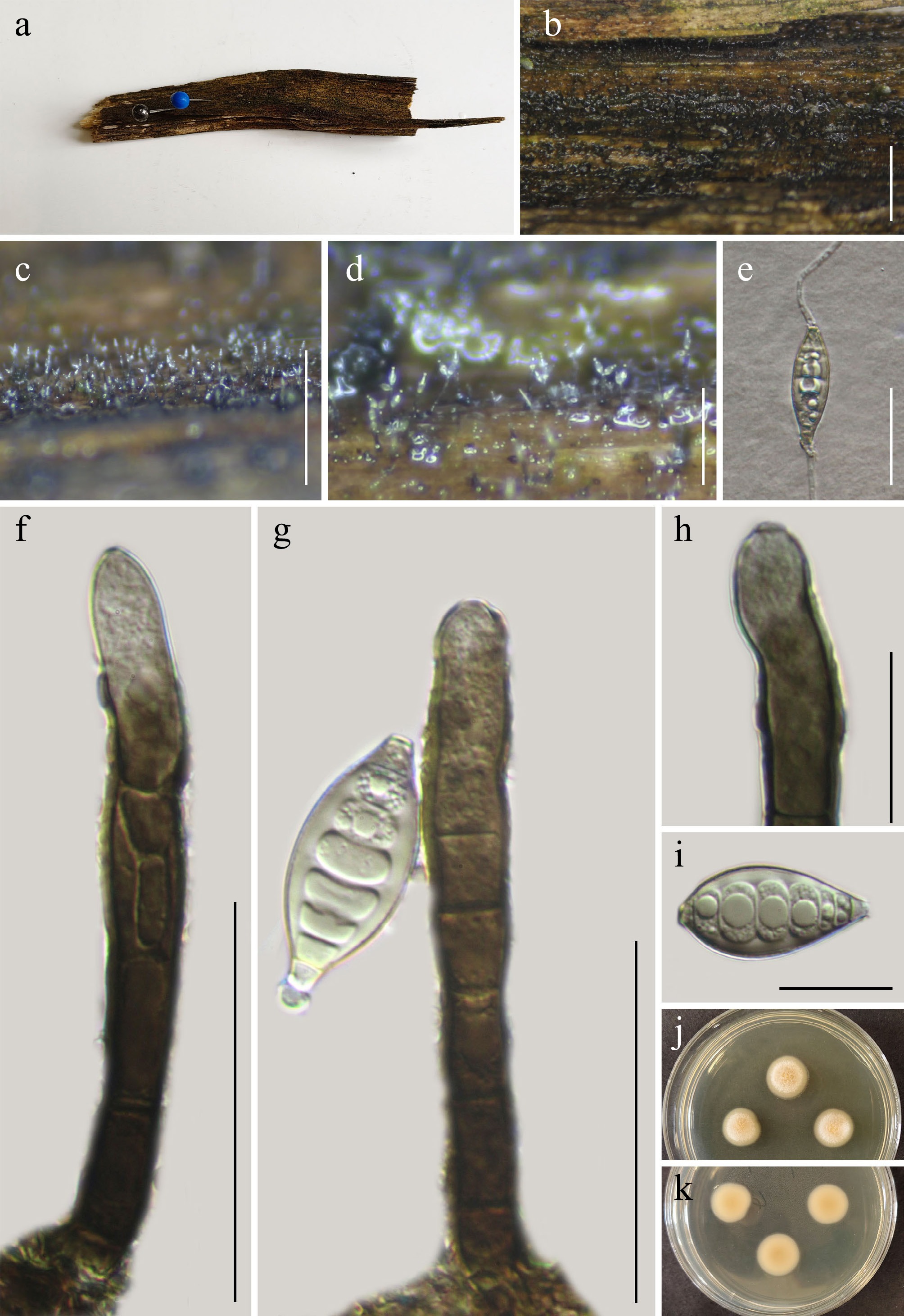
Figure 21.
Strossmayeria multiseptata (GMB5166 holotype). (a) Host. (b)–(d) Colonies on natural substrate. (e) Germinating conidium. (f), (g) Conidiophores and conidia. (h) Conidiogenous cells. (i) Conidia. (j), (k) Surface and reverse view of culture on PDA. Scale bars: b = 3 mm, c =1 mm, d = 0.3 mm, e = 30 μm, f, g = 50 μm, h, i= 20 μm.
MycoBank number: MB863028
Etymology – The specific epithet refers to its multisptate conidia.
Saprobic on submerged decaying wood in a freshwater wetland. Sexual morph: not observed. Asexual morph: hyphomycetous. Colonies are dark brown to black, effuse, and hairy. Mycelium mostly immersed, composed of brown, smooth- to slightly rough and thick-walled, hyphae. Conidiophores 66.7–117.2 × 7.1–10.9 μm (x = 90.6 × 8.9 μm, n = 15), mononematous, macronematous, solitary to loosely fasciculate, erect, straight to slightly flexuous, cylindrical, septate with 5–8 septa, unbranched or rarely branched, brown to dark brown, paler toward the apex, surface smooth to finely verrucose. Conidiogenous cells 20.4–41.9 × 6.9–10.8 μm (x = 31.5 × 8.8 μm, n = 15), polyblastic, holoblastic, integrated, terminal or intercalary, indeterminate, pale brown to brown, cylindrical to subcylindrical, with percurrent proliferations, conidiogenous loci inconspicuous to slightly prominent. Conidia 24.6–41.5 × 12.7–18.3 μm (x = 36.5 × 15.1 μm, n = 30), solitary, dry, acropleurogenous, pale olivaceous to pale brown, 4–7 pseudoseptate, broad fusiform to ellipsoidal, smooth-walled, with a mucilaginous cap at the apex, secession schizolytic.
Culture characteristics – Conidia germinating on PDA within 24 h. After 21 d at 25 °C, colonies on PDA reach 15–25 mm in diameter; circular, raised, with dense mycelium, fluffy, margins entire, surface grayish white; reverse light brown.
Material examined – CHINA, Yunnan Province, Yiliang County, Haiziping, 27°36′07.45″ N, 104°15′54.32″ E, elevation 2,150 m, on submerged decaying wood in a freshwater wetland, 18 March 2025, Qin-Fang Zhang, 2025HZP16-1 (GMB5166, holotype; GMBC5166, ex-type); ibid., KUN-HKAS 152900, isotype.
Additional specimens examined – CHINA, Yunnan Province, Yiliang County, Haiziping, 27°37′32.45″ N, 104°16′54.32″ E, elevation 2,250 m, on submerged decaying wood in a freshwater wetland, 18 March 2025, Qin-Fang Zhang, 2025HZP93 (GMB5167, GMBC5167).
Notes – In the phylogenetic analysis (Fig. 22), Strossmayeria multiseptata forms an independent lineage in a clade containing S. narathiwatensis, S. basitricha, and S. bakeriana. The latter two species are known only from their sexual morphs, whereas S. multiseptata is represented solely by a hyphomycetous morph, making direct morphological comparison impossible. Morphologically, S. multiseptata resembles S. narathiwatensis, sharing a hyphomycetous asexual morph, effuse and hairy colonies, macronematous and mononematous conidiophores, holoblastic and polyblastic conidiogenous cells with percurrent proliferations, and pale olivaceous to pale brown, fusiform conidia. However, S. multiseptata can be distinguished from S. narathiwatensis by having much shorter conidiophores (66.7–117.26–10 vs 7–8 septa μm vs up to 577 μm) and much larger conidia (24.6–41.5 × 12.7–18.3 μm vs 15–30 × 8–12 μm)[71].
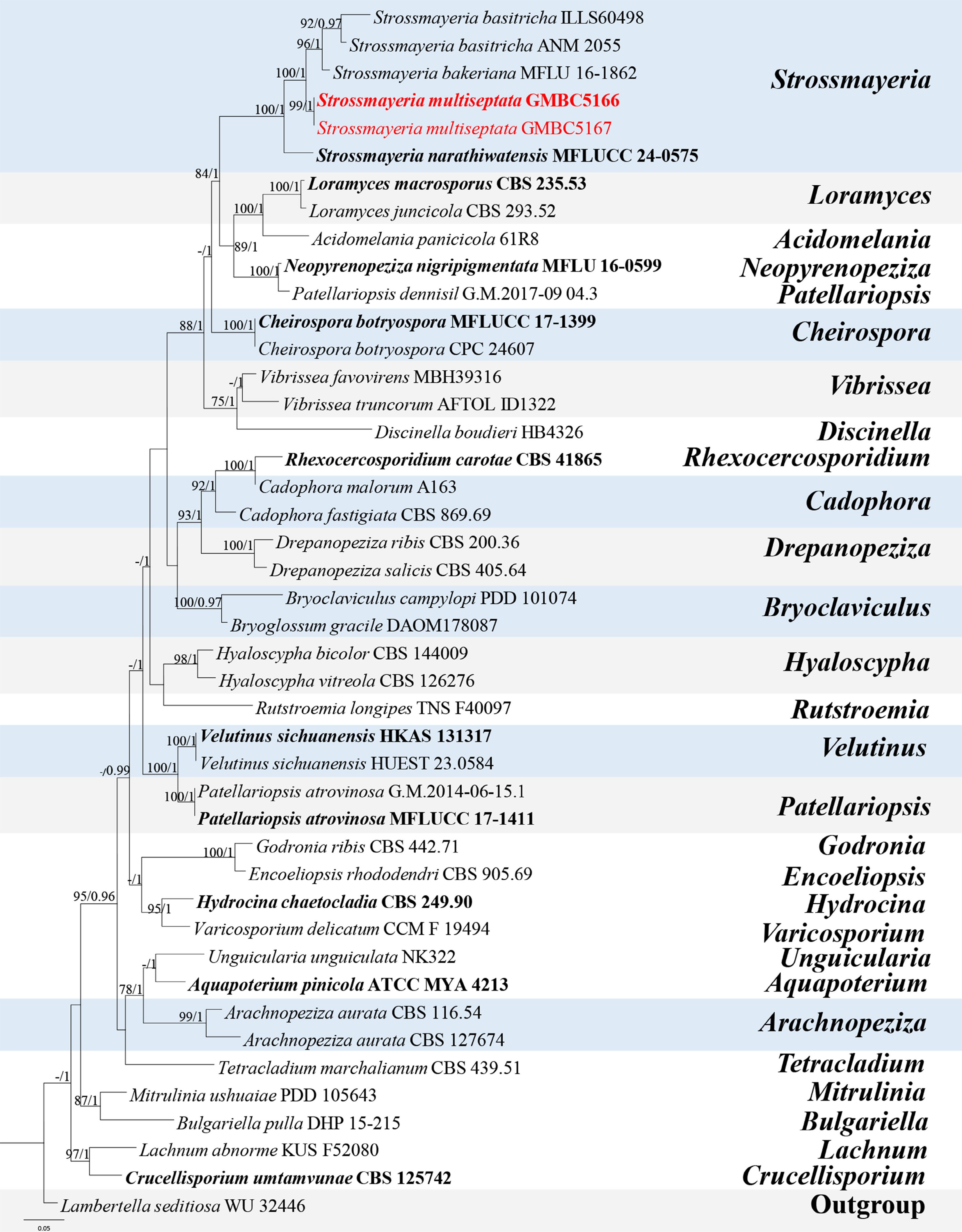
Figure 22.
ML tree of selected species of Strossmayeria and related genera inferred from a combined ITS-LSU dataset. The combined dataset comprised 44 taxa, 1,447 aligned sites (ITS:1–542 bp; LSU: 543–1,447 bp) including indel regions. Maximum likelihood (lnL = −11,968.462866) and Bayesian analyses produced consistent tree topologies. Lambertella seditiosa (WU 32446) was selected as the outgroup taxon. Bayesian posterior probabilities (PP) ≥ 0.90 and RAxML bootstrap support values ≥ 70 % are shown on the branches. The newly described species is marked in red. Ex-type/type strains are indicated in bold.
Comparative analysis of nucleotide base pairs showed that S. multiseptata differs from the type strain of S. narathiwatensis (MFLUCC 24-0575) by 10.8% (51/474 bp) in the ITS locus and 1.4% (12/840 bp) in the LSU locus.
Based on its distinct phylogenetic placement and clear morphological differences from closely related taxa, S. multiseptata is herein described as a new species.
Orbiliomycetes O.E. Erikss. & Baral, Myconet 9: 96 (2003).
Orbiliales Baral, O.E. Erikss., G. Marson & E. Weber, Myconet 9: 96 (2003).
Arthrobotryaceae Corda [as 'Arthrobotrydeae'], Icon. fung. (Prague) 5: 14 (1842).
Arthrobotrys Corda, Pracht-Fl. Eur. Schimmelbild. (Leipzig und Dresden): 43 (1839).
Notes – Arthrobotrys was established by Corda, typified by A. superba[1,88,89]. Species of Arthrobotrys are widely distributed worldwide and primarily occur in soil or sediment across a broad range of ecosystems, including farmland, forests, mangroves, and freshwater habitats. They have also been reported from hot springs, animal waste, and decaying wood or tree trunks[17,90−93]. The genus is characterized by branched or simple conidiophores bearing elliptic to pyriform, obovoid conidia that are 0–3-septate and produced asynchronously on nodes or short denticles of the conidiophores[94,95]. Arthrobotrys is regarded as the largest and most morphologically complex genus of nematode trapping fungi in Orbiliaceae and is distinguished by the production of adhesive networks used to capture nematodes. According to Species Fungorum (accessed 25 March 2026), the genus currently comprises 72 accepted species. Herein, a new species of Arthrobotrys is described based on a combination of morphological characteristics and phylogenetic analyses.
Arthrobotrys bambusicola L.L. Liu, W.M. Li, & Q.R. Li, sp. nov. (Fig. 23)
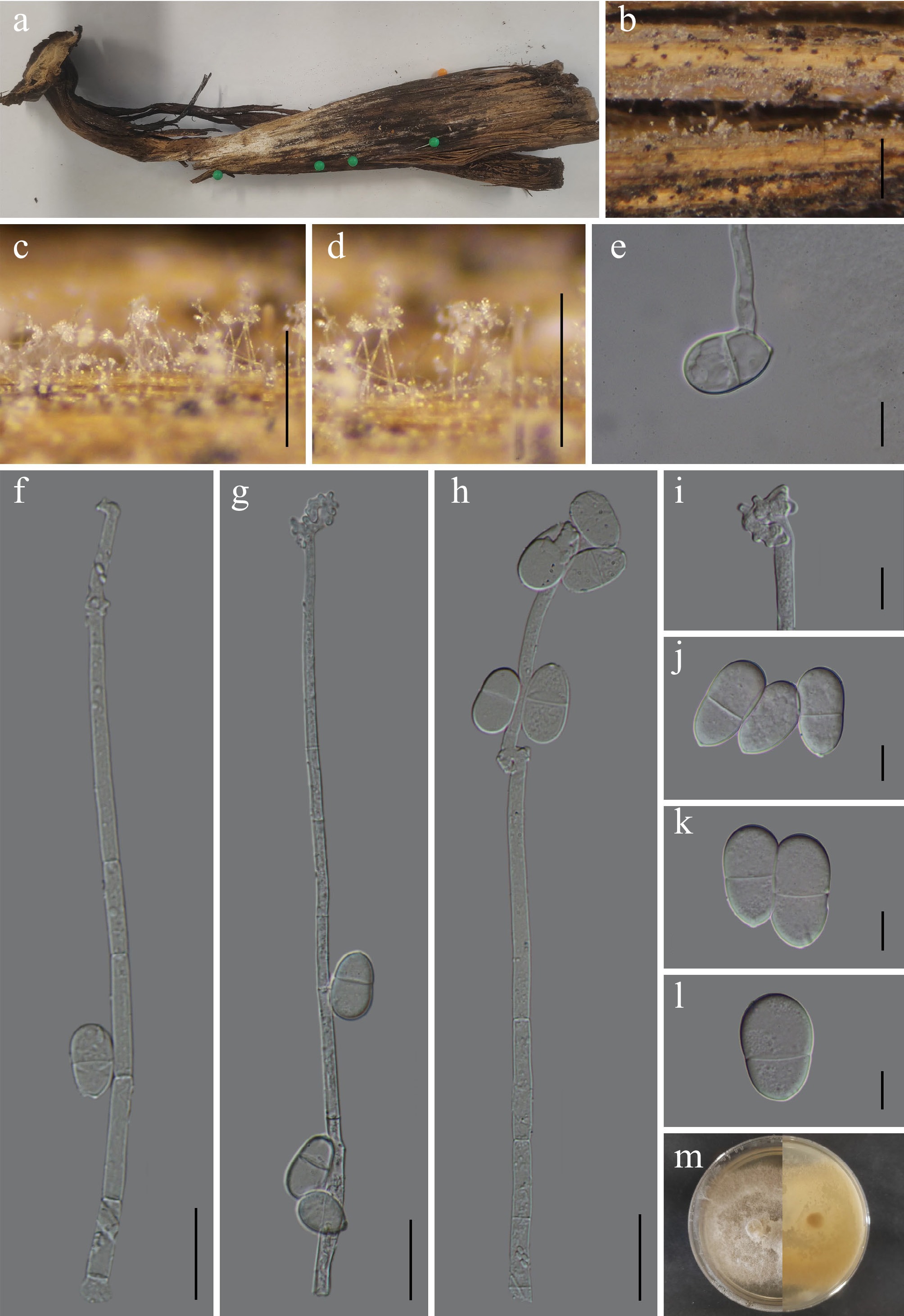
Figure 23.
Arthrobotrys bambusicola (GMB6933 holotype). (a) Host. (b)–(d) Colonies on natural substrate. (e) Germinating conidium. (f)–(h) Conidiophores and conidia. (i) Conidiogenous cells. (j)–(l) Conidia. (m) Surface and reverse view of culture on PDA. Scale bars: b = 1 mm, c, d = 0.5 mm, f– h = 30 μm, e, i–l = 10 μm.
MycoBank No: MB862959
Etymology –The epithet refers to its occurrence on bamboo.
Saprobic on dead bamboo culm in terrestrial habitats. Sexual morph: not observed. Asexual morph: hyphomycetous. Colonies effuse, superficial, hyaline, producing conspicuous whitish conidial masses at the apices of conidiophores. Mycelium a partly immersed, partly superficial, formed by branched, smooth-walled, hyaline, septate hyphae. Conidiophores 194–319 µm long, 3.5–7.1 µm wide at the base (x = 241.4 × 4.9 µm, n = 30), mononematous, solitary, macronematous, erect, straight to slightly flexuous, unbranched, cylindrical, septate, smooth-walled, hyaline. Conidiogenous cells integrated, polyblastic, formed by repeated elongation of the conidiophore, producing several discrete nodes; denticles polyblastic, short, hyaline. Conidia 23.4–28.3 × 12.1–16.3 µm (x = 25.8 × 14.2 µm, n = 30), solitary, 1-septate, septum median, rarely slightly constricted at the septum, broadly ellipsoid to pyriform, apex rounded, base narrowed, sometimes truncate, smooth-walled, guttulate, hyaline.
Culture characteristics – Conidia germinate on PDA within 24 h. After 14 d at 25 °C, colonies on PDA reach 20–30 mm in diameter; circular, raised, cottony to floccose, fluffy, margins entire, surface pale grey to whitish; reverse light brown.
Material examined – CHINA, Guizhou Province, Guiyang City, Panlong Mountain Park, 26°43′39.88″ N, 106°49′46.76″ E, elevation 1,267 m, on dead bamboo culm, 24 May 2025, Wen-Mei Li, 2025GY33-2 (GMB6933, holotype; GMBC6933, ex-type); ibid., KUN-HKAS 152901, isotype.
Additional examined specimens – CHINA, Guizhou Province, Guiyang City, Panlong Mountain Park, 26°43′41.76″ N, 106°49′49.73″ E, elevation 1,310 m, on dead bamboo culm, 24 May 2025, Wen-Mei Li, 2025GY57 (GMB6934, GMBC6934).
Notes – In the phylogenetic analysis (Fig. 24), Arthrobotrys bambusicola forms a well-supported sister clade (BS = 92%, PP = 0.98) with the type strain of A. hyrcanus (IRAN 3650C). Comparative nucleotide analysis shows that A. bambusicola (GMBC6933) differs from A. hyrcanus (IRAN 3650C) by 4.7% (24/507 bp) in the ITS region.
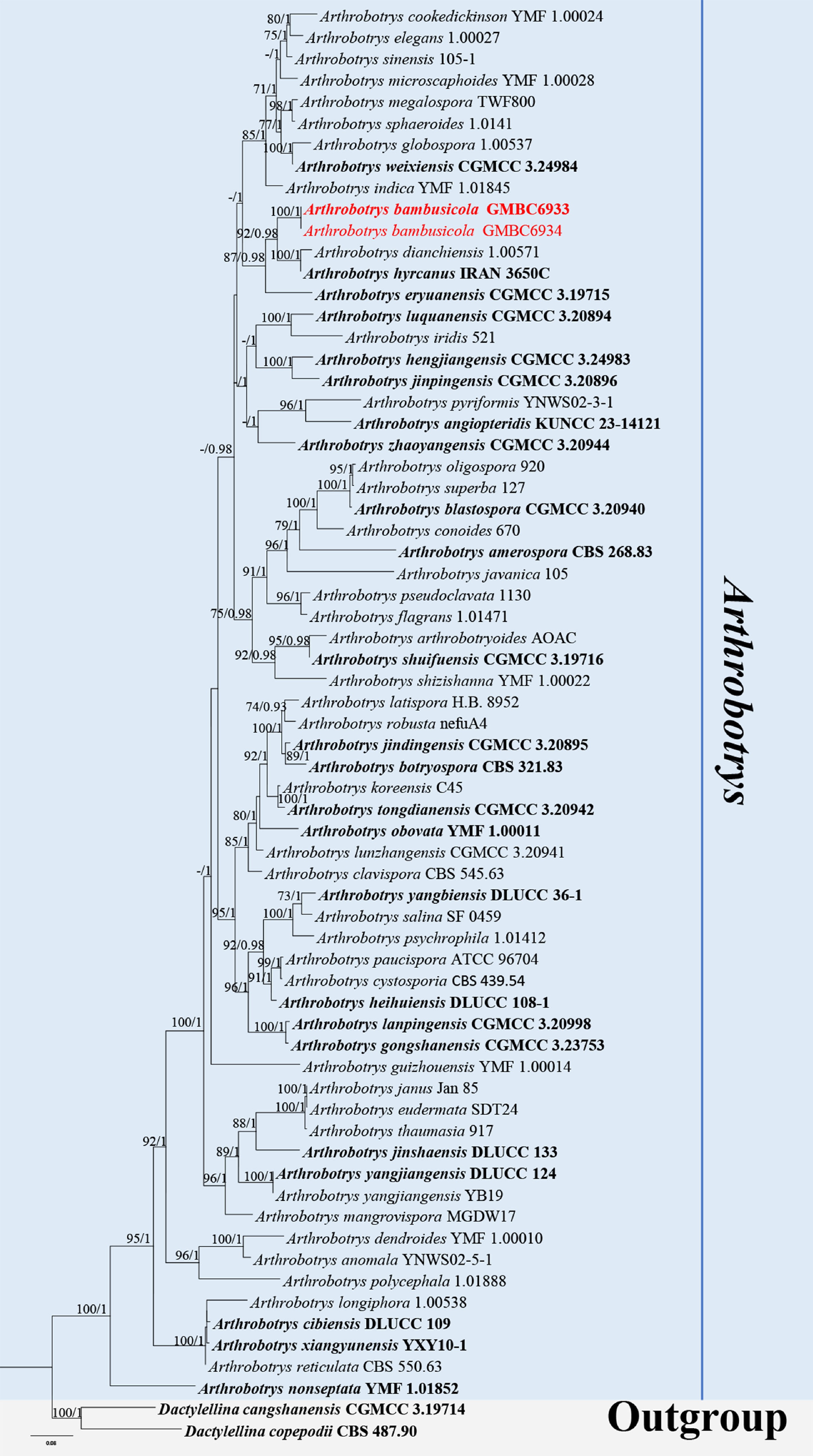
Figure 24.
ML tree of selected Arthrobotrys species based on the ITS -tef1-α-rpb2 dataset. The combined dataset comprised 67 taxa, 2,405 characters (ITS:1–613 bp; tef1-α: 614–1,554 bp; rpb2: 1,555–2,405 bp), including indel regions. Maximum likelihood (lnL = −29,133.876958) and Bayesian analyses produced consistent tree topologies. Dactylellina cangshanensis (CGMCC 3.19714) and D. copepodii (CBS 487.90) were selected as the outgroup taxa. Bayesian posterior probabilities (PP) ≥ 0.90 and RAxML bootstrap support values ≥ 70 % are shown on the branches. The newly described species is marked in red. Ex-type/type strains are indicated in bold.
Morphologically, A. bambusicola differs from A. hyrcanus (asexual morph is known only from culture) in having shorter conidiophores (194–319 µm vs 70–312 µm) and producing solitary, broadly ellipsoid to pyriform, 1-septate conidia (23.4–28.3 × 12.1–16.3 µm). In contrast, A. hyrcanus is characterized by larger, clavate to spindle-shaped conidia that are predominantly multi-septate (2–9 septa; 44.2–135.2 × 10–14.4 µm), and by the presence of secondary conidiophores, secondary conidia, and chlamydospores, which were not observed in A. bambusicola. Ecologically, A. bambusicola was collected on dead bamboo, whereas A. hyrcanus has been reported from decaying leaves[96].
Based on these distinct morphological characters and its well-supported phylogenetic placement, A. bambusicola is described here as a new species.
Sordariomycetes O.E. Erikss. & Winka, Myconet 1(1): 10 (1997).
Chaetosphaeriales Huhndorf, A.N. Mill. & F.A. Fernández, Mycologia 96(2): 378 (2004).
Australiascaceae Réblová & W. Gams, Stud. Mycol. 68: 171 (2011).
Monilochaetes Halst. ex Harter, J. Agric. Res., Washington 5: 791 (1916).
Notes – Monilochaetes was established by Harter, typified by M. infuscans[97]. The sexual morph is known in a few species and is characterized by dark brown, obpyriform, superficial ascomata, ostioles periphysate, cylindrical to clavate, short-pedicellate, unitunicate asci, and 0–3-septe hyaline ascospores[98]. The asexual morph is hyphomycetous and dimorphic, with mononematous, macronematous, conidiophores, conidiogenous cells monophialidic, with a shallow collarette, and cylindrical to ellipsoid, aseptate or rarely septate, hyaline conidia[99,100]. Species of Monilochaetes are cosmopolitan, occurring as endophytes, pathogens, or saprobes on diverse hosts, with a notable preference for ferns, especially tree ferns (Cyatheaceae)[16,99,101]. According to Species Fungorum (accessed 2 January 2026), the genus comprised 10 accepted species. In this study, we collected M. camelliae from a freshwater habitat, representing a new record for China.
Monilochaetes camelliae (Alcorn & Sivan.) Réblová, W. Gams & Seifert, Stud. Mycol. 68: 175 (2011). (Fig. 25)
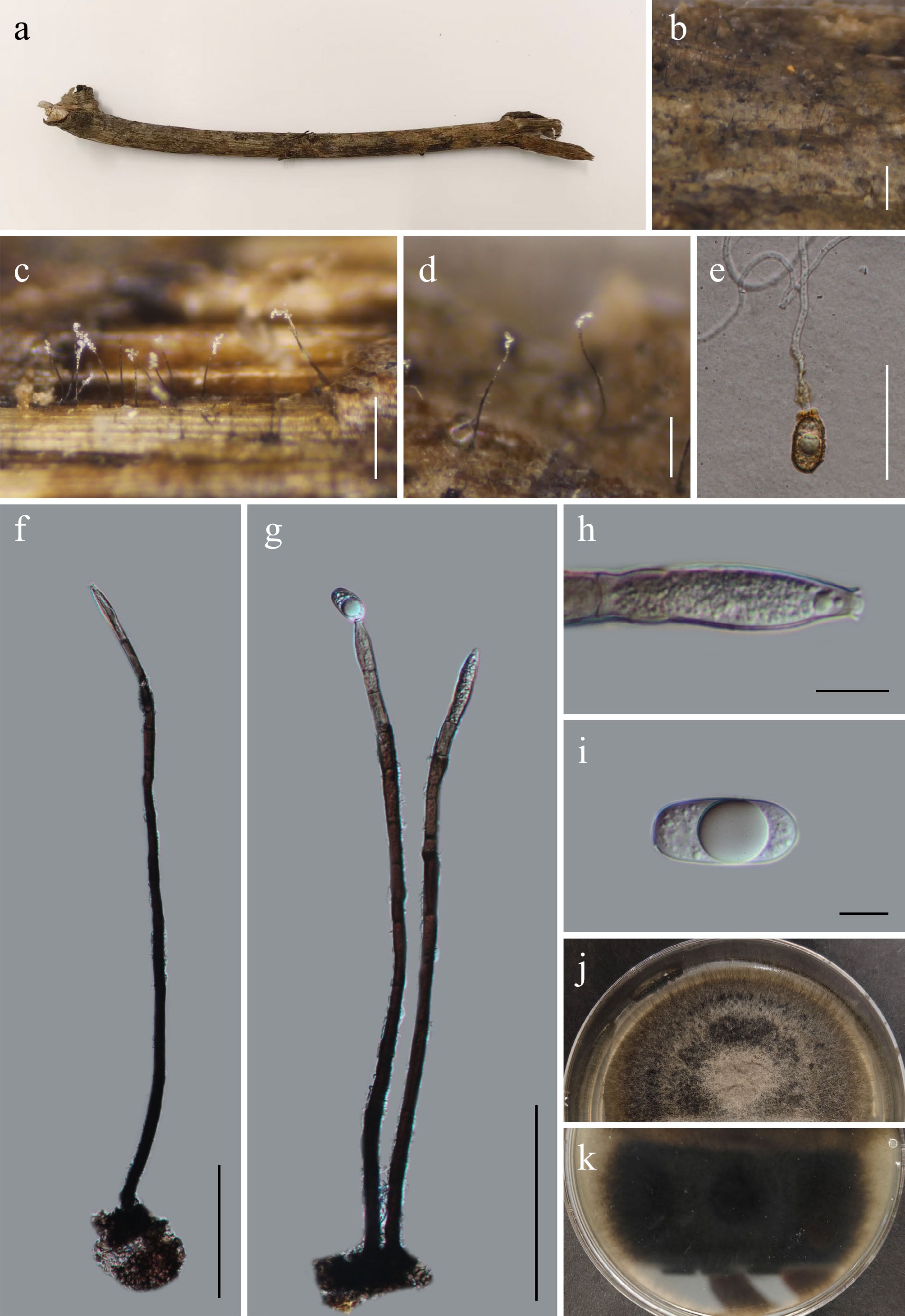
Figure 25.
Monilochaetes camelliae (GMB5170). (a) Host. (b)–(d) Colony on natural substrate. (e) Germinating conidium. (f), (g) Conidiophores and conidia. (h) Conidiogenous cells. (i) Conidium. (j), (k) Surface and reverse view of culture on PDA. Scale bars: b = 1 mm, c = 0.5 mm, d = 0.25 mm, e = 50 μm, f, g = 100 μm, h, i = 10 μm.
MycoBank: MB518385
Basionym: Dischloridium camelliae Sivan. & Alcorn, Aust. Syst. Bot. 15: 743 (2002).
Sexual morph: Australiasca queenslandica Sivan. & Alcorn, Aust. Syst. Bot. 15(5): 742 (2002).
Saprobic on submerged dead branch along the riverbank. Sexual morph: not observed. Asexual morph: hyphomycetous. Colonies scattered to gregarious, superficial, composed of erect, dark brown to black conidiophores arising singly or in small groups from the host surface, giving a hairy appearance. Mycelium rarely superficial, mostly immersed, formed by hyaline to pale brown, septate, smooth-walled hyphae. Conidiophores 251–520 × 5.4–14.4 µm (x = 390.6 × 9.7 µm, n = 30), mononematous, macronematous, straight to slightly flexuous erect, pale brown to dark brown, gradually becoming subhyaline towards the apex, simple, smooth, and thick-walled at base, multiseptated, basal cell slightly swollen, firmly attached to substrate, apical region possesses a single terminal conidiogenous cell. Conidiogenous cells 38–82 × 3.8–11.6 µm (x = 53.7 × 6.7 µm, n = 30), monophialidic, terminal, integrated, ampulliform to cylindrical, slightly swollen at mid-region, tapering gradually towards apex, subhyaline to pale brown, smooth-walled, collarette conspicuous, flared, cup-shaped, hyaline. Conidia 20–29.5 × 8.7–16 µm (x = 25.2 × 13.3 µm, n = 30), solitary, hyaline, smooth-walled, 0–1-septate, ellipsoidal to broadly cylindrical-ellipsoidal; apex broadly rounded, guttulate, often containing a large central guttule.
Culture characteristics – Conidia germinate on PDA within 24 h. After 21 d at 25 °C, colonies on PDA reach 40–55 mm in diameter; circular, margin entire, floccose to velvety, surface dark brown, with sparse white mycelium; reverse bluish black.
Material examined – CHINA, Yunnan Province, Xishuangbanna Dai Autonomous Prefecture, 24°02'24.48" N, 100°48'38.25" E, elevation 1,450 m, on submerged dead branch along the riverbank, 18 August 2025, Qin-Fang Zhang, 2025XSBN36 (GMB5170, GMBC5170).
Notes – In the phylogenetic analysis (Fig. 26), Monilochaetes camelliae GMBC5170 clusters with the reference strain M. camelliae BRIP 24607 with strong support (99% BS/1.0 PP), confirming their conspecific identity.
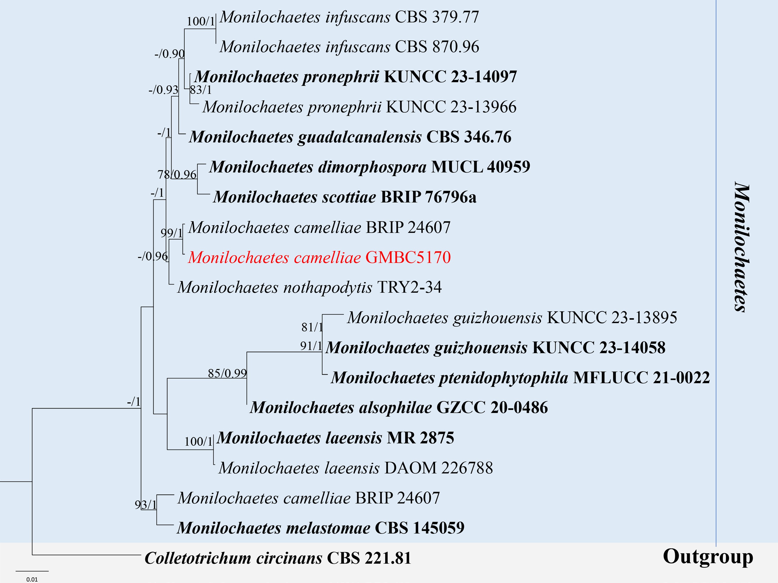
Figure 26.
ML tree of selected Monilochaetes species based on the ITS-LSU-SSU dataset. The combined dataset comprised 19 taxa, 2,435 characters (ITS: 1–505 bp; LSU: 506–1,400 bp; SSU: 1,401–2,435 bp), including indel regions. Maximum likelihood (lnL = −5292.494395) and Bayesian analyses produced consistent tree topologies. Colletotrichum circinans (CBS 221.81) was selected as the outgroup taxon. Bayesian posterior probabilities (PP) ≥ 0.90 and RAxML bootstrap support values ≥ 70 % are shown on the branches. The new record is marked in red. Ex-type/type strains are indicated in bold.
Morphologically, our collection GMBC5170 agrees with the type description of M. camelliae, except in the dimensions of conidiophores (251–520 µm long vs up to 420 µm long), conidiogenous cells (38–82 µm long vs 25–52 µm long), and conidia (20–29.5 × 8.7–16 µm vs 18–35 × 8–13 µm)[98].
Based on the phylogeny and molecular similarity, GMBC5170 is identified as M. camelliae and is reported here as a new record for China.
Chaetosphaeriaceae Locq., Mycol. gén. struct. (Paris): 174 (1984).
Notes – Chaetosphaeriaceae was introduced to accommodate Chaetosphaeria and its relatives[102]. The circumscription of the family has been considerably expanded through integrative morphological and molecular studies. Comprehensive treatments by Lin et al.[103] and Hyde et al.[103] recognized 43 genera within Chaetosphaeriaceae. Subsequent phylogenetic investigations have led to the introduction of additional genera, including Achrochaeta, Phialolunulospora, Arcuatospora, Flectospora, Phialoturbella, Tubulicolla, and Ericiosphaeria, and segregated multiple lineages from Catenularia- and Codinaea-like taxa[4,104]. Chaetosphaeriaceae currently comprises more than 108 genera[1]. In the present study, a new genus, Radiaticonidium, is proposed based on distinct morphological characteristics and phylogenetic evidence.
Chaetosphaeria Tul. & C. Tul., Select. fung. carpol. (Paris) 2: 252 (1863).
Notes – Chaetosphaeria was established by Tulasne, with C. innumera designated as the type species[105]. Members of the genus are characterized by black, smooth to setose ascomata, eight-spored asci with a distinct, refractive J- apical apparatus, and hyaline, septate ascospores. Asexual morphs are hyphomycetous, showing considerable variation in conidiophore morphology (mononematous or macronematous) and conidiogenous cells (polyphialidic or monophialidic), and conidia aseptate to multi-septate, sometimes with appendages or guttules[5,102,106]. Species of the genus are cosmopolitan saprobes, commonly occurring on dead plant material in both freshwater and terrestrial habitats[71,107,108]. Recent multigene phylogenetic studies have improved the understanding of species relationships within the genus and highlighted its considerable morphological diversity and ecological significance. In this study, a novel species of Chaetosphaeria is described.
Chaetosphaeria tongrenensis L.L. Liu, W.M. Li, & Q.R. Li, sp. nov. (Fig. 27)
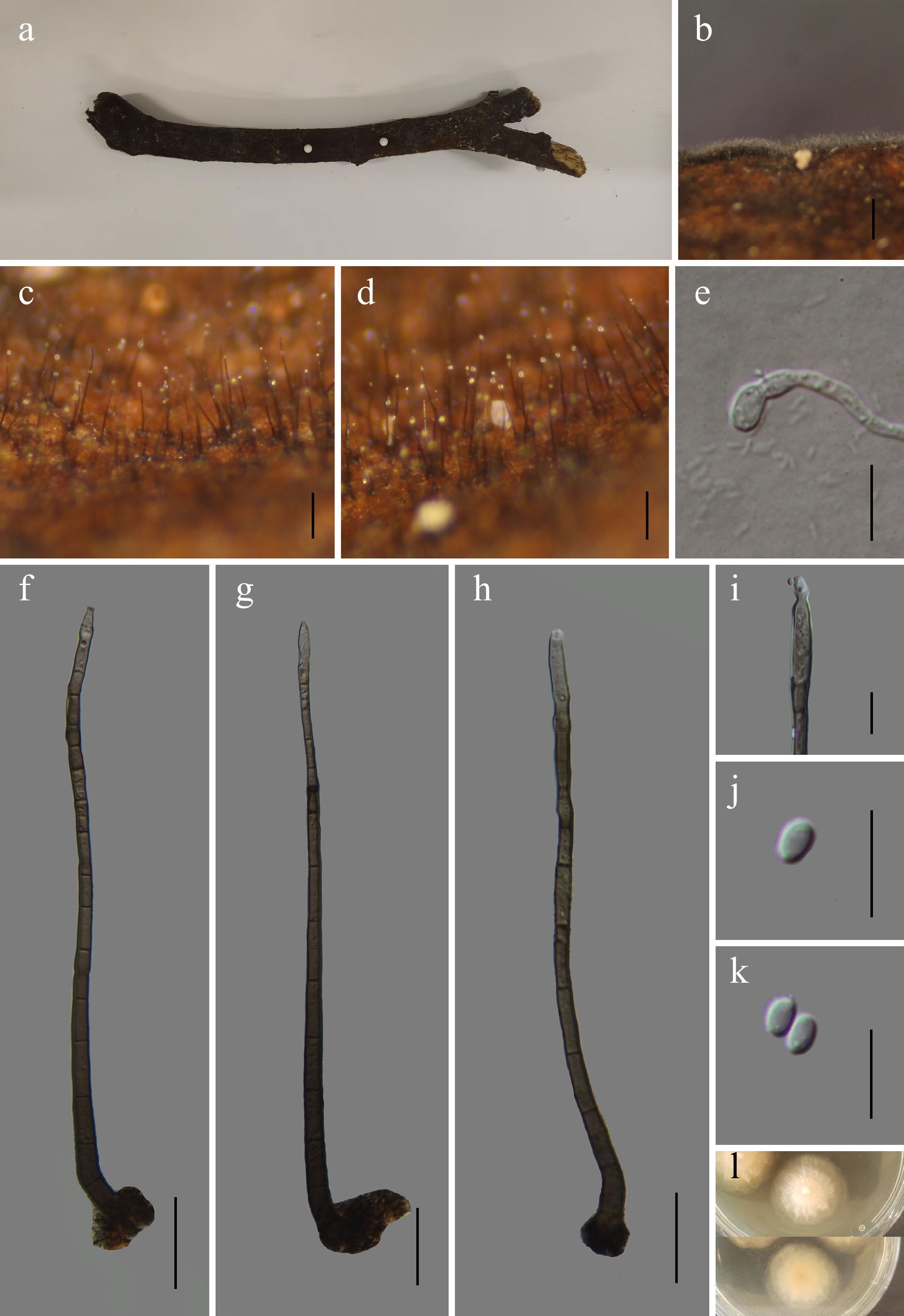
Figure 27.
Chaetosphaeria tongrenensis (GMB6921 holotype). (a) Host. (b)–(d) Colonies on natural substrate. (e) Germinating conidium. (f)–(h) Conidiophores and conidia. (i) Conidiogenous cells. (j), (k) Conidia. (l) Surface and reverse view of culture on PDA. Scale bars: b = 1 mm, c, d = 0.5 mm, f–h = 30 μm, e, i–k = 10 μm.
MycoBank number: MB862964
Etymology – The specific epithet tongrenensis refers to Tongren City, China, where the type specimen was collected.
Saprobic on submerged dead branches of an unknown plant in a lake. Sexual morph: not observed. Asexual morph: hyphomycetous. Colonies on natural substrate scattered to gregarious, appearing as minute dark brown to blackish dots, bearing conspicuous, erect, hair-like conidiophores; conidial masses inconspicuous or scanty. Mycelium rarely superficial, mostly immersed, composed of brown, smooth, thick-walled, septate hyphae. Conidiophores 107–249 × 3.6–6.7 µm (x = 172.8 × 5.13 µm, n = 30), mononematous, macronematous, solitary, straight to slightly curved, unbranched, cylindrical, 9–15-septate, smooth, thick-walled, dark brown to brown, becoming paler towards the apex, basal cell slightly swollen or irregularly enlarged, firmly attached to the substrate; apex rounded to slightly tapered. Conidiogenous cells 16.2–29.9 × 3.2–6.3 µm (x = 23.23 × 4.17 µm, n = 30), terminal, integrated, monophialidic, cylindrical to slightly lageniform, smooth, thin- to slightly thick-walled, pale brown to subhyaline; collarette present, short and inconspicuous. Conidia 3.4–4.8 × 1.8–2.7 µm (x = 4.18 × 2.21 µm, n = 30), produced singly, acrogenous, aseptate, ellipsoidal to broadly ovoid, thin-walled, hyaline, smooth, often observed aggregated in small mucilaginous masses.
Culture characteristics – Conidia germinate on PDA within 24 h. After seven weeks at 25 °C, colonies on PDA reach 15–25 mm in diameter; circular, raised, floccose to velvety, margin entire, surface white to creamy; reverse pale yellow.
Material examined – CHINA, Guizhou Province, Tongren City, Fodingshan National Nature Reserve, 27°23′0.42″ N, 108°7′30.20″ E, elevation 678 m, on submerged dead branches of an unknown plant in a lake, 29 July 2025, Wen-Mei Li, 2025FDS7 (GMB6921, holotype; GMBC6921, ex-type); ibid., KUN-HKAS 152904, isotype.
Additional specimens examined – CHINA, Guizhou Province, Tongren City, Fodingshan National Nature Reserve, 27°23′1.18″ N, 108°7′30.21″ E, elevation 914 m, on submerged dead branches of an unknown plant in a lake, 29 July 2025, Wen-Mei Li, 2025FDS48 (GMB6922, GMBC6922).
Notes – Phylogenetically (Fig. 28), Chaetosphaeria tongrenensis clusters closely with C. aseptata (KUNCC 10401), forming a well-supported sister clade. Comparative analysis of nucleotide base pairs showed that C. tongrenensis (GMBC6921) differs from the type strain of C. aseptata (KUNCC 10401) by 3.6% (16/450 bp) in the ITS locus.
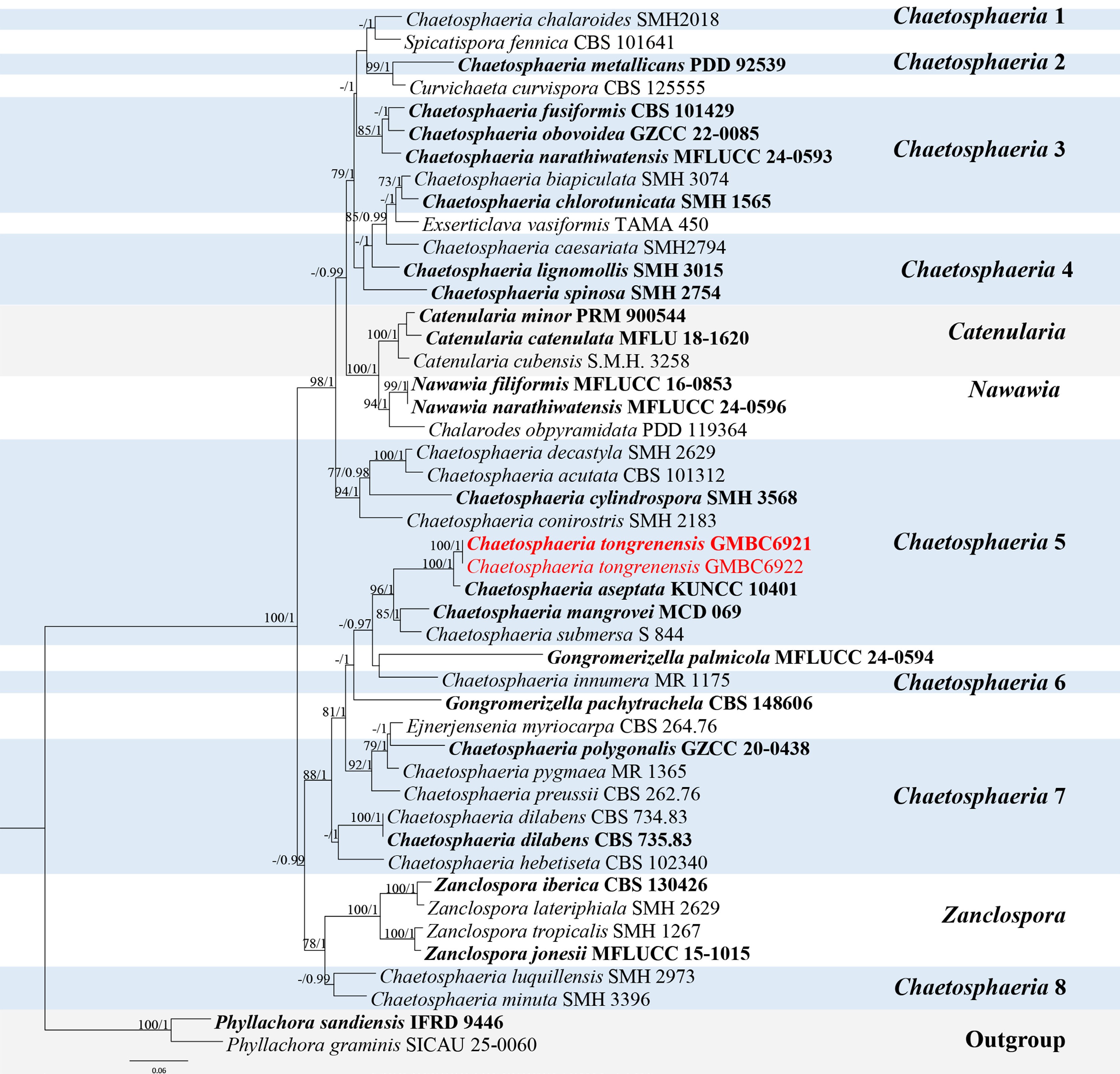
Figure 28.
ML tree of selected species of Chaetosphaeria and related genera in Chaetosphaeriaceae based on the ITS-LSU- tef1-αdataset. The combined dataset comprised 46 taxa, 2,292 characters (ITS: 1–498 bp; LSU: 499–1,383 bp; tef1-α: 1,384–2,292 bp), including indel regions. Maximum likelihood (lnL = −14,251.487607) and Bayesian analyses produced consistent tree topologies. Phyllachora graminis (SICAU 25-0060) and P. sandiensis (IFRD 9446) were selected as the outgroup taxa. Bayesian posterior probabilities (PP) ≥ 0.90 and RAxML bootstrap support values ≥ 70 % are shown on the branches. The novel species is marked in red, and ex-type/type strains are in bold.
Morphologically, C. tongrenensis (GMB6921) shares several characteristics with C. aseptata, including macronematous, mononematous conidiophores and aseptate, hyaline conidia. However, C. tongrenensis can be distinguished from C. aseptata by its larger conidiophores (107–249 µm vs 101–137 µm), higher number of septa in conidiophores (9–15-septate vs 5–7-septate), monophialidic conidiogenous cells (vs polyblastic conidiogenous cells) and smaller conidia (smaller (3.4–4.8 × 1.8–2.7 µm vs 4–6 × 2–3 µm)[5].
Based on these morphological differences and phylogeny, C. tongrenensis is introduced as a novel species.
Melanopsamma Niessl, Verh. Naturf. Vereins Brünn 14 (Abh.): 200 (1876).
Notes – Melanopsamma was established by Niessl[109]. However, a type species was not designated at that time. Later, Höhnel designated Melanopsamma pomiformis as the type species[110]. Wang et al. synonymized the genus under Stachybotrys based on ITS sequence analysis[111]. However, Lombard et al. reinstated it based on molecular phylogenetic analysis and designated CBS 101322 as the ex-type strain for the generic type species, M. pomiformis[112]. The anamorph of Melanopsamma is characterized by macronematous, erect, mononematous, unbranched, solitary or grouped conidiophores, conidiogenous cells phialidic, smooth, hyaline, elongated doliiform or subcylindrical to clavate, and aseptate, hyaline to dark brown, obovoid, globose, limoniform, or ellipsoidal condia[112].
According to Species Fungorum (accessed 3 January 2026), the genus currently comprises 60 accepted species. Herein, we introduce a new hyphomycetous Melanopsamma species found in a freshwater habitat.
Melanopsamma wumengshanensis L.L. Liu, W.M. Li, & Q.R. Li, sp. nov. (Fig. 29)
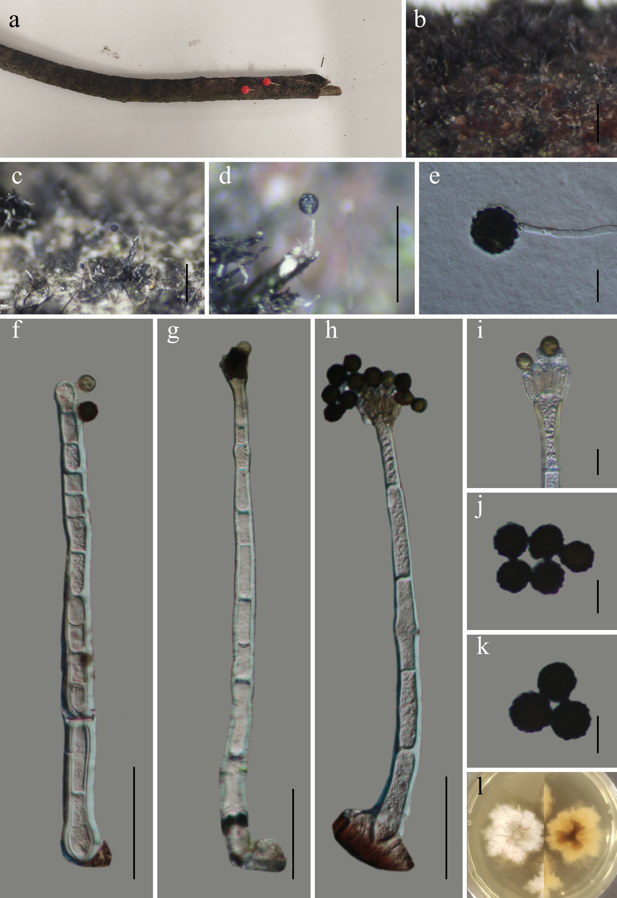
Figure 29.
Melanopsamma wumengshanense (GMB6943 holotype). (a) Host. (b)–(d) Colonies on natural substrate. (e) Germinating conidium. (f)–(h) Conidiophores and conidia. (i) Conidiogenous cells with conidia. (j), (k) Conidia. (l) Surface and reverse view of culture on PDA. Scale bars: b = 1 mm, c, d = 0.5 mm, f–h = 30 μm, e, i–k = 10 μm.
MycoBank number: MB862965
Etymology –The epithet refers to Wumengshan National Nature Reserve, where the holotype was collected.
Saprobic on submerged dead branches of an unknown plant along the riverbank. Sexual morph: not observed. Asexual morph: hyphomycetous. Colonies velvety, effuse, black. Conidiophores 183–301 × 8–13 µm (x = 232 × 11 μm, n = 30), mononematous, macronematous, single or in groups, simple, unbranched, straight to slightly flexuous erect, hyaline, smooth, with a bulbous apex, 4–10-septate, with a whorl of four to ten conidiogenous cells. Conidiogenous cells 10–17 µm long, phialidic, hyaline, smooth, elongate doliiform to subcylindrical. Conidia 10.6–12.5 × 9.5–12.2 µm (x = 11.4 × 10.7 µm, n = 30), aseptate, acrogenous, globose to subglobose, verrucose, smooth-walled, hyaline at young, brown to dark at maturity, separating schizolytically.
Culture characteristics – Conidia germinate on PDA within 24 h. After 21 d at 25 °C, colonies on PDA reach 20–30 mm in diameter; irregular, raised, floccose to cottony, aerial mycelium abundant, margin lobate, surface white; reverse pale yellow to brown, with a darker central zone.
Material examined – CHINA, Yunnan Province, Zhaotong City, Wumengshan National Nature Reserve, 25°21′45.29″ N, 103°15′30.66″ E, elevation 2,508 m, on submerged dead branches of an unknown plant along the riverbank, 26 October 2024, Wen-Mei Li, 2024WMS199 (GMB6943, holotype; GMBC6943, ex-type); ibid., KUN-HKAS 152905, isotype.
Additional examined specimens – CHINA, Yunnan Province, Zhaotong City, Wumengshan National Nature Reserve, 25°36.91.15″ N, 103°15′30.56″ E, elevation 2,671 m, on submerged dead branches of an unknown plant along the riverbank, 26 October 2024, Wen-Mei Li, 2024WMS206 (GMB6944, GMBC6944).
Notes – In the phylogenetic analyses (Fig. 30), Melanopsamma wumengshanensis clusters with M. sichuanensis (CC.SLC.S72), forming a well-supported sister clade. Comparative analysis of nucleotide base pairs showed that M. wumengshanensis (GMBC6943) differs from the type strain of M. sichuanensis (CC.SLC.S72) by 1.6% (8/493 bp) in the ITS locus and 1.8% (16/882 bp) in the rpb2 gene.
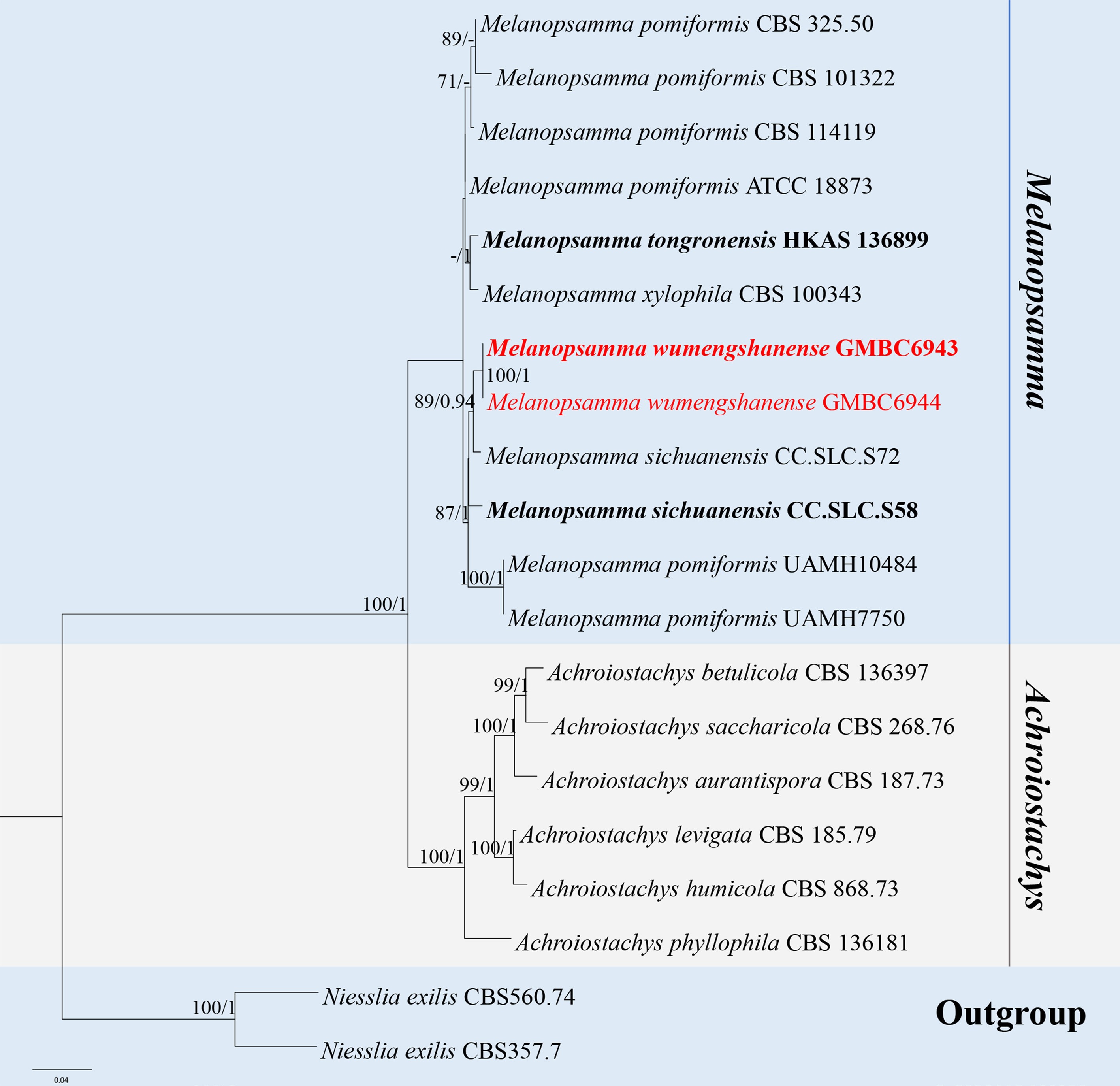
Figure 30.
ML tree of selected species of Melanopsamma and Achroiostachys inferred from a combined ITS-LSU-rpb2 dataset. The combined dataset comprised 20 taxa, 2,174 characters (ITS: 1–574 bp; LSU: 575–1,415 bp; rpb2: 1,416–2,174 bp), including indel regions. Maximum likelihood (lnL = –6686.302130) and Bayesian analyses produced consistent tree topologies. Niesslia exilis (CBS 560.74) and N. exilis (CBS 357.7) were selected as the outgroup taxa. Bayesian posterior probabilities (PP) ≥ 0.90 and RAxML bootstrap support values ≥ 70 % are shown on the branches. The novel described species is indicated in red and ex-type/type strains in bold.
Morphologically, M. wumengshanensis differs from M. sichuanensis in having larger, unbranched conidiophores with a distinctly bulbous apex (183–301 × 8–13 µm), whereas the latter exhibits irregularly or sympodially branched conidiophores tapering toward the apex (192.2–349.4 × 8.1–15.4 µm). Conidiogenous cells in M. wumengshanensis are elongate-doliiform to subcylindrical, arranged in a regular whorl of four to ten cells and measuring 10–17 µm long, whereas M. sichuanensis has obovoid, monophialidic cells measuring 10.6–13.5 × 5–9.7 µm (in press).
Based on these molecular and morphological differences, together with the phylogenetic support, we introduce M. wumengshanensis as a novel species.
Radiaticonidium L.L. Liu, Q.F. Zhang, & Q.R. Li, gen. nov.
MycoBank number: MB862970
Etymology – From Latin radiatus, meaning radiating, and conidium, referring to the conidia surrounded by numerous radiating/curled appendages.
On decaying submerged wood in freshwater habitats. Sexual morph unknown. Asexual morph hyphomycetous. Colonies effuse, superficial. Conidiophores mononematous, macronematous, darkly pigmented, erect, arising singly or in small groups from a compact stroma-like basal tissue, each terminating in a single conidiogenous cell. Conidiogenous cells are terminal, holoblastic, monoblastic, integrated, determinate; conidiogenous loci are inconspicuous. Conidia solitary, acrogenous, dry, globose to subglobose, aseptate, smooth-walled, hyaline to pale brown, surrounded by numerous radiating/curled, hyaline, filamentous appendages, producing a characteristic stellate to echinulate appearance.
Type species – Radiaticonidium aquaticum L.L. Liu, Q.F. Zhang, & Q.R. Li.
Notes – Phylogenetically (Fig. 31), Radiaticonidium forms a well-supported, independent lineage within Chaetosphaeriaceae and is resolved as sister to Pseudostriatosphaeria. Despite this phylogenetic affinity, Radiaticonidium is morphologically distinct from Pseudostriatosphaeria, which is characterized by a coelomycetous asexual morph[113], whereas Radiaticonidium exhibits a hyphomycetous asexual morph. This clear difference in asexual morph type and well-resolved phylogenetic position clearly separates Radiaticonidium from Pseudostriatosphaeria and other morphologically allied genera in Chaetosphaeriaceae.
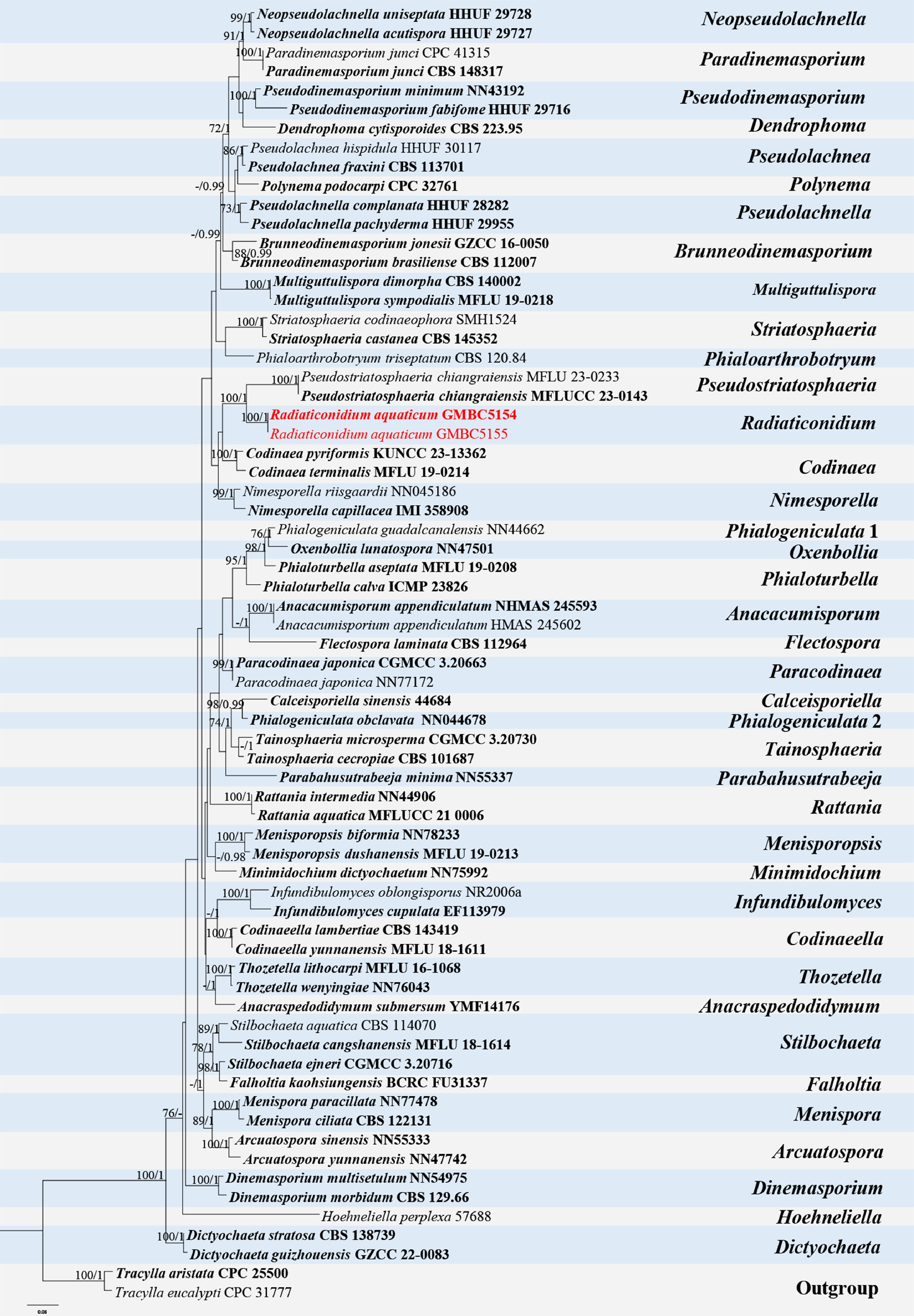
Figure 31.
ML tree of selected Chaetosphaeriaceae species based on the ITS-LSU dataset. The combined dataset comprised 68 taxa, 1,371 characters (ITS: 1–512 bp; LSU: 513–1,371 bp), including indel regions. Maximum likelihood (lnL = −15,747.664593) and Bayesian analyses produced consistent tree topologies. Tracylla aristata (CPC 25500) and T. eucalypti (CPC 31777) were selected as the outgroup taxa. Bayesian posterior probabilities (PP) ≥ 0.90 and RAxML bootstrap support values ≥ 70 % are shown on the branches. The newly described species is marked in red. Ex-type/type strains are indicated in bold.
Radiaticonidium aquaticum L.L. Liu, Q.F. Zhang, & Q.R. Li, sp. nov. (Fig. 32)
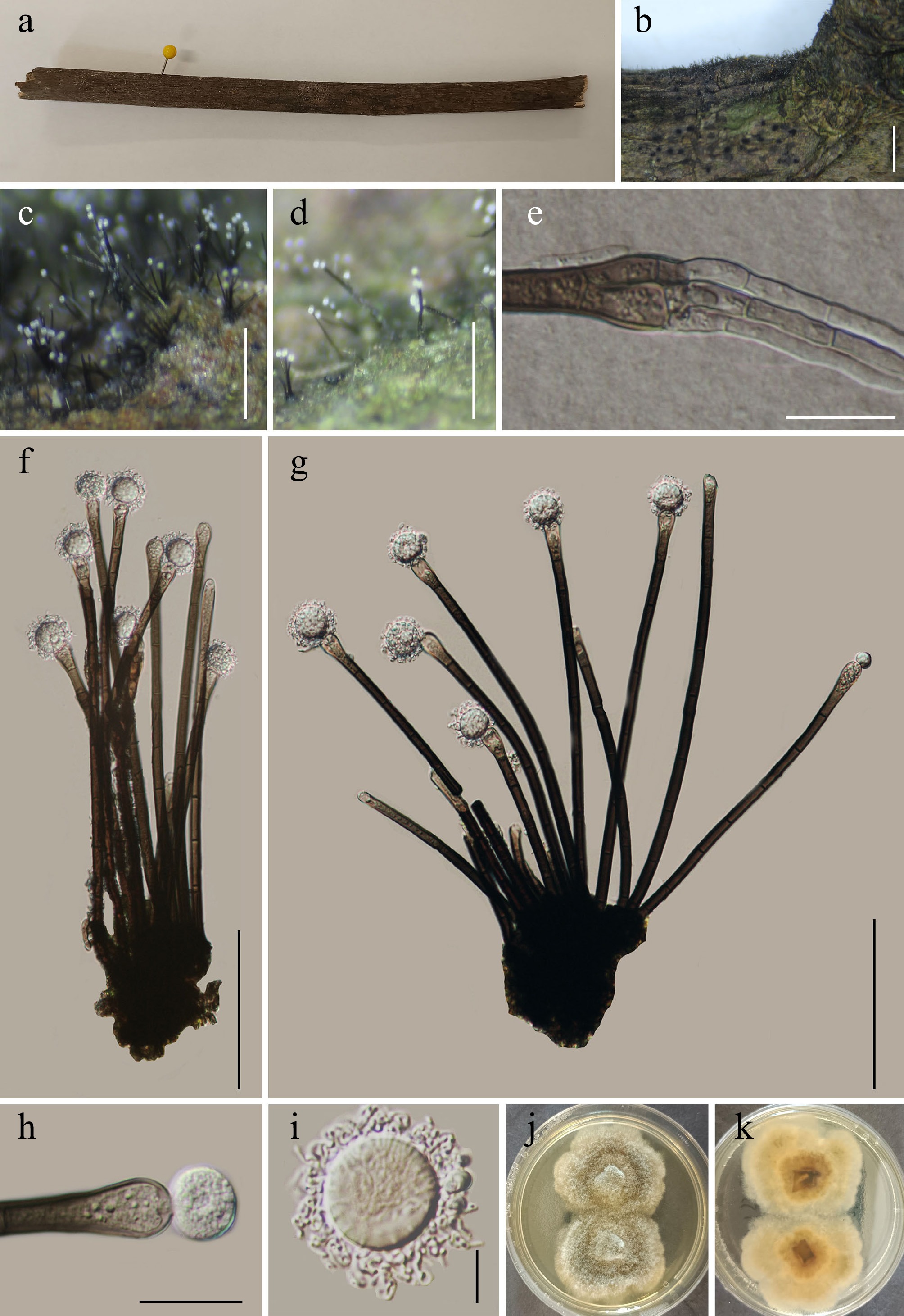
Figure 32.
Radiaticonidium aquaticum (GMB5154 holotype). (a) Host. (b)–(d) Colonies on natural substrate. (e) Germinating conidiophore. (f), (g) Conidiophores and conidia. (h) Conidiogenous cells and conidium. (i) Conidium. (j), (k) Surface and reverse view of culture on PDA. Scale bars: b = 1 mm, c, d = 2.5 mm, e–g = 20 μm, h = 20 μm, i = 10 μm.
MycoBank number: MB862971
Etymology – The epithet refers to the freshwater habitat from which the specimen was collected.
Saprobic on submerged dead branches of an unknown plant in a freshwater wetland. Sexual morph: not observed. Asexual morph: hyphomycetous. Colonies are effuse, hairy, superficial, gregarious, dark brown to black, scattered to aggregated in small groups on the host surface. Mycelium partly immersed, superficial, composed of septate, branched, pale brown to dark brown hyphae. Conidiophores 137–267 × 10–21 µm (x = 211 × 15 µm, n = 30), mononematous, macronematous, solitary to gregarious, erect, straight to slightly flexuous, cylindrical, dark brown to blackish, smooth-walled, thin- to slightly thick-walled, septate, unbranched, arising from a compact, dark basal stroma-like tissue; basal cell slightly swollen; apical portion bearing a single terminal conidiogenous cell. Conidiogenous cells 24–35 × 2–9 µm (x = 29 × 6 µm, n = 30), monoblastic, holoblastic, terminal, integrated, determinate, cylindrical to subcylindrical, pale brown to brown, smooth-walled; conidiogenous loci inconspicuous. Conidia 18–22 µm diam. (x = 20 µm, n = 30), sun-shaped globose to subglobose, solitary, dry, acrogenous, hyaline to pale brown, thin-walled, aseptate, smooth-walled; conidial body surrounded by numerous radiating, hyaline, conidial appendages 4–7 µm long (x = 6 µm, n = 30), irregularly arranged, giving the conidium a stellate to echinulate appearance in optical view; appendages persistent, non-septate, slightly flexuous.
Culture characteristics – Conidia germinate on PDA within 24 h. After 14 d at 25 °C, colonies on PDA reach 30–50 mm in diameter; circular to irregular, raised in the center, floccose to cottony, aerial mycelium abundant, margins lobate and thin, surface white to greyish white; reverse brown at center fading toward margins.
Material examined – CHINA, Guangxi Zhuang Autonomous Region, Jiuwan Mountain National Nature Reserve, 25°9'50.49" N, 108°47'39.39" E, elevation 984 m, on submerged dead branches of an unknown plant in a freshwater wetland, 18 August 2024, Qin-Fang Zhang, 2024JWS126-1 (GMB5154, holotype; GMBC5154, ex-type); ibid., KUN-HKAS 152906, isotype.
Additional examined specimens – CHINA, Guangxi Zhuang Autonomous Region, Jiuwan Mountain National Nature Reserve, 25°12'24.61" N, 102°40'29.24" E, elevation 1,209 m, on submerged dead branches of an unknown plant in a freshwater wetland, 18 August 2024, Qin-Fang Zhang, 2024JWS159 (GMB5155, GMBC5155).
Notes – In the phylogenetic analysis (Fig. 31), Radiaticonidium aquaticum (GMBC5154) forms a well-supported (BS = 100; PP = 1), independent lineage within Chaetosphaeriaceae, sister to Pseudostriatosphaeria chiangraiensis (MFLUCC 23-0143). Comparative analysis of nucleotide base pairs showed that R. aquaticum (GMBC5154) differs from the type strain of P. chiangraiensis (MFLUCC 23-0143) by 19% (88/463 bp) in the ITS locus and 2.8% (22/796 bp) in the LSU locus, indicating a clear genetic divergence between the two taxa.
Morphologically, R. aquaticum differs from P. chiangraiensis by having a hyphomycetous asexual morph, whereas the latter is characterized by a coelomycetous asexual morph[113]. Radiaticonidium can be distinguished from other genera in the family by its distinctive, sun-shaped conidia surrounded by numerous radiating, curled appendages.
Given its distinct phylogenetic position and distinct morphological characters, Radiaticonidium aquaticum is introduced as a new genus and species.
Glomerellales Chadef. ex Réblová, W. Gams & Seifert, Stud. Mycol. 68: 170 (2011).
Plectosphaerellaceae W. Gams, Summerb. & Zare, Nova Hedwigia 85(3-4): 476 (2007).
Plectosphaerella Kleb., Phytopath. Z. 1: 43 (1929).
Notes – Plectosphaerella was established by Klebahn, with P. cucumeris designated as the type species. Species of Plectosphaerella are characterized by hyaline to pale, septate hyphae, simple to branched conidiophores, and cylindrical to ellipsoidal, aseptate to septate, hyaline conidia formed singly or in slimy heads. Species of the genus occur as plant pathogens, soil-borne or saprobic fungi, and are widely distributed in temperate and tropical regions, being isolated from plants, soil, and occasionally animals[114−116]. Initially, only five species were recognized in the genus, but following the transfer of species from Plectosporium and the introduction of several new taxa, the number of accepted species has increased[14,117−119]. According to Species Fungorum (accessed 5 January 2026), Plectosphaerella currently comprises 24 accepted species. In the present study, a new species of Plectosphaerella is introduced.
Plectosphaerella guangxiensis L.L. Liu, Q.F. Zhang, & Q.R. Li, sp. nov. (Fig. 33)
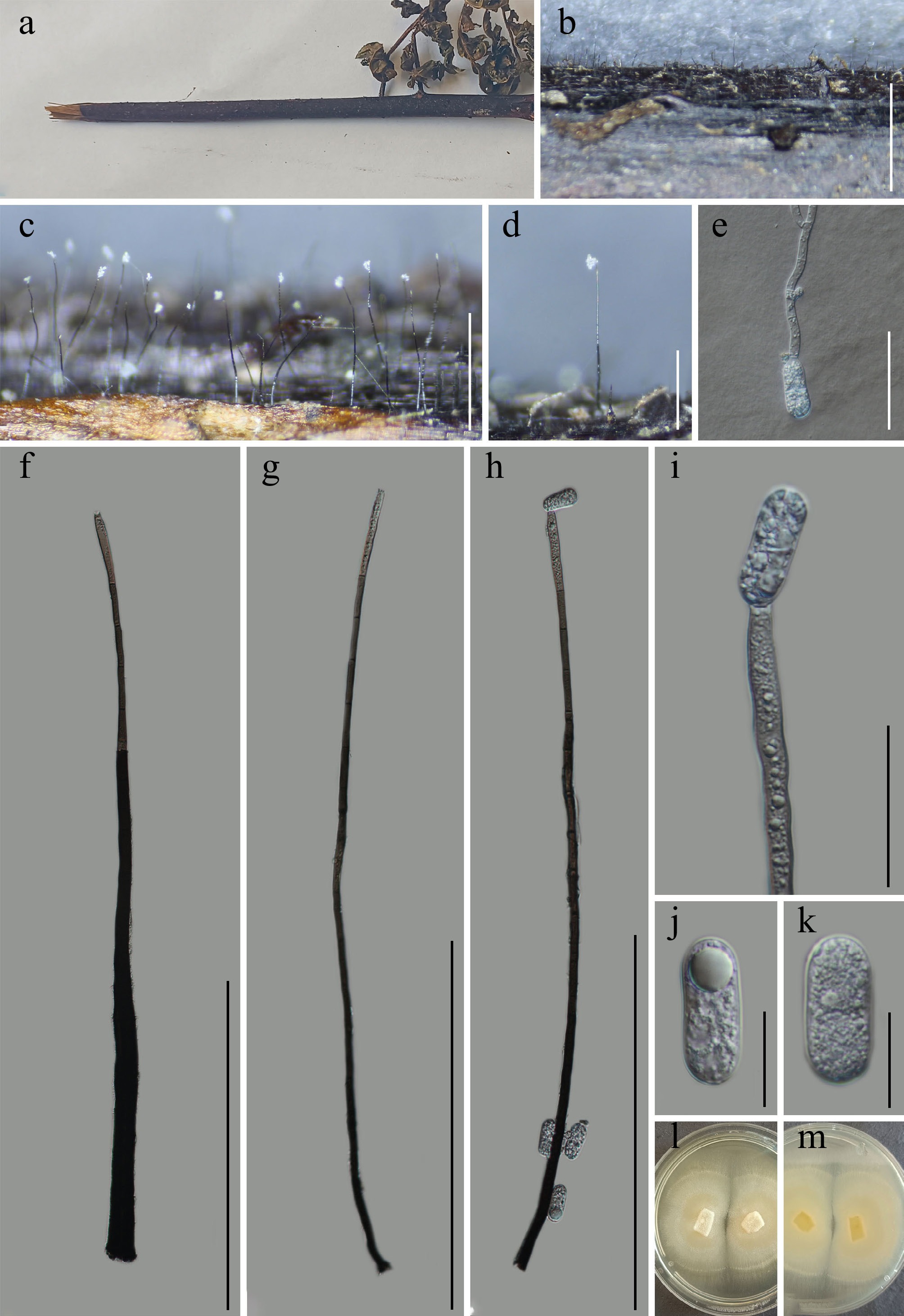
Figure 33.
Plectosphaerella guangxiensis (GMB5156 holotype). (a) Host. (b)–(d) Colonies on natural substrate. (e) Germinating conidium. (f)–(h) Conidiophores and conidia. (i) Conidiogenous cells. (j), (k) Conidia. (l), (m) Surface and reverse view of culture on PDA. Scale bars: b = 1 mm, c, d = 2.5 mm, e = 50 μm, f–h = 300 μm, i = 40 μm, j, k = 20 μm.
MycoBank number: MB862972
Etymology –The epithet refers to Guangxi Zhuang Autonomous Region, China, where the holotype specimen was collected.
Saprobic on submerged decaying wood in an aquatic habitat. Sexual morph: not observed. Asexual morph: hyphomycetous. Colonies effuse, scattered to gregarious, superficial, hair-like conidiophores arising singly or in small groups, with hyaline apical conidial masses. Mycelium hyaline, smooth, septate, branched, thin-walled, mostly immersed to superficial, lacking distinct hyphal coils. Conidiophores 99.5–1,574.0 × 6.1–46.0 µm (x = 659.2 × 17.5 µm, n = 30), solitary, mononematous, macronematous, straight to slightly flexuous erect, unbranched, rarely with short lateral branches; hyaline to pale brown at the base, smooth, cylindrical, thick- to thin-walled, arising directly from vegetative hyphae. Conidiogenous cells 28.6–148.2 × 4.3–21.1 µm (x = 77.1 × 4.3 µm, n = 30), phialidic, monophialidic, integrated, terminal or occasionally lateral, discrete, cylindrical to subcylindrical or slightly ampulliform, hyaline, smooth, thin-walled, straight or slightly curved, gradually tapering towards the apex, collarette cylindrical to slightly flared, short but distinct. Conidia 25.4–35.9 × 11.5–13.9 µm (x = 28.9 × 12.7 µm, n = 30), produced singly, accumulating in slimy heads; hyaline, smooth, thin-walled, mostly ellipsoidal to broadly cylindrical, occasionally slightly navicular, with rounded to obtuse ends, aseptate.
Culture characteristics – Conidia germinate on PDA within 24 h. After 14 d at 25 °C, colonies on PDA reach 35–50 mm in diameter; circular, raised in the center, floccose to cottony, margins entire and thin, surface grayish white; reverse light yellow at the center, gradually fading to white towards the outside.
Material examined – CHINA, Guangxi Zhuang Autonomous Region, Jiuwan Mountain National Nature Reserve, 25°9'50.49" N, 108°47'39.39" E, elevation 984 m, on submerged decaying wood in aquatic habitat, 18 August 2024, Qin-Fang Zhang, 2024JWS139 (GMB5156, holotype; GMBC5156, ex-type); ibid., KUN-HKAS 152907, isotype.
Additional examined specimens – CHINA, Guangxi Zhuang Autonomous Region, Jiuwan Mountain National Nature Reserve, 25°12'24.61" N, 102°40'29.24" E, elevation 1,209 m, on submerged decaying wood in aquatic habitat, 18 August 2024, Qin-Fang Zhang, 2024JWS175 (GMB5157, GMBC5157).
Notes – Phylogenetically (Fig. 34), Plectosphaerella guangxiensis forms a well-supported sister clade (BS = 100; PP = 0.94) with an unidentified Plectosphaerella sp. (LLC116). Comparative nucleotide sequence analyses indicate that P. guangxiensis (GMBC5156) differs from the isolate LLC116 by 1.83% (9/492 bp) in the ITS region and 0.88% (7/792 bp) in the LSU region.
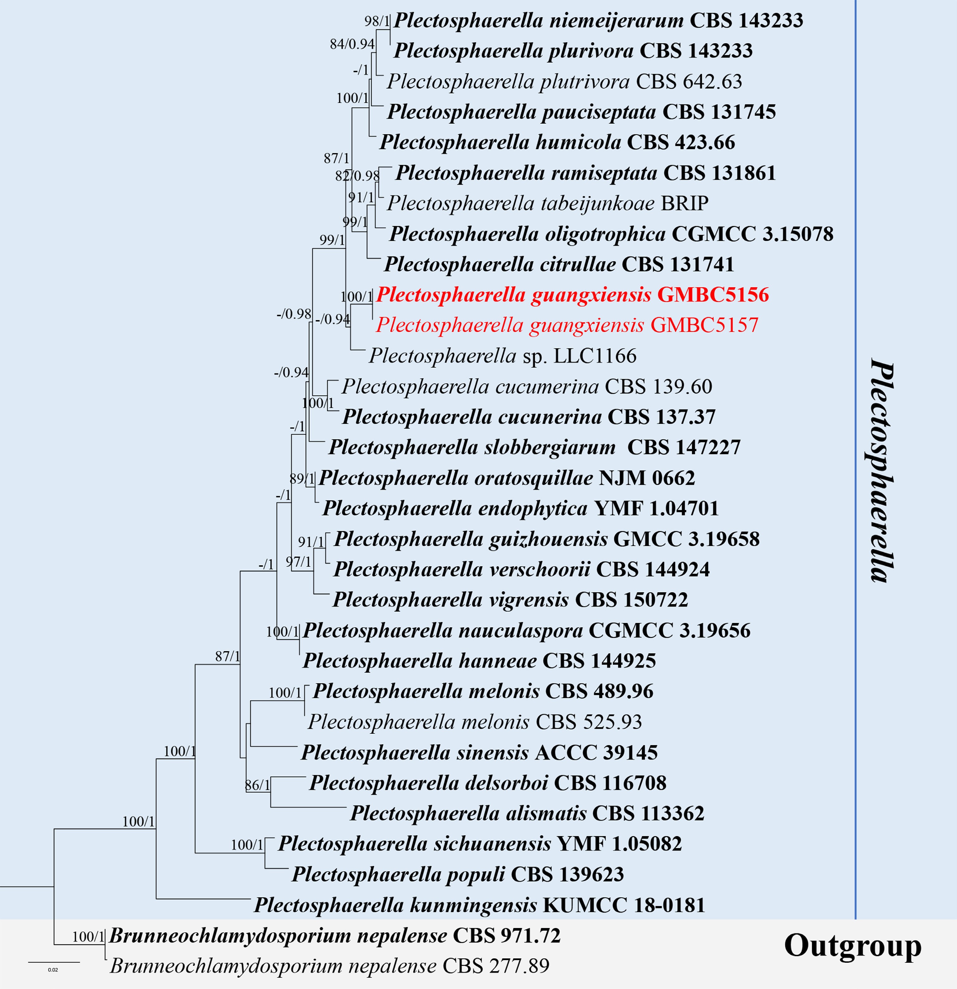
Figure 34.
ML tree of selected Plectosphaerella species based on the ITS-LSU-tef1-α-rpb2 dataset. The combined dataset comprised 32 taxa, 2,944 characters (ITS: 1–503 bp; LSU: 504–1,360 bp; tef1-α: 1,361–2,201 bp; rpb2: 2,201–2,944 bp), including indel regions. Maximum likelihood (lnL = −8,548.505177) and Bayesian analyses produced consistent tree topologies. Brunneochlamydosporium nepalense (CBS 277.89) and B. nepalense (CBS 971.72) were selected as the outgroup taxa. Bayesian posterior probabilities (PP) ≥ 0.90 and RAxML bootstrap support values ≥ 70 % are shown on the branches. The novel species is indicated in red, and ex-type/type strains in bold.
This study represents the first report of a hyphomycetous asexual morph occurring on a natural substratum within the genus, and its morphology is distinct from all previously known species. Given these distinct morphological characteristics and strong phylogenetic support, we introduce P. guangxiensis as a novel species.
Musicillium Zare & W. Gams, Nova Hedwigia 85(3-4): 482 (2007).
Notes – Musicillium was established by Zare et al., typified by M. theobromae[120]. The genus is characterized by solitary, pale brown conidiophores that are verticillate towards the apex, bearing whorls of phialidic conidiogenous cells that produce aseptate, hyaline conidia[115,120]. Species of Musicillium are mainly known as saprobes or weak associates on plant substrates, with records from woody and herbaceous hosts, including economically important plants. The genus has been reported from tropical and subtropical regions, with occasional records from temperate areas, suggesting a relatively wide but sparsely documented distribution[13,77,115]. According to Species Fungorum (accessed 5 January 2026), the genus currently comprises five accepted species. Herein, a new species of Musicillium is introduced based on morphological characteristics and multigene phylogenetic analyses.
Musicillium verticillatum L.L. Liu, Q.F. Zhang, & Q.R. Li, sp. nov. (Fig. 35)
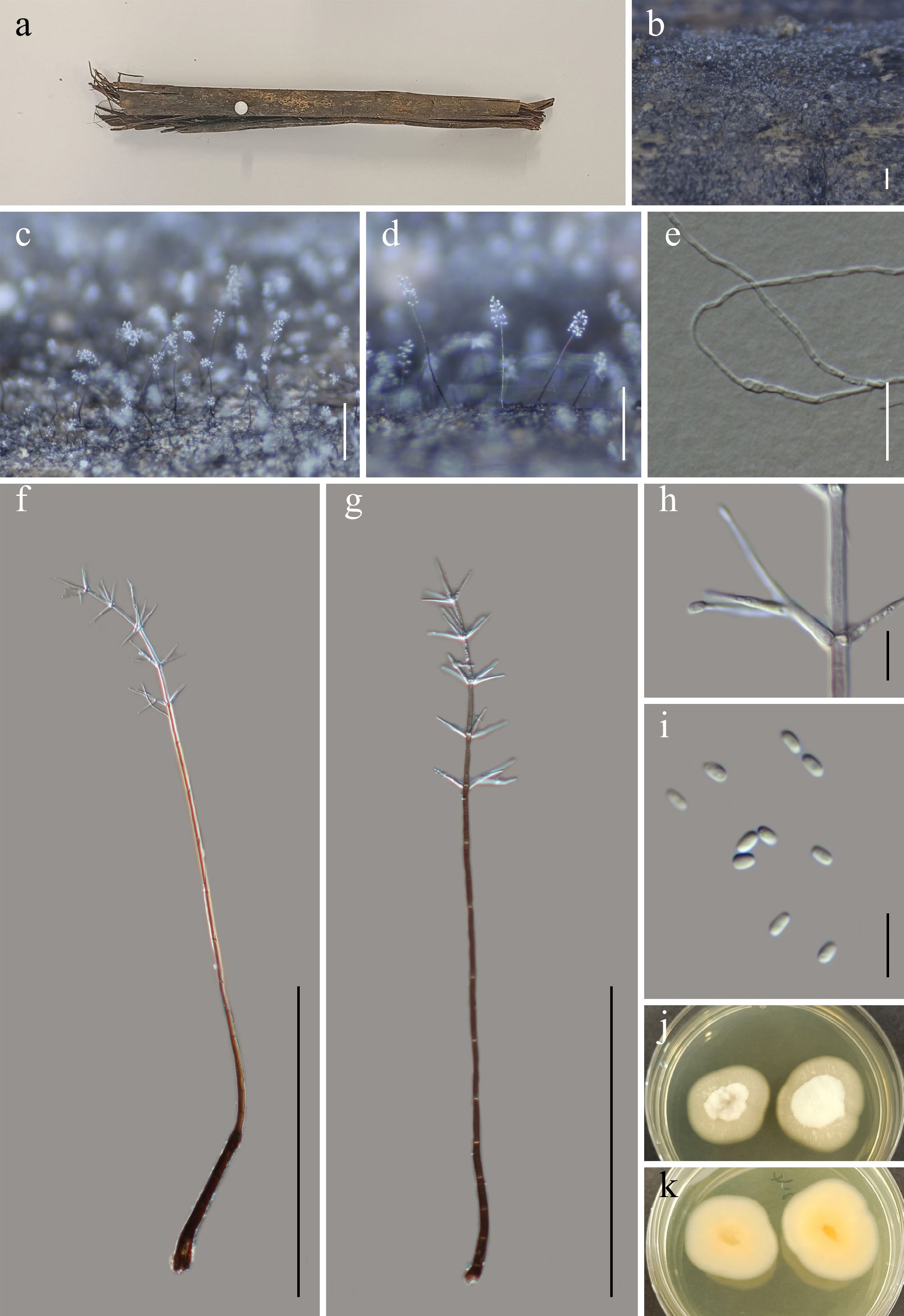
Figure 35.
Musicillium verticillatum (GMB5180 holotype). (a) Host. (b)–(d) Colonies on natural substrate. (e) Germinating conidium. (f), (g) Conidiophores and conidia. (h) Conidiogenous cells with conidia. (i) Conidia. (j), (k) Surface view of culture on PDA. Scale bars: b = 1 mm, c, d = 0.5 mm, f, g = 50 μm, e = 20 μm, h, i = 10 μm.
MycoBank number: MB862974
Etymology –The epithet verticillatum comes from the Latin verticillatus, meaning 'verticillate', referring to the whorled arrangement of phialides on the conidiophores.
Saprobic on submerged decaying wood in a freshwater wetland. Sexual morph: not observed. Asexual morph: hyphomycetous. Colonies superficial, effuse, scattered to gregarious, forming minute whitish to pale grey, synnematous-like tufts; surface appearing powdery to floccose due to abundant conidiophores. Mycelium partly superficial, mostly immersed, consisting of smooth-walled, branched, hyaline to pale brown, septate, thin-walled hyphae, 1.8–3.2 µm wide. Conidiophores 195–782 × 3–9 µm (x = 478 × 5 µm, n = 15), mononematous, solitary, macronematous, straight to slightly flexuous erect, cylindrical, dark brown at the base, paler towards the apex, smooth, thick-walled, 13–18-septate; upper portion verticillately branched, bearing five whorls of phialides; basal cell slightly swollen, apex tapering. Conidiogenous cells (phialides) 17–38 × 1–3 µm (x = 25.5 × 1.8 µm, n = 30), arranged in divergent whorls of three to six per node, terminal and lateral, subulate to acicular, hyaline, smooth- and thick-walled, apex with minute collarette. Conidia 2–5 × 1–2.7 µm (x = 3.9 × 2.2 µm, n = 30), ellipsoidal to ovoid, aseptate, hyaline, smooth- and thick-walled, accumulating in small slimy heads at phialide apices.
Culture characteristics – Conidia germinate on PDA within 24 h. After 28 d at 25 °C, colonies on PDA reach 10–30 mm in diameter; circular, raised in the center, thin toward margins, floccose to cottony, margins entire, surface white to creamy white; reverse whitish-yellow.
Material examined – CHINA, Yunnan Province, Yuxi City, Ailaoshan National Nature Reserve, 24°5'7.01" N, 101°31'30.44" E, elevation 1,169 m, on submerged decaying wood in a freshwater wetland, 15 September 2024, Qin-Fang Zhang, 2024ALS153-1 (GMB5180, holotype; GMBC5180, ex-type); ibid., KUN-HKAS 152908, isotype.
Additional examined specimens – CHINA, Yunnan Province, Yuxi City, Ailaoshan National Nature Reserve, 28°1'7.20" N, 101°32'50.29" E, elevation 1,795 m, on submerged decaying wood in a freshwater wetland, 15 September 2024, Qin-Fang Zhang 2024ALS179 (GMB5181, GMBC5181).
Notes – Phylogenetically (Fig. 36), Musicillium verticillatum forms a well-supported (BS = 97; PP = 1) independent lineage within the genus Musicillium, supporting the recognition of an undescribed taxon.
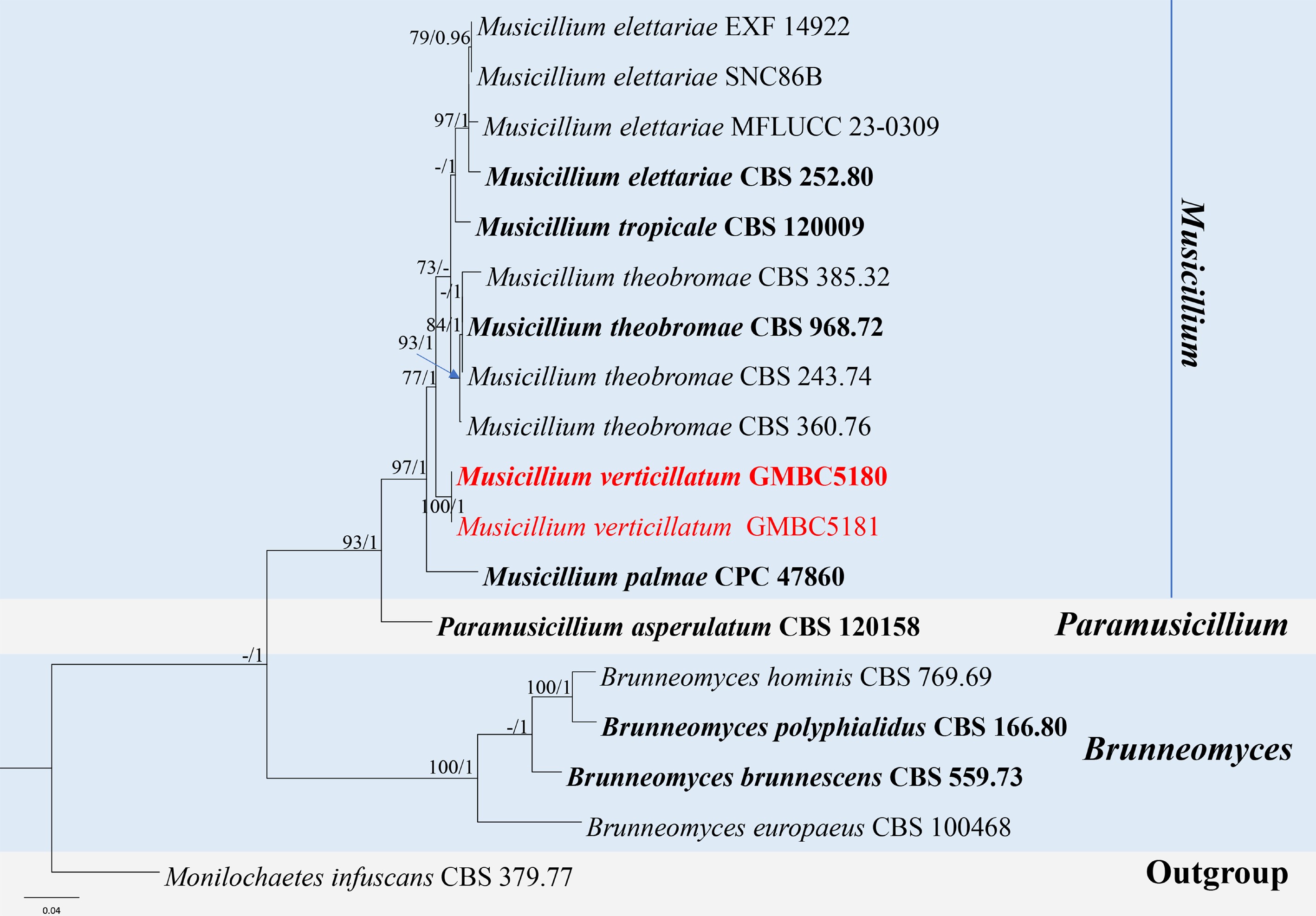
Figure 36.
ML tree of selected Musicillium species based on the ITS-LSU-rpb2 dataset. The combined dataset comprised 18 taxa, 2,086 characters (ITS:1–515 bp; LSU: 516–1,340 bp; rpb2: 1,341–2,086 bp), including indel regions. Maximum likelihood (lnL = −7,061.024465) and Bayesian analyses produced consistent tree topologies. Monilochaetes infuscans (CBS 379.77) was selected as the outgroup taxon. Bayesian posterior probabilities (PP) ≥ 0.90 and RAxML bootstrap support values ≥ 70 % are shown on the branches. The novel species is indicated in red and ex-type/type strains in bold.
Morphologically, M. verticillatum resembles M. theobromae in having brown macronematous conidiophores with verticillate phialides arranged in whorls of three to six per node and hyaline conidia produced in slimy heads. However, M. verticillatum differs in having longer conidiophores (up to 782 µm vs 500 µm), with 13–18 septa (vs 4–9), and smaller conidia (2–5 × 1–2.7 µm vs 4–9 × 1.0–2.0 µm). Ecologically, M. verticillatum was collected from freshwater habitats, whereas M. theobromae has been reported from terrestrial habitats[120].
Based on its clear phylogenetic distinctness and morphological and ecological differences, M. verticillatum is proposed here as a new species.
Hypocreales Lindau, Nat. Pflanzenfam., Teil. I (Leipzig) 1(1): 343 (1897).
Hypocreaceae De Not. [as 'Hypocreacei'], G. bot. ital. 2(1): 48 (1845) [1844].
Hypomyces (Fr.) Tul. & C. Tul., Select. Fung. Carp. 3: 15 (1860) protected name; (Index Fungorum, current name).
≡ Cladobotryum Nees, Syst. Pilze (Würzburg): 56 (1816) [1816–17].
Notes – Hypomyces was originally introduced as a subgenus of Hypocrea[121]. Tulasne & Tulasne later elevated it to generic rank and designated H. lactifluorum from the USA as the type species[121−123]. Historically, Cladobotryum was treated as the asexual morph genus of Hypomyces and numerous studies have demonstrated a close connection between the two genera based on morphological characteristics and life cycle observations[124]. Crous et al. protected the name Hypomyces over Cladobotryum under the 'one fungus, one name' principle, with Cladobotryum now treated as its synonym[123]. Species of the genus are mainly mycoparasitic, occurring on basidiomata or mycelia of other fungi, although some taxa have also been reported as saprobes[125]. The asexual morph of Hypomyces is characterized by hyaline to pale-colored, septate hyphae, erect and branched conidiophores, and holoblastic, sympodially proliferating conidiogenous cells producing hyaline, septate conidia, often ellipsoidal to fusiform in shape[126,127]. Species of Hypomyces are widely distributed worldwide, occurring in temperate, subtropical, and tropical regions, and are commonly found in forest ecosystems associated with decaying fungal hosts[128,129].
Hypomyces aquatilis L.L. Liu, W.M. Li, & Q.R. Li, sp. nov. (Fig. 37)
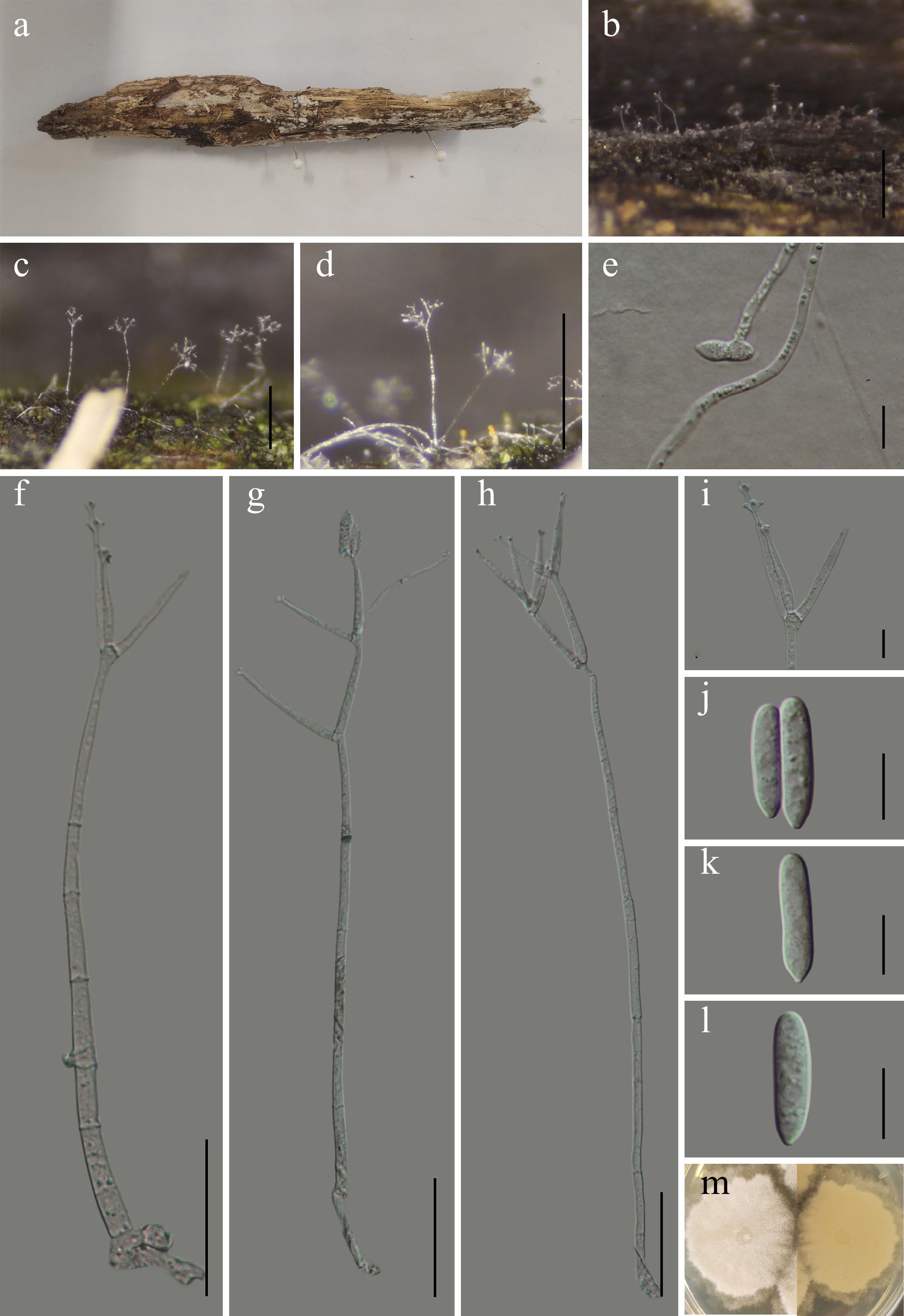
Figure 37.
Hypomyces aquatilis (GMB6929 holotype). (a) Host. (b)–(d) Colonies on natural substrate. (e) Germinating conidium. (f)–(h) Conidiophores and conidia. (i) Conidiogenous cells. (j)–(l) Conidia. (m) Surface and reverse view of culture on PDA. Scale bars: b = 1 mm, c, d = 0.5 mm, f–h = 50 μm, e, i–l= 10 μm.
MycoBank number: MB862976
Etymology – The epithet refers to the aquatic habitat from which holotype was collected.
Saprobic on submerged decaying wood in a freshwater wetland. Sexual morph: not observed. Asexual morph: hyphomycetous. Colonies effuse, superficial, forming scattered to gregarious, delicate, whitish to pale hyaline synnemata-like tufts; conidiophores arising directly from submerged mycelium, visible as minute whitish erect structures on wood surface. Mycelium partly immersed, rarely superficial, formed by hyaline, smooth-walled, septate, branched hyphae. Conidiophores 199–414 × 3.6–6.1 µm (x = 305.56 × 4.89 µm, n = 30), erect, straight to slightly flexuous, hyaline, smooth-walled, 3–7-septate, unbranched to sparsely branched; branching mainly in the upper part, producing lateral branches that function as conidiogenous units; apex often bearing verticillate conidiogenous cells. Conidiogenous cells 25.6–54.5 × 2.4–4.7 µm (x = 37.19 × 3.57 µm, n = 30), formed terminally or laterally on conidiophores, occurring singly or in verticils of two to six cells; hyaline, smooth-walled, elongated cylindrical to subulate, gradually attenuated towards the apex; monoblastic, producing a single conidiogenous locus at the tip. Conidia 14.9–23.8 × 3.7–5.4 µm (x = 18.45 × 4.64 µm, n = 30), hyaline, smooth-walled, aseptate, ellipsoidal to narrowly fusiform, straight or slightly curved, apex rounded, base attenuated with a distinct, slightly refractive hilum, formed singly or in short chains from the apical locus of conidiogenous cells.
Culture characteristics – Conidia germinate on PDA within 24 h. After 14 d at 25 °C, colonies on PDA reach 20–30 mm in diameter; circular to irregular, floccose, dense mycelium, margins entire, surface white; reverse light brown.
Material examined – CHINA, Guizhou Province, Guiyang City, Panlong Mountain Park, 26°43′53.23″ N, 106°49′54.17″ E, elevation 1,156 m, on submerged decaying wood in a freshwater wetland, 24 May 2025, Wen-Mei Li, 2025GY9 (GMB6929, holotype; GMBC6929, ex-type); ibid., KUN-HKAS 152909, isotype.
Additional specimens examined – CHINA, Guizhou Province, Guiyang City, Panlong Mountain Park, 26°43′54.13″ N, 106°49′54.19″ E, elevation 1,286 m, on submerged decaying wood in a freshwater wetland, 24 May 2025, Wen-Mei Li, 2025GY54 (GMB6930, GMBC6930).
Notes – Phylogenetically (Fig. 38), Hypomyces aquatilis forms a well-supported sister clade (BS = 100; PP = 1) with Cladobotryum arthrobotryoides (TFC 97-16). Comparative analysis of nucleotide base pairs showed that H. aquatilis (GMBC6929) differs from the type strain of Cladobotryum arthrobotryoides (TFC 97-16) by 1.1% (9/851 bp) in the LSU locus.
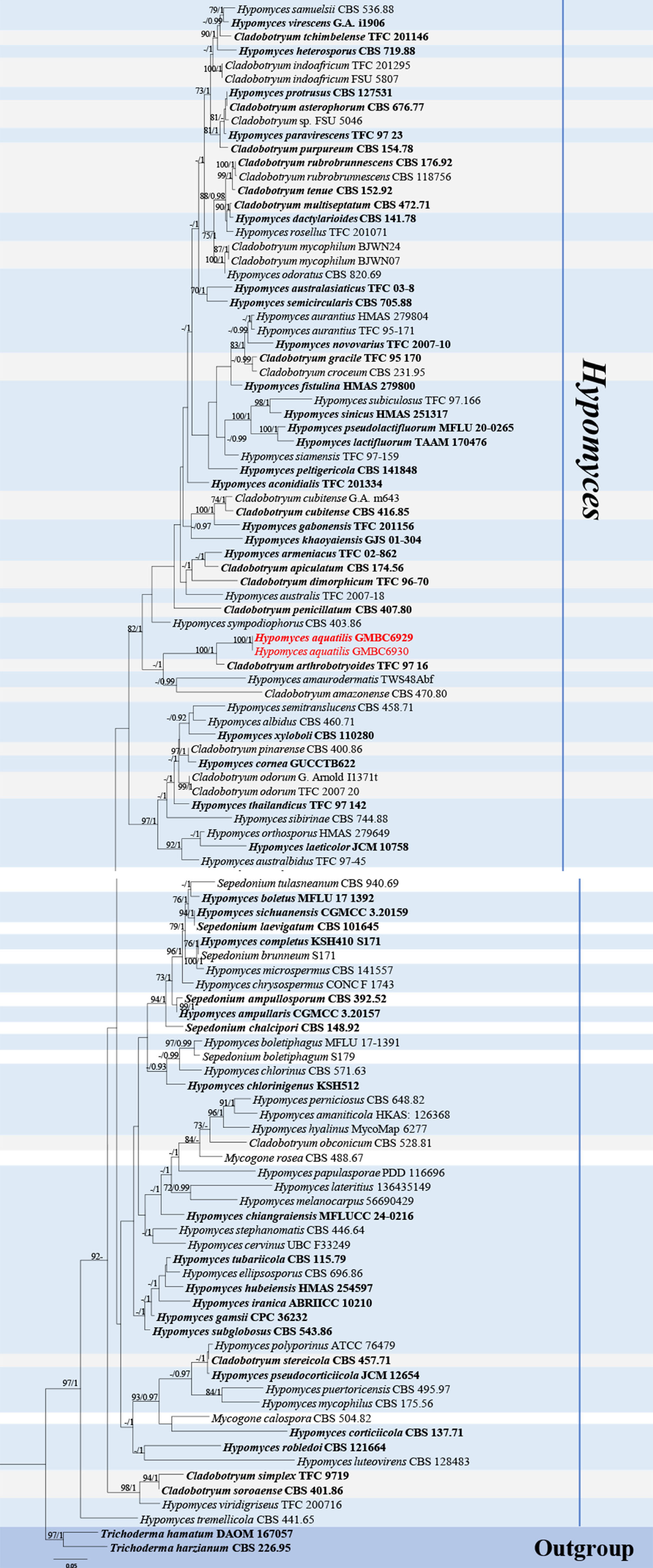
Figure 38.
ML tree of selected Hypomyces species based on a combined ITS-LSU-tef1-α-rpb2 dataset. The combined dataset comprised 109 taxa, 3,383 characters (ITS:1–537 bp; LSU: 538–1,390 bp; tef1-α: 1,391–2,314 bp; rpb2: 2,315–3,383 bp), including indel regions. Maximum likelihood (lnL = −31,431.265333) and Bayesian analyses produced consistent tree topologies. Trichoderma harzianum (CBS 226.95) and T. hamatum (DAOM 167057) were selected as the outgroup taxa. Values of the Bayesian PP ≥ 0.90 and bootstrap support RAxML ≥ 70% are shown on the branches. The novel species is indicated in red and ex-type/type strains in bold.
Morphologically, H. aquatilis differs from C. arthrobotryoides in having longer conidiogenous cells (25.6–54.5 μm vs 30–41 μm) and longer, aseptate conidia (14.9–23.8 × 3.7–5.4 µm), whereas C. arthrobotryoides produces shorter, 1–3-septate conidia (12.7–19.5 × 4.8–7 µm)[127].
Based on these difference and phylogenetic placement, H. aquatilis is introduced here as a novel species.
Hypocreales Lindau, Teil. I (Leipzig) 1(1): 343 (1897).
Niessliaceae Kirschst., Annls mycol. 37(1/2): 89 (1939).
Niesslia Auersw., Pilze Eur. 5-6: 30 (1869).
Notes – Niesslia is a speciose genus typified by N. Chaetomium, characterized by small, mostly spinulose perithecia that collapse into a discoid form when dry. The asexual morphs are morphologically reduced and were previously placed in Monocillium. Following the 'one fungus, one name' principle, Niesslia has nomenclatural priority over Monocillium[130]. Morphological characters alone offer limited diagnostic value, so reliable species delimitation in Niesslia relies on DNA barcode data and phylogenetic analyses[131−133].
Asexual morphs typically produce hyaline, smooth-walled, aseptate conidia, usually aggregated in slimy or nearly dry heads; sporodochium-like conidiophore aggregations may occur. Conidial chains are rare, reported only in N. catenata and N. indica, while thick-walled or slightly pigmented conidia occur in a few species[134]. Members of Niesslia are predominantly saprotrophic and widely distributed[135]. According to Species Fungorum (accessed 22 March 2026), the genus comprises 118 accepted species.
Niesslia waitemataensis W. Gams, Samuels, Gräfenhan & Schroers, Mycol. Progr. 18(1-2): 71 (2019). (Fig. 39).
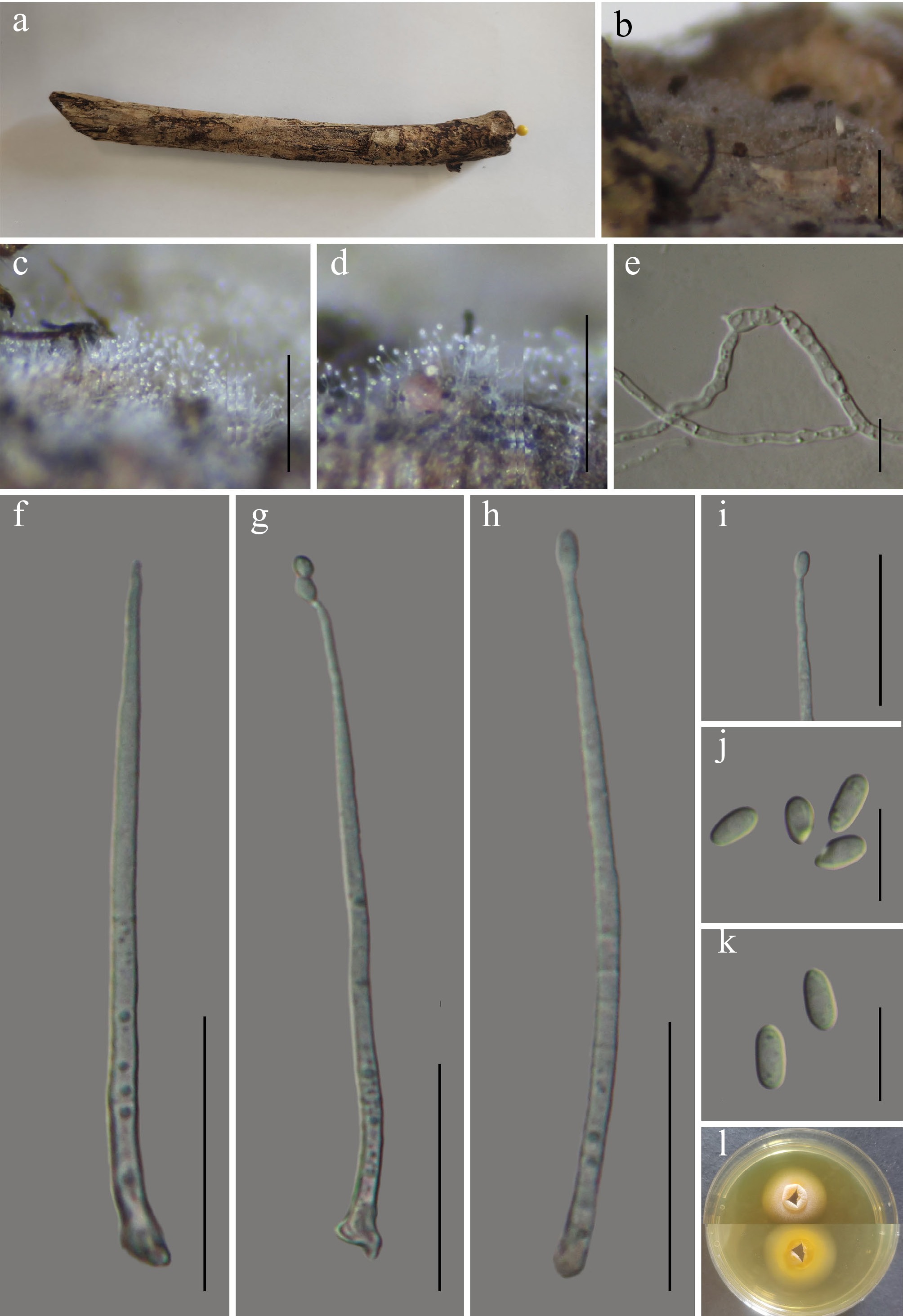
Figure 39.
Niesslia waitemataensis (GMB6949). (a) Host. (b)–(d) Colonies on natural substrate. (e) Germinating conidium. (f)–(h) Conidiophores and conidia. (i) Conidiogenous cells and conidium. (j), (k) Conidia. (l) Surface and reverse view of culture on PDA. Scale bars: b = 1 mm, c, d = 0.5 mm, f–h = 30 μm, e, i–k = 10 μm.
MycoBank number: MB827325
Saprobic on submerged decaying wood in freshwater habitats. Sexual morph: see Gams et al.[131]. Asexual morph: hyphomycetous. Colonies superficial, effuse, scattered to gregarious, appearing as minute whitish to translucent points on the surface of submerged decaying wood; forming sparse to moderately dense conidiophores emerging from the substrate surface. Mycelium partly immersed, formed by hyaline, septate, branched, smooth, thin- to slightly thick-walled hyphae. Conidiophores 60–126 × 2.3–3.9 µm (x = 86.78 × 2.78 µm, n = 30), mononematous, solitary, macronematous, erect to slightly flexuous, straight or gently curved, unbranched or very rarely branched in the lower part, often 2–3-septate in the lower portion; basal part thick-walled (up to ca. 0.5 µm), slightly roughened to minutely warted, gradually tapering towards the apex; apical portion narrowing into an integrated conidiogenous region. Conidiogenous cells (phialides) integrated, terminal, phialidic, cylindrical to slightly lageniform, thin-walled, smooth, tapering towards a narrow apex, collarette inconspicuous; phialides continuous with the conidiophore apex. Conidia 4–7.4 × 2.5–3.7 µm (x = 6.11 × 2.97 µm, n = 30) holoblastic, phialoconidia, hyaline, smooth-walled, aseptate, ellipsoidal to broadly ellipsoidal, symmetrically rounded at both ends, lacking appendages or sheaths.
Culture characteristics – Conidia germinate on PDA within 48 h. After 21 d at 25 °C, colonies on PDA reach 10–15 mm in diameter; circular, centrally umbonate to pulvinate, floccose, velvety, margins entire, surface white to pinkish white; reverse pale yellow.
Material examined – CHINA, Yunnan Province, Zhaotong City, Wumengshan National Nature Reserve, 25°23′03.25″ N, 103°15′23.67″ E, elevation 2,333 m, on dead branches, 26 October 2024, Wen-Mei Li, 2024WMS276 (GMB6949, GMBC6949).
Notes – Niesslia waitemataensis was originally described from New Zealand on stems of Lupinus arboreus[131]. In the phylogenetic analysis (Fig. 40), the isolate of our collection GMBC6949 clustered with N. waitemataensis (CBS 324.77) with strong support (BS = 100; PP = 1). Morphologically, the original asexual morph of N. waitemataensis was described from culture, whereas our collection represents the asexual morph on a natural substratum. The conidiophores of our collection (GMB6949) are slightly larger than those in the original holotype description, 60–126 μm long (vs 65–100 μm), and the conidia are slightly smaller, 4–7.4 × 2.5–3.7 μm [vs 4.5–9(–13) × (1.7–)2–2.5(–3) μm]. These minor differences may be attributed to the different growth conditions (natural substratum vs culture) rather than representing distinct taxonomic variation. This represents the first documentation of the asexual morph of N. waitemataensis occurring on a natural substratum and also constitutes a new record for China.
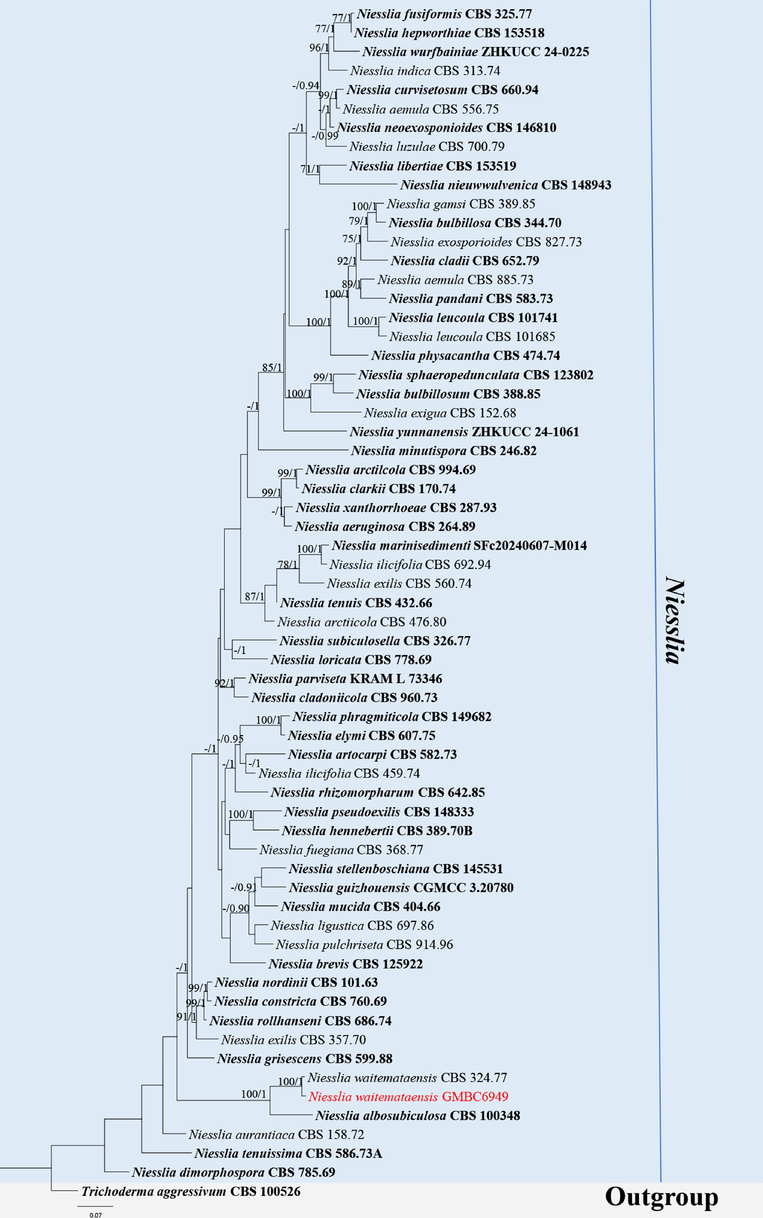
Figure 40.
ML tree of selected Niesslia species based on the ITS-LSU- tef1-α-tub2 dataset. The combined dataset comprised 63 taxa, 2,330 characters (ITS:1–533 bp; LSU: 534–1,416 bp; tef1-α: 1,417–1,899 bp; tub2: 1,900–2,330 bp), including indel regions. Maximum likelihood (lnL = −25,543.032496) and Bayesian analyses produced consistent tree topologies. Trichoderma aggressivum (CBS 100526) was selected as the outgroup taxon. Bayesian posterior probabilities (PP) ≥ 0.90 and RAxML bootstrap support values ≥ 70 % are shown on the branches. The new record is marked in red. Ex-type/type strains are indicated in bold.
Stachybotryaceae L. Lombard & Crous [as 'Stachybotriaceae'], Persoonia 32: 283 (2014).
Myxospora L. Lombard & Crous, Persoonia 36: 202 (2016).
MycoBank number: MB816011
Notes – Myxospora was established by Lombard et al., with Myxospora masonii designated as the type species[112]. The genus is known only from its asexual morph and is characterized by sporodochial or synnematous conidiomata; synnemata unbranched, cylindrical to pyriform, composed of compact, longitudinally arranged conidiophores; conidiophores macronematous; conidiogenous cells phialidic, with conspicuous collarettes and periclinal thickenings; and aseptate, fusiform conidia, with an apical hilum, lacking a funnel-shaped mucoid appendage[112,136,137].
Species of Myxospora occur as saprobes or weak pathogens on a broad range of plant substrates, including dead twigs, decaying grass leaves, leaf litter, leaves, and tar spot lesions on undetermined hosts. The genus shows a wide geographical distribution, with records from Asia, Europe, Africa, and North America, including China, England, Japan, Madagascar, and the USA[17,112]. According to Species Fungorum (accessed 3 January 2026), it currently comprises 11 accepted species. Herein, a new species of Myxospora is introduced.
Myxospora aquatica L.L. Liu, Q.F. Zhang, & Q.R. Li, sp. nov. (Fig. 41)
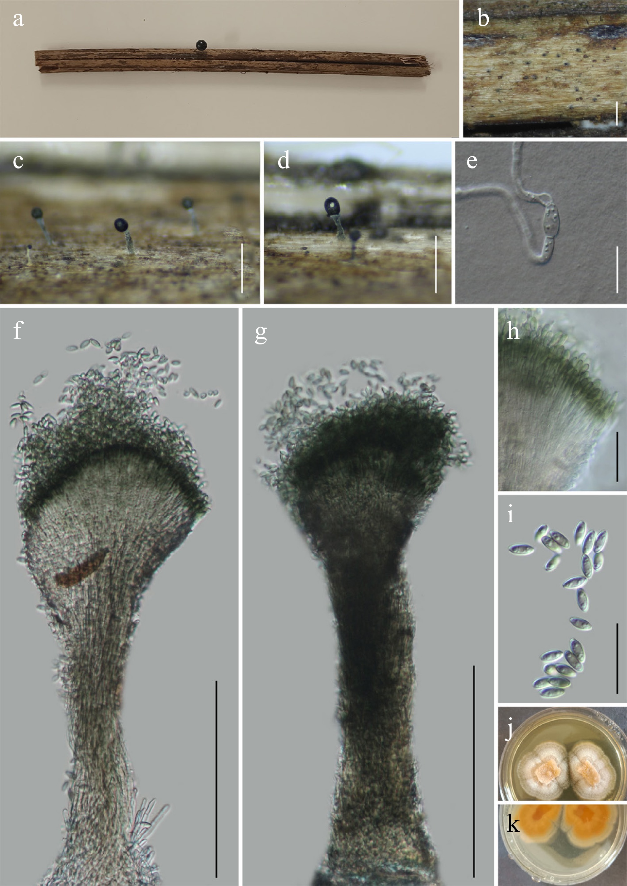
Figure 41.
Myxospora aquatica (GMB5158 holotype). (a) Host. (b)–(d) Colonies on natural substrate. (e) Germinating conidium. (f), (g) Conidiophores and conidia. (h) Conidiogenous cells. (i) Conidia. (j), (k) Surface and reverse view of culture on PDA. Scale bars: b = 1 mm, c, d = 0.5 mm, f, g = 120 μm, e, h, i = 20 μm.
MycoBank number: MB862978
Etymology – Referring to its occurrence on submerged decaying wood in freshwater habitats.
Saprobic on submerged decaying wood along a stream. Sexual morph: not observed. Asexual morph: hyphomycetous. Conidiomata synnematous, scattered to solitary on the host surface, erect or slightly curved, composed of densely compacted, longitudinal hyphae, terminating in a distinct black fertile head. Synnemata 190–270 × 32–73 μm (x = 235 × 45 μm, n = 30), hyaline to pale brownish green, smooth-walled, compact, firmly attached to substrate. Conidiophores reduced, integrated within synnemata, septate, hyaline to pale brownish green. Conidiogenous cells 11.7–23 × 1.2–2.6 μm (x = 16 × 1.7 μm, n = 30), phialidic, cylindrical, hyaline, smooth-walled, tapering apically, collarettes inconspicuous. Conidia 5.9–8.8 × 2.6–3.7 μm (x = 7.6 × 3.1 μm, n = 30) aseptate, short cylindrical to broadly ellipsoidal, smooth-walled, pale brown, produced in compact heads.
Culture characteristics – Conidia germinating on PDA within 48 h. Colonies on PDA reaching 15–25 mm after 21 d at 25 °C, circular, dense, centrally thick, velvety, margins entire, surface light brown at center, white toward margins; reverse light brown to brown.
Material examined – CHINA, Guangxi Zhuang Autonomous Region, Jiuwan Mountain National Nature Reserve, 25°9'50.49" N, 108°47'39.39" E, elevation 984 m, on submerged decaying wood along a stream, 18 August 2024, Qin-Fang Zhang, 2024JWS132 (GMB5158, holotype; GMBC5158, ex-type); ibid., KUN-HKAS 152911, isotype.
Additional specimens examined – CHINA, Guangxi Zhuang Autonomous Region, Liuzhou City, Jiuwanshan National Nature Reserve, 25°12′21″ N, 108°40′17″ E, elevation 1,322 m, on submerged decaying wood along a stream, 15 August 2024, Qin-Fang Zhang, 2024JWS160 (GMB5159, GMBC5159).
Notes – In the phylogenetic analysis (Fig. 42), Myxospora aquatica forms a well-supported (BS = 100; PP = 1) sister clade with M. synnematosa (KMNCC 23-14017). Comparative analysis of nucleotide base pairs showed that M. aquatica (GMBC5158) differs from the type strain of M. synnematosa (KMNCC 23-14017) by 0.5% (3/521 bp) in the ITS locus and 0.4% (3/785 bp) in the LSU locus.
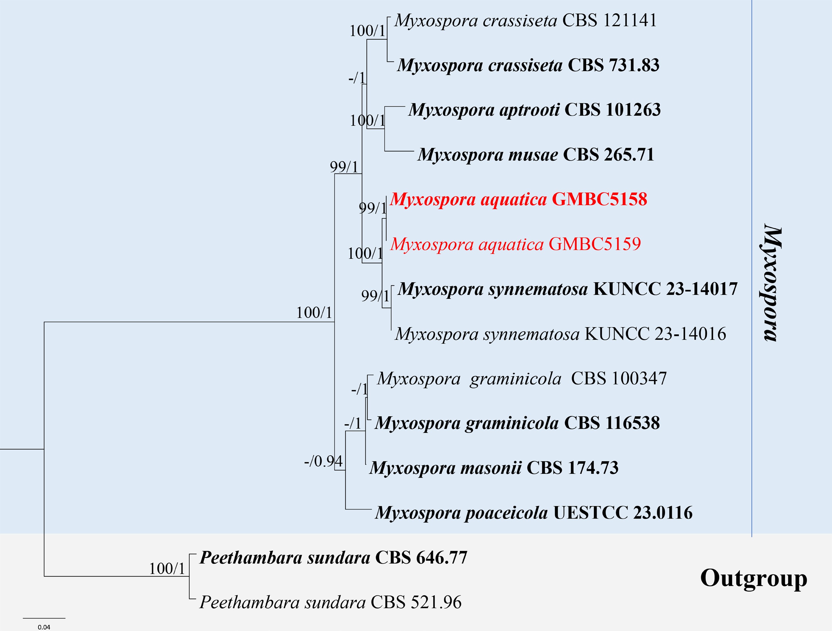
Figure 42.
ML tree of selected Myxospora species based on the ITS-LSU-tef1-α-rpb2-tub2 dataset. The combined dataset comprised 14 taxa, 2,895 characters (ITS:1–531 bp; LSU: 532–1,360 bp; tef1-α: 1,361–1,839 bp; rpb2: 1,840–2,618 bp; tub2: 2,619–2,895 bp), including indel regions. Maximum likelihood (lnL = −7,963.613215) and Bayesian analyses produced consistent tree topologies. Peethambara sundara (CBS 521.96) and P. sundara (CBS 646.77) were selected as the outgroup taxa. Bayesian posterior probabilities (PP) ≥ 0.90 and RAxML bootstrap support values ≥ 70 % are shown on the branches. The novel species is indicated in red and ex-type/type strains in bold.
Morphologically, M. aquatica resembles M. synnematosa in having synnematous conidiomata, phialidic conidiogenous cells, and aseptate conidia. However, M. aquatica differs by having much shorter synnemata (190–270 μm vs 405–550 μm in M. synnematosa) with longer conidiogenous cells (11.7–23 μm vs 5.8–13 μm). Moreover, the conidia of M. synnematosa often bear a small prominence when young and are narrower (2.4–2.9 μm wide) compared to those of M. aquatica (2.6–3.7 μm wide). Ecologically, M. synnematosa has been reported from terrestrial habitats, whereas M. aquatica was collected from a freshwater habitat[17].
Based on morphological, ecological, and phylogenetic differences, M. aquatica is described here as a new species.
Microascales Luttr. ex-Benny & R.K. Benj., Mycotaxon 12(1): 40 (1980).
Ceratocystidaceae Locq. [as 'Ceratocystaceae'], Syn. gen. fung. (Paris): [1] (1972).
Ophiostoma Syd. & P. Syd., Annls mycol. 17(1): 43 (1919).
=Ceratocystis Ellis & Halst., New Jers. agric. Exp. Sta. Bull. 76: 14 (1890).
Notes – Ophiostoma was established by Sydow and Sydow (with O. piliferum as the type species[1,138,139]. Species of Ophiostoma are characterized by dark, flask-shaped perithecia with elongated necks, often bearing ostiolar hyphae, and by diverse asexual morphs that are typically hyphomycetous, mononematous or synnematous, producing hyaline to pigmented conidia[139,140].
Members of the genus are predominantly saprobic or pathogenic on woody substrates and living trees, where many species cause blue stain of sapwood or serious tree diseases, particularly in conifers and broadleaf hosts[138]. Ecologically, Ophiostoma species are well known for their close associations with bark- and wood-inhabiting beetles and mites, which act as important vectors facilitating dispersal and host colonization[62,141]. The genus is widely distributed worldwide, occurring mainly in temperate and boreal regions but also reported from tropical areas, largely reflecting the distribution of their insect vectors and host trees. According to Species Fungorum (accessed 25 March 2026), the genus currently comprises 143 accepted species. Herein, a new species of Ophiostoma is introduced based on morphological characteristics and phylogenetic evidence.
Ophiostoma balanophorae L.L. Liu, Q.F. Zhang, & Q.R. Li, sp. nov. (Fig. 43)
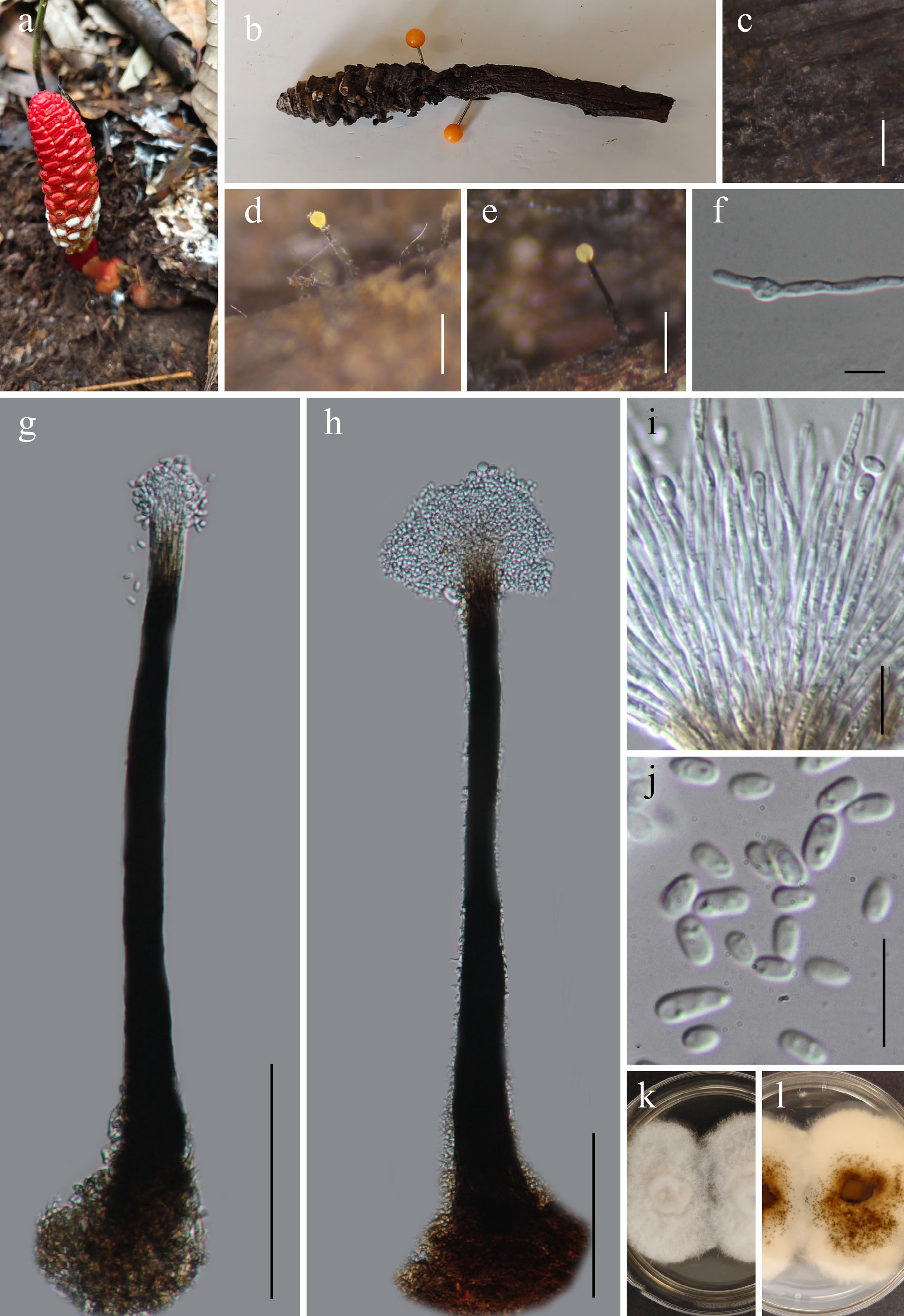
Figure 43.
Ophiostoma balanophorae (GMB5176 holotype). (a), (b) Host. (c)–(e) Synnemata on natural substrate. (f) Germinating conidium. (g), (h) Conidiophores and conidia. (i) Conidiogenous cells. (j) Conidia. (k), (l) Surface and reverse view of culture on PDA. Scale bars: b =1 mm, c = 0.5 mm; d = 0.25 mm; g, h= 100 μm; e, i, j = 10 μm.
MycoBank number: MB862994
Etymology – The epithet refers to the host genus Balanophora, from which this fungus was isolated.
Saprobic on dead stems of Balanophora laxiflora. Sexual morph: not observed. Asexual morph: hyphomycetous, pesotum-like. Colonies forming erect, dark synnemata, solitary to scattered, arising from the substrate surface. Synnemata 322–597 × 17.8–31.4 µm (x = 436.4 × 24.0 µm, n =15), unbranched, straight to slightly flexuous, composed of parallel conidiophores; stipe dark brown to black, basally swollen; apex with hemispherical to subglobose conidial head and mucilaginous spore mass. Mycelium partly superficial to mostly immersed, formed by septate, smooth-walled, hyaline to pale brown hyphae. Conidiophores macronematous, synnematous, densely aggregated into a single synnema, straight, cylindrical, smooth-walled, dark brown to black in the lower part, paler toward the apex. Conidiogenous cells 15.2–44.5 × 1.0–2.5 µm (x = 28.5 × 1.7 µm, n = 15) discrete, terminal, arising from the apical region of the synnema, hyaline, smooth-walled, cylindrical to narrowly lageniform, tapering slightly toward the apex, Conidia 3.7–7.3 × 2.2–3.2 µm (x = 5.2 × 2.6 µm, n = 30) hyaline, smooth-walled, ellipsoidal to obovoid, ends rounded, sometimes slightly truncated at the base, aseptate, often containing one or two guttules, accumulating in a hyaline to pale mucilaginous mass at the apex of the synnema.
Culture characteristics – Conidia germinate on PDA within 48 h. After 21 d at 25 °C, colonies on PDA reach 20–30 mm in diameter; circular, dense, centrally thick, cottony to floccose, margins entire, surface white; reverse pale to light brown, centrally dark brown.
Material examined – CHINA, Yunnan Province, Wuliangshan National Nature Reserve, 24°25'33.66" N, 101°23'55.67" E, elevation 1,455 m, on dead stems of Balanophora laxiflora Hemsl. (Balanophoraceae), 18 August 2025, Qin-Fang Zhang, 2025WLS57-1 (GMB5176, holotype; GMBC5176, ex-type); ibid., KUN-HKAS 152912, isotype.
Additional examined specimens – CHINA, Yunnan Province, Wuliangshan National Nature Reserve, 24°35'56.42" N, 100°25'55.67" E, elevation 2,180 m, on dead stems of Balanophora laxiflora Hemsl. (Balanophoraceae), 18 August 2025, Qin-Fang Zhang, 2025WLS85 (GMB5177, GMBC5177).
Notes – The ITS sequence of Ophiostoma balanophorae showed the closest match to O. araucariae in BLASTn search. The two species are also phylogenetically closely related (Fig. 44). However, the asexual morph of O. araucariae, described from culture, differs by having smaller conidia (2.5–4.5 × 2–2.5 µm), whereas O. balanophorae is characterized by a hyphomycetous morph on the natural substratum and has larger conidia (3.7–7.3 × 2.2–3.2 µm)[142]. Comparative analysis of nucleotide base pairs showed that O. balanophorae (GMBC5176) differs from the type strain of O. araucariae (CBS 114.68) by 4.5% (20/446 bp) in the ITS locus. Another phylogenetically related species, O. juglandis, differs in having a Hyalorhinocladiella-like asexual morph with mononematous, micronematous conidiophores[143], whereas O. balanophorae possesses a pesotum-like asexual morph, synnematous, macronematous conidiophores. Ophiostoma balanophorae is phylogenetically distinct from all other species of the genus, supporting its recognition as a novel taxon.
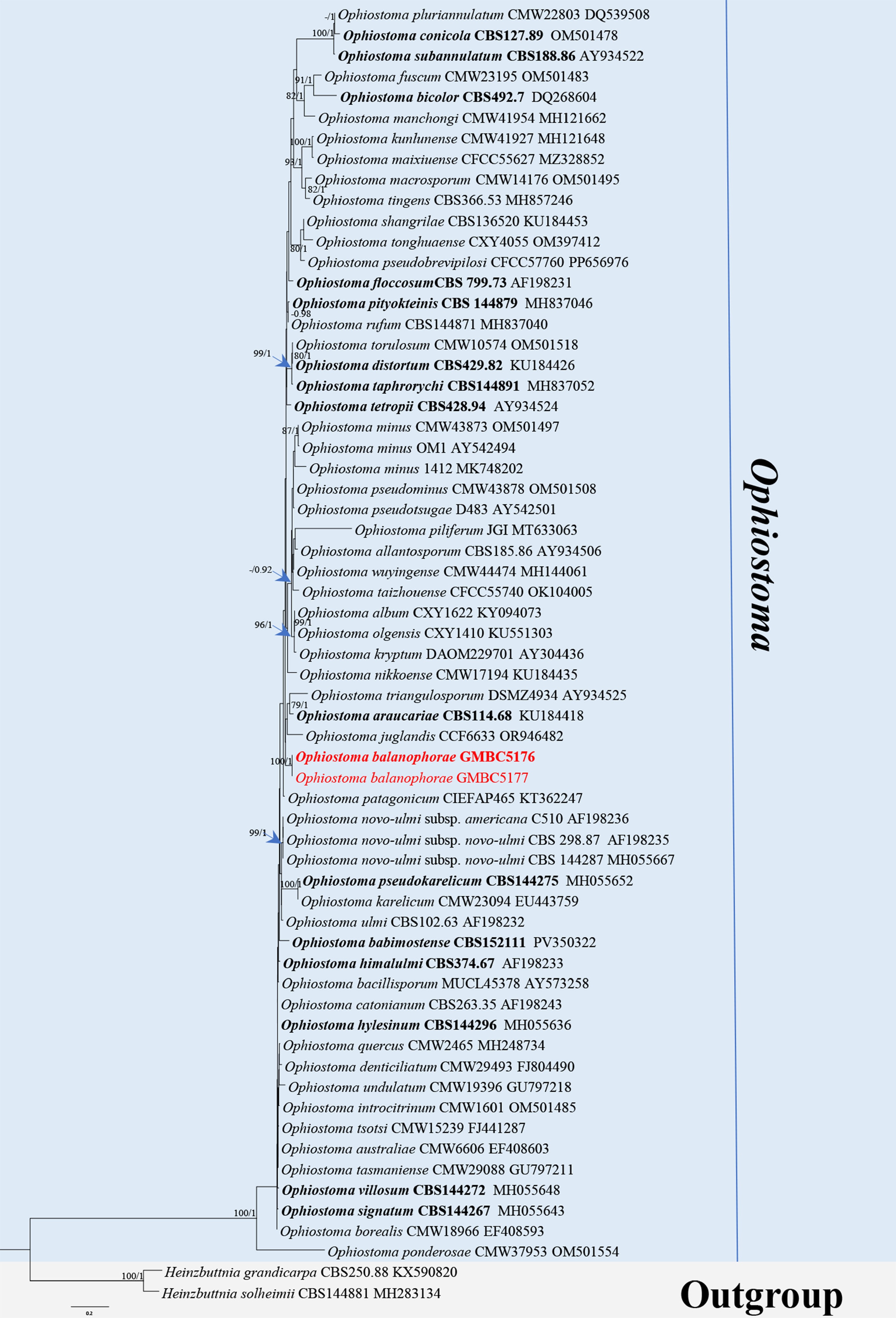
Figure 44.
ML tree of selected Ophiostoma species based on ITS. The combined dataset comprised 63 taxa, 573 characters, including indel regions. Maximum likelihood (lnL = −5,167.162764) and Bayesian analyses produced consistent tree topologies. Heinzbuttnia solheimii (CBS144881) and H. grandicarpa (CBS250.88) were selected as the outgroup taxa. Bayesian posterior probabilities (PP) ≥ 0.90 and RAxML bootstrap support values ≥ 70 % are shown on the branches. The novel species is indicated in red and ex-type/type strains in bold.
Microascaceae Luttr. ex-Malloch, Mycologia 62(4): 734 (1970).
Parascedosporium Gilgado, Gené, Cano & Guarro, Int. J. Syst. Evol. Microbiol. 57(9): 2176 (2007).
Notes – Parascedosporium was introduced by Gilgado et al., with P. tectonae as the type species[144]. The genus is characterized by sympodially proliferating, denticulate conidiogenous cells producing hyaline to pale conidia, and by synanamorphs with solitary conidiophores arising from aerial mycelium and forming graphium-like synnemata[144,145]. Species of Parascedosporium are saprobic and have been reported mainly from plant-associated substrates, including seeds and decaying plant material[144,146,147]. Sexual morph has not been observed for the genus to date[144,146,148]. According to Species Fungorum (accessed 1 January 2026), the genus currently comprises three accepted species.
Parascedosporium juglandicola L.L. Liu, Q.F. Zhang, & Q.R. Li, sp. nov. (Fig. 45)
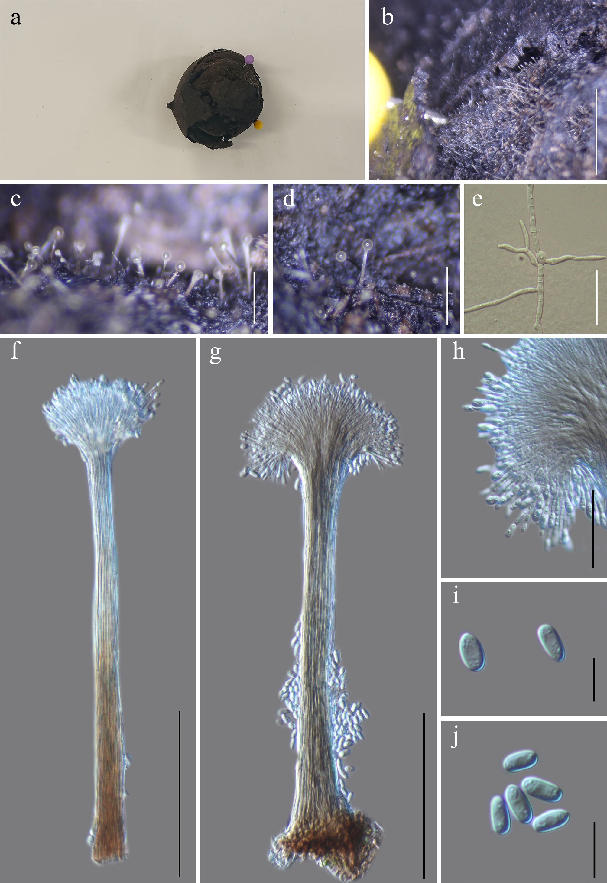
Figure 45.
Parascedosporium juglandicola (GMB5160 holotype). (a) Decaying walnut fruit. (b)–(d) Colonies on natural substrate. (e) Germinating conidium. (f), (g) Conidiophores and conidia. (h) Conidiogenous cells. (i), (j) Conidia. Scale bars: b = 3 mm, c = 4 mm, d = 0.5 mm, e, h = 30 μm, f, g = 100 μm, i, j = 10 μm.
MycoBank number: MB862995
Etymology –The specific epithet juglandicola is derived from Juglans (host genus), referring to its occurrence on walnut fruit pericarps.
Saprobic on decaying pericarps of Juglans regia. Sexual morph: not observed. Asexual morph: hyphomycetous, forming graphium-like synnemata on natural substrate. Colonies on natural substrate superficial and inconspicuous, not forming effuse mycelial mats, represented by scattered, erect, hyaline to whitish synnemata; apices bearing a transparent mucilaginous conidial droplet. Mycelium partly immersed to superficial, formed by smooth-walled, septate, branched, pale brown to olivaceous brown hyphae. Conidiophores aggregated into synnemata 87–367.5 µm tall, erect, solitary to gregarious, composed of parallel, compacted conidiophores. Stipe cylindrical, straight, smooth-walled, hyaline to light brown, widening slightly towards the base, terminating in a conspicuous, hyaline mucilaginous conidial head. Solitary conidiophores 5.7–27.7 µm (x = 18.6 µm, n = 30) wide, mononematous, macronematous, emerge from aerial mycelium or form synnemata unbranched, thick-walled, septate, darker at the base, paler towards the apex. Conidiogenous cells 13.6–27 × 0.9–2.2 µm (x = 18.8 × 1.6 µm, n = 30), enteroblastic, percurrent, cylindrical, integrated, terminal, smooth-walled, hyaline to subhyaline, proliferating percurrently to form inconspicuous annellations, producing conidia from the apex; collarettes absent; conidia accumulating in a slimy mass at the synnematal apex. Conidia 4.8–7.9 × 2.3–4.0 µm (x = 6.5 × 3.4 µm, n = 30), solitary, cylindrical to ellipsoid, aseptate, straight to slightly curved, smooth-walled, hyaline, apex rounded, base rounded to slightly attenuated.
Material examined – CHINA, Yunnan Province, Wumengshan National Nature Reserve 25°21′36.87″ N, 103°15′29.76″ E, elevation 2,473 m, on decaying pericarps of Juglans regia, 22 July 2024, Qin-Fang Zhang, 2024WMS31-1 (GMB5160, holotype; GMBC5160, ex-type); ibid., KUN-HKAS 152913, isotype.
Additional specimens examined – CHINA, Yunnan Province, Wumengshan National Nature Reserve 25°36.91.15″ N, 103°15′30.56″ E, elevation 2,584 m, on decaying pericarps of Juglans regia, 22 July 2024, Qin-Fang Zhang, 2024WMS56 (GMB5161, GMBC5161).
Notes – Phylogenetically (Fig. 46), Parascedosporium juglandicola is closely related to P. tectonae and P. germanicum. Morphologically, P. juglandicola resembles both in having graphium-like synnemata, percurrently proliferating conidiogenous cells, and aseptate conidia that accumulate in a mucilaginous apical head. However, P. juglandicola can be distinguished from P. tectonae by its shorter synnemata (87–367.5 µm vs 325–450 µm in P. tectonae), and by its conidia, which are hyaline, cylindrical to ellipsoid, and 2.3–4 µm wide, whereas those of P. tectonae are subglobose to obovate, brown, and 4.5–6 µm wide. Ecologically, P. juglandicola was isolated from the pericarp of decaying walnut fruits, whereas P. tectonae was originally described from woody substrates[144]. Parascedosporium germanicum, isolated from llama dung, is characterized by brown to dark brown synnemata that terminate in a gray-brown to black slimy conidial head, and larger conidia 7–11 × 2.5–4 µm[145], whereas P. juglandicola has hyaline to light brown synnemata bearing a hyaline mucilaginous conidial head. No species of Parascedosporium has previously been reported from the pericarp of walnut or from Juglans. Based on the combined morphological, ecological, and phylogenetic analyses, P. juglandicola is introduced here as a novel species.
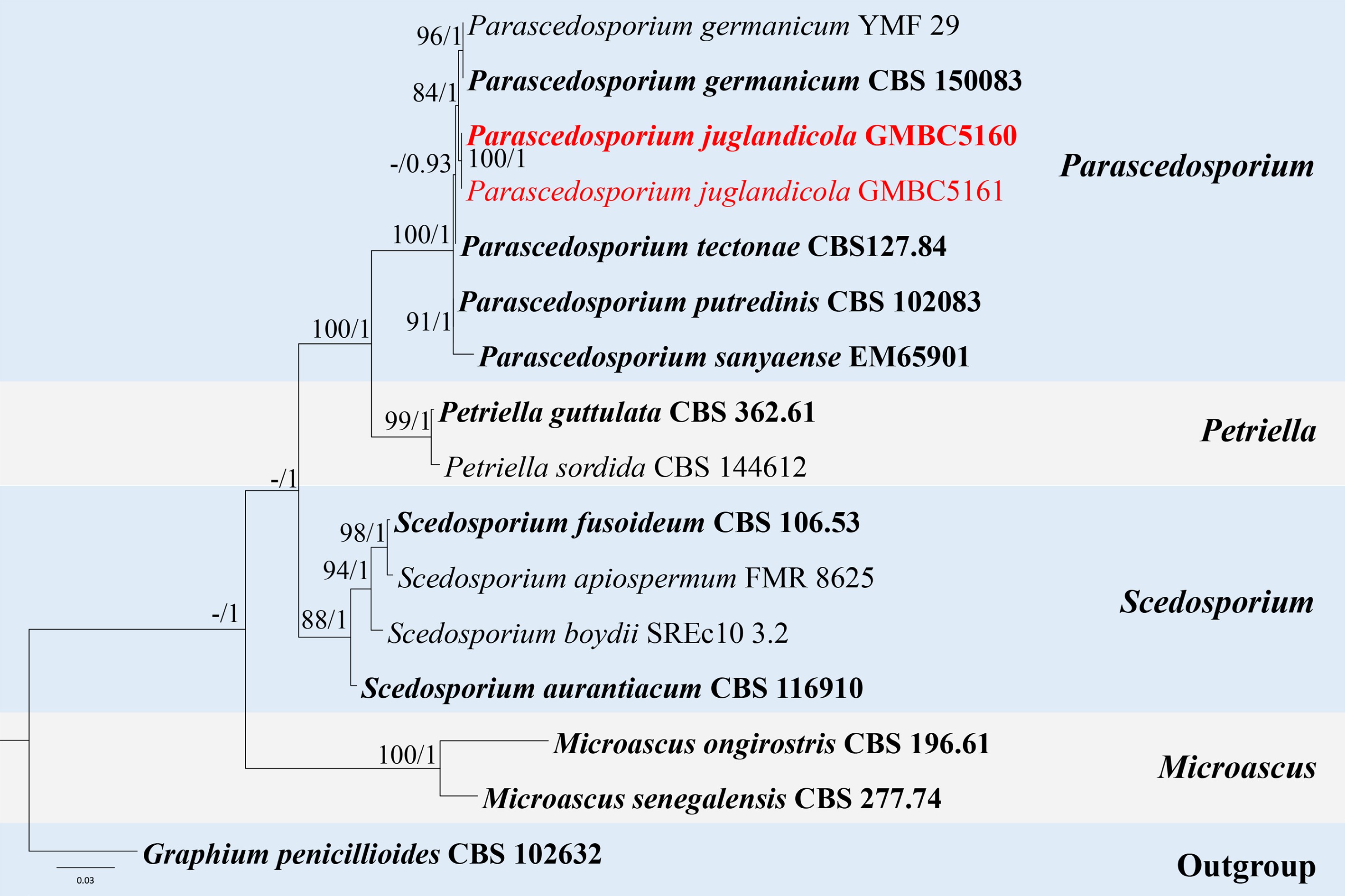
Figure 46.
ML tree of selected species of Parascedosporium and related genera based on the ITS-LSU dataset. The combined dataset comprised 14 taxa, 1,329 characters (ITS: 1–523 bp; LSU: 524–1,329 bp), including indel regions. Maximum likelihood (lnL = −4076.672862) and Bayesian analyses produced consistent tree topologies. Graphium penicillioides (CBS 102632) was selected as the outgroup taxon. Bayesian posterior probabilities (PP) ≥ 0.90 and RAxML bootstrap support values ≥ 70 % are shown on the branches. The novel species is indicated in red and ex-type/type strains in bold.
Myrmecridiales Crous, Persoonia 34: 219 (2015).
Myrmecridiaceae Crous, Persoonia 34: 219 (2015).
Myrmecridium Arzanlou, W. Gams & Crous, Stud. Mycol. 58: 84 (2007).
Notes – Myrmecridium was established by Arzanlou et al., typified by M. schulzeri[149]. The genus was previously treated as an incertae sedis genus of Sordariomycetes, but later multigene phylogenetic analyses demonstrated that Myrmecridium represents a distinct, monophyletic lineage within Sordariomycetes[150]. Consequently, the order Myrmecridiales and the family Myrmecridiaceae were introduced to accommodate species of Myrmecridium.
Morphologically, Myrmecridium is characterized by macronematous conidiophores and terminal, integrated conidiogenous cells bearing distinctive, pimple-like denticles. Conidia are solitary, aseptate, obovoid to fusiform, tapering towards a narrowly truncate base, and smooth to finely verrucose. The mycelium is hyaline to pale, with conidiogenous structures usually subhyaline to lightly pigmented[149]. Species of the genus are widely distributed and are commonly isolated from soil and plant-associated substrates, including decaying plant tissues, indicating a predominantly saprobic lifestyle across diverse ecological niches[1,113,151,152]. According to Species Fungorum (accessed 10 January 2026), the genus currently comprises 24 accepted species. Herein, we describe a novel species of the genus from bamboo in a freshwater habitat.
Myrmecridium bambusicola L.L. Liu, W.M. Li, & Q.R. Li, sp. nov. (Fig. 47)
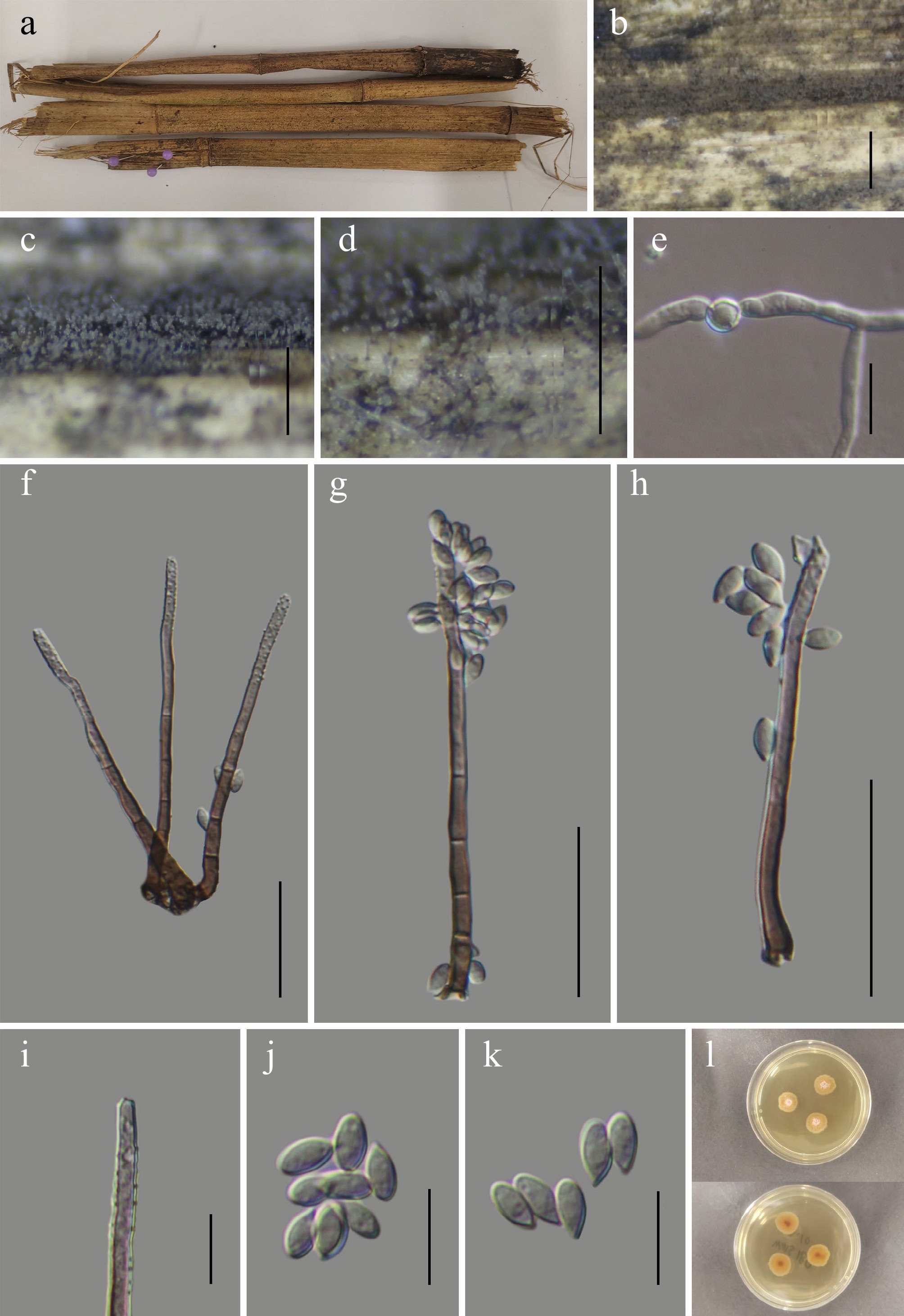
Figure 47.
Myrmecridium bambusicola (GMB6939 holotype). (a) Decaying bamboo host. (b)–(d) Colonies on natural substrate. (e) Germinating conidium. (f)–(h) Conidiophores and conidia. (i) Conidiogenous cells. (j), (k) Conidia. (l) Surface and reverse view of culture on PDA. Scale bars: b = 1 mm, c, d = 0.5 mm, f–h = 30 μm, e, i–k = 10 μm.
MycoBank number: MB862996
Etymology –bambusicola (Latin bambus = bamboo; suffix -cola = inhabiting), referring to the host bamboo.
Saprobic on dead bamboo culm in a freshwater stream. Sexual morph: not observed. Asexual morph: hyphomycetous. Colonies on natural substrate are effuse, greyish white, velvety. Mycelium immersed, composed of brown, branched, septate hyphae. Conidiophores 53–84 × 2–3.5 μm (x = 68.7 × 2.9 μm, n = 30), mononematous, solitary, macronematous, straight or slightly flexuous erect, subcylindrical, unbranched, 3–5-septate, paler towards apex, smooth, thick-walled. Conidiogenous cells 6–25 × 2– 4 μm (x = 16 × 3.1 μm, n = 30), integrated, terminal, polyblastic, subcylindrical, subhyaline, verrucose, with several denticles at the apex. Conidia 5–8 × 3–4 μm (x = 6.2 × 3.2 μm, n = 30), ellipsoid, obovoid or fusiform, solitary, aseptate, subhyaline, smooth or finely verrucose-walled.
Culture characteristics – Conidia germinate on PDA within 48 h. After five weeks at 25 °C, colonies on PDA reach 10–15 mm in diameter; circular, centrally thick, with a distinct central umbo, margins entire, thick, surface white; reverse orange-brown, pigmentation concentrated in the central zone.
Material examined – CHINA, Yunnan Province, Zhaotong City, Wumengshan National Nature Reserve, 25°21′36.87″ N, 103°15′29.76″ E, elevation 2,473 m, on dead bamboo culm in a freshwater stream, 26 October 2024, Wen-Mei Li, 2024WMS180 (GMB6939, holotype; GMBC6939, ex-type); ibid., KUN-HKAS 152914, isotype.
Additional specimens examined – CHINA, Yunnan Province, Zhaotong City, Wumengshan National Nature Reserve, 25°36.91.15″ N, 103°15′30.56″ E, elevation 2,584 m, on dead bamboo culm in a freshwater stream, 26 October 2024, Wen-Mei Li, 2024WMS183 (GMB6940, GMBC6940).
Notes – Phylogenetically (Fig. 48), Myrmecridium bambusicola forms a well-supported (BS = 92; PP = 1) sister clade to the type strain of M. schulzeri (CBS 325.74). Pairwise comparison of nucleotide sequences revealed that M. bambusicola (GMBC6939) differs from the type strain of M. schulzeri (CBS 325.74) by 1.3 % (6/469 bp) in the ITS locus and 0.9 % (7/814 bp) in the LSU locus.
Morphologically, M. bambusicola is distinguishable from M. schulzeri by its much shorter conidiophores (53–84 µm long vs up to 250 µm in M. schulzeri) and by its conidia, which are smaller (5–8 µm long) and lack a mucilaginous sheath. In contrast, M. schulzeri produces conidia (6–)9–10(–12) µm long, surrounded by a wing-like, gelatinous sheath up to 0.5 µm thick[149,153].
Given these morphological, molecular, and phylogenetic differences, M. bambusicola is introduced here as a novel species.
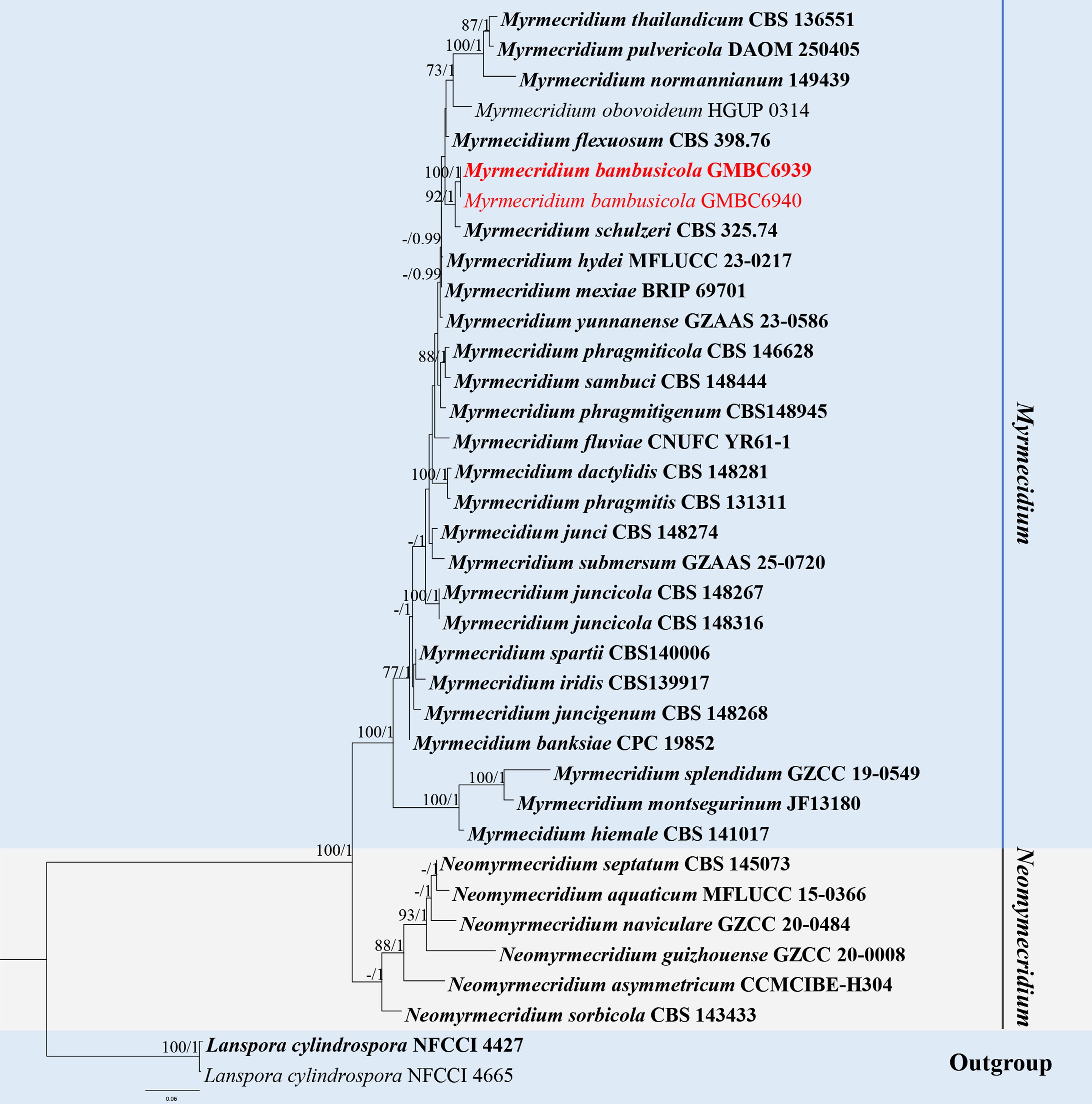
Figure 48.
ML tree of selected species of Myrmecridium and related genera inferred from a combined ITS-LSU dataset. The combined dataset comprised 36 taxa, 1,329 characters (ITS: 1–491 bp; LSU: 491–1,329 bp), including indel regions. Maximum likelihood (lnL = –6,247.180591) and Bayesian analyses produced consistent tree topologies. Lanspora cylindrospora (NFCCI 4427) and L. cylindrospora (NFCCI 4665) were selected as the outgroup taxa. Bayesian posterior probabilities (PP) ≥ 0.90 and RAxML bootstrap support values ≥ 70 % are shown on the branches. The novel species is indicated in red and ex-type/type strains in bold.
Pleurotheciales Réblová & Seifert, Persoonia 37: 63 (2015) [2016].
Pleurotheciaceae Réblová & Seifert, Persoonia 37: 63 (2015) [2016].
Pleurothecium Höhn., Berichte der Deutschen Botanischen Gesellschaft 37: 154 (1919).
Notes – Pleurothecium was established by Höhnel, typified by P. recurvatum[110]. The genus was previously linked to the sexual genus Carpoligna. However, following the one fungus–one name principle, Carpoligna has been synonymized with Pleurothecium[152,154]. Pleurothecium is mainly characterized by macronematous, brown conidiophores; sympodially, polyblastic, proliferating, denticulate conidiogenous cells; and solitary, hyaline to pigmented, unicellular or septate, and cylindrical to fusiform or clavate conidia. The sexual morph, known in only a few species, is similar to Chaetosphaeria but differs in having holoblastic rather than phialidic conidiogenous cells[2,22,152,155−157].
Species of Pleurothecium are saprobic on dead wood and plant debris in terrestrial habitats and are widely distributed in tropical and temperate regions. Phylogenetically, the genus forms a well-supported monophyletic clade within Pleurotheciaceae, except for P. obovoideum, which represents a distinct lineage[3,158]. According to Species Fungorum (accessed 3 January 2026), Pleurothecium currently comprises 19 accepted species. Herein, a new species of the genus is described.
Pleurothecium longisetosum L.L. Liu, W.M. Li, & Q.R. Li, sp. nov. (Fig. 49)
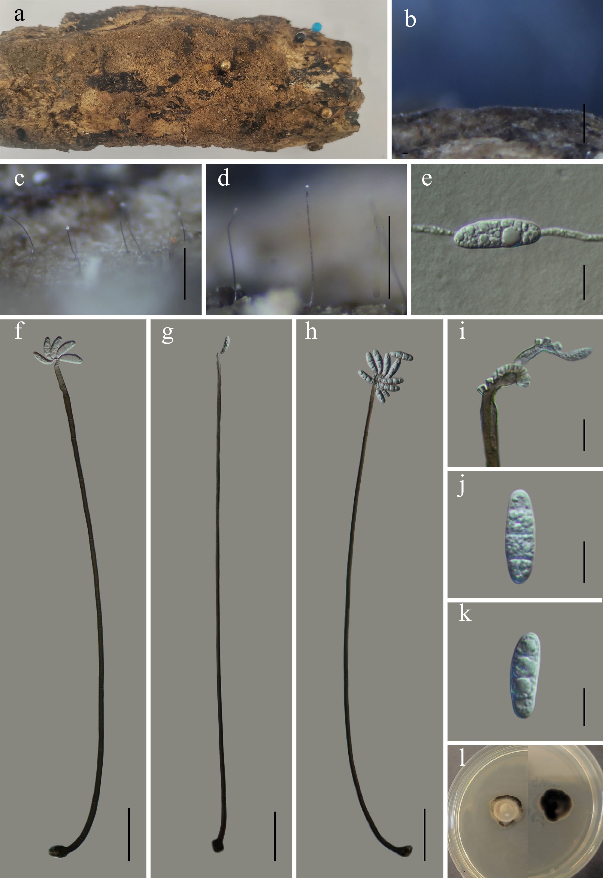
Figure 49.
Pleurothecium longisetosum (GMB6937 holotype). (a) Host. (b)–(d) Colonies on natural substrate. (e) Germinating conidium. (f)–(h) Conidiophores and conidia. (i) Conidiogenous cells. (j), (k) Conidia. (l) Surface and reverse view of culture on PDA. Scale bars: b = 1 mm, c, d = 0.5 mm, f–h = 50 μm, e, i–k = 10 μm.
MycoBank number: MB863007
Etymology – The epithet refers to the long seta-like conidiophores of the species.
Saprobic on dead wood in a freshwater stream. Sexual morph: not observed. Asexual morph: hyphomycetous. Colonies superficial, effuse, brown to dark brown, velvety to smooth. Mycelium partly superficial, partly immersed, formed by septate, smooth, brown to pale brown hyphae. Conidiophores 310–685 × 2.8–6.3 µm (x = 461 × 4.5 µm, n = 30), macronematous, mononematous, arising from superficial hyphae, solitary or loosely aggregated, long, filiform, cylindrical to subcylindrical, straight to slightly flexuous, unbranched, smooth, septate, brown, gradually paler toward the apex; basal cell slightly swollen, attached directly to substrate. Conidiogenous cells 2.5–5.1 × 0.9–3.9 µm (x = 2.7 × 1.9 µm, n = 30), terminal, integrated, polyblastic, sympodial, subcylindrical to narrowly clavate, holoblastic, bearing conspicuous denticles, subhyaline to pale brown. Conidia 21.2–25.6 × 6.8–7.3 µm (x = 22.8 × 7.1 µm, n = 30), acrogenous, straight to slightly curved, solitary, oblong to ellipsoidal, 3-septate, slightly constricted at septa, hyaline to pale green, smooth-walled, apices rounded, base truncate to slightly tapering.
Culture characteristics – Conidia germinate on PDA within 24 h. After five weeks at 25 °C, colonies on PDA reach 10–20 mm in diameter; circular, centrally thick, margins entire, thick, surface light grey to creamy; reverse dark brown to black.
Material examined – CHINA, Yunnan Province, Zhaotong City, Wumengshan National Nature Reserve, 25°21′15.13″ N, 103°15′05.18″ E, elevation 2,700 m, on decaying wood in freshwater stream, 26 October 2024, Wen-Mei Li, 2024WMS170-1 (GMB6937, holotype; GMBC6937, ex-type); ibid., KUN-HKAS 152915, isotype.
Additional specimens examined – CHINA, Yunnan Province, Zhaotong City, Wumengshan National Nature Reserve, 25°21′15.15″ N, 103°15′05.94″ E, elevation 2,750 m, on decaying wood in freshwater stream, 26 October 2024, Wen-Mei Li, 2024WMS182 (GMB6938, GMBC6938).
Notes – In BLAST searches of ITS and LSU sequences, Pleurothecium longisetosum showed the closest similarity to P. hyalosporum, and the two species are also phylogenetically closely related (Fig. 50). Morphologically, P. longisetosum resembles P. hyalosporum in having mononematous, macronematous conidiophores, sympodial, polyblastic conidiogenous cells, and hyaline conidia. However, P. longisetosum can be distinguished from P. hyalosporum by its longer conidiophores (310–685 µm vs 205–578 µm), and slightly longer, consistently 3-septate conidia (21.2–25.6 µm long) compared to the predominantly aseptate (rarely 3-septate) conidia of P. hyalosporum (14.6–22.1 µm long)[159,160]. Another phylogenetically close species, P. lignicola, differs in having much shorter, branched conidiophores (154–280 µm vs 310–685 µm) and smaller conidia (14–21.5 × 4.5–6.5 µm vs 21.2–25.6 × 6.8–7.3 µm)[157]. The combination of distinct morphological characteristics and phylogenetic placement supports the description of P. longisetosum as a novel species.
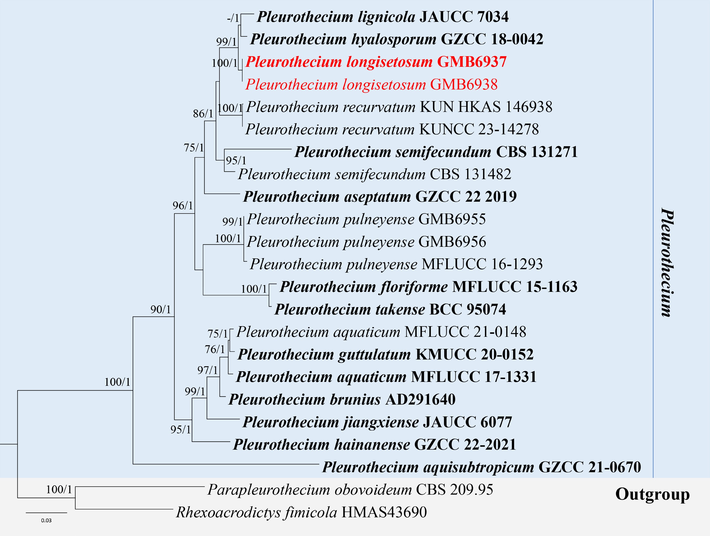
Figure 50.
ML tree of selected Pleurothecium species based on the ITS-LSU-SSU dataset. The combined dataset comprised 23 taxa, 2,442 characters (ITS: 1–555 bp; LSU: 556–1,425 bp; SSU: 1,426–2,442 bp), including indel regions. Maximum likelihood (lnL = –8,244.303005) and Bayesian analyses produced consistent tree topologies. Parapleurothecium obovoideum (CBS 209.95) and Rhexoacrodictys fimicola (HMAS43690) were selected as the outgroup taxa. Bayesian posterior probabilities (PP) ≥ 0.90 and RAxML bootstrap support values ≥ 70 % are shown on the branches. The novel species is indicated in red and ex-type/type strains in bold.
Rhamphoriales K.D. Hyde & Hongsanan, Fungal Diversity 107: 94 (2021).
Rhamphoriaceae Réblová, in Réblová & Štěpánek, Mycologia 110(4): 754 (2018).
Rhamphoriopsis Réblová & Gardiennet, Mycologia 11(4): 755 (2018).
Notes – Rhamphoriopsis was established by Réblová & Štěpánek, with R. muriformis as the type species, isolated from dead wood of Buxus sempervivens in France[161]. Rhamphoriopsis is characterized by perithecial, nonstromatic, immersed ascomata with cylindrical necks, unitunicate asci bearing eight ascospores and a distinct non-amyloid apical apparatus, and smooth-walled, ellipsoidal to fusiform ascospores. The asexual morph comprises macronematous, mononematous conidiophores that may occur singly or in loose fascicles. Conidiogenous cells are integrated, terminal, cylindrical, and apically tapering, polyblastic, producing numerous denticles. Conidia are ellipsoidal to obovoid, hyaline, and aseptate[161,162]. Rhamphoriopsis species are saprobes on dead wood, mostly collected from moist, river-associated habitats in temperate Europe and Asia[162−164]. According to Species Fungorum (accessed 25 March 2026), 13 species are currently accepted in Rhamphoriopsis. In this work, we introduce a novel species of the genus isolated from decaying wood collected in a riverine habitat.
Rhamphoriopsis flabelliformis L.L. Liu, W.M. Li, & Q.R. Li, sp. nov. (Fig. 51)
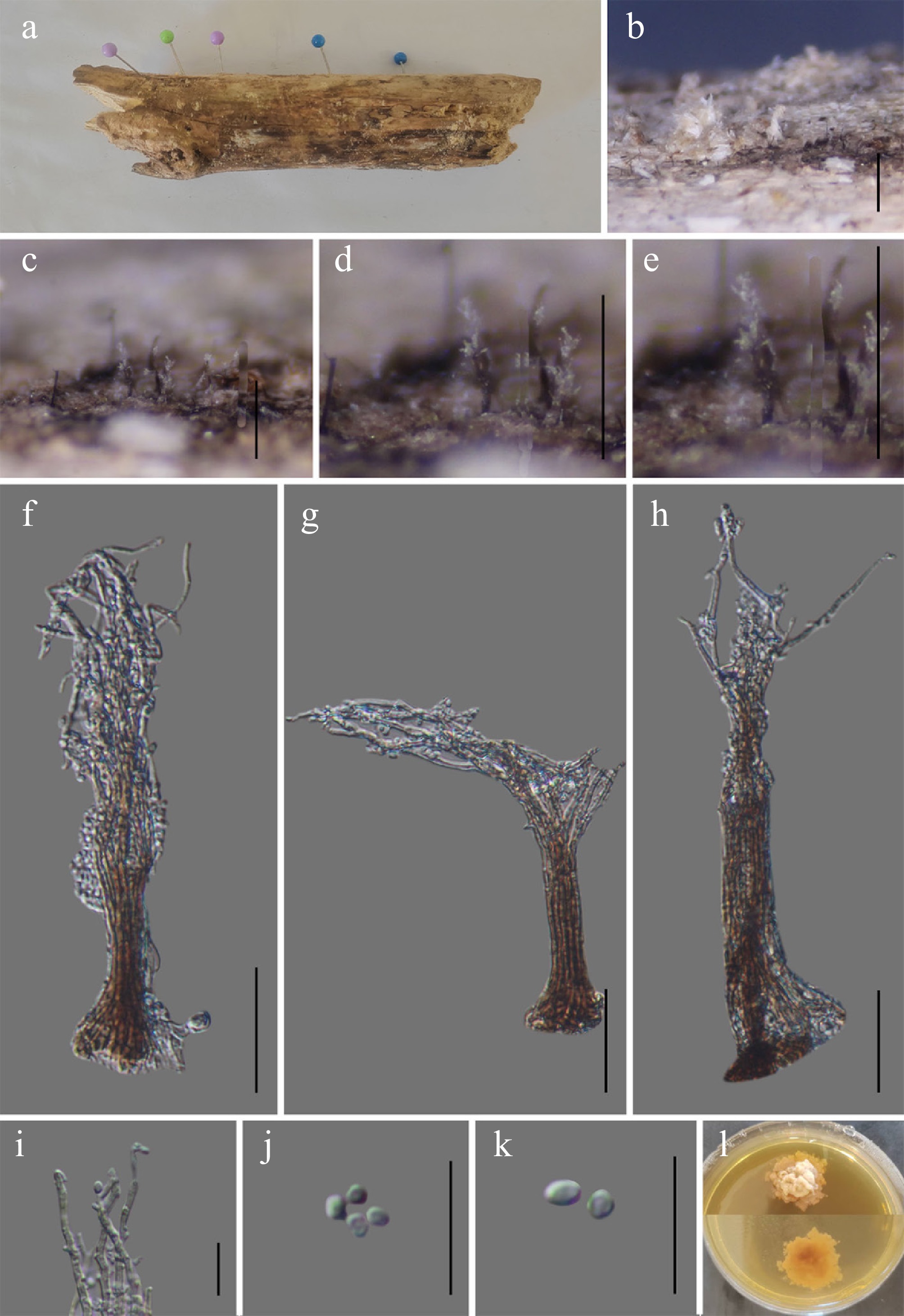
Figure 51.
Rhamphoriopsis flabelliformis (GMB6923 holotype). (a) Host. (b)–(e) Colonies on natural substrate. (f)–(h) Conidiophores and conidia. (i) Conidiogenous cells. (j), (k) Conidia. (l) Surface and reverse view of culture on PDA. Scale bars: b = 1 mm, c, d = 0.5 mm, f–h = 30 μm, i–k = 10 μm.
MycoBank number: MB863008
Etymology – The specific epithet flabelliformis refers to the apically splayed, fan-like synnematal head.
Saprobic on submerged dead wood in riverine habitats. Sexual morph: not observed. Asexual morph: hyphomycetous. Colonies superficial, effuse, scattered to gregarious, hairy, forming solitary to aggregated synnemata, with apical white conidial masses. Mycelium partly immersed, partly superficial, formed by septate, pale brown to hyaline, smooth-walled hyphae. Conidiophores synnematous, erect, macronematous, cylindrical, septate, smooth-walled, compactly parallel. Synnemata 68–184 µm long (x = 125.3 µm, n = 30) and 8.3–18.6 µm wide at mid-region (x = 11.6 µm, n = 30); base distinctly widened, reddish brown, apex paler; basal portion flared; apex expanded, spread widely, composed of divergent conidiophore tips; terminal region hyaline to subhyaline. Conidiogenous cells 1.3–2.4 µm wide (x = 1.7 µm, n = 30), integrated, sympodial, terminal, polyblastic, pale brown basally, hyaline apically, smooth-walled, denticulate, denticles aggregated at synnematal apex. Conidia 2.0–3.4 × 1.2–2.0 µm (x = 2.5 × 1.5 µm, n = 30), ellipsoidal to broadly ellipsoidal, aseptate, hyaline, smooth-walled, forming mucilaginous masses at synnematal apex.
Culture characteristics – Conidia germinate on PDA within 24 h. After five weeks at 25 °C, colonies on PDA reach 10–20 mm in diameter; irregular, centrally thick, aerial mycelium sparse to moderate, margins lobate, surface white, light brow at margins; reverse pale yellow to light brown.
Material examined – CHINA, Guizhou Province, Tongren City, Fodingshan National Nature Reserve, 27°23′0.35″ N, 108°7′30.27″ E, elevation 598 m, on submerged decaying wood in riverine habitats, 10 August 2024, Wen-Mei Li, 2025FDSDCY36-3 (GMB6923, holotype; GMBC6923, ex-type); ibid., KUN-HKAS 152917, isotype.
Additional examined specimens – CHINA, Guizhou Province, Tongren City, Fodingshan National Nature Reserve, 27°23′2.56″ N, 108°7′30.17″ E, elevation 966 m, on submerged decaying wood in riverine habitats, 10 August 2024, Wen-Mei Li, 2025FDSDCY80 (GMB6924, GMBC6924).
Notes – Phylogenetically (Fig. 52), Rhamphoriopsis flabelliformis forms a well-supported (BS = 100; PP = 1) sister clade with R. aquimicrospora (GZCC 20-0515). Pairwise comparison of nucleotide sequences revealed that R. flabelliformis (GMBC6923) differs from the type strain of R. aquimicrospora (GZCC 20-0515) by 1.8 % (9/495 bp) in the ITS locus and 0.12 % (1/833 bp) in the LSU locus. Morphologically, R. flabelliformis can be distinguished from R. aquimicrospora by its broader synnemata (8.3–18.6 µm wide vs 5–11 µm wide) and by its more conspicuously flared, fan-shaped synnematal apex, whereas R. aquimicrospora has a compact, less expanded apical head[16].
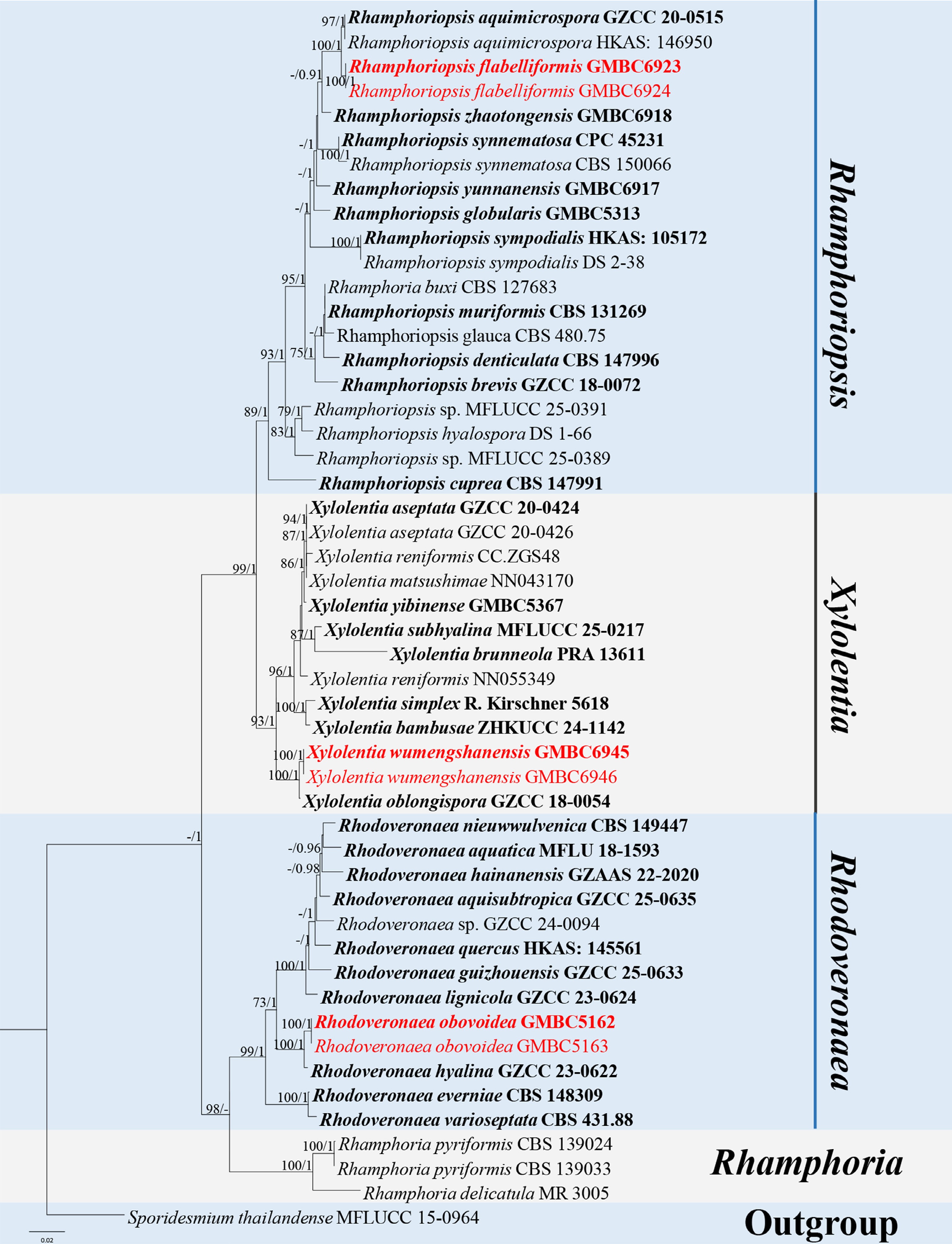
Figure 52.
ML tree of selected Rhamphoriaceae species based on the ITS-LSU-SSU dataset. The combined dataset comprised 50 taxa, 2,373 characters (ITS: 1–512 bp; LSU: 513–1,372 bp; SSU: 1,373–2,373 bp), including indel regions. Maximum likelihood (lnL = –9,769.852619) and Bayesian analyses produced consistent tree topologies. Sporidesmium thailandense (MFLUCC 15-0964) was selected as the outgroup taxon. Bayesian posterior probabilities (PP) ≥ 0.90 and RAxML bootstrap support values ≥ 70 % are shown on the branches. The novel species are indicated in red and ex-type/type strains in bold.
Based on the combined molecular, morphological, and phylogenetic differences, R. flabelliformis is herein introduced as a new species.
Rhodoveronaea Arzanlou, W. Gams & Crous, Stud. Mycol. 58: 89 (2007).
Notes – Rhodoveronaea was established by Arzanlou et al., typified by R. varioseptata[149]. The genus is mainly known from its asexual morphs, characterized by reddish-brown, septate, straight to flexuous conidiophores with inflated basal cells. Conidiogenous cells are terminal, bearing crowded, slightly conspicuous denticles. Conidia are ellipsoidal to obovoidal, pale brown, septate, with a protruding basal hilum and a marginal basal frill[149,165−167]. The sexual morph, described by Réblová, has immersed ascomata with a subglobose to conical venter and a conical neck, filamentous septate paraphyses longer than the asci, unitunicate cylindrical asci with long stipes, and fusiform, septate, hyaline ascospores[167]. Species of Rhodoveronaea are saprobic on dead wood in both terrestrial and freshwater habitats and are distributed from tropical to temperate regions[168,169]. According to Species Fungorum (accessed 5 January 2026), the genus currently comprises 10 accepted species. In the present study, a new species of Rhodoveronaea is introduced.
Rhodoveronaea obovoidea L.L. Liu, Q.F. Zhang, & Q.R. Li, sp. nov. (Fig. 53)
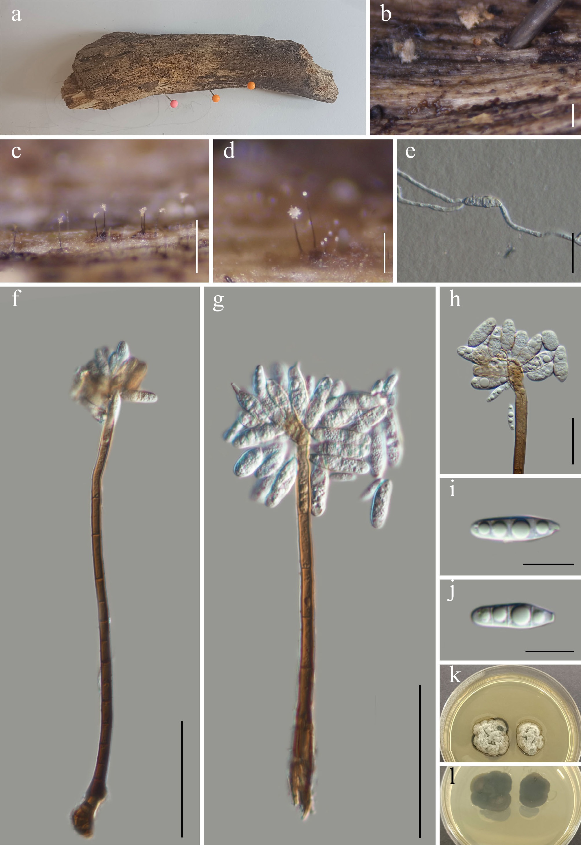
Figure 53.
Rhodoveronaea obovoidea (GMB5162 holotype). (a) Host. (b)–(d) Colonies on natural substrate. (e) Germinating conidium. (f), (g) Conidiophores and conidia. (h) Conidiogenous cells and conidia. (i), (j) Conidia. (k), (l) Surface and reverse view of culture on PDA. Scale bars: b = 1 mm, c = 0.5 mm, d = 0.2 mm, f, g = 50 μm, e, h = 20 μm, i, j = 10 μm.
MycoBank number: MB863010
Etymology – The epithet refers to the obovoid shape of the conidia.
Saprobic on submerged dead wood in a freshwater wetland. Sexual morph: not observed. Asexual morph: hyphomycetous. Colonies effuse, inconspicuous, hairy, brown. Conidiophores 147–207 × 4–6 μm (x = 184 × 5 μm, n=30), mononematous, macronematous, arising vertically from creeping hyphae, cylindrical, straight or flexuose, simple, red-brown, paler at apex, thick-walled, septate. Conidiogenous cells 24–49 × 5–7 μm (x = 37 × 6 μm, n = 30), terminally integrated, sympodial, polyblastic, pale brown at the base, paler towards the apex, smooth, thick-walled. Conidia 9–21 × 4–8 μm (x = 17 × 6 μm, n = 30), ellipsoid to obovoid, acropleurogenous, 2–4-septate, hyaline to pale brown, apically rounded with a flat basal scar, guttulate, smooth-walled.
Culture characteristics – Conidia germinate on PDA within 24 h. After 28 d at 25 °C, colonies on PDA reach 20–30 mm in diameter; circular to irregular, raised in the middle, aerial mycelium dense, margins entire to lobate, surface white, light brown at margins; reverse bluish-black to dark brown.
Material examined – CHINA, Guangxi Zhuang Autonomous Region, Cenwang Laoshan National Nature Reserve, 24°17′05.04″ N, 106°13′51.25″ E, elevation 1,583 m, on submerged decaying wood in a freshwater wetland, 2 August 2024, Qin-Fang Zhang, 2024CWLS43-1 (GMB5162, holotype; GMBC5162, ex-type); ibid., KUN-HKAS 152918, isotype.
Additional examined specimens – CHINA, Guangxi Zhuang Autonomous Region, Cenwang Laoshan National Nature Reserve, 24°16′57.10″ N, 106°13′32.76″ E, elevation 1,536 m, on submerged decaying wood in a freshwater wetland, 2 August 2024, Qingfang Zhang, 2024CWLS77 (GMB5163, GMBC5163).
Notes – Phylogenetically (Fig. 52), Rhodoveronaea obovoidea forms a well-supported sister clade with R. hyaline (GZCC 23-0622) (BS = 100; PP = 1). Comparative analysis of nucleotide base pairs showed that R. obovoidea (GMBC5162) differs from the type strain of R. hyaline (GZCC 23-0622) by 2.4% (12/499 bp) in the ITS locus and 1.6% (10/624 bp) in the LSU locus.
Morphologically, R. obovoidea can be readily distinguished from R. hyalina by its aquatic habitat, longer conidiophores (147–207 µm, x = 184 vs 95–165 µm, x = 133), and larger, 2–4-septate conidia (9–21 × 4–8 µm). In contrast, R. hyalina produces aseptate, hyaline conidia (14–18 × 4.5–5.5 µm) and occurs in terrestrial habitats[168].
Phylogenetic analyses clearly separate R. obovoidea from all other species within the genus. Given these molecular, morphological, and phylogenetic differences, R. obovoidea is herein introduced as a novel species.
Xylolentia Réblová, in Réblová & Štěpánek, Mycologia 110(4): 759 (2018).
Notes – Xylolentia was introduced by Réblová & Štěpánek, typified by X. brunneola[161]. Its sexual morph is characterized by black ascomata with a cylindrical neck, unitunicate, cylindric to clavate, long-pedicellate asci with an inamyloid apical ring, and ellipsoidal to obovoid, brown, septate ascospores. The asexual morph has septate, macronematous, unbranched, conidiophores; sympodial, polyblastic conidiogenous cells; and ellipsoidal to subglobose, aseptate conidia that are hyaline when young and become brown at maturity[161,170]. Species of Xylolentia are saprobic on dead wood, mainly in freshwater habitats[3,161]. According to Species Fungorum (accessed 3 January 2026), the genus currently comprises 11 accepted species. In the present study, a new species of Xylolentia is introduced.
Xylolentia wumengshanensis L.L. Liu, W.M. Li, & Q.R. Li, sp. nov. (Fig. 54)
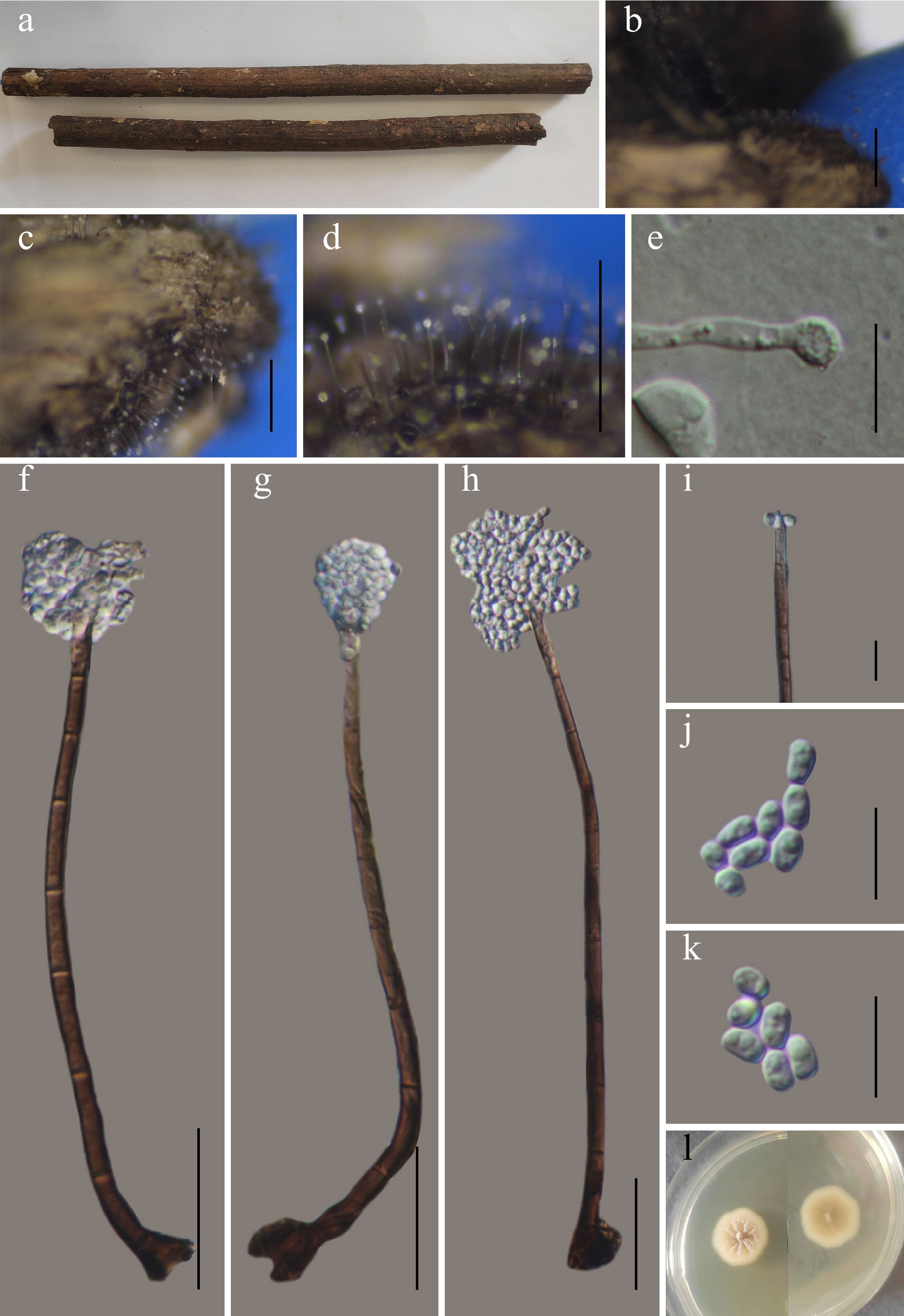
Figure 54.
Xylolentia wumengshanensis (GMB6945 holotype). (a) Host. (b)–(d) Colonies on natural substrate. (e) Germinating conidium. (f)–(h) Conidiophores and conidia. (i) Conidiogenous cells. (j), (k) Conidia. (l) Surface and reverse view of culture on PDA. Scale bars: b = 1 mm, c, d = 0.5 mm, f–h = 30 μm, e, i–k = 10 μm.
MycoBank number: MB863011
Etymology – The epithet refers to Wumengshan National Nature Reserve, Yunnan Province, China, where the type specimen was collected.
Saprobic on dead branches of an unknown plant in a terrestrial habitat. Sexual morph: not observed. Asexual morph: hyphomycetous. Colonies superficial, effuse, scattered or aggregated, brown, hairy, with glistening conidial masses at apex. Mycelium partly immersed and superficial, formed by smooth-walled, brown to hyaline, septate hyphae. Conidiophores 124–150 × 3.7–6 µm (x = 145 × 4.8 µm, n = 30) mononematous, erect, macronematous, straight to slightly flexuous, cylindrical, smooth-walled, septate, unbranched, 6–7-septate, light to dark brown, pale brown to subhyaline at the apex. Conidiogenous cells 17.7–34 × 3–4.5 µm (x = 22.1 × 3.7 µm, n = 30), determinate, integrated, terminal, elongating percurrently, polyblastic, cylindrical to cylindrical-lageniform, subhyaline to hyaline towards the apex, pale brown near the base, sympodially extending. Conidia acrogenous, 3–5.5 × 2.4–2.7 µm (x = 4.4 × 2.4 µm, n = 30), ellipsoidal to reniform, smooth-walled, hyaline, aseptate, guttulate, thin walled, aggregated in slimy masses.
Culture characteristics – Conidia germinate on PDA within 24 h. After 21 d at 25 °C, colonies on PDA reach 10–15 mm in diameter; circular, centrally umbonate to slightly depressed with a star-like radial furrowed pattern, aerial mycelium dense, glabrous to finely velvety margins entire, surface cream to pale buff; reverse concolorous to slightly darker.
Material examined – CHINA, Yunnan Province, Zhaotong City, Wumengshan National Nature Reserve, 25°21′15.13″ N, 103°15′05.18″ E, elevation 2,550 m, on dead branches of an unknown plant, 26 October 2024, Wen-Mei Li, 2024WMS273 (GMB6945, holotype; GMBC6945, ex-type); ibid., KUN-HKAS 152919, isotype.
Additional specimens examined – CHINA, Yunnan Province, Zhaotong City, Wumengshan National Nature Reserve, 25°21′15.17″ N, 103°16′09.34″ E, elevation: 2,635m, on dead branches of an unknown plant, 26 October 2024, Wen-Mei Li, 2024WMS289 (GMB6946, GMBC6946).
Notes – In the phylogenetic analysis (Fig. 52), Xylolentia wumengshanensis forms a well-supported (BS = 100; PP = 1) sister clade with X. oblongispora (GZCC 18-0054) at the base of the genus, distinct from all other species. Comparative analysis of nucleotide base pairs showed that X. wumengshanensis (GMBC6945) differs from the type strain of X. oblongispora (GZCC 18-0054) by 2.8% (15/535 bp) in the ITS locus, 0.1% (1/804 bp) in the LSU locus, and 1.6% (15/921 bp) in the tef1-α gene.
Morphologically, X. wumengshanensis can be distinguished from X. oblongispora by its shorter conidiophores (124–150 µm vs 91–219 µm) and ellipsoidal to reniform conidia with rounded ends, whereas those of X. oblongispora are oblong with obtuse ends[3]. Given these molecular, morphological, and phylogenetic differences, X. wumengshanensis is introduced as a novel species.
Sporidesmiales Crous, Persoonia 40: 377 (2018).
Sporidesmiaceae Fr. [as 'Sporidesmiacei'], Summa veg. Scand., Sectio Post. (Stockholm): 504 (1849).
Notes – Sporidesmiaceae was introduced by Fries but remained largely overlooked in traditional classification systems until the advent of molecular phylogenetics and the re-examination of sporidesmium-like fungi[171]. The family is typified by Sporidesmium, which was introduced by Link with S. atrum as the type species[172]. Su et al. reinstated Sporidesmiaceae to accommodate taxa supported by molecular data and exhibiting morphological features comparable to Sporidesmium ehrenbergii (the lectotype species of the genus)[173]. Members of the Sporidesmiaceae are predominantly saprobic or occasionally mycoparasitic and commonly occur on dead wood and plant debris in freshwater and terrestrial habitats, and are widely distributed from tropical to temperate regions[43,173−175].
Sporidesmium was long regarded as the sole genus within Sporidesmiaceae until Subramanian separated Ellisembia to include species characterized by proliferating conidiophores and distoseptate (pseudoseptate) conidia[176]. Su et al.[173] treated Ellisembia as a synonym of Sporidesmium sensu stricto due to the lack of phylogenetic support for the taxonomic significance of euseptate vs distoseptate conidia among sporidesmium-like fungi. This interpretation was subsequently challenged by Delgado et al.[177], who reassessed the group based on newly collected material of the type species Ellisembia coronata, from its original locality in Germany. Their phylogenetic analyses revealed that E. coronata forms a well-supported independent clade within the family, distinct from Sporidesmium. By confined Ellisembia to this group, characterized by pseudoseptate conidia and conidiophores with few or no percurrent extensions, the genus become consistent with the concept original proposed by Subramanian[176].
Morphologically, Sporidesmiaceae is characterized by hyphomycetous asexual morphs, macronematous conidiophores, and solitary, dry conidia that are euseptate or distoseptate, obclavate to cylindrical, often pigmented, and holoblastically produced. Although the taxonomic significance of conidial septation has long been debated, recent studies integrating detailed morphology with multigene phylogenetic analyses have led to a refined circumscription of the family, which is currently recognized to include Ellisembia and Sporidesmium[1,175].
Herein, phylogenetic analyses based on ITS, LSU, and rpb2 sequence data (Fig. 55), together with comparative morphological evidence, support the introduction of one new genus, Acroappendicula, in Sporidesmiaceae, further clarifying generic boundaries and enhancing the understanding of diversity within this family[171].
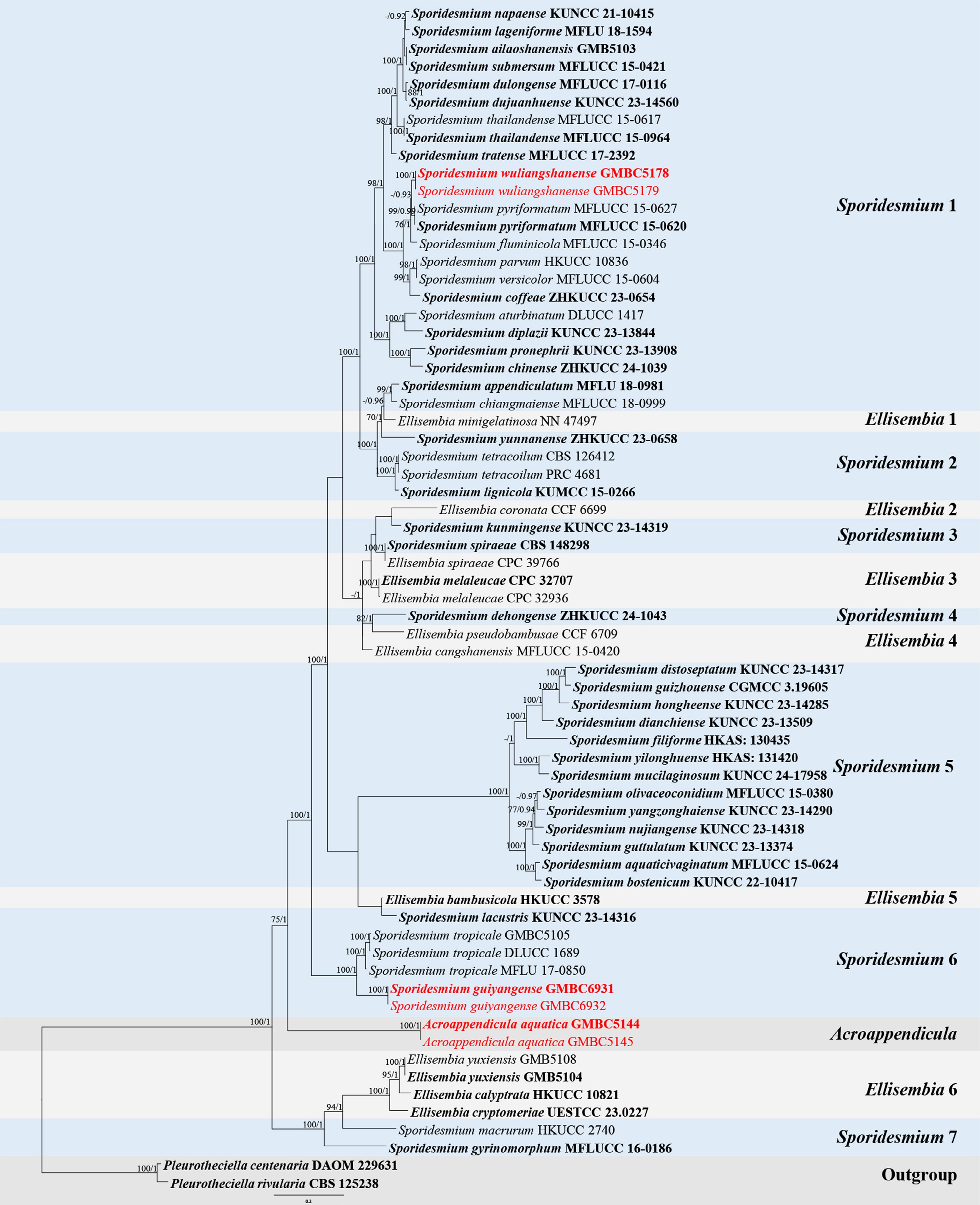
Figure 55.
ML tree of selected Sporidesmiaceae species based on the ITS-LSU-rpb2 dataset. The combined dataset comprised 67 taxa, 2,387 characters (ITS: 1–499 bp; LSU: 500–1,367 bp; rpb2: 1,368–2,387 bp), including indel regions. Maximum likelihood (lnL = –29,293.227036) and Bayesian analyses produced consistent tree topologies. Pleurotheciella centenaria (DAOM 229631) and P. rivularia (CBS 125238) were selected as the outgroup taxa. Bayesian posterior probabilities (PP) ≥ 0.90 and RAxML bootstrap support values ≥ 70 % are shown on the branches. The novel species are indicated in red and ex-type/type strains in bold.
Acroappendicula L.L. Liu, Q.F. Zhang & Q.R. Li, gen. nov.
MycoBank number: MB863012
Etymology – The generic name refers to the appendages at the tip of conidia.
On submerged dead wood in freshwater habitat. Sexual morph unknown. Asexual morph hyphomycetous. Colonies effuse and darkly pigmented. Conidiophores macronematous, mononematous, solitary, erect, dark brown to black. Conidiogenous cells integrated, terminal, monoblastic, determinate. Conidia solitary, acrogenous, dry, fusiform to obclavate, dark brown, distoseptate, with a conspicuous hyalin, cap-like to filiform, gelatinous apical appendage; basal cell truncate.
Typification – Acroappendicula aquatica L.L. Liu, Q.F. Zhang, & Q.R. Li.
Notes – Acroappendicula is placed in Sporidesmiaceae based on a combination of morphological characters and multigene phylogenetic analyses (Fig. 55). The genus shares general sporidesmium-like features with Ellisembia and Sporidesmium, including macronematous, mononematous, darkly pigmented conidiophores and solitary, dry, distoseptate conidia[175,178,179]. However, Acroappendicula is readily distinguished from Ellisembia and Sporidesmium by the presence of a well-developed, hyaline, cap-like to filiform, gelatinous apical appendage on the conidia. Phylogenetically (Fig. 55), Acroappendicula forms a well-supported independent clade within Sporidesmiaceae, further supporting its recognition as a new genus. Based on these morphological and phylogenetic differences, Acroappendicula is introduced as a new genus within Sporidesmiaceae.
Acroappendicula aquatica L.L. Liu, Q.F. Zhang, & Q.R. Li, sp. nov. (Fig. 56)
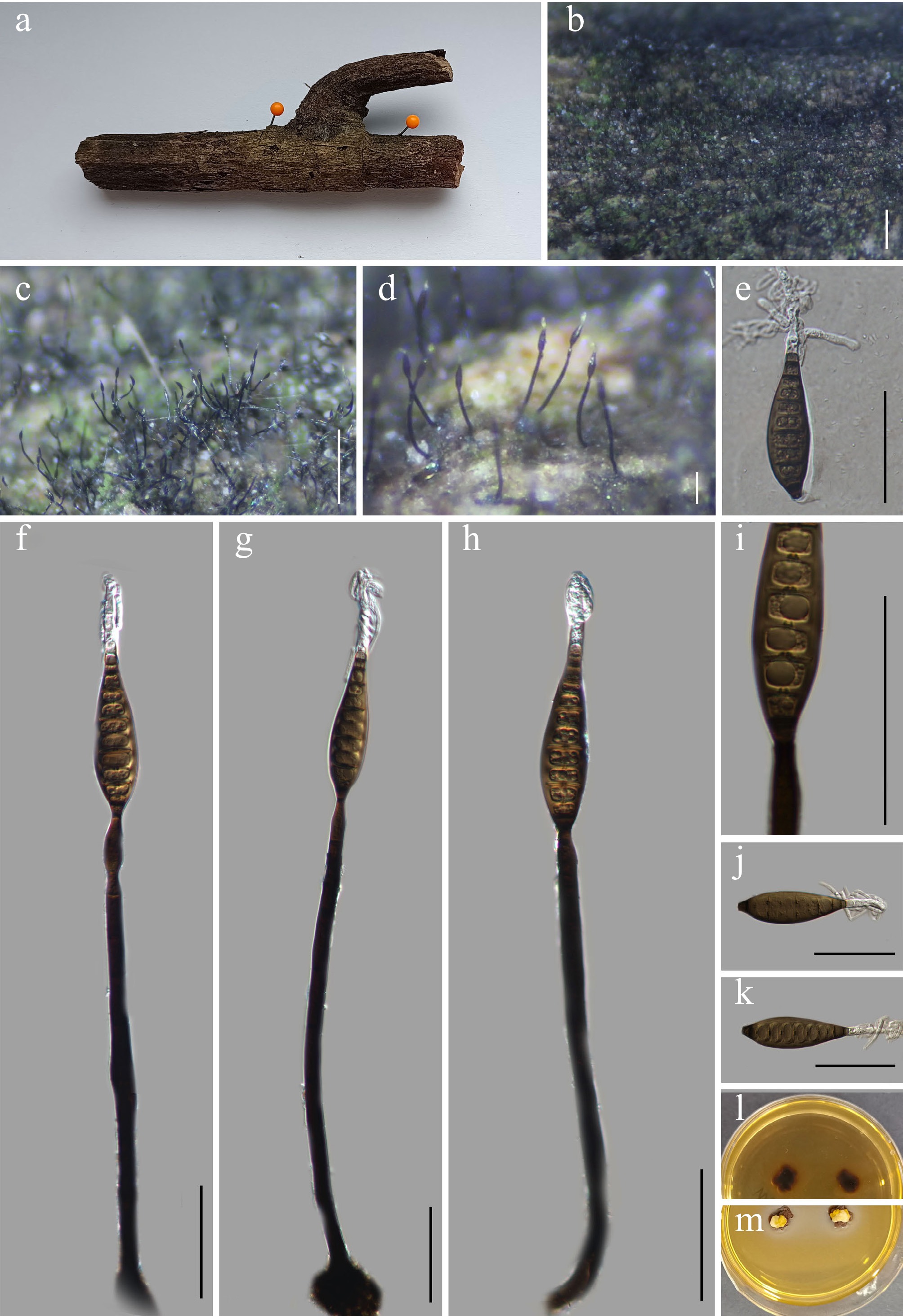
Figure 56.
Acroappendicula aquatica (GMB5144 holotype). (a) Host. (b)–(d) Colonies on natural substrate. (e) Germinating conidium. (f)–(h) Conidiophores and conidia. (i) Conidiogenous cells with conidia. (j), (k) Conidia. (l), (m) Surface view of culture on PDA. Scale bars: b = 1 mm, c = 0.5 mm, d = 0.1 mm, e–k = 50 μm.
MycoBank number: MB863013
Etymology – The epithet refers to its aquatic habitat.
Saprobic on submerged dead branches of an unknown plant in a freshwater lake. Sexual morph: not observed. Asexual morph: hyphomycetous. Colonies on natural substrate effuse, superficial, dark olivaceous to black, forming scattered to gregarious, dark, setiform conidiophores emerging from the substrate surface. Mycelium superficial to mostly immersed, consist of smooth-walled, branched, hyaline to pale brown, septate hyphae. Conidiophores 186.5–269.3 × 10.9–44.2 µm (x = 215.8 × 24.3 µm, n = 15) mononematous, solitary, macronematous, straight to slightly flexuous erect, unbranched, septate, dark brown to black, paler towards the apex, base slightly swollen, smooth-walled. Conidiogenous cells 20.0–32.7 × 4.5–5.2 µm (x = 25.0 × 4.9 µm, n = 15), monoblastic, determinate, integrated, terminal, cylindrical to slightly lageniform, smooth-walled, hyaline to subhyaline. Conidia 77.4–104.1 × 14.6–20.4 µm (x = 90.2 × 17.8 µm, n = 30) solitary, acrogenous, dry, fusiform to obclavate, straight to slightly curved, dark brown, smooth- and thick-walled, 8–11-distoseptate, septa conspicuous, a pical cell bearing a hyaline, gelatinous, cap-like to filiform appendage, sometimes elongate or coiled; basal cell truncate.
Culture characteristics – Conidia germinate on PDA within 24 h. After six weeks at 25 °C, colonies on PDA reach 10–15 mm in diameter; circular to irregular, centrally thick, aerial mycelium dense, glabrous to finely velvety, margins entire to lobate, surface yellow-brown to brown; reverse dark brown to black.
Material examined – CHINA, Yunnan Province, Wuliangshan National Nature Reserve, 24.213366° N, 101.224567° E, elevation 1,800 m, on submerged dead branches of an unknown plant in a freshwater lake, 2 August 2024, Qin-Fang Zhang, 2024WLSY72 (GMB5144, holotype; GMBC5144, ex-type); ibid., KUN-HKAS 152920, isotype.
Additional examined specimens – CHINA, Yunnan Province, Wuliangshan National Nature Reserve, 24°25'33.66" N, 101°23'55.67" E, elevation 1,455 m, on submerged dead branches of an unknown plant in a freshwater lake, 2 August 2024, Qin-Fang Zhang, 2024WLSY152 (GMB5145, GMBC5145).
Notes – Phylogenetically, Acroappendicula aquatica forms a well-supported independent lineage within the family, clearly separated from other genera (Fig. 55).
Morphologically, Acroappendicula aquatica is distinguished from other members of the family by its large, dark brown, fusiform to obclavate, distoseptate conidia, which bear a conspicuous, cap-like to filiform, gelatinous apical appendage, a character not observed in related taxa[173,175]. These morphological and phylogenetic differences support the recognition of A. aquatica as a new species.
Sporidesmium Link, Mag. Gesell. naturf. Freunde, Berlin 3(1-2): 41 (1809).
Notes – Sporidesmium was established by Link, typified by S. atrum[172]. It is characterized by mononematous, macronematous conidiophores and holoblastic, monoblastic, percurrent conidiogenous cells. Conidia are variable in shape (ovoid to rostrate), euseptate or distoseptate, sometimes with an apical appendage or mucilaginous sheath[5,16,173,180]. Sporidesmium sensu lato was historically treated as a broad and heterogeneous assemblage, later shown to be polyphyletic within the Ascomycota[181−188]. Subramanian[183] revised the genus and restricted Sporidesmium sensu stricto to species with euseptate conidia and conidiophores showing none or irregular percurrent proliferation, while segregating related taxa into genera such as Repetophragma and Stanjehughesia. Phylogenetic studies confirmed that sporidesmium-like taxa are distributed across multiple lineages, particularly within Dothideomycetes and Sordariomycetes. To resolve this taxonomic inconsistency, Su et al.[173] redefined the genus and restricted it to a monophyletic lineage corresponding to Sporidesmium sensu stricto. Index Fungorum (accessed 27 March 2026) lists 523 epithets of Sporidesmium, approximately 83% of which were established prior to 1992. At present, 252 species are recognized as accepted.
Sporidesmium guiyangense L.L. Liu, W.M. Li, & Q.R. Li, sp. nov. (Fig. 57)
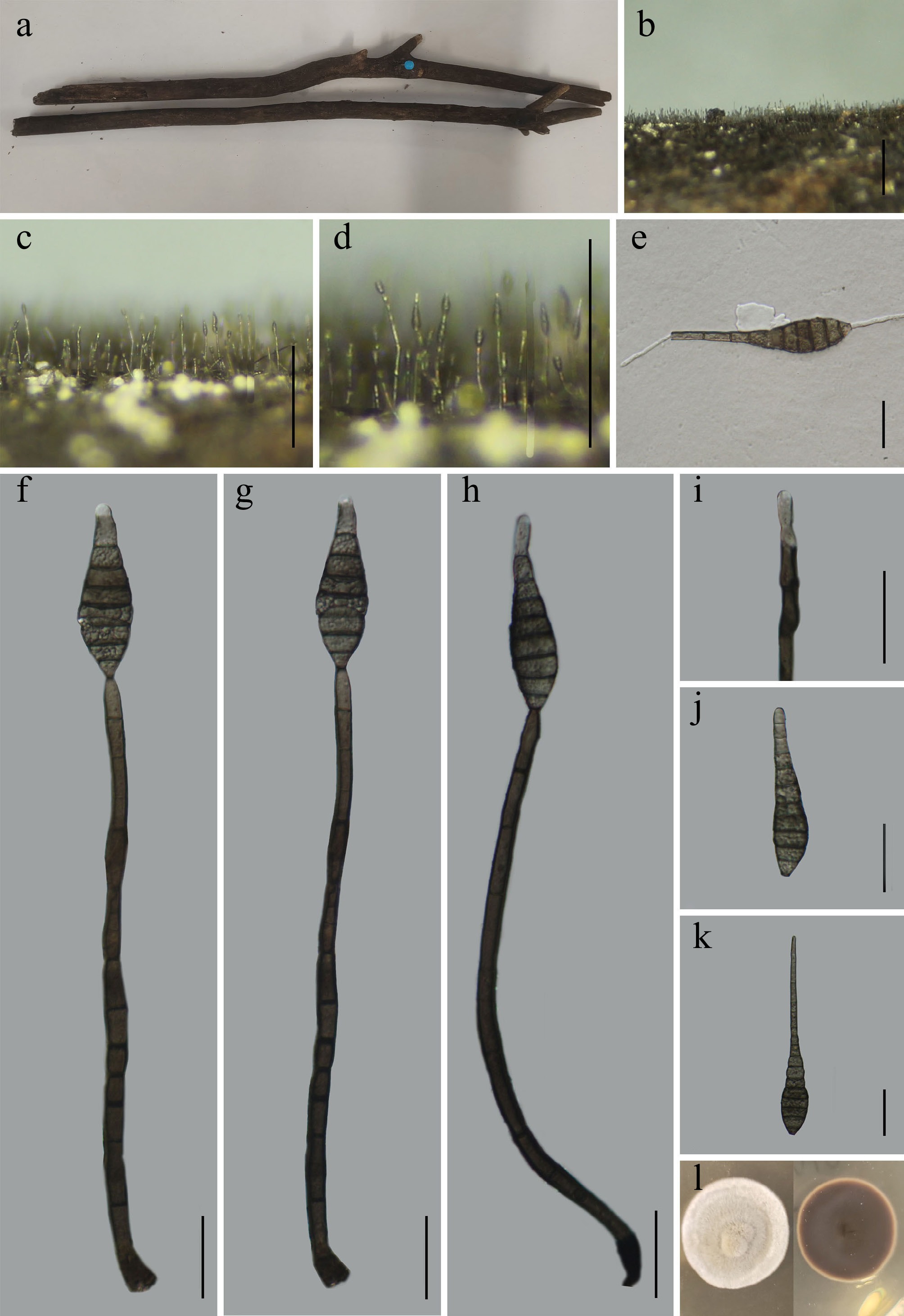
Figure 57.
Sporidesmium guiyangense (GMB6931 holotype). (a) Host. (b)–(d) Colonies on natural substrate. (e) Germinating conidium. (f)–(h) Conidiophores and conidia. (i) Conidiogenous cells. (j), (k) Conidia. (l) Surface and reverse view of culture on PDA. Scale bars: b = 1 mm, c, d = 0.5 mm, e–k = 30 μm.
MycoBank number: MB863014
Etymology – The specific epithet guiyangense refers to Guiyang City, Guizhou Province, China, where the holotype was collected.
Saprobic on dead branches of an unknown plant. Sexual morph: not observed. Asexual morph: hyphomycetous. Colonies superficial, effuse, dark brown to black, hairy. Mycelium immersed, septate, smooth, brown. Conidiophores 77–307 × 4.1–7.9 µm (x = 165.9 × 6.1 µm, n = 30), mononematous, solitary, macronematous, straight to slightly flexuous erect, cylindrical, dark brown, paler apically, smooth, thick-walled, 4–12-septate, unbranched. Conidiogenous cells 10–22 × 4–7 µm (x = 16.7 × 5.4 µm, n = 30), monotretic, terminal, integrated, cylindrical to lageniform, pale brown to subhyaline apically, smooth. Conidia 55.1–146.1 × 6.6–18 µm (x = 91.3 × 12.3 µm, n = 30), solitary, dry, acrogenous, obclavate to fusiform, dark brown, straight to slightly curved, smooth, 6–15-septate; apex rounded to subacute; base truncate to slightly tapered.
Culture characteristics – Conidia germinate on PDA within 24 h. After five weels at 25 °C, colonies on PDA reach 40–50 mm in diameter; circular, even, aerial mycelium dense, glabrous to finely velvety, margins entiree, surface pale grey to whitish; reverse brown to dark brown.
Material examined – CHINA, Guizhou Province, Guiyang City, Panlong Mountain Park, 26°43′27.83″ N, 106°49′53.66″ E, elevation 1,235 m, on dead branches of an unknown plant., 24 May 2025, Wen-Mei Li, 2025GY11 (GMB6931, holotype; GMBC6931, ex-type); ibid., KUN-HKAS 152921, isotype.
Additional examined specimens – CHINA, Guizhou Province, Guiyang City, Panlong Mountain Park, 26°43′33.56″ N, 106°49′35.78″ E, elevation 1,275 m, on dead branches of an unknown plant., 24 May 2025, Wen-Mei Li, 2025GY55 (GMB6932, GMBC6932).
Notes – Phylogenetically (Fig. 55), Sporidesmium guiyangense forms a well-supported sister clade (BS = 100; PP = 1) with S. tropicale (MFLU 17-0850). Comparative analysis of nucleotide sequences revealed that S. guiyangense (GMBC6931) differs from the type strain of S. tropicale (MFLU 17-0850) by 6.4% (32/498 bp) in the ITS locus and 0.6% (5/822 bp) in the LSU locus. Morphologically, S. guiyangense can be distinguished from S. tropicale by its shorter and less septate conidiophores (77–307 µm long, 4–12-septate), compared to the longer and 5–14-septate septate conidiophores of S. tropicale (40–340 µm long). In addition, S. guiyangense has smooth-walled, smaller conidia (55.1–146.1 × 6.6–18 µm), whereas S. tropicale produces larger conidia (80–230 × 12–15 µm, 7–19-septate) with a verrucose proximal wall[189]. Based on the combined morphological, molecular, and phylogenetic evidence, S. guiyangense is proposed here as a new species.
Sporidesmium wuliangshanense L.L. Liu, Q.F. Zhang, & Q.R. Li, sp. nov. (Fig. 58)
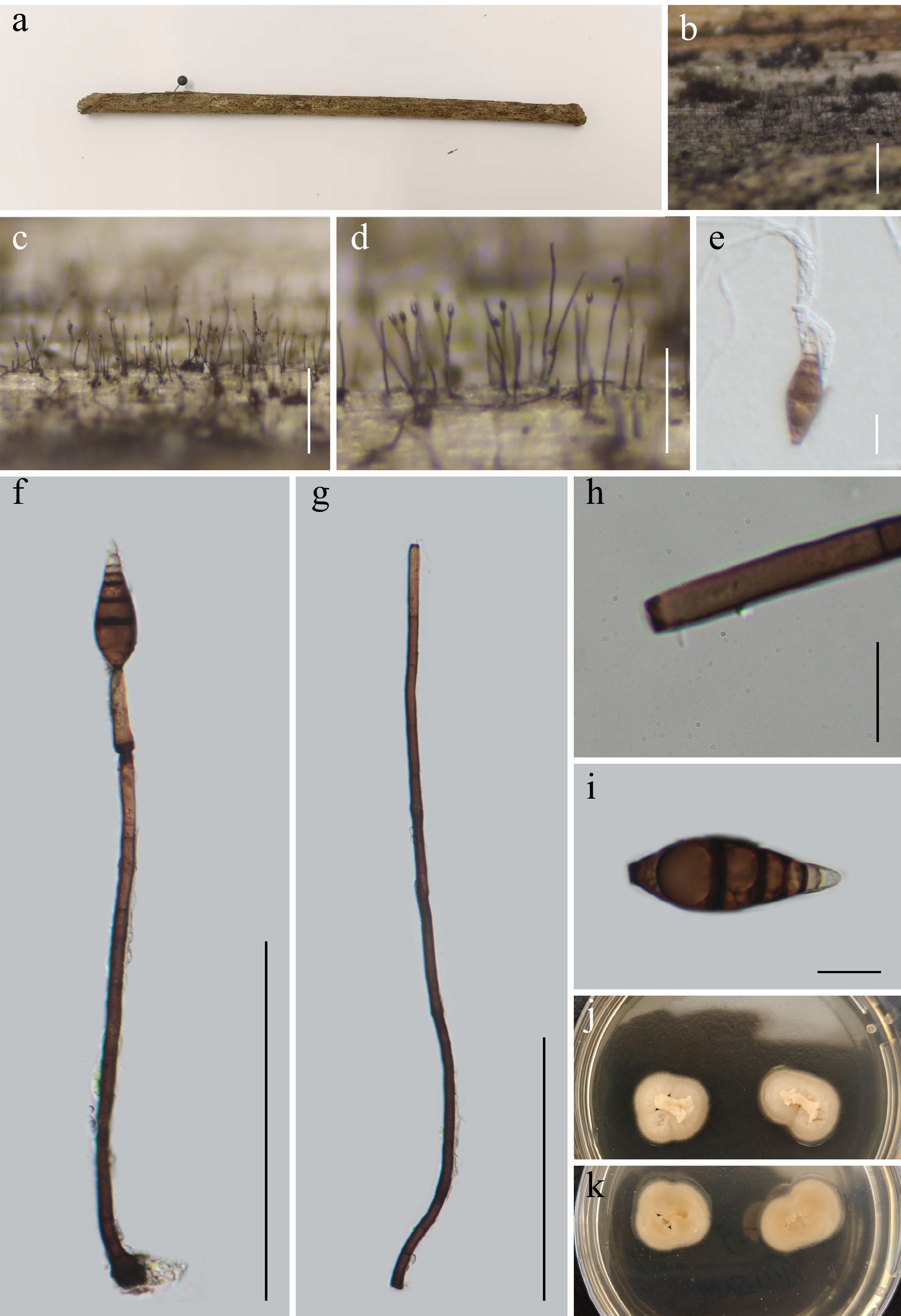
Figure 58.
Sporidesmium wuliangshanense (GMB5178 holotype). (a) Host. (b)–(d) Colonies on natural substrate. (e) Germinating conidium. (f), (g) Conidiophores and conidia. (h) Conidiogenous cells. (i) Conidium. (j), (k) Surface and reverse view of culture on PDA. Scale bars: b = 1 mm, c = 0.5 mm, d = 0.25 mm, f, g = 100 μm, e, h, i = 10 μm.
MycoBank number: MB863029
Etymology – The epithet refers to Wuliangshan National Nature Reserve, Yunnan Province, China, where the type specimen was collected.
Saprobic on submerged dead branches of an unknown plant in a freshwater lake. Sexual morph: not observed. Asexual morph: hyphomycetous. Colonies effuse, superficial, hairy, scattered to gregarious, dark brown to black. Mycelium mostly immersed, formed by smooth-walled, pale brown to brown, septate hyphae. Conidiophores 97–277.5 × 3.5–5 µm (x = 157 × 4.2 µm, n = 15), mononematous, solitary, macronematous, straight to slightly flexuous erect, cylindrical, unbranched, 4–10-septate, smooth, brown to dark brown, slightly paler toward the apex, base slightly swollen. Conidiogenous cells 16.2–25 × 2.5–3.8 µm (x = 21.8 × 3.2 µm, n = 15), holoblastic, integrated, monoblastic, terminal, smooth, cylindrical, pale brown to brown. Conidia 29–38.5 × 10.5–12.7 µm (x = 33.9 × 11.9 µm, n = 20), solitary, acrogenous, obclavate to fusiform, rostrate, straight to slightly curved, smooth, brown to dark brown, 5–8-septate, constricted at septa, 2.7–3.2 µm wide, base truncate, apex attenuated, 2–4 µm wide.
Culture characteristics – Conidia germinate on PDA within 24 h. After five weeks at 25 °C, colonies on PDA reach 20–30 mm in diameter; circular to irregular, floccose to velvety, aerial mycelium sparse to moderate, margins entire to lobate, surface whitish-grey to cream; reverse pale brown.
Material examined – CHINA, Yunnan Province, Wuliangshan National Nature Reserve, 24°25'33.66" N, 101°23'55.67" E, elevation 1,455 m, on submerged dead branches of an unknown plant in a freshwater lake, 18 August 2025, Qin-Fang Zhang, 2025WLS62A (GMB5178, holotype; GMBC5178, ex-type); ibid., KUN-HKAS 152922, isotype.
Additional examined specimens – CHINA, Yunnan Province, Wuliangshan National Nature Reserve, 24°25'56.42" N, 101°23'55.74" E, elevation 2,180 m, on submerged dead branches of an unknown plant in a freshwater lake, 18 August 2025, Qin-Fang Zhang, 2025WLS95 (GMB5179, GMBC5179).
Notes – Phylogenetically (Fig. 55), Sporidesmium wuliangshanense forms a clade in a sister relationship with the type strain of S. pyriformatum (MFLUCC 15-0620). Comparative analysis of nucleotide base pairs showed that S. wuliangshanense (GMBC5178) differs from the type strain of S. pyriformatum (MFLUCC 15-0620) by 3.3% (17/514 bp) in the ITS locus. Morphologically, S. wuliangshanense can be easily distinguished from S. pyriformatum by its longer, 4–10-septate conidiophores (97–277.5 µm vs 65–190 µm, 2–5-septate) and larger, 5–8-euseptate, rostrate conidia (29–38.5 × 10.5–12.7 µm vs 3–4-euseptate, non-rostrate conidia 8.5–32.5 × 8–15.5 µm)[190].
Based on the combined morphological, molecular, and phylogenetic differences, S. wuliangshanense is proposed here as a new species.
Trichosphaeriales M.E. Barr, Mycologia 75(1): 11 (1983).
Trichosphaeriaceae G. Winter [as 'Trichosphaerieae'], Rabenh. Krypt.-Fl., Edn 2 (Leipzig) 1.2: 191 (1885).
Stachylidium Link, Mag. Gesell. naturf. Freunde, Berlin 3(1-2): 15 (1809).
Notes – Stachylidium was established by Link, typified by S. bicolor[171,172]. Species of the genus are saprobic, mainly occurring on herbaceous debris and decaying wood, with occasional records from soil[191]. Only the asexual morph of the genus is known, characterized by septate, verticillate conidiophores that are brown at the base and paler towards the apex; conidiogenous cells are hyaline to pale brown, arranged in whorls; and cylindrical, pale brown to brown conidia[192]. Stachylidium is a small, globally distributed genus. According to Species Fungorum (accessed 3 January 2026), the genus currently comprises 32 accepted species; however, only two species of the genus have molecular data at NCBI GenBank. The remaining taxa require further revision, highlighting a significant gap in our understanding of the genus's phylogenetic relationships.
Stachylidium bicolor Link, Mag. Gesell. naturf. Freunde, Berlin 3(1-2): 15 (1809) (Fig. 59).
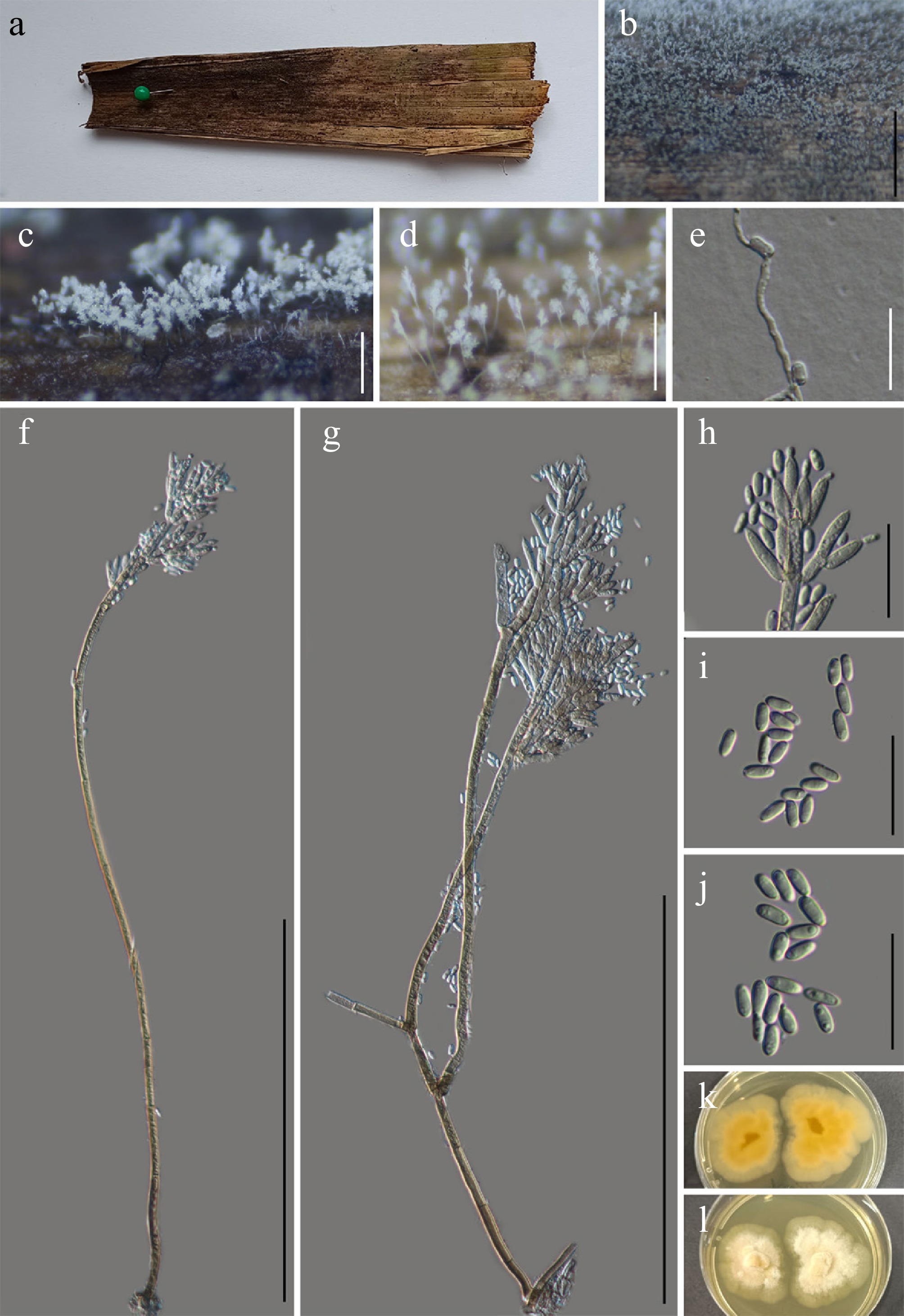
Figure 59.
Stachylidium bicolor (GMB5148). (a) Host. (b)–(d) Colonies on natural substrate. (e) Germinating conidium. (f), (g) Conidiophores and conidia. (h) Conidiogenous cells. (i), (j) Conidia. (k), (l) Surface and reverse view of culture on PDA. Scale bars: b = 3 mm, c = 0.5 mm, d = 2 mm, f, g = 200 μm, e, h–j = 20 μm.
MycoBank number: MB166350
Saprobic on submerged dead wood in a freshwater lake. Sexual morph: not observed. Asexual morph: hyphomycetous. Colonies effuse, superficial, whitish to pale grey. Mycelium immersed; hyphae septate, branched, smooth-walled, hyaline to pale brown. Conidiophores 194.4–427.1 × 3.4–5.8 µm (x = 322.4 × 4.4 µm, n = 20) mononematous, erect, solitary, macronematous, unbranched, septate, smooth-walled, pale brown, paler apically. Conidiogenous cells 8.7–21.6 × 2.6–3.7 µm (x = 13.0 × 3.1 µm, n = 30) phialidic, monophialidic, cylindrical to narrowly clavate, hyaline to subhyaline, verticillate. Conidia 4.4–6.9 × 2.3–3.2 µm (x = 5.6 × 2.7 µm, n = 30) aseptate, ellipsoidal to oblong, hyaline to subhyaline, smooth-walled.
Culture characteristics – Conidia germinate on PDA within 12 h. After 14 d at 25 °C, colonies on PDA reach 30–40 mm in diameter; irregular, floccose, aerial mycelium sparse, margins lobate, surface white to light yellow; reverse pale brown.
Material examined – CHINA, Yunnan Province, Yuxi City, Ailaoshan National Nature Reserve 24°5'7.01" N, 101°31'30.44" E, elevation 1,169 m, on submerged decaying wood in a freshwater lake, 15 September 2024, Qin-Fang Zhang, 2024ALS63 (GMB5148, GMBC5148).
Notes – The original description of Stachylidium bicolor by Link was based on morphology from European specimens[172]. The description of our collection closely matches this original description[172]. In our phylogenetic analyses, GMBC5148 clusters with previously reported S. bicolor strains in a well-supported clade (Fig. 60). Given that the type material has never been sequenced, we base the identification of GMBC5148 on a combination of morphological consistency and phylogenetic placement. This study reports S. bicolor for the first time from China, marking a new country record.
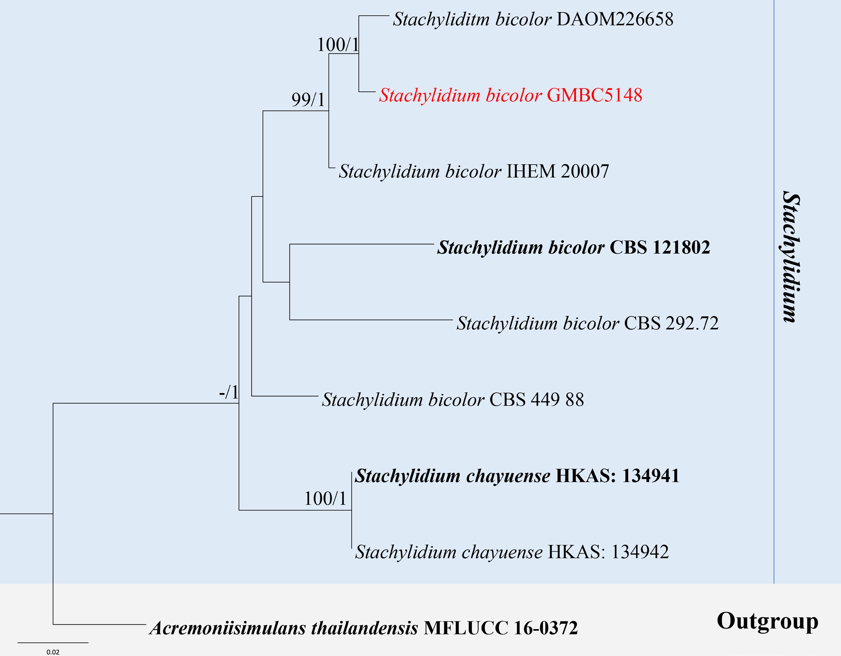
Figure 60.
ML tree of selected Stachylidium species based on the ITS-LSU-tef1-αdataset. The combined dataset comprised nine taxa, 2,221 characters (ITS: 1–515 bp; LSU: 516–1,361 bp; tef1-α: 1,362–2,221 bp), including indel regions. Maximum likelihood (lnL = –5,080.289634) and Bayesian analyses produced consistent tree topologies. Acremoniisimulans thailandensis (MFLUCC 16-0372) was selected as the outgroup taxon. Bayesian posterior probabilities (PP) ≥ 0.90 and RAxML bootstrap support values ≥ 70 % are shown on the branches. The new record is indicated in red and ex-type/type strains in bold.
Brachysporium Sacc., Syll. Fung. (Abellini) 4: 423 (1886).
Notes – Brachysporium was established by Sacc, typified by B. obovatum[193]. Species of the genus are saprobic, occurring mainly on decaying wood and tree bark, and are also reported from both terrestrial and freshwater habitats. Bactrodesmium has a wide distribution in temperate, tropical, and subtropical regions of both hemispheres[5,43,112]. Recent multigene analyses demonstrated that Bactrodesmium sensu stricto forms a well-supported monophyletic clade within Savoryellales[79]. Morphologically, it is characterized by sporodochial conidiomata, mononematous conidiophores, holoblastic conidiogenesis, and acrogenous, dry, pigmented conidia with transverse or longitudinal septa[5,174]. According to Species Fungorum (accessed 27 March 2026), the genus currently comprises 43 accepted species.
Brachysporium wumengshanense L.L. Liu, W.M. Li, & Q.R. Li, sp. nov. (Fig. 61)
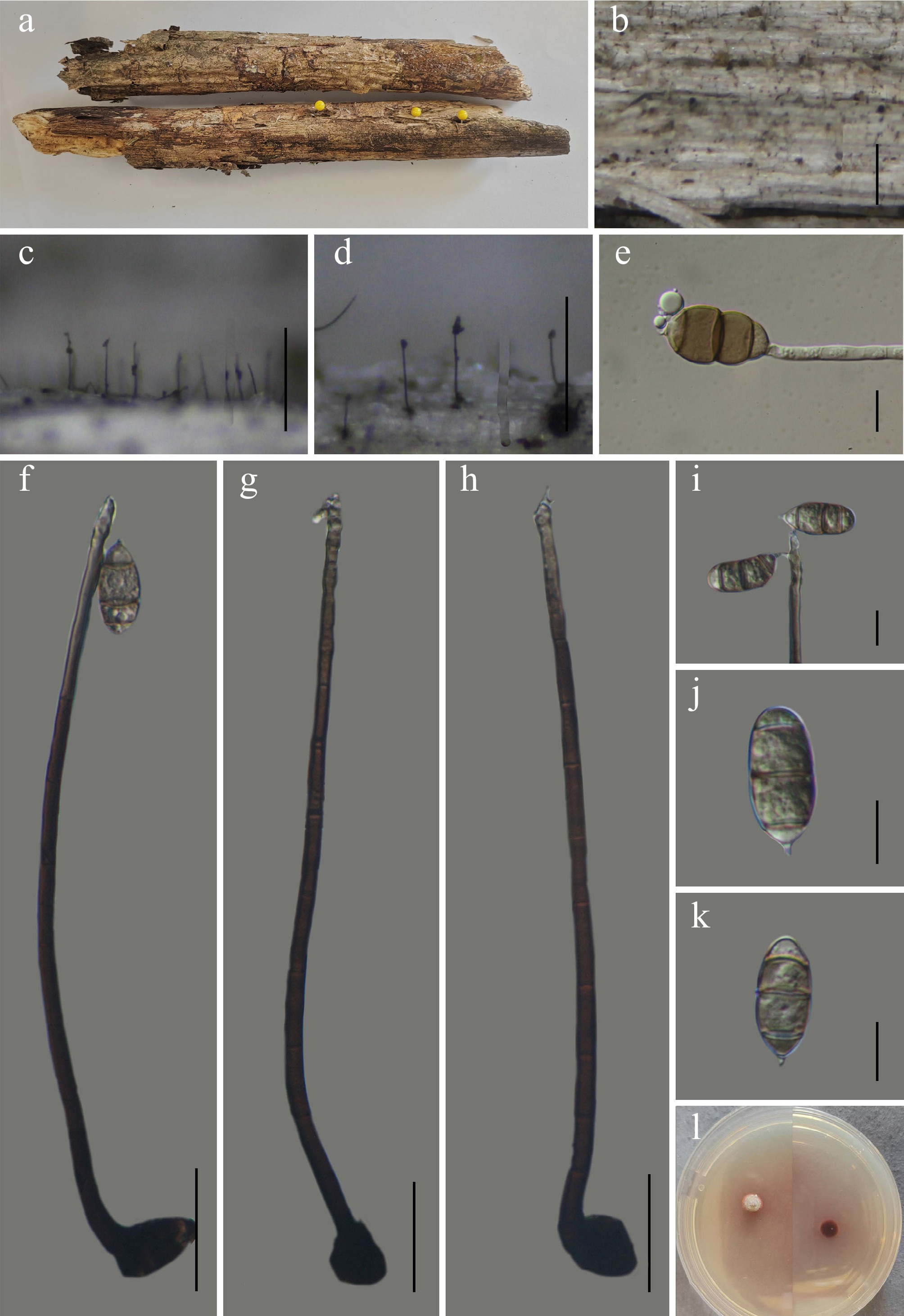
Figure 61.
Brachysporium wumengshanense (GMB6947 holotype). (a) Host. (b)–(d) Colonies on natural substrate. (e) Germinating conidium. (f)–(h) Conidiophores and conidia. (i) Conidiogenous cells and conidia. (j)–(k) Conidia. (l) Surface and reverse view of culture on PDA. Scale bars: b = 1 mm, c, d = 0.5 mm, f–h = 30 μm, e, i–k = 10 μm.
MycoBank number: MB863030
Etymology – The epithet refers to Wumengshan National Nature Reserve, the type locality where the species was collected.
Saprobic on submerged dead wood in a freshwater wetland. Sexual morph: not observed. Asexual morph: hyphomycetous. Colonies on natural substrate superficial, effuse, scattered, hairy, composed of erect conidiophores arising directly from the substratum. Mycelium partly superficial, partly immersed; hyphae septate, pale brown, smooth-walled. Conidiophores macronematous, solitary, mononematous, unbranched, erect, straight to flexuous, cylindrical, dark brown, gradually paler towards the apex, smooth-walled, septate, 7–8 septate, 146–206 × 3.5–5 µm (x = 171.1 × 4.2 µm, n = 30); basal cell slightly swollen. Conidiogenous cells integrated, terminal, holoblastic, sympodially proliferating, monoblastic, pale brown to subhyaline, smooth-walled, 11.2–24 × 3–4 µm (x = 16.7 × 3.6 µm, n = 30). Conidia 20–25 × 9–11.5 µm (x = 22.5 × 10.5 µm, n = 30), acrogenous, solitary, occasionally producing secondary conidia, oblong to ellipsoid, 3-septate at maturity, light brown to brown, dry, rough-walled, apex rounded, connected to the conidiogenous cells by a single denticle.
Culture characteristics – Conidia germinate on PDA within 12 h. After six weeks at 25 °C, colonies on PDA reach 5–10 mm in diameter; circular, umbonate, margins entire, surface whitish pink; pink to reddish brown.
Material examined – CHINA, Yunnan Province, Zhaotong City, Wumengshan National Nature Reserve, 25°23′09.86″ N, 103°15′21.66″ E, elevation: 2,698 m, on submerged decaying in a freshwater wetland, 26 October 2024, Wen-Mei Li, 2024WMS275 (GMB6947, holotype; GMBC6947, ex-type); ibid., KUN-HKAS 152924, isotype.
Additional specimens examined – CHINA, Yunnan Province, Zhaotong City, Wumengshan National Nature Reserve, 25°21′12.11″ N, 103°16′10.56″ E, elevation: 2,745 m, on submerged decaying in a freshwater wetland, 26 October 2024, Wen-Mei Li, 2024WMS211 (GMB6948, GMBC6948).
Notes – Phylogenetically (Fig. 62), Brachysporium wumengshanense forms a well-supported clade with B. nigrum (SH12 and MR 1346) (BS = 88, PP = 1). Morphologically, B. wumengshanense (GMB6947) resembles B. nigrum in having effuse, hairy colonies; solitary, erect, dark brown conidiophores that become paler toward the apex; and 3-septate conidia. However, it can be easily distinguished from B. nigrum by its shorter conidiophores (146–206 µm vs 140–420 µm) with 7–8 septa (vs 1–7 septa), as well as by its monoblastic conidiogenous cells (vs polyblastic and denticulate)[194,195].
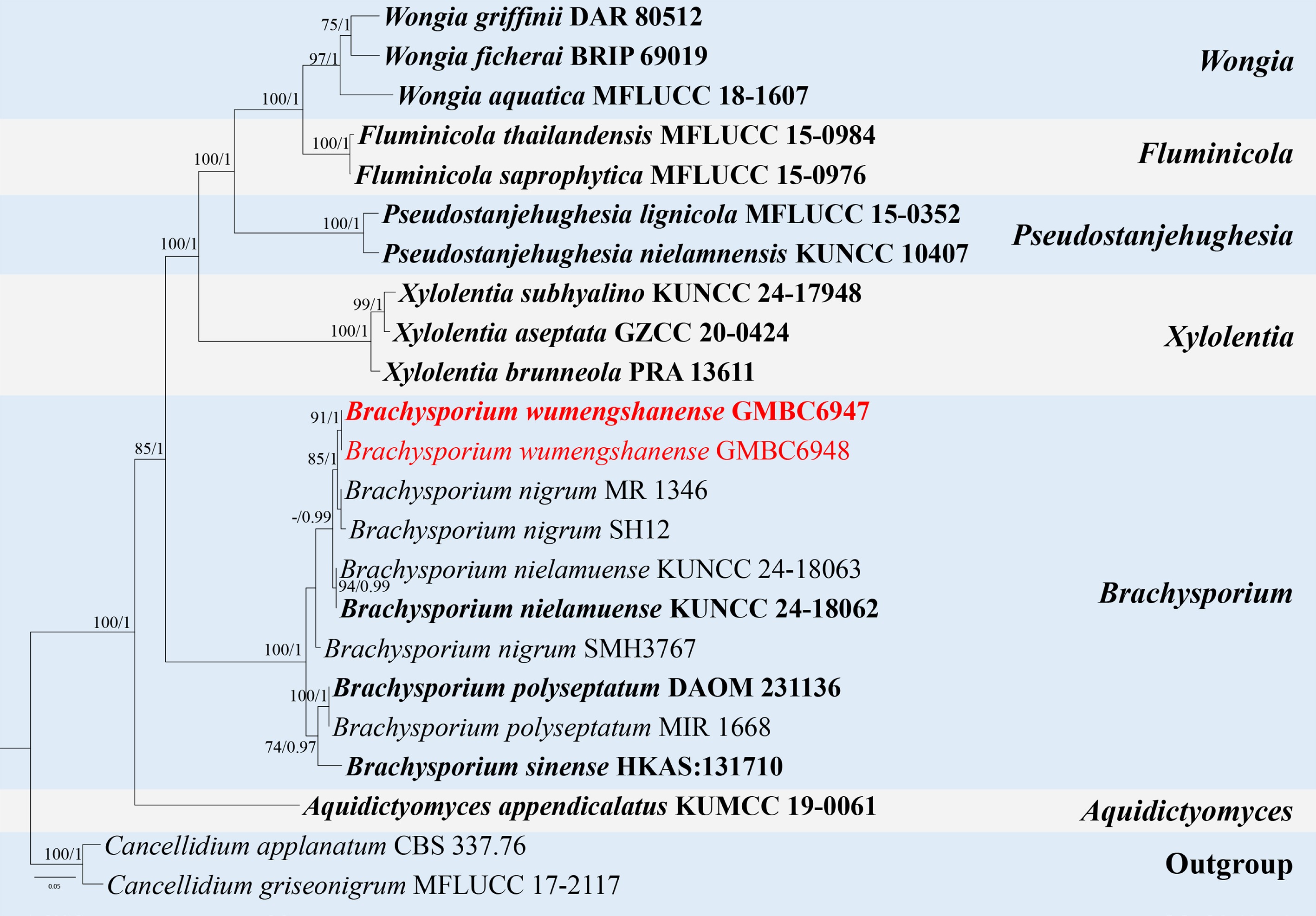
Figure 62.
ML tree of selected species of Brachysporium and related genera inferred from a combined ITS-LSU-rpb2 dataset. The combined dataset comprised 23 taxa, 2,531 characters (ITS: 1–513 bp; LSU: 514–1,388 bp; rpb2: 1,389–2,531 bp), including indel regions. Maximum likelihood (lnL = –11,943.803387) and Bayesian analyses produced consistent tree topologies. Cancellidium griseonigrum (MFLUCC 17-2117) and C. applanatum (CBS 337.76) were selected as the outgroup taxa. Bayesian posterior probabilities (PP) ≥ 0.90 and RAxML bootstrap support values ≥ 70 % are shown on the branches. The novel species is marked in red and ex-type/type strains in bold.
In terms of conidiophore morphology, size, and monoblastic conidiogenous cells, B. wumengshanense is similar to B. sinense, but differs in having larger conidia (22–47 × 21–25 µm) that are fusoid to limoniform, verrucose, and 4-septate[196].
Based on these morphological differences and its phylogenetic distinctiveness, B. wumengshanense is introduced here as a new species.
Xylariales Nannf., Nova Acta R. Soc. Scient. upsal., Ser. 4 8(no. 2): 66 (1932).
Vamsapriyaceae Y.R. Sun, Yong Wang bis & K.D. Hyde, Journal of Fungi 7(no. 891): 7 (2021).
Vamsapriya Gawas & Bhat, Mycotaxon 94: 150 (2006) [2005].
Notes – Vamsapriya was established by Gawas & Bhat for synnematous hyphomycetes occurring on bamboo in India, with V. indica as the type species[197]. Species of Vamsapriya are mainly bamboo-associated and lignicolous, and are distributed predominantly in tropical to subtropical regions, especially in Asia. The asexual morph of the genus is characterized by black colonies with immersed mycelium and dark brown, erect, synnematous conidiophores bearing monotretic conidiogenous cells and catenate, brown conidia[20,181]. The sexual morph is characterized by black, immersed, ostiolate perithecial ascomata, eight-spored asci with an amyloid apical ring, and hyaline, fusiform to broadly fusiform ascospores[182,183]. According to Species Fungorum (accessed 22 March 2026), the genus currently comprises 22 accepted species. In this study, the asexual morph of V. tongluobaensis is described for the first time, based on specimens collected from decaying wood. This report provides a more complete understanding of the life cycle of this species and facilitates accurate identification.
Vamsapriya tongluobaensis X.Y. Luo, K. Habib & Q.R. Li, MycoKeys 120: 181 (2025). (Fig. 63)
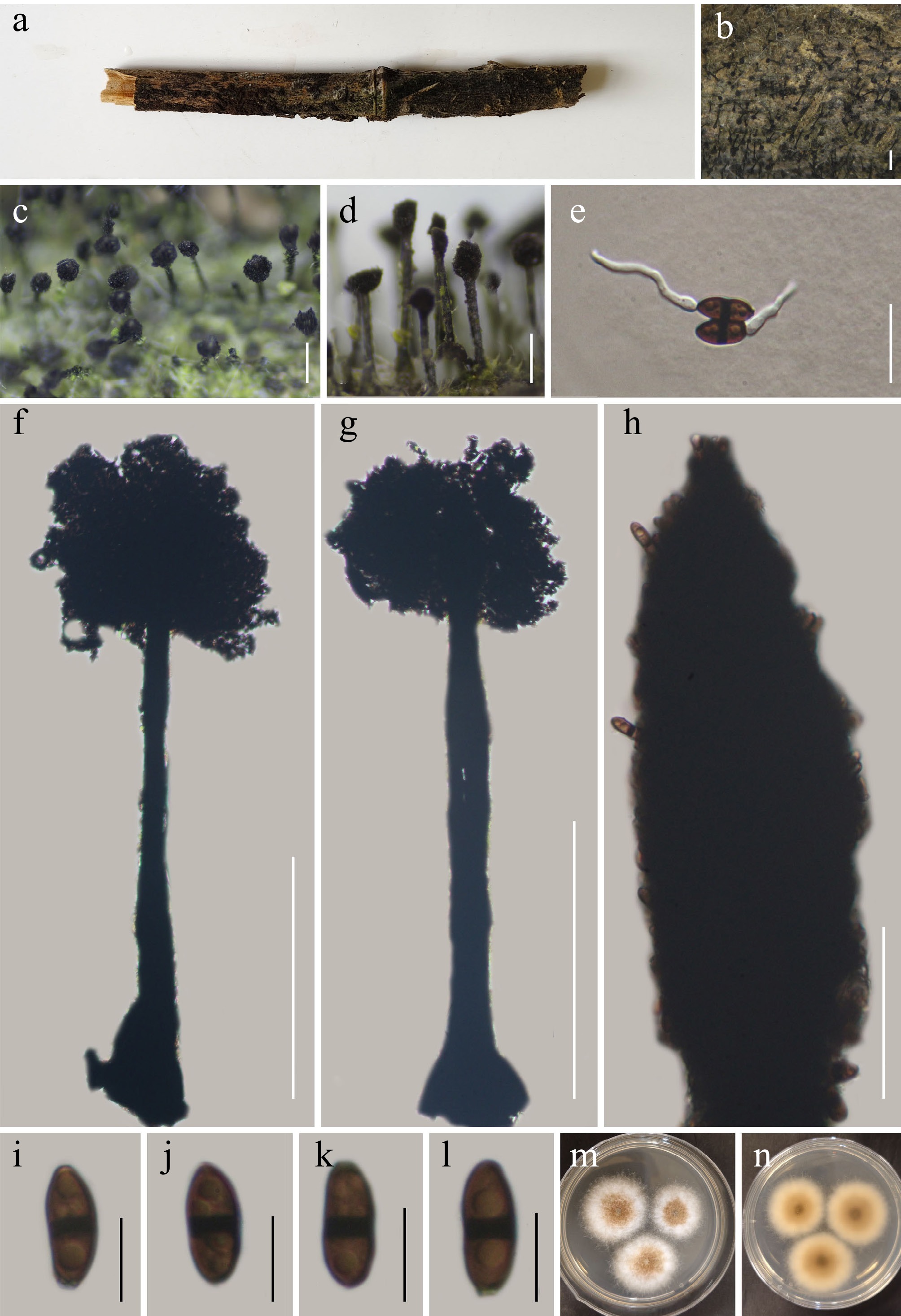
Figure 63.
Vamsapriya tongluobaensis (GMB5164). (a) Host. (b)–(d) Colonies on natural substrate. (e) Germinating conidia. (f), (g) Conidiophores and conidia. (h) Conidiogenous cells with conidia. (i)–(l) Conidia. (m), (n) Surface and reverse view of culture on PDA. Scale bars: b = 1 mm, c, d = 0.5 mm; f, g = 500 μm, e = 20 μm, i–l = 10 μm.
MycoBank number: MB859144
Saprobic on dead bamboo culms. Sexual morph: see Luo et al.[182]. Asexual morph: hyphomycetous. Colonies superficial, effuse, dark brown to black, hairy. Mycelium mostly immersed, composed of septate, branched, brown hyphae. Conidiophores macronematous, synnematous, solitary, erect, straight to slightly flexuous, cylindrical, dark brown, smooth-walled. Synnemata 905.3–1,127.5 × 40.3–70.8 µm (x = 1,008.8 × 49.5 µm, n = 15) rigid, dark brown, basal portion immersed in substrate, apically expanded into a cylindrical to clavate fertile head composed of densely aggregated conidiophores. Conidiogenous cells monotretic, integrated, terminal, cylindrical to clavate, brown, apically rounded, and smooth-walled. Conidia 13.5–16.9 × 4.3–7.5 µm (x = 14.9 × 6.2 µm, n = 20), catenate, acrogenous, ellipsoidal to cylindrical, apex rounded, base slightly tapering to subtruncate, brown to dark brown, septate, slightly constricted at septa, smooth-walled.
Culture characteristics – Conidia germinate on water agar within 12 h. After four weeks at 25 °C, colonies on PDA reach 30–40 mm in diameter; cottony to woolly, slightly elevated, nearly white with irregular, fimbriate margins; reverse brown centrally, olive-brown to colourless towards the periphery.
Material examined – CHINA, Yunnan Province, Yiliang County, Haiziping, 27°36′07.45″ N, 104°15′54.32″ E, elevation 2,150 m, on the withered bamboo, 18 March 2025, Qin-Fang Zhang, 2025HZP13 (GMB5164, GMBC5164).
Additional specimens examined – CHINA, Yunnan Province, Yiliang County, Haiziping, 27°37′32.45″ N, 104°16′54.32″ E, elevation 2,250 m, on the withered bamboo, 18 March 2025, Qin-Fang Zhang, 2025HZP92 (GMB5165, GMBC5165).
Notes – Vamsapriya tongluobaensis was originally described from its sexual morph[182]. In our phylogenetic analyses (Fig. 64), isolates GMBC5164 and GMBC5165 of our collections form a strongly supported clade (100% ML/1.00 PP) with the type strains of V. tongluobaensis (GMB6404 and GMB6405). The LSU and rpb2 sequences show 100% identity to the type strains of V. tongluobaensis as well. Together, the molecular and phylogenetic evidence confirms that our collections represent the asexual morph of V. tongluobaensis, which is described here for the first time.
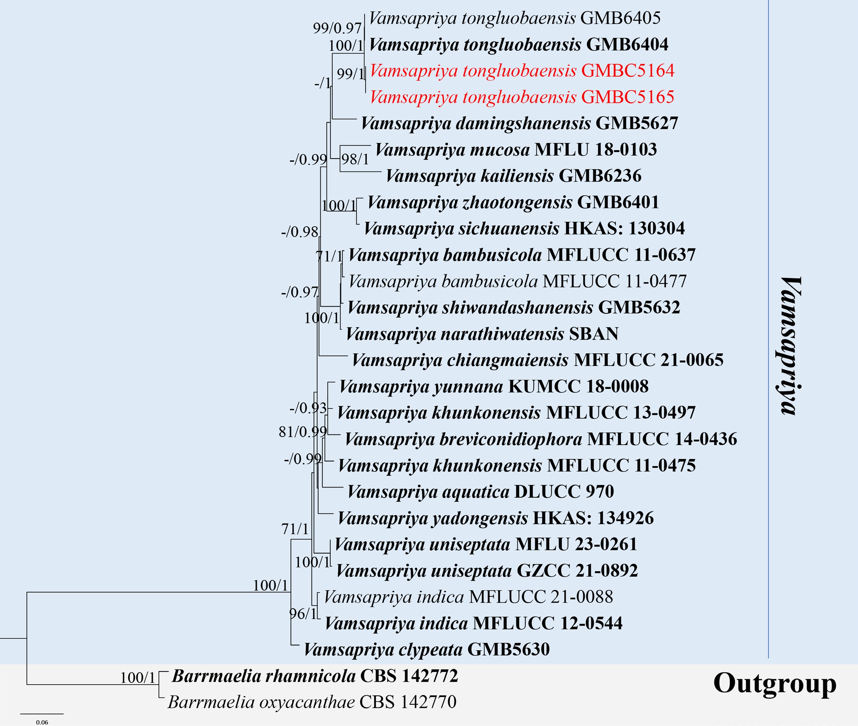
Figure 64.
ML tree of selected Vamsapriya species based on the ITS-LSU-SSU-rpb2-tub2 dataset. The combined dataset comprised 27 taxa, 2,947 characters (ITS: 1–535 bp; LSU: 536–1,406 bp; rpb2: 1,407–2,483 bp; tub2: 2,484–2,947 bp) including indel regions. Maximum likelihood (lnL = −10,545.792640) and Bayesian analyses produced consistent tree topologies. Barrmaelia oxyacanthae (CBS 142770) and B. rhamnicola (CBS 142772) were selected as the outgroup taxon. Bayesian posterior probabilities (PP) ≥ 0.90 and RAxML bootstrap support values ≥ 70 % are shown on the branches. The new strains are marked in red. Ex-type/type strains are indicated in bold.
-
The present study considerably expands the known diversity of hyphomycetous fungi in southwestern China by introducing three new genera, 31 new species, three new geographic records, and the first report of an asexual morph for Vamsapriya tongluobaensis. These findings indicate that the diversity of hyphomycetous fungi in this region remains far from fully documented and highlight the need for continued taxonomic investigations in biodiversity-rich subtropical areas.
The introduction of Acroappendicula, Aquaclavispora, and Radiaticonidium reflects the presence of distinct evolutionary lineages that cannot be accommodated within existing generic frameworks. The recognition of these genera is supported by consistent morphological characters in conjunction with robust phylogenetic evidence, a pattern increasingly observed in recent taxonomic studies of hyphomycetous fungi[150,163,184−186]. The high number of newly described species further demonstrates the extent of previously unrecognized diversity within asexual fungi, particularly in regions that have received limited systematic attention[1,187].
Phylogenetically, Aquaclavispora clusters closely with morphologically unrelated genera within Mollisiaceae. This unexpected grouping suggests either incomplete lineage sorting, convergent evolution, or, more likely, an insufficient understanding of morphological plasticity within the family. Compounding this issue is the limited taxon sampling across these groups, which indicates that fungal diversity in these clades remains substantially underexplored and in urgent need of targeted investigation.
The problem is not limited to Mollisiaceae. In several morphologically diverse hyphomycetous genera, molecular data are still available for only a small subset of species. This scarcity severely complicates reliable species identification, comparative morphological studies, and robust phylogenetic reconstructions, as sequence information is often entirely absent or paired with only minimal, rough descriptions that hinder meaningful comparisons. For example, the genus Stachylidium currently comprises 32 accepted species (Species Fungorum, accessed 27 March 2026), yet molecular data are available in NCBI GenBank for only two of them. Similarly, Index Fungorum (accessed 27 March 2026) records 523 epithets under Sporidesmium, with approximately 83% of these names established prior to 1992. Although 252 species are presently accepted, molecular data exist for merely about one-quarter of the Sporidesmium. The genus Brachysporium, which includes 43 accepted species (Species Fungorum, accessed 27 March 2026), has sequence data for just five species.
In some genera, morphological differences between species are very subtle and are best distinguished by molecular data. In such cases, the absence of molecular data hinders accurate species delimitation, reliable identification, and meaningful taxonomic comparisons. For example, species within the genus Pleurothecium exhibit very subtle morphological differences, making identification challenging. For Pleurothecium pulneyense, only LSU sequence data are available for phylogeny, but this marker is highly conserved and often nearly identical even across closely related species, providing limited resolution for distinguishing taxa or reconstructing reliable phylogenies.
These gaps highlight the critical need for a concerted effort to sequence older herbarium collections, particularly type specimens. Generating molecular data from historical material would not only stabilize the taxonomy of these genera but also enable robust phylogenetic placement of names that have long remained in morphological isolation. Integrating molecular data from herbarium specimens with those from fresh collections is essential for clarifying evolutionary relationships. This would aid in stabilizing names, connecting sexual and asexual morphs, refining generic boundaries, and uncovering hidden diversity that may still be overlooked in various groups.
Multigene phylogenetic analyses in this study reveal that morphological convergence remains widespread among hyphomycetous fungi, frequently resulting in artificial groupings when morphology is considered alone. Similar discrepancies between morphology-based classifications and molecular phylogenies have been documented across multiple hyphomycete lineages, emphasizing the limitations of traditional character sets[188,198]. The congruence observed here between molecular data and carefully evaluated morphological traits underscores the importance of integrative approaches for stabilizing species concepts and refining generic boundaries[199,200].
The recognition of Monilochaetes camelliae, Niesslia waitemataensis, and Stachylidium bicolor as new geographic records for China expands their known distribution ranges and contributes to a more comprehensive understanding of hyphomycetous fungal biogeography[99,131,172]. Such findings suggest that many species may possess broader ecological tolerances and wider distributions than currently documented, particularly among saprobic taxa[201,202]. In addition, the first documentation of the asexual morph of Vamsapriya tongluobaensis provides valuable morphological information that complements its sexual description and facilitates future identification and comparison[182].
The discovery of numerous novel taxa from Guizhou, Yunnan, and Guangxi further supports the view that southwestern China represents a major reservoir of fungal diversity. The complex topography, diverse vegetation types, and variety of microhabitats in this region create favorable conditions for fungal diversification, particularly among saprobic hyphomycetes. Continued sampling across different substrates and habitats, coupled with integrative taxonomic methods, is expected to reveal additional undescribed taxa and further clarify evolutionary patterns within hyphomycetous fungi.
-
This study provides a comprehensive assessment of hyphomycetous fungal diversity in southwestern China based on extensive sampling and integrative taxonomic analyses. By examining over 150 specimens using detailed morphological observations and multi-locus phylogenetic approaches, three new genera, 31 new species, three new geographic records for China, and the first asexual morph of Vamsapriya tongluobaensis are documented. These findings substantially expand current knowledge of hyphomycetous fungi in this biodiversity-rich region and demonstrate that their diversity remains far from fully explored. The results further highlight the necessity of combining morphological and molecular data to resolve species boundaries and stabilize generic concepts. Continued integrative taxonomic investigations across diverse habitats and substrates are essential for revealing the full extent of fungal diversity and understanding evolutionary patterns within hyphomycetous fungi.
-
Not applicable.
-
The authors confirm contribution to the paper as follows: study conception and design: Liu H, Li Q; data collection: Liu L, Zhang Q, Li W, Ren Y, Luo X; formal analysis and interpretation of results: Liu L, Lu C, Yang Q, Zou S; draft manuscript preparation: Liu L, Zhang Q, Li W; manuscript review and editing: Samarakoon MC, Wijayawardene NN, Elgorban AM, Al-Shwaiman HA. All authors reviewed the results and approved the final version of the manuscript.
-
All DNA sequence data generated in this study have been deposited in GenBank. Voucher specimens and living cultures are deposited in the recognized herbaria and culture collections cited in the Taxonomy section.
-
This research was supported by the National Natural Science Foundation of China (Grant Nos 32460051, 12132006); the Funded Research Projects of the State Key Laboratory of Discovery and Utilization of Functional Components in Traditional Chinese Medicine (Grant No. GMUSKL-202510, GMUSKL-202601); Guizhou Key Laboratory of Microbio and Infectious Disease Prevention & Control (Grant No. ZDSYS[2023]004); The Central Government Guided Local Science Foundation of Guizhou Province (Grant No. Qiankehe[2025]024); Guizhou Science and Technology Department (Grant Nos CXPTXM [2025]010/[2025]021/KXJZ[2025]014); The High-level Innovation Talents of Guizhou (Grant No. GCC [2023]048); the Science and Technology Program of Guizhou Province (Grant No. Qian Ke He Foundation-ZK [2022] General 364), and special funds from the central finance to support the development of local universities (Grant No. Qian Jiao Ji No [2023]036); This work was supported by the National '111' Project (Grant No. D20009), Scientists Workstation Guizhou Province (Grant No. KXJZ[2024]009/KXJZ[2024]007), High-level Innovation Talent Project of Guizhou Province (Grant No. GCC[2022]036-1). The authors extend their appreciation to the Ongoing Research Funding Program (ORF-RICSP-2026-6), King Saud University, Riyadh, Saudi Arabia.
-
The authors have declared that no competing interests exist.
-
accompanies this paper online at: https://doi.org/10.48130/mycosphere-0026-0008.
- Supplementary Table S1 GenBank accessions obtained in this study.
- Copyright: © 2026 by the author(s). Published by Maximum Academic Press, Fayetteville, GA. This article is an open access article distributed under Creative Commons Attribution License (CC BY 4.0), visit https://creativecommons.org/licenses/by/4.0/.
-
About this article
Cite this article
Liu L, Zhang Q, Li W, Ren Y, Luo X, et al. 2026. Morphological and phylogenetic analyses of hyphomycetous fungi from Guizhou, Yunnan, and Guangxi, China. Mycosphere 17: e008 doi: 10.48130/mycosphere-0026-0008 

Morphological and phylogenetic analyses of hyphomycetous fungi from Guizhou, Yunnan, and Guangxi, China
- Received: 26 January 2026
- Revised: 20 April 2026
- Accepted: 11 May 2026
- Published online: 03 July 2026
Abstract: Hyphomycetous fungi represent an ecologically important and a widely distributed fungal group in natural environments. In this study, over 150 specimens of hyphomycetous fungi were collected from Guangxi Zhuang Autonomous Region, Guizhou Province, and Yunnan Province, China, and investigated using integrated morphological observations and multi-locus phylogenetic analyses based on ITS, SSU, LSU, rpb2, tub2, and tef1-α regions. A total of 35 species were identified, of which 22 species belong to Sordariomycetes, followed by Dothideomycetes (eight species), Leotiomycetes (three species), Orbiliomycetes (one species), and Eurotiomycetes (one species). Three novel genera, Acroappendicula, Aquaclavispora, and Radiaticonidium, are proposed, and 31 new species are described, namely Acroappendicula aquatica, Aquaclavispora lignicola, Arthrobotrys bambusicola, Brachysporium wumengshanense, Chaetosphaeria tongrenensis, Corynespora guizhouensis, Dematioscypha aquatica, Gamsomyces guangxiensis, Helicoma wuliangshanense, Hermatomyces hyalodimorphus, Hypomyces aquatilis, Kirschsteiniothelia chinensis, Melanopsamma wumengshanensis, Musicillium verticillatum, Myrmecridium bambusicola, Myxospora aquatica, Neohelicomyces wuliangshanensis, Nigrograna chinensis, Ophiostoma balanophorae, Parascedosporium juglandicola, Plectosphaerella guangxiensis, Pleurothecium longisetosum, Radiaticonidium aquaticum, Rhamphoriopsis flabelliformis, Rhodoveronaea obovoidea, Sporidesmium guiyangense, S. wuliangshanense, Strossmayeria multiseptata, Xenopleopunctum dictyosporum, Xenopleopunctum yunnanense, and Xylolentia wumengshanensis. Furthermore, Monilochaetes camelliae, Niesslia waitemataensis, and Stachylidium bicolor are reported as new geographic records for China, and the asexual morph of Vamsapriya tongluobaensis is documented for the first time. Nigrograna borsei (= Biatriospora borsei) is proposed as a new combination. These results reveal a high and previously undocumented diversity of hyphomycetous fungi in southwestern China.
-
Key words:
- Asexual morphs /
- Fungal systematics /
- Freshwater fungi /
- Karst ecosystem /
- Saprobic fungi