











-
Thelephora Ehrh. ex Willd., a member of the family Thelephoraceae Chevall. (Thelephorales, Basidiomycota), was established by Willdenow (1787)[1]. The genus is typified by T. terrestris Ehrh. ex Fr. and is characterized by the following features. The basidiomata are diverse in shape (resupinate, effused-reflexed, pileate or coralloid) and color. The hymenophore may be smooth, granulose, arachnoid, pelliculose, mucedinoid, crustose, colliculose, or hydnoid. Rhizomorphs are present in some species. The hyphal system is monomitic. Cystidia are absent in most species; when present, cystidia shapes are clavate, capitate, or hyphoid. Basidia are clavate or utriform and have two or four sterigmata. Basidiospores are verruculose, bi- and trifurcate or echinulate[2−5]. According to the records of Index Fungorum, the number of currently accepted species in the genus Thelephora is approximately 499.
As ectomycorrhizal fungi, Thelephora species are vital to forest functioning[6,7]. They form symbiotic associations with trees, improving host uptake of water and nutrients, particularly limiting elements like nitrogen (N) and phosphorus (P) in nutrient-poor soils[8−12]. Their extensive mycelial networks interconnect plants, facilitating nutrient and signal transfer, thereby modulating plant interactions and promoting community stability[13−16]. In addition, by contributing to soil carbon sequestration and driving nutrient cycling, these networks become vital to sustaining forest productivity and long-term ecosystem health[17−20]. In addition, some species of Thelephora are edible, especially in southwestern China, such as T. aurantiotincta and T. vialis[21]; others are medicinal; for example, T. ganbajun serves as a potent source of natural antioxidants, and its extract displays antiproliferative activity against human lung, breast, liver, and other cancer cells at the cellular level[22].
The genus Thelephora was first established by Ehrhart in 1787. Fries (1821) circumscribed Thelephora as a broad collective group that even included five tribes, as well as the subgenera Stereum, Phylacteria, and Leiostroma[23]. Subsequently, Patouillard (1900) and other taxonomists made continuous revisions to the generic boundaries and defined Thelephora as erect, often branched or pileate macrofungi[24]. Tomentella was first proposed by Persoon in 1799 as an infrageneric taxon under Corticium, comprising two species: C. ferrugineum and C. chalibaeum; it was not until 1887 that Patouillard formally raised it to generic rank. Tomentella was defined as strictly resupinate, corticioid fungi closely adhering to the substratum[25,26]. Even before the widespread application of molecular techniques, researchers noted the high morphological similarity in microscopic features between Tomentella and Thelephora, particularly in the verruculose to echinulate spore ornamentation. The advent of molecular phylogenetics fundamentally reshaped our understanding of Thelephora and its allied taxa. Based on molecular phylogenetic analyses, Tedersoo et al.[27] demonstrated that species of Thelephora and Tomentella form a monophyletic clade, commonly referred to as the 'tomentella-thelephora' clade. Ramírez-López et al. found that some Thelephora species, T. versatilis Ram.-Lóp. & Villegas and T. pseudoversatilis Ram.-Lóp. & Villegas, also possess resupinate basidiomata, indicating that a generic delimitation based solely on basidiomata morphology (erect vs resupinate) does not reflect natural evolutionary relationships[28,29]. In 2024, Kõljalg et al., following the principle of nomenclatural priority, proposed merging Tomentella into Thelephora, with 191 new combinations[29]. Concurrent studies clarified that the genus Odontia Pers., previously thought to be closely related to Tomentella, is in fact a non-mycorrhizal sister group to this clade and should be retained as a distinct genus[30−34]. In addition, some other generic-level taxonomy of Thelephorales was revised recently. For example, Bankera Coker & Beers ex Pouzar was merged into Phellodon P. Karst.[35−38], the large-scale transfer of species occurred between the genera Hydnellum P. Karst. and Sarcodon Quél. ex P. Karst.[39−45], and most species of Pseudotomentella Svrček et al. were transferred into Polyozellus Murrill[46,47].
This study focuses on the species diversity of Thelephora in China. In recent years, a number of new species of the genus Thelephora from China have been discovered and described based on morphology, ecology, and multi-gene phylogenetic analyses. For example, Yuan et al.[48] revealed 46 new species from China; Zhang et al.[49] revealed four new species from China; Song et al.[34] revealed two new species from China; Zhu et al.[5] revealed 12 new species and provided two keys to the species of resupinate and upright Thelephora from China; Yuan et al.[50] revealed two new resupinate species from China. To date, 88 known species of Thelephora have been discovered and described in China, and on this basis, this study has added another 20 new species of Thelephora found in China. Although molecular systematics has clarified the merger of Tomentella into Thelephora and promoted the development of the taxonomy of this group, systematic surveys targeting specific geographic regions and hosts are still lacking. Based on morphological studies and analyses of DNA sequences, including the internal transcribed spacer (ITS) and the large subunit of nuclear ribosomal RNA (LSU), we present 20 new species from China, each with an illustrated description and comparison with morphologically similar or related species.
-
The examined specimens are deposited in the Fungarium of the Institute of Microbiology, Beijing Forestry University (BJFC), Beijing, China. Macromorphological characteristics, including the color, thickness of the basidiomata, hymenophoral surface, and sterile margin, were examined under a stereomicroscope (Nikon SMZ 1000: Tokyo, Japan) at 4 × magnification. Colors used follow Anonymous[51] and Petersen[52]. Samples for microscopic examination were stained in Cotton Blue, Melzer's reagent, 5% potassium hydroxide, and distilled water, and sections or tissues of basidiomata were studied in Cotton Blue at a magnification of 1,000 × using a Nikon E80i microscope with phase contrast illumination (Nikon, Tokyo, Japan), and line drawings were made with the aid of a drawing tube. The surface morphology of the basidiospores was observed with a SU8010 Cold Field Emission Scanning Electron Microscope (SEM, SU8010, Hitachi, Japan) at an accelerating voltage of 15 kV. The working distance was 12.2 mm. A thin layer of gold was coated on the samples to avoid charging. Additionally, the scientific names at all ranks in italics are written in italics, following Thines et al.[53].
The following abbreviations were used: L = mean spore length (arithmetical average of all spores), W = mean spore width (arithmetical average of all spores), Q = values of the length/width ratios among the studied specimens, n (a/b) = number of basidiospores (a) measured from given number of specimens (b), IKI = Melzer's reagent, IKI− = negative in Melzer's reagent, KOH = 5% potassium hydroxide, CB = Cotton Blue, CB+ = cyanophilous, CB− = acyanophilous.
DNA extraction, PCR amplification, and sequencing
-
A CTAB plant genome rapid extraction kit-DN14 (Aidlab Biotechnologies Co., Ltd, Beijing, China) was used for DNA extraction and PCR amplification from dried specimens according to the manufacturer's instructions, with some modifications[54]. The ITS region was amplified with the primers ITS5 (5′-GGAAGTAAAAGTCGTAACAAGG-3′) and ITS4 (5′-TCCTCCGCTTATTGATATGC-3′). The LSU gene was amplified with the primers LR0R (5′-ACCCGCTGAACTTAAGC-3′) and LR7 (5′-TACTACCACCAAGATCT-3′). The Polymerase Chain Reaction (PCR) procedure for ITS was as follows: initial denaturation at 95 °C for 5 min, followed by 39 cycles at 95 °C for 30 s, 54 °C for 30 s, and 72 °C for 20 s, and a final extension at 72 °C for 10 min. The PCR procedure for LSU was as follows: initial denaturation at 95 °C for 5 min, followed by 39 cycles at 95 °C for 30 s, 48 °C for 30 s, and 72 °C for 20 s, and a final extension at 72 °C for 10 min[55]. The PCR products were purified and sequenced with the same primers at the Beijing Genomics Institute, China. The newly generated sequences were deposited in GenBank. All sequences[56−91]analyzed in this study were listed in Supplementary Table S1.
Phylogenetic analyses
-
The phylogenetic relationships of Thelephora were analyzed using datasets of ITS + LSU sequences. Amaurodon aquicoeruleus Agerer and A. viridis (Alb. & Schwein.) J. Schröt were used as the outgroups in our phylogenetic analyses[34].
The DNA sequences were aligned by MAFFT v.7 with the G-INS-i option[92]. Alignments were optimized manually in BioEdit v7.0.9[93] and spliced in Mesquite v3.2.[94]. The partition homogeneity test (PHT)[95] of the gene dataset was tested by PAUP v4.0b10[96] under 1,000 homogeneity replicates. The best-fit evolutionary model was selected with AIC (Akaike information criterion) using MrModelTest v2.3[97,98]. Sequences were analysed using maximum likelihood (ML) with RAxML-HPC2 through the CIPRES Science Gateway[99]. Branch support (BS) for ML analysis was determined by 1,000 bootstrap replicates. The Bayesian inference (BI) analysis with MrBayes v3.2.7[100] implemented the Markov chain Monte Carlo (MCMC) technique. Four simultaneous Markov chains were run with 15, 10, 5, and 5 million generations for the ITS and LSU datasets, respectively, starting from random trees and keeping one tree every 100th generation until the average standard deviation of split frequencies was below 0.01. The first 25% of the sampled trees were discarded as burn-in, and the remaining ones were used to reconstruct a majority-rule consensus and calculate Bayesian posterior probabilities (BPP) of the clades. Phylogenetic trees were visualized with FigTree v1.4.2 (
http://tree.bio.ed.ac.uk/software/figtree ), and additional layout was carried out using Adobe Illustrator CS v5. Branches that received bootstrap support ML ≥ 75% and BPP ≥ 0.95 were considered significantly supported. The ML bootstrap support ≥ 50% and BPP ≥ 0.90 are presented on topologies from ML analysis, respectively. -
All new species ITS sequences BLAST results in this study are listed in Supplementary Table S2.
Molecular phylogeny
-
In this study, 512 sequences derived from 335 fungal samples representing 171 species were used to reconstruct phylogenetic trees, including 327 sequences of ITS and 185 sequences of LSU. Among them, 65 sequences were newly generated, including 39 sequences of ITS and 26 sequences of LSU. The dataset had an aligned length of 2,142 characters, of which 1,243 (58%) characters were constant, 200 (9%) were variable and parsimony-uninformative, and 699 (33%) were parsimony-informative. The phylogenetic reconstruction performed with ML and BI analyses for two combined datasets showed similar topology and few differences in statistical support. The best model fit applied in the Bayesian analysis was GTR + I + G, lset nst = 6, rates = invgamma, and prset statefreqpr = dirichlet (1, 1, 1, 1). The phylogenetic tree topology resulting from Bayesian analyses was similar to that obtained from ML analysis, with the Bayesian analysis yielding an average standard deviation of split frequencies = 0.009966; thus, only the ML tree is provided (Fig. 1). The ML topology was presented and annotated with ML bootstraps ≥ 50% and BI bootstraps ≥ 0.90.

Figure 1.
Maximum Likelihood tree illustrating the phylogeny of Thelephora based on combined ITS + LSU sequence data. Branches are labeled with maximum-likelihood bootstrap proportions higher than 50% and Bayesian posterior probabilities greater than 0.90, respectively. * Indicate the type species. The new species are in bold.
In the phylogenetic tree, 22 clades with moderate to strong support were marked (Fig. 1). The support values for each clade are as follows: 99% ML/1.00 BPP for clade 1, 100% ML/1.00 BPP for clade 2, 97% ML/1.00 BPP for clade 3, 100% ML/1.00 BPP for clade 4, 98% ML/1.00 BPP for clade 5, 100%/0.94 BPP ML for clade 6, 100% ML/0.99 BPP for clade 7, 100% ML/1.00 BPP for clade 8, 82%/0.95 BPP ML for clade 9, 100% ML/1.00 BPP for clade 10, 73% ML/0.90 BPP for clade 11, 92% ML/0.95 BPP for clade 12, 92% ML/0.997 BPP for clade 13, 75% ML/0.90 BPP for clade 14, 100% ML/1.00 BPP for clade 15, 97% ML/0.90 BPP for clade 16, 94% ML/0.94 BPP for clade 17, 95% ML/0.96 BPP for clade 18, 96% ML/0.97 BPP for clade 19, 98% ML/1.00 BPP for clade 20, 98% ML/0.97 BPP for clade 21, and 92% ML/1.00 BPP for clade 22. Among these 22 clades, 19 of the 20 new species were distributed across 13 clades (clades 1, 2, 3, 5, 6, 10, 11, 12, 13, 18, 19, 20, and 21), whereas one new species, Thelephora violaceardesiaca, was not placed in any of these numbered clades and instead formed a separate isolated lineage with strong support (100% ML/1.00 BPP), suggesting a distinct phylogenetic position.
Taxonomy
-
Thelephora bambusicola X. Li, Y.C. Dai, X. Zhang & Yuan Yuan, sp. nov. Fig. 2
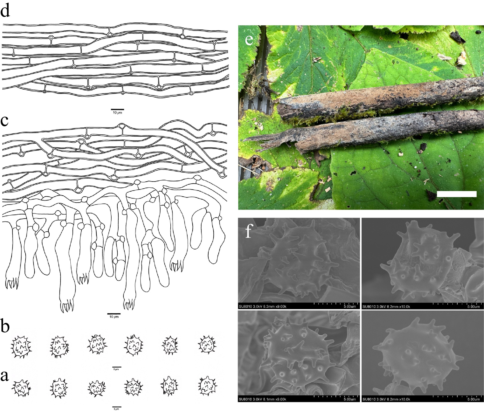
Figure 2.
Basidiomata and microscopic structures of Thelephora bambusicola (holotype, Dai 31713). (a) Basidiospores in lateral view. (b) Basidiospores in frontal view. (c) A section through a basidioma. (d) Hyphae from a rhizomorph. (e) Basidiomata on the host substrates. (f) Scanning electron microscope (SEM) of basidiospores. Scale bars: (a), (b) = 5 µm; (c), (d) = 10 µm; (e) = 1 cm.
MycoBank number: MB 864301
Diagnosis – Thelephora bambusicola is distinguished by resupinate basidiomata, olivaceous buff to clay-buff hymenophoral surface when fresh and grayish brown when dry, presence of rhizomorphs, fusiform cystidioles, and broadly ellipsoid to bi-, tri-, or quadra-lobed basidiospores.
Type – CHINA. Xizang Autonomous Region, Yadong County, forests from Pasha Falls to Nathu La Gate, on dead bamboo, 16 October 2024, Dai 31713 (holotype BJFC051972).
Etymology – Bambusicola (Lat.): refers to the species growing on bamboo.
Basidiomata – Annual, resupinate, adherent to the substrate, mucedinoid, soft, without odor or taste when fresh, continuous, up to 4 cm long, 2 cm wide, and 0.9 mm thick at the center; hymenophoral surface smooth, olivaceous buff to clay-buff when fresh, grayish brown and concolorous with subiculum when dry; sterile margin determinate, byssoid, whitish, paler than hymenophore.
Rhizomorphs – Present in subiculum and margin, 25–55 µm diam; rhizomorphic surface more or less smooth; hyphae in rhizomorph monomitic, undifferentiated, individual hyphae with clamp connections and rarely with simple septa, 4.5–7 µm diam, unbranched, thin- to slightly thick-walled with a wide lumen, more or less regularly arranged, pale brown in KOH and distilled water.
Subicular hyphae – Monomitic; generative hyphae mostly with clamp connections and rarely with simple septa, occasionally branched, thin- to slightly thick-walled, without encrustation, more or less interwoven, yellowish in KOH and distilled water, IKI−, CB−, 4–7 µm diam; subhymenial hyphae frequently with clamp connections, thin-walled, smooth, 4–6 µm diam; hyphal cells more or less uniform, yellowish in KOH and in distilled water, IKI−, CB−.
Cystidia – Absent; cystidioles present, arising at a level with the basidia, fusiform, 35–45 × 8–10 µm, yellowish in KOH and distilled water, with a clamp connection at the base.
Basidia – Clavate, without transverse septa, yellowish in KOH and distilled water, not stalked, with four sterigmata and a clamp connection at the base, 35–65 µm long, 7–10 µm diam at the apex and 4–6 µm at the base; sterigmata 5–8 µm long and 2–4 µm diam at the base.
Basidiospores – Broadly ellipsoid to bi-, tri- or quadra-lobed in frontal and lateral faces, echinulate, yellowish in KOH and distilled water, IKI−, CB−, (6.1–)6.4–9.5(–9.1) × (4.9–)5.0–7.7(–7.8) µm, L = 7.65 µm, W = 6.15 µm, Q = 1.20–1.25 (n = 60/2); echinuli usually grouped in two or more, up to 2 µm long.
Additional specimen (paratype) examined – CHINA. Xizang Autonomous Region, Yadong County, forests from Pasha Falls to Nathu La Gate, on dead bamboo, 16 October 2024, Dai 31719 (BJFC051978).
Habitat – Thelephora bambusicola was found in southwest China with a subtropical semi-humid monsoon climate.
Notes – Thelephora bambusicola is more or less related to T. extendens Kõljalg, I. Saar & Svant. (60% in ML) in the phylogenetic tree (Fig. 1). Thelephora extendens is similar to T. bambusicola by its continuous basidiomata adherent to the substrate, clamped subhymenial hyphae, and the absence of cystidia. However, T. extendens is distinguished from T. bambusicola by having a crustose hymenophoral surface and basidiospores often with oil drops[48,101].
Thelephora bambusicola shares similar morphological characteristics with the species T. bomiensis by sharing a smooth and grayish-brown hymenophoral surface. But phylogenetically, they are distantly related (Fig. 1), and T. bomiensis differs from T. bambusicola by having narrower subhymenium hyphae (2.5–4 vs 4–6 µm) and lacking rhizomorphs[5].
Thelephora caeruleoalba X. Li, Y.C. Dai & Yuan Yuan, sp. nov. Fig. 3
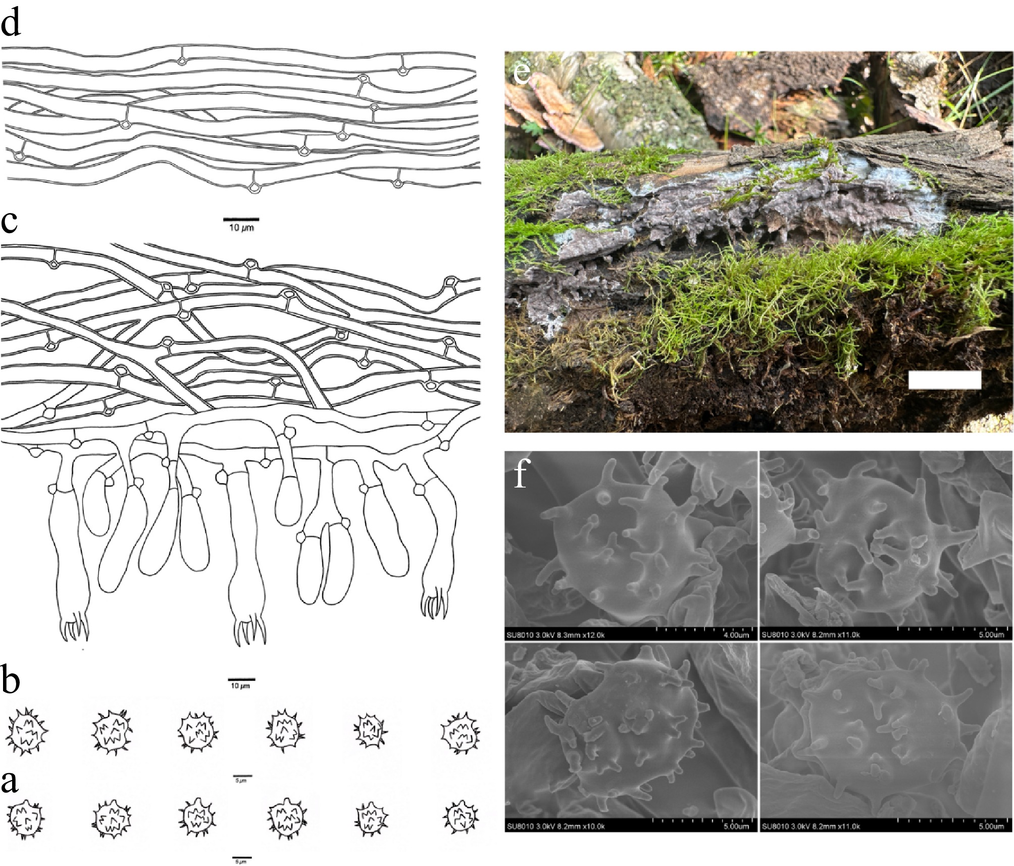
Figure 3.
Basidiomata and microscopic structures of Thelephora caeruleoalba (holotype, Dai 29452). (a) Basidiospores in lateral view. (b) Basidiospores in frontal view. (c) A section through a basidioma. (d) Hyphae from a rhizomorph. (e) Basidiomata on the host substrates. (f) Scanning electron microscope (SEM) of basidiospores. Scale bars: (a), (b) = 5 µm; (c), (d) = 10 µm; (e) = 1 cm.
MycoBank number: MB 864302
Diagnosis – Thelephora caeruleoalba is distinguished by resupinate basidiomata, grayish-blue hymenophoral surface when fresh and light vinaceous gray when dry, presence of rhizomorphs, long sterigmata (8–11 µm), broadly ellipsoid to subglobose to bi-, tri-lobed basidiospores.
Type – CHINA. Qinghai Province, Huzhu County, Beishan Forest Park, Langshidang, on rotten wood of Salix, 27 July 2024, Dai 29452 (holotype BJFC049711).
Etymology – Caeruleoalba (Lat.): refers to the basidiomata of the species having a bluish-white margin when fresh.
Basidiomata – Annual, resupinate, separable from the substrate, pelliculose, soft, without odor or taste when fresh, continuous, up to 6 cm long, 3 cm wide, and 1 mm thick at the center; hymenophoral surface smooth, grayish blue when fresh, and light vinaceous gray and paler than subiculum when dry; sterile margin determinate, byssoid, bluish white, paler than hymenophore.
Rhizomorphs – Present in subiculum and margin, 15–40 µm diam; rhizomorphic surface more or less smooth; hyphae in rhizomorph monomitic, undifferentiated, individual hyphae with clamp connections, 3.5–5.5 µm diam, unbranched, slightly thick-walled, more or less regularly arranged, hyaline in KOH and distilled water.
Subicular hyphae – Monomitic; generative hyphae frequently with clamp connections, occasionally branched, thick-walled with a wide lumen, without encrustation, more or less interwoven, hyaline in KOH and distilled water, IKI−, CB−, 4–6.5 µm diam; subhymenial hyphae frequently with clamp connections, thin-walled, smooth, 3–5 µm diam; hyphal cells more or less uniform, hyaline in KOH and in distilled water, IKI−, CB−.
Cystidia – Absent.
Basidia – Clavate, constricted in the middle, without transverse septa, hyaline in KOH and distilled water, not stalked, with four sterigmata and a clamp connection at the base, 35–55 µm long, 8–10 µm diam at the apex and 4–7 µm at the base; sterigmata 8–11 µm long and 2–3 µm diam at the base.
Basidiospores – Broadly ellipsoid to subglobose to bi-, tri-lobed in frontal face and subglobose in lateral face, echinulate, yellowish in KOH and distilled water, IKI−, CB−, (5.8–)6.2–9.0(–9.7) × (5.3–)5.4–8.0(–8.5) µm, L = 7.89 µm, W = 6.61 µm, Q = 1.15–1.22 (n = 60/2); echinuli usually grouped in two or more, up to 2 µm long.
Additional specimen (paratype) examined – CHINA. Qinghai Province, Huzhu County, Beishan Forest Park, Langshidang, on a fallen branch of Betula, 10 August 2025, Dai 35205 (BJFC056466);
Habitat – Thelephora caeruleoalba was found in northwest China with a plateau continental climate. Additionally, T. caeruleoalba was also known from one eDNA sequence from Austria (Fig. 1).
Notes – Thelephora caeruleoalba is closely related to Thelephora murina (100% in ML) in the phylogenetic tree (Fig. 1) and then clustered with Thelephora rotundata H.S. Yuan & Y. Q. Zhu (100% in ML and 1.00 BPP). Thelephora murina shares similar characteristics with T. caeruleoalba, including continuous pelliculose basidiomata, the absence of cystidia, and clamped hyphae. However, T. murina is distinguished from T. caeruleoalba by the absence of rhizomorphs. Thelephora rotundata is similar to T. caeruleoalba by sharing a smooth hymenophoral surface and subhymenial hyphae with clamp connections. However, T. rotundata is distinguished from T. caeruleoalba by its basidiomata not separable from the substrate, the absence of rhizomorphs, and bigger basidiospores (10.2–13.1 × 9.5–12.1 vs 6.2–9.0 × 5.4–8.0 µm)[74].
In morphology, T. caeruleoalba shares similar characteristics with T. fuscopelliculosa, by sharing pelliculose basidiomata, a smooth hymenophoral surface, and lacking cystidia. However, phylogenetically, they are distantly related (Fig. 1), and T. fuscopelliculosa is distinguished from T. caeruleoalba by its wider basidiospores (8.0–9.0 vs 5.4–8.0 µm) and the lack of rhizomorphs[48].
Thelephora ceracea X. Li, Y.C. Dai, X. Zhang & Yuan Yuan, sp. nov. Fig. 4
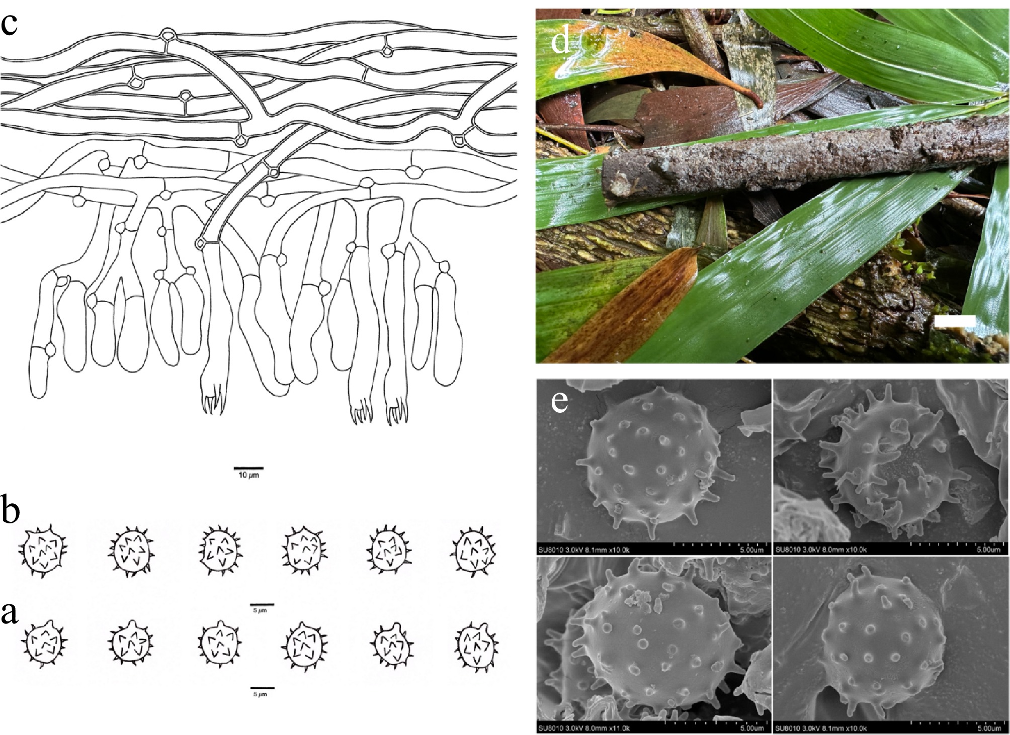
Figure 4.
Basidiomata and microscopic structures of Thelephora ceracea (holotype, Dai 30115). (a) Basidiospores in lateral view. (b) Basidiospores in frontal view. (c) A section through a basidioma. (d) Basidiomata on the host substrates. (e) Scanning electron microscope (SEM) of basidiospores. Scale bars: (a), (b) = 5 µm; (c) = 10 µm; (d) = 1 cm.
MycoBank number: MB 864303
Diagnosis – Thelephora ceracea is distinguished by resupinate basidiomata, a pale mouse gray to vinaceous gray hymenophoral surface when both fresh and dry, broad subicular hyphae (5–8 µm), and subglobose basidiospores.
Type – CHINA. Hainan Province, Wuzhishan County, Shuiman, on rotten bamboo, 23 September 2024, Dai 30115 (holotype BJFC050374).
Etymology – Ceracea (Lat.): refers to the species having waxy basidiomata when fresh.
Basidiomata – Annual, resupinate, separable from the substrate, soft, without odor or taste when fresh, continuous, up to 10 cm long, 3 cm wide, and 1.5 mm thick at the center; hymenophoral surface smooth, pale mouse gray to vinaceous gray when both fresh and dry; sterile margin determinate, farinaceous, concolorous with hymenophore.
Rhizomorphs – Absent.
Subicular hyphae – Monomitic; generative hyphae mostly with clamp connections and rarely with simple septa, occasionally branched, thick-walled with a wide lumen, without encrustation, more or less interwoven, hyaline in KOH and distilled water, IKI−, CB−, 5–8 µm diam; subhymenial hyphae frequently with clamp connections and rarely with simple septa, thin-walled, smooth, 4–7.5 µm diam; hyphal cells more or less uniform, hyaline in KOH and in distilled water, IKI−, CB−.
Cystidia – Absent.
Basidia – Clavate, more or less constricted in the middle, without transverse septa, hyaline in KOH and distilled water, not stalked, with four sterigmata and a clamp connection at the base, 25–55 µm long, 6–10 µm diam at the apex and 4–8 µm at the base; sterigmata 4–7 µm long and 2–4 µm at the base.
Basidiospores – Subglobose in frontal and lateral faces, echinulate, yellowish in KOH and distilled water, IKI−, CB−, (6.9–)7.1–9.0(–9.1) × (6.0–)6.7–7.8(–8.5) µm, L = 7.94 µm, W = 6.99 µm, Q = 1.10–1.14 (n = 60/2); echinuli usually isolated, up to 1.5 µm long.
Additional specimen (paratype) examined – CHINA. Hainan Province, Wuzhishan County, Shuiman, on rotten bamboo, 23 September 2024, Dai 30117 (BJFC050376).
Habitat – Thelephora ceracea was found in southern China with a tropical monsoon climate.
Notes – Thelephora ceracea is closely related to Thelephora hjortstamiana Suvi & Kõljalg (100% in ML and 1.00 BPP) in the phylogenetic tree (Fig. 1), and both species share a smooth hymenophoral surface, clamped subhymenial hyphae, and the absence of cystidia. However, T. hjortstamiana is distinguished from T. ceracea by having discontinuous basidiomata not separable from the substrate and the presence of rhizomorphs[67,75].
In morphology, T. ceracea shares similar characteristics with T. nivea, including separable basidiomata from the substrate, a vinaceous gray hymenophoral surface, and a lack of rhizomorphs. However, phylogenetically, they are distantly related (Fig. 1), and T. nivea is distinguished from T. ceracea by narrower basidiospores (4.6–6.4 vs 6.7–7.8 µm).
Thelephora griseosepiacea X. Li, Y.C. Dai & Yuan Yuan, sp. nov. Fig. 5
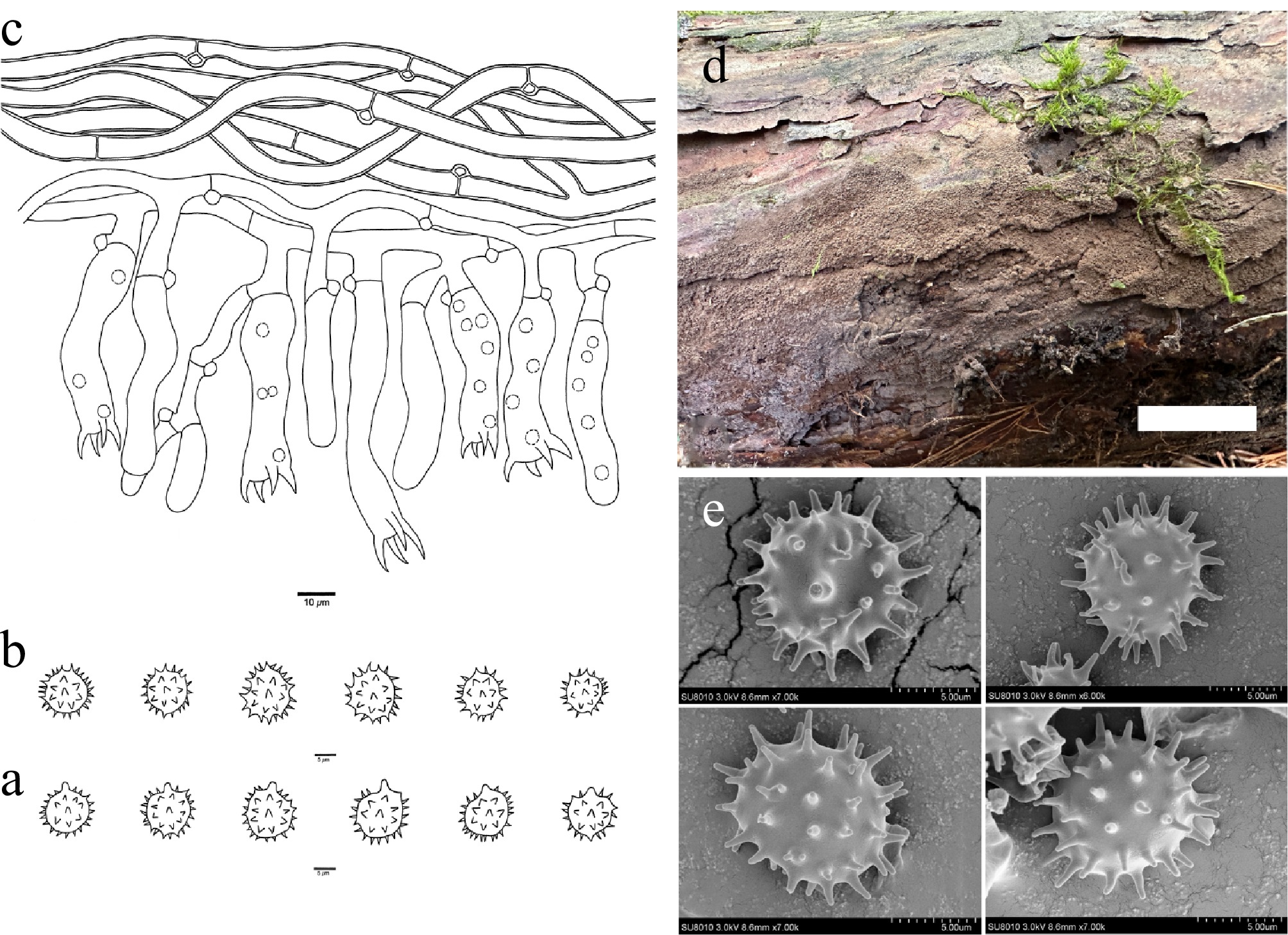
Figure 5.
Basidiomata and microscopic structures of Thelephora griseosepiacea (holotype, Dai 37654). (a) Basidiospores in lateral view. (b) Basidiospores in frontal view. (c) A section through a basidioma. (d) Basidiomata on the host substrates. (e) Scanning electron microscope (SEM) of basidiospores. Scale bars: (a), (b) = 5 µm; (c) = 10 µm; (d) = 1 cm.
MycoBank number: MB 864304
Diagnosis – Thelephora griseosepiacea is distinguished by resupinate basidiomata, a grayish-brown hymenophoral surface when fresh and vinaceous gray when dry, long sterigmata (9–13 µm), and subglobose to globose basidiospores.
Type – CHINA. Jilin Province, Jiaohe County, Experimental Forest Reserve, on a fallen trunk of Pinus koraiensis, 11 September 2025, Dai 37654 (holotype BJFC058913).
Etymology – Griseosepiacea (Lat.): refers to the species having a grayish-brown hymenophoral surface when fresh.
Basidiomata – Annual, resupinate, separable from the substrate, mucedinoid, soft, without odor or taste when fresh, continuous, up to 9 cm long, 2 cm wide, and 1 mm thick at the center; hymenophoral surface granulose, grayish brown when fresh, and vinaceous gray and concolorous with subiculum when dry; sterile margin determinate, farinaceous, concolorous with hymenophore.
Rhizomorphs – Absent.
Subicular hyphae – Monomitic; generative hyphae mostly with clamp connections and rarely with simple septa, occasionally branched, thick-walled with a wide lumen, without encrustation, more or less interwoven, hyaline in KOH and distilled water, IKI−, CB−, 4–7.5 µm diam; subhymenial hyphae mostly with clamp connections and rarely with simple septa, thin-walled, smooth, 4–6.5 µm diam; hyphal cells more or less uniform, hyaline in KOH and in distilled water, IKI−, CB−.
Cystidia – Absent.
Basidia – Clavate, distinctly constricted at the middle, without transverse septa, hyaline in KOH and distilled water, not stalked, usually with oil droplets, with four sterigmata and a clamp connection at the base, 30–70 µm long, 8–12 µm diam at the apex and 5–10 µm at the base; sterigmata 9–13 µm long and 2–4 µm diam at the base.
Basidiospores – Subglobose to globose in frontal and lateral faces, echinulate, yellowish in KOH and distilled water, IKI−, CB−, (7.8–)8.6–11.0(–11.1) × (7.6–)7.7–10.4(–10.5) µm, L = 9.27 µm, W = 8.57 µm, Q = 1.02–1.08 (n = 30/1); echinuli usually isolated, up to 2 µm long.
Habitat – Thelephora griseosepiacea was found in northeast China with a temperate continental monsoon climate.
Notes – Thelephora griseosepiacea is more or less related to Thelephora coffeae (H.S. Yuan & Y.C. Dai) Kõljalg et al. (88% in ML) in the phylogenetic tree (Fig. 1), and both species have continuous and mucedinoid basidiomata, the absence of cystidia and rhizomorphs, and thick-walled subicular hyphae. However, T. coffeae is distinguished from T. griseosepiacea by a smooth hymenophoral surface, stalked basidia, and shorter sterigmata (3–5 vs 9–13 µm)[48].
In morphology, T. griseosepiacea is similar to T. duplexa by sharing a grayish-brown to brown hymenophoral surface, subglobose to globose basidiospores, and lacking rhizomorphs. Phylogenetically, T. griseosepiacea and T. duplexa are placed in the same highly supported clade (99% in ML) (Fig. 1). However, T. duplexa differs from T. griseosepiacea by shorter basidia sterigmata (4–7 vs 9–13 µm) and shorter basidiospores (7.5–8.5 vs 8.6–11.0 µm)[48].
Thelephora keteleeriae X. Li, Y.C. Dai & Yuan Yuan, sp. nov. Fig. 6
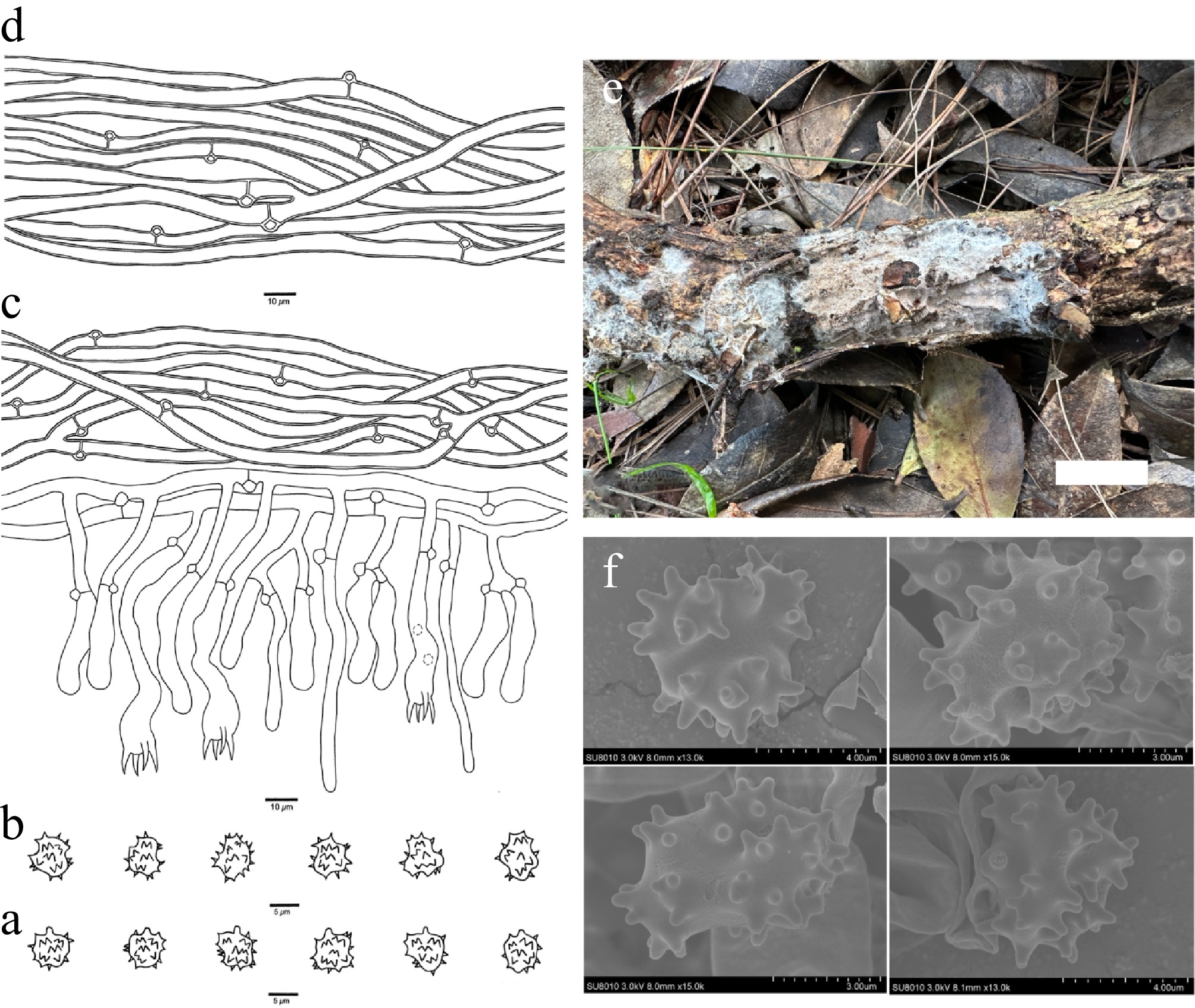
Figure 6.
Basidiomata and microscopic structures of Thelephora keteleeriae (holotype, Dai 28478). (a) Basidiospores in lateral view. (b) Basidiospores in frontal view. (c) A section through a basidioma. (d) Hyphae from a rhizomorph. (e) Basidiomata on the host substrates. (f) Scanning electron microscope (SEM) of basidiospores. Scale bars: (a), (b) = 5 µm; (c), (d) = 10 µm; (e) = 1 cm.
MycoBank number: MB 864305
Diagnosis – Thelephora keteleeriae is distinguished by resupinate basidiomata, white to salmon hymenophoral surface when fresh and white to light vinaceous gray when dry, presence of rhizomorphs, clavate cystidia, and broadly ellipsoid to subglobose to bi-, tri-, or quadra-lobed basidiospores.
Type – CHINA. Guangxi Autonomous Region, Leye County, Huangjingdong Forest Park, on a fallen branch of Keteleeria fortunei, 29 June 2024, Dai 28478 (holotype BJFC048737).
Etymology – Keteleeriae (Lat.): refers to the species growing on K. fortunei.
Basidiomata – Annual, resupinate, separable from the substrate, arachnoid, soft, without odor or taste when fresh, continuous, up to 5 cm long, 3 cm wide, and 1.5 mm thick at the center; hymenophoral surface irregularly poroid, white to salmon when fresh, and white to light vinaceous gray and darker than subiculum when dry; sterile margin determinate, fimbriate, white, paler than hymenophore.
Rhizomorphs – Present in subiculum and margin, 30–70 µm diam; rhizomorphic surface more or less smooth; hyphae in rhizomorph monomitic, undifferentiated, individual hyphae with clamp connections, 4–7 µm diam, unbranched, thick-walled with a wide lumen, more or less regularly arranged, hyaline in KOH and distilled water.
Subicular hyphae – Monomitic; generative hyphae frequently with clamp connections, occasionally branched, thick-walled with a wide lumen, without encrustation, more or less interwoven, hyaline in KOH and distilled water, IKI−, CB−, 3–6 µm diam; subhymenial hyphae frequently with clamp connections, thin-walled, smooth, 3.5–6.0 µm diam; hyphal cells more or less uniform, hyaline in KOH and in distilled water, IKI−, CB−.
Cystidia – Cystidia arising from subhymenial hyphae, clavate, projecting above the hymenium, aseptate, without encrustation, 40–75 µm long, 2–5 µm diam at the apex and 2–4 µm at the base, hyaline in KOH and in distilled water, IKI−, CB−; cystidioles absent.
Basidia – Clavate, strongly winding, without transverse septa, hyaline in KOH and distilled water, not stalked, occasionally with oil droplets, with four sterigmata and a clamp connection at the base, 40–70 µm long, 7–10 µm diam at the apex and 3–5 µm at the base; sterigmata 5–7 µm long and 1–3 µm diam at the base.
Basidiospores – Broadly ellipsoid to subglobose to bi-, tri- or quadra-lobed in frontal face and subglobose to bi-, tri- or quadra-lobed in lateral face, echinulate, yellowish in KOH and distilled water, IKI−, CB−, (4.9–)5.0–7.0(–7.1) × (4.1–)4.3–6.1(–6.5) µm, L = 5.97 µm, W = 5.05 µm, Q = 1.17–1.22 (n = 60/2); echinuli usually grouped in two or more, up to 1 µm long.
Additional specimen (paratype) examined – CHINA. Guangxi Autonomous Region, Leye County, Huangjingdong Forest Park, on a fallen branch of K. fortunei, 29 June 2024, Dai 28514 (BJFC048773).
Habitat – Thelephora keteleeriae was found in southern China with a subtropical monsoon climate.
Notes – Thelephora keteleeriae is closely related to Thelephora pallidocastanea (X. Lu, Y.H. Mu & H.S. Yuan) Kõljalg et al. (100% in ML and 1.00 BPP) in the phylogenetic tree (Fig. 1) and then clustered with Thelephora niveomarginata X. Li et al. (97% in ML and 0.99 BPP). Thelephora pallidocastanea resembles T. keteleeriae by sharing thin-walled subhymenial hyphae, thick-walled subicular hyphae, and basidia with a clamp connection at the base. However, T. pallidocastanea is differentiated from T. keteleeriae by having a smooth hymenophoral surface and the absence of rhizomorphs[78,102]. Thelephora niveomarginata is similar to T. keteleeriae by sharing continuous basidiomata separable from the substrate and clamp connections on the subhymenial hyphae. However, T. niveomarginata is distinguished from T. keteleeriae by having a smooth hymenophoral surface, the absence of cystidia, and longer sterigmata (7–9 vs 5–7 µm).
In morphology, T. keteleeriae is similar to T. efibulis by sharing arachnoid basidiomata, the presence of rhizomorphs, and up to 1 µm-long basidiospore echinuli. However, phylogenetically they are distantly related (Fig. 1), and T. efibulis is differentiated from T. keteleeriae by having shorter basidia (15–35 vs 40–70 µm) and lacking cystidia[48].
Thelephora koraiensis X. Li, Y.C. Dai & Yuan Yuan, sp. nov. Fig. 7
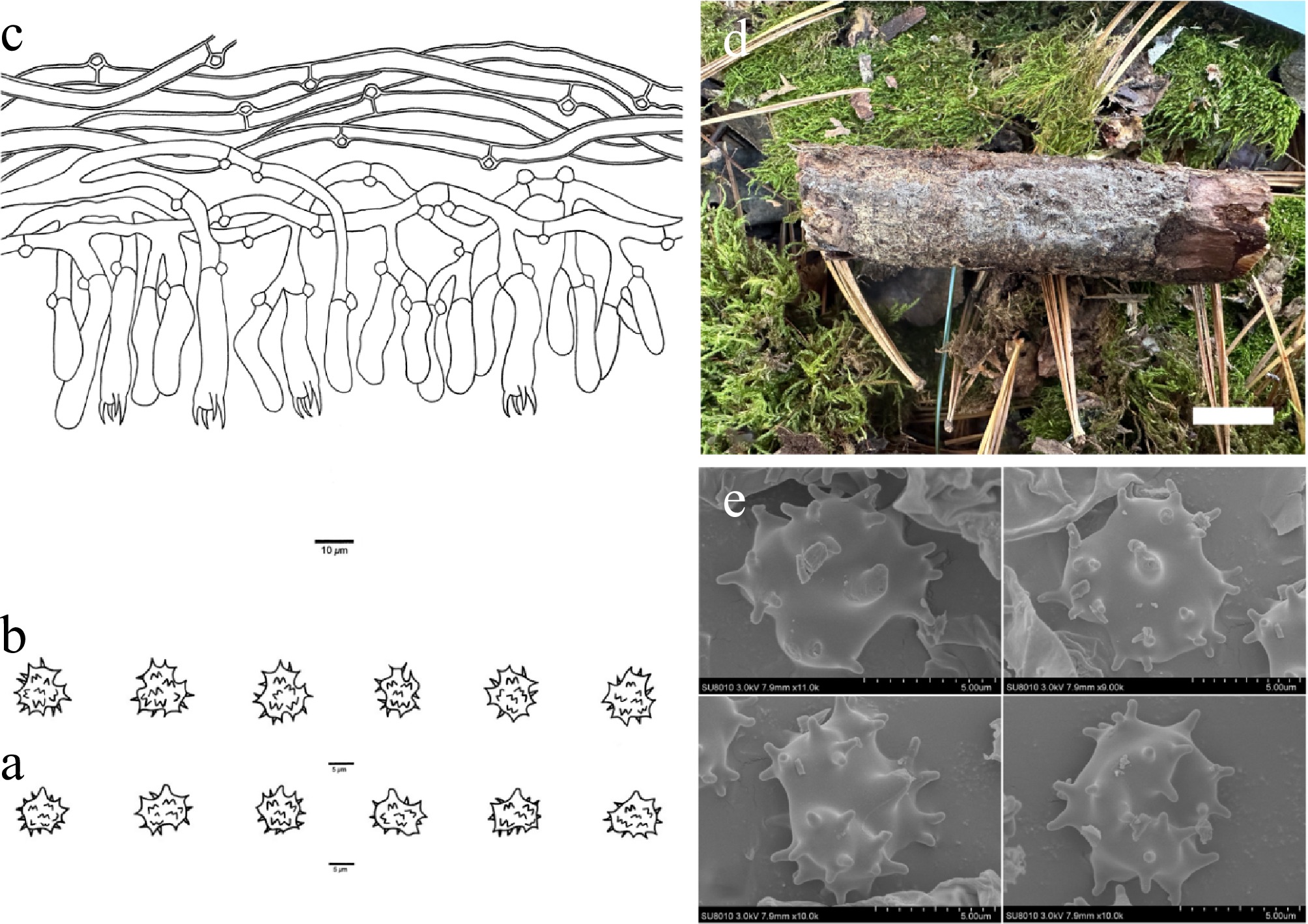
Figure 7.
Basidiomata and microscopic structures of Thelephora koraiensis (holotype, Dai 38336). (a) Basidiospores in lateral view. (b) Basidiospores in frontal view. (c) A section through a basidioma. (d) Basidiomata on the host substrates. (e) Scanning electron microscope (SEM) of basidiospores. Scale bars: (a), (b) = 5 µm; (c) = 10 µm; (d) = 1 cm.
MycoBank number: MB 864306
Diagnosis – Thelephora koraiensis is distinguished by resupinate basidiomata, an ash-gray to grayish-white hymenophoral surface when fresh and dry, slightly winding basidia, and broadly ellipsoid to bi-, tri-, or quadra-lobed basidiospores.
Type – CHINA. Heilongjiang Province, Fangzheng County, Shuangzishan Forest Park, on a fallen branch of Pinus koraiensis, 12 October 2025, Dai 38336 (holotype BJFC059595).
Etymology – Koraiensis (Lat.): refers to the species growing on P. koraiensis.
Basidiomata – Annual, resupinate, adherent to the substrate, arachnoid, soft, without odor or taste when fresh, continuous, up to 5 cm long, 1.5 cm wide, and 1 mm thick at the center; hymenophoral surface colliculose, ash gray to grayish white when both fresh and dry; sterile margin determinate, fimbriate, concolorous with hymenophore.
Rhizomorphs – Absent.
Subicular hyphae – Monomitic; generative hyphae frequently with clamp connections, occasionally branched, thick-walled with a wide lumen, without encrustation, more or less interwoven, yellowish in KOH and distilled water, IKI−, CB+, 3–6.5 µm diam; subhymenial hyphae frequently with clamp connections, thin-walled, smooth, 3–5.5 µm diam; hyphal cells more or less uniform, yellowish in KOH and in distilled water, IKI−, CB+.
Cystidia – Absent.
Basidia – Clavate, some slightly winding, without transverse septa, yellowish in KOH and distilled water, not stalked, with four sterigmata and a clamp connection at the base, 15–40 µm long, 5–8 µm diam at the apex, and 3–6 µm at the base; sterigmata 4–7 µm long and 1–3 µm diam at the base.
Basidiospores – Broadly ellipsoid to bi-, tri- or quadra-lobed in frontal and lateral faces, echinulate, dark brown in KOH and distilled water, IKI−, CB−, (6.8–)7.0–9.1(–9.4) × (6.0–)6.2–8.4(–8.5) µm, L = 8.01 µm, W = 7.14 µm, Q = 1.10–1.14 (n = 60/2); echinuli usually grouped in two or more, up to 2 µm long.
Additional specimen (paratype) examined – CHINA. Liaoning Province, Xinbin County, Laotudingzi Nature Reserve, Liudaogou, on a fallen gymnosperm trunk, 14 October 2025, Dai 38459 (BJFC059718).
Habitat – Thelephora koraiensis was found in northeast China with a temperate monsoon climate.
Notes – Thelephora koraiensis is related to Thelephora galzinii (Bourdot) Kõljalg et al. (100% in ML) in the phylogenetic tree (Fig. 1) and then clustered with Thelephora viridula (Bourdot & Galzin) Svrček and Thelephora pyrolae (Ellis & Halst.) Kõljalg et al. (100% in ML). Thelephora galzinii is similar to T. koraiensis by sharing basidiomata adherent to the substrate and the absence of rhizomorphs. However, the former is distinguished from the latter by having an olivaceous hymenophoral surface and the presence of cystidia[26]. Thelephora viridula shares similar characteristics with T. koraiensis by having basidiomata adherent to the substrate, the absence of rhizomorphs, and clamped subhymenial and subicular hyphae. However, T. viridula is distinguished from T. koraiensis by having discontinuous basidiomata and the presence of cystidia[26]. Thelephora pyrolae is similar to T. koraiensis by sharing clamped subicular hyphae. However, T. pyrolae is distinguished from T. koraiensis by having basidiomata adherent to the substrate and the presence of cystidia[3].
In morphology, T. koraiensis is similar to T. mongolica and T. niveomarginata by sharing an ash-gray to grayish-white hymenophoral surface, a fimbriate sterile margin, and a lack of rhizomorphs. However, phylogenetically they are distantly related (Fig. 1), and T. mongolica differs from T. koraiensis by having simple septa subicular hyphae and narrower basidiospores (4.9–6.2 vs 6.2–8.4 µm); T. niveomarginata is distinguished from T. koraiensis by having longer basidia (45–70 vs 15–40 µm) and narrower basidiospores (4.2–5.9 vs 6.2–8.4 µm).
Thelephora longissima X. Li, Y.C. Dai & Yuan Yuan, sp. nov. Fig. 8
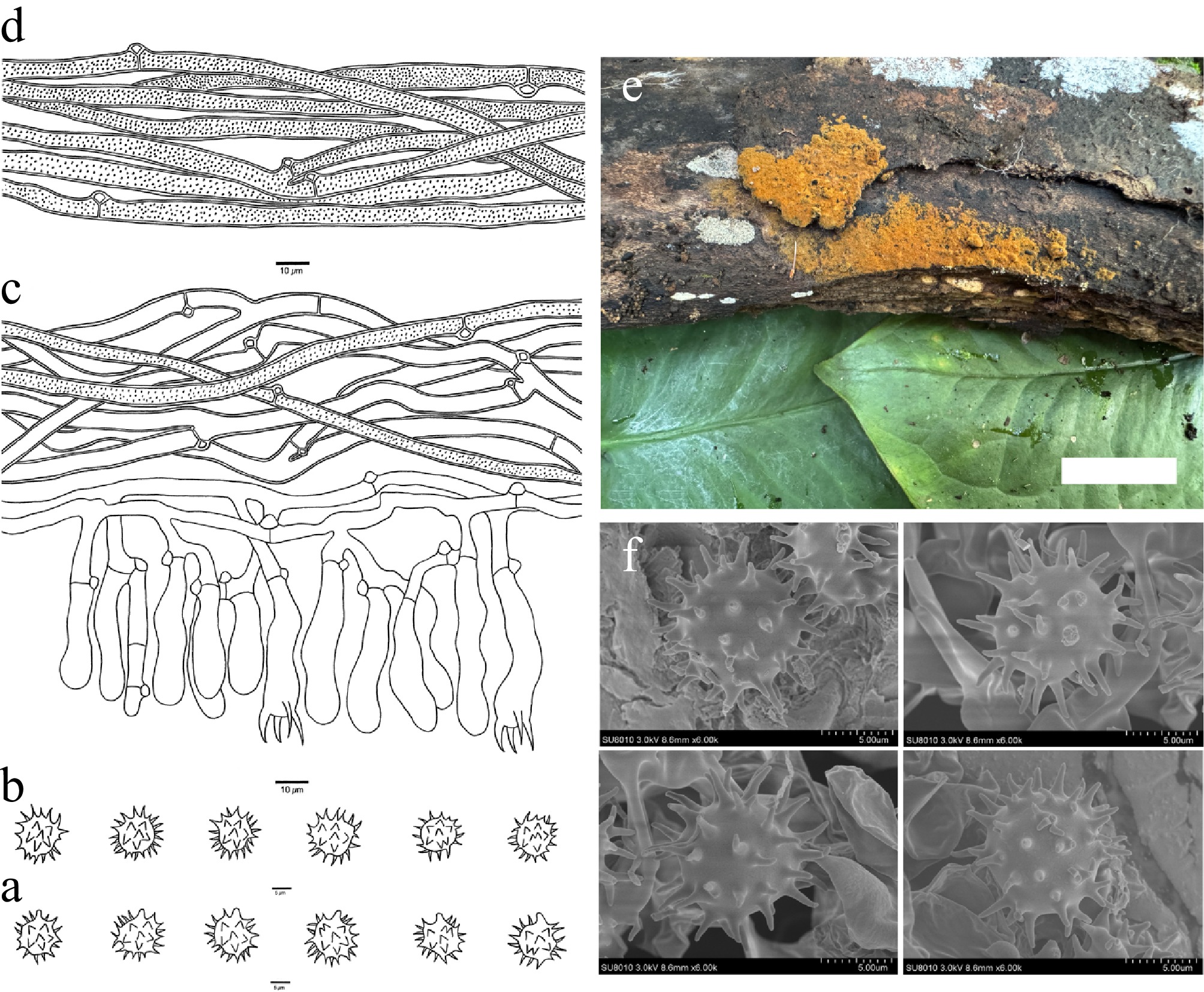
Figure 8.
Basidiomata and microscopic structures of Thelephora longissima (holotype, Dai 39708). (a) Basidiospores in lateral view. (b) Basidiospores in frontal view. (c) A section through a basidioma. (d) Hyphae from a rhizomorph. (e) Basidiomata on the host substrates. (f) Scanning electron microscope (SEM) of basidiospores. Scale bars: (a), (b) = 5 µm; (c), (d) = 10 µm; (e) = 1 cm.
MycoBank number: MB 864307
Diagnosis – Thelephora longissima is distinguished by resupinate basidiomata, presence of rhizomorphs, occasionally fine encrustation of subicular hyphae, long echinulate (up to 3 µm).
Type – CHINA. Guangxi Autonomous Region, Tian'e County, Longtan Forest Park, on a fallen angiosperm branch, 9 June 2025, Dai 39708 (holotype BJFC060967).
Etymology – Longissima (Lat.): refers to the basidiospores of the species having very long echinuli.
Basidiomata – Annual, resupinate, separable from the substrate, mucedinoid, soft, without odor or taste when fresh, continuous, up to 3 cm long, 2 cm wide, and 0.9 mm thick at the center; hymenophoral surface smooth, orange-yellow when fresh, and yellowish brown and concolorous with subiculum when dry; sterile margin determinate, farinaceous, concolorous with hymenophore.
Rhizomorphs – Present in subiculum and margin, 20–50 µm diam; rhizomorphic surface more or less smooth; hyphae in rhizomorph monomitic, undifferentiated, individual hyphae with clamp connections, 5–8 µm diam, rarely branched, more or less regularly arranged, thick-walled with a wide lumen, usually with fine encrustation, dark brown in KOH and distilled water.
Subicular hyphae – Monomitic; generative hyphae mostly with clamp connections and rarely with simple septa, occasionally branched, thick-walled with a wide lumen, occasionally with fine encrustation, more or less interwoven, dark brown in KOH and distilled water, IKI−, CB−, 4.0–6.5 µm diam; subhymenial hyphae frequently with clamp connections, thin-walled, smooth, 3–5.0 µm diam; hyphal cells more or less uniform, yellowish in KOH and in distilled water, IKI−, CB−.
Cystidia – Absent.
Basidia – Clavate, slightly winding, without transverse septa, yellowish in KOH and distilled water, not stalked, with four sterigmata and a clamp connection at the base, 35–55 µm long, 7–10 µm diam at the apex and 2–6 µm at the base; sterigmata 8–10 µm long and 2–4 µm diam at the base.
Basidiospores – Subglobose to bi-, tri-, or quadra-lobed in frontal and lateral faces, echinulate, yellowish in KOH and distilled water, IKI−, CB−, (8.3–)8.8–10.9(–11.0) × (7.6–)7.8–10.2(–10.8) µm, L = 9.88 µm, W = 9.13 µm, Q = 1.06–1.08 (n = 60/2); echinuli usually isolated, sometimes grouped in two, up to 3 µm long.
Additional specimen (paratype) examined – CHINA. Guangxi Autonomous Region, Tian'e County, Longtan Forest Park, on a fallen angiosperm branch, 9 June 2025, Dai 39727 (BJFC060986).
Habitat – Thelephora longissima was found in southern China with a subtropical monsoon climate.
Notes – Thelephora longissima, Thelephora pallidobrunnea (H.S. Yuan, X. Lu & Y.C. Dai) Kõljalg, I. Saar & Svant., Thelephora changbaiensis (H.S. Yuan, X. Lu & Y.C. Dai) Kõljalg, I. Saar & Svant., Thelephora megaspora (H.S. Yuan, X. Lu & Y.C. Dai) Kõljalg, I. Saar & Svant., Thelephora interrupta (H.S. Yuan & Y.C. Dai) Kõljalg, I. Saar & Svant., and Thelephora longiaculeifera (H.S. Yuan & Y.C. Dai) Kõljalg & Svant. clustered together with moderate support (97% in ML and 0.96 BPP). Thelephora pallidobrunnea is similar to T. longissima by sharing a smooth hymenophoral surface, clamped subhymenial hyphae, and lacking cystidia. But T. pallidobrunnea differs from T. longissima by lacking rhizomorphs, having shorter basidial sterigmata (3–6 vs 8–10 µm) and shorter basidiospores (7.5–8.5 vs 8.8–10.9 µm)[48]. Thelephora changbaiensis resembles T. longissima by being separable from the substrate, a smooth hymenophoral surface, and clamped subhymenial hyphae. However, T. changbaiensis differs from T. longissima by lacking rhizomorphs and having shorter basidial sterigmata (4–5.5 vs 8–10 µm)[48]. Thelephora megaspora is similar to T. longissima by sharing a smooth hymenophoral surface and lacking cystidia. But T. megaspora differs from T. longissima by basidiomata adherent to the substrate and lacking rhizomorphs[48]. Thelephora interrupta is similar to T. longissima by being separable from the substrate, mostly clamped and rarely simple septate subicular hyphae, and the presence of rhizomorphs. However, T. interrupta differs from T. longissima by subicular hyphae without encrustation and shorter basidiospores (3–5 vs 8–10 µm)[48]. Thelephora longiaculeifera resembles T. longissima by sharing mostly clamped and rarely simple septate subicular hyphae and the absence of cystidia. But T. longiaculeifera is distinguished from T. longissima by lacking rhizomorphs[48].
In morphology, T. longissima is similar to T. flavidobadia in having a yellowish-brown hymenophoral surface. However, phylogenetically, they are distantly related (Fig. 1), and T. flavidobadia is distinguished from T. longissima by shorter basidial sterigmata (3–5 vs 8–10 µm) and narrower subicular hyphae (3.0–4.0 vs 4.0–6.5 µm)[48].
Thelephora mongolica X. Li, Y.C. Dai & Yuan Yuan, sp. nov. Fig. 9
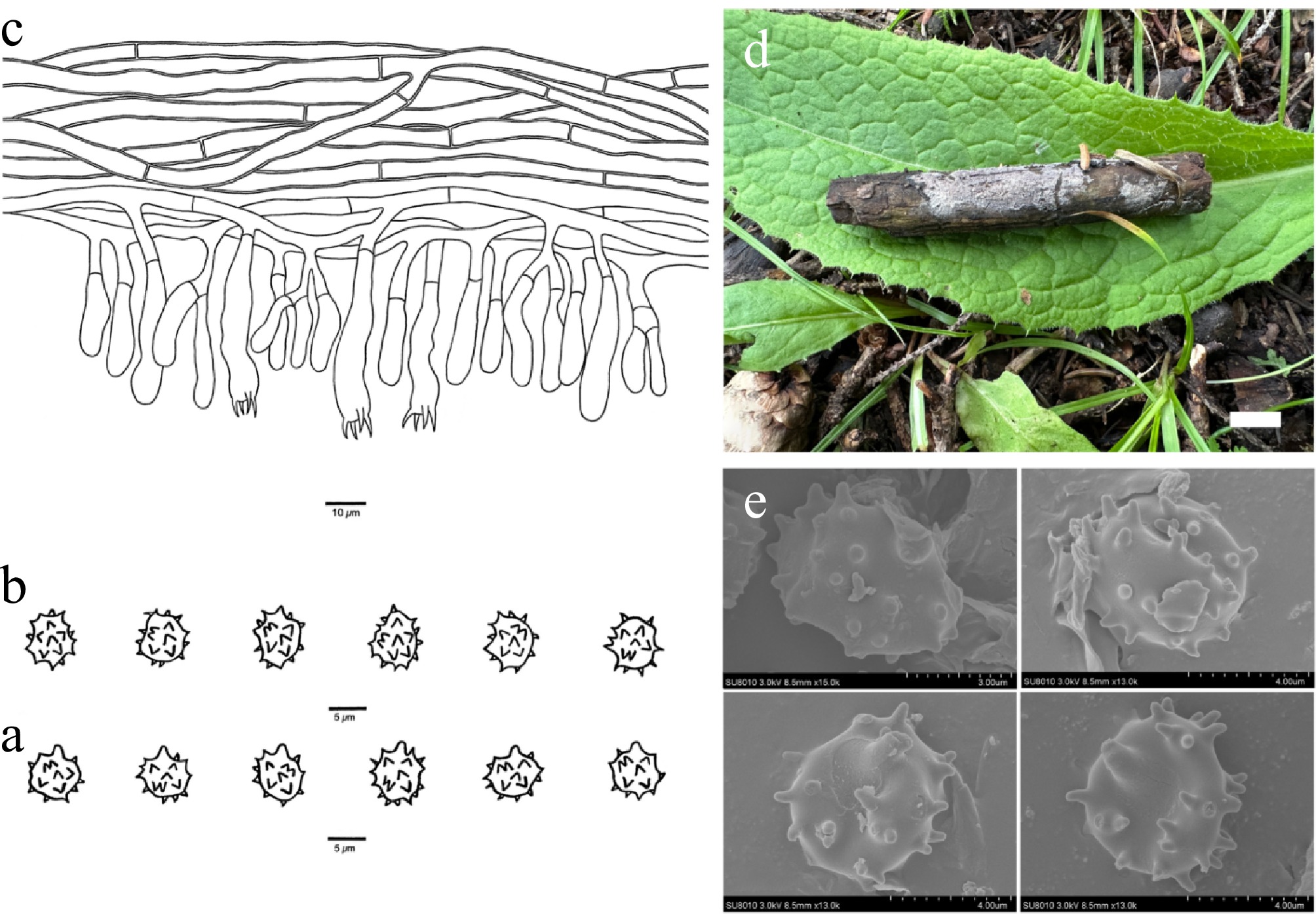
Figure 9.
Basidiomata and microscopic structures of Thelephora mongolica (holotype, Dai 37245). (a) Basidiospores in lateral view. (b) Basidiospores in frontal view. (c) A section through a basidioma. (d) Basidiomata on the host substrates. (e) Scanning electron microscope (SEM) of basidiospores. Scale bars: (a), (b) = 5 µm; (c) = 10 µm; (d) = 1 cm.
MycoBank number: MB 864308
Diagnosis – Thelephora mongolica is distinguished by resupinate basidiomata, a grayish-white hymenophoral surface when fresh and ash gray when dry, broadly ellipsoid to bi-, tri-, or quadra-lobed basidiospores, and short echinuli (up to 1.0 µm).
Type – CHINA. Inner Mongolia Autonomous Region, Horqin Right County, Yongfeng Reservoir, on a fallen branch of Quercus mongolica, 28 August 2025, Dai 37245 (holotype BJFC058504).
Etymology – Mongolica (Lat.): refers to the species being found in Inner Mongolia, China.
Basidiomata – Annual, resupinate, adherent to the substrate, arachnoid, soft, without odor or taste when fresh, continuous, up to 4 cm long, 2.5 cm wide, and 2 mm thick at the center; hymenophoral surface smooth, grayish-white when fresh, and ash gray and concolorous with subiculum when dry; sterile margin determinate, fimbriate, concolorous with hymenophore.
Rhizomorphs – Absent.
Subicular hyphae – Monomitic; generative hyphae with simple septa, occasionally branched, slightly thick-walled with a wide lumen, without encrustation, more or less interwoven, hyaline in KOH and distilled water, IKI−, CB−, 3–5.5 µm diam; subhymenial hyphae with simple septa, thin-walled, smooth, 3.5–5 µm diam; hyphal cells more or less uniform, hyaline in KOH and in distilled water, IKI−, CB−.
Cystidia – Absent.
Basidia – Clavate, without transverse septa, hyaline in KOH and distilled water, not stalked, with four sterigmata and a simple septum at the base, 20–50 µm long, 4–7.5 µm diam at the apex and 1–3 µm at the base; sterigmata 2–4 µm long and 1–3 µm diam at the base.
Basidiospores – Broadly ellipsoid to bi-, tri-, or quadra-lobed in frontal and lateral faces, echinulate, pale brown in KOH and distilled water, IKI−, CB−, (6.9–)5.6–7.5(–7.6) × (4.7–)4.9–6.2(–6.3) µm, L = 6.18 µm, W = 5.50 µm, Q = 1.09–1.16 (n = 60/2); echinuli usually isolated, sometimes grouped in two or more, up to 1.0 µm long.
Additional specimen (paratype) examined – CHINA. Inner Mongolia Autonomous Region, Horqin Right County, Yongfeng Reservoir, on a fallen branch of Quercus mongolica, 28 August 2025, Dai 37246 (BJFC058505) & Dai 37253 (BJFC058512).
Habitat – Thelephora mongolica was found in northeast China with a temperate continental climate. Additionally, T. mongolica was also known from one eDNA sequence from the Czech Republic (Fig. 1).
Notes – The phylogenetic tree indicates that T. mongolica formed a sister lineage with T. quercicola and T. eucoerulea Kõljalg & Svant. with 92% ML (Fig. 1). Thelephora quercicola resembles T. mongolica by sharing continuous basidiomata and the absence of cystidia. But T. quercicola differs in the presence of rhizomorphs, clamped subicular hyphae, and basidia occasionally with oil droplets. Thelephora eucoerulea is similar to T. mongolica by sharing continuous basidiomata adherent to the substrate and the absence of cystidia. However, T. eucoerulea differs from T. mongolica by having clamped subhymenial hyphae and the presence of rhizomorphs[26,103,104].
Thelephora mongolica shares similar morphological characteristics with the species T. latihypha by sharing an ash-gray hymenophoral surface and basidiomata adherent to the substrate. However, phylogenetically, they are distantly related (Fig. 1), and T. latihypha differs from T. mongolica by clamped and simple-septa generative hyphae and wider basidiospores (6.6–8.2 vs 4.9–6.2 µm)[5].
Thelephora murina X. Li, Y.C. Dai & Yuan Yuan, sp. nov. Fig. 10
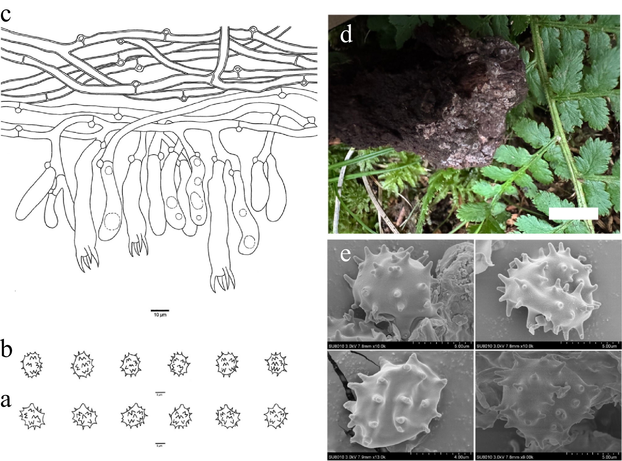
Figure 10.
Basidiomata and microscopic structures of Thelephora murina (holotype, Dai 35286). (a) Basidiospores in lateral view. (b) Basidiospores in frontal view. (c) A section through a basidioma. (d) Basidiomata on the host substrates. (e) Scanning electron microscope (SEM) of basidiospores. Scale bars: (a), (b) = 5 µm; (c) = 10 µm; (d) = 1 cm.
MycoBank number: MB 864311
Diagnosis – Thelephora murina is distinguished by resupinate basidiomata, a mouse-gray to light vinaceous gray hymenophoral surface when fresh and vinaceous gray when dry, long sterigmata (7–12 µm), and ellipsoid to bi-, tri-, or quadra-lobed basidiospores.
Type – CHINA. Qinghai Province, Xunhua County, Galeng Forest Farm, on a fallen angiosperm branch, 20 August 2025, Dai 35286 (holotype BJFC056547).
Etymology – Murina (Lat.): refers to the species having a mouse-gray hymenophoral surface when fresh.
Basidiomata – Annual, resupinate, adherent to the substrate, pelliculose, soft, without odor or taste when fresh, continuous, up to 2.5 cm long, 1.0 cm wide, and 1.0 mm thick at the center; hymenophoral surface smooth, mouse-gray to light vinaceous gray when fresh, and vinaceous gray and darker than subiculum when dry; sterile margin determinate, fimbriate, whitish, paler than hymenophore.
Rhizomorphs – Absent.
Subicular hyphae – Monomitic; generative hyphae with clamp connections and rarely with simple septa, occasionally branched, thick-walled with a wide lumen, without encrustation, more or less interwoven, hyaline in KOH and distilled water, IKI−, CB−, 3.5–6.5 µm diam; subhymenial hyphae frequently with clamp connections, thin-walled, smooth, 3.5–5.5 µm diam; hyphal cells more or less uniform, hyaline in KOH and in distilled water, IKI−, CB−.
Cystidia – Absent.
Basidia: Clavate, more or less constricted in the middle, without transverse septa, hyaline in KOH and distilled water, not stalked, with four sterigmata and a clamp connection at the base, 40–70 µm long, 8–11 µm diam at the apex and 4–7 µm at the base; sterigmata 7–12 µm long and 2–3 µm diam at the base.
Basidiospores – Ellipsoid to bi-, tri-, or quadra-lobed in frontal and lateral faces, echinulate, yellowish in KOH and distilled water, IKI−, CB−, (8.2–)8.4–10.8(–11.2) × (6.1–)6.2–8.9(–9.3) µm, L = 9.47 µm, W = 7.55 µm, Q = 1.24–1.27 (n = 60/2); echinuli usually grouped in two or more, up to 2 µm long.
Additional specimen (paratype) examined – CHINA. Qinghai Province, Xunhua County, Galeng Forest Farm, on a fallen angiosperm branch, 20 August 2025, Dai 35306 (BJFC056567).
Habitat – Thelephora murina was found in northwest China with a plateau continental climate.
Notes – Thelephora murina is closely related to Thelephora caeruleoalba (100% in ML and 1.00 BPP) in the phylogenetic tree (Fig. 1) and then clustered with Thelephora rotundata H.S. Yuan & Y. Q. Zhu (100% in ML and 1.00 BPP). However, T. caeruleoalba is distinguished from T. murina by being separable from the substrate and the presence of cystidia. Thelephora rotundata shares similar characteristics with T. murina by having continuous basidiomata adherent to the substrate, the absence of rhizomorphs and cystidia. However, T. rotundata is distinguished from T. murina by its wider basidiospores (9.5–12.1 vs 6.2–8.9 µm), and longer echinuli (4 vs 2 µm)[74].
In morphology, T. murina resembles T. vinosogrisea by having a vinaceous gray hymenophoral surface. However, phylogenetically, they are distantly related (Fig. 1), and T. vinosogrisea is distinguished from T. murina by being separable from the substrate and by the presence of rhizomorphs.
Thelephora niveomarginata X. Li, Y.C. Dai & Yuan Yuan, sp. nov. Fig. 11
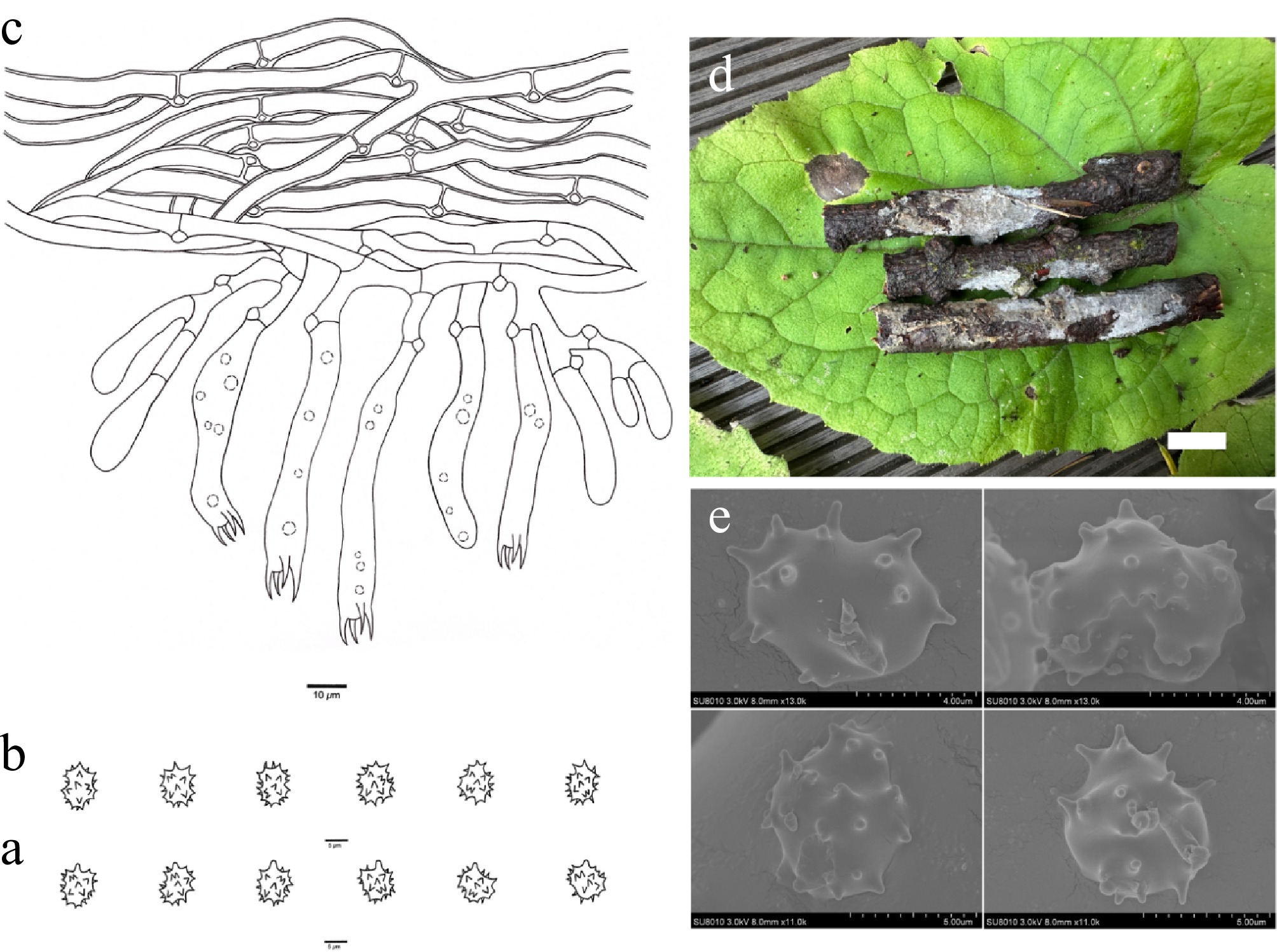
Figure 11.
Basidiomata and microscopic structures of Thelephora niveomarginata (holotype, Dai 31735). (a) Basidiospores in lateral view. (b) Basidiospores in frontal view. (c) A section through a basidioma. (d) Basidiomata on the host substrates. (e) Scanning electron microscope (SEM) of basidiospores. Scale bars: (a), (b) = 5 µm; (c) = 10 µm; (d) = 1 cm.
MycoBank number: MB 864312
Diagnosis – Thelephora niveomarginata is distinguished by resupinate basidiomata, ash-gray to white hymenophoral surface when fresh and pinkish buff when dry, usually oil-droplet basidia, ellipsoid to bi-, tri-, or quadra-lobed basidiospores, and short echinuli (up to 1 μm).
Type – CHINA. Xizang Autonomous Region, Yadong County, forests from Pasha Falls to Nathu La Gate, on a fallen twig of Abies fabri, 16 October 2024, Dai 31735 (holotype BJFC051994).
Etymology – Niveomarginata (Lat.): refers to the basidiomata of the species having a white margin when fresh.
Basidiomata – Annual, resupinate, separable from the substrate, pelliculose, soft, without odor or taste when fresh, continuous, up to 6 cm long, 1.5 cm wide, and 1.6 mm thick at the center; hymenophoral surface smooth, ash-gray to white when fresh, and pinkish buff and concolorous with subiculum when dry; sterile margin determinate, fimbriate, concolorous with hymenophore.
Rhizomorphs – Absent.
Subicular hyphae – Monomitic; generative hyphae mostly with clamp connections, occasionally branched, thick-walled with a wide lumen, without encrustation, more or less interwoven, yellowish in KOH and distilled water, IKI−, CB−, 4–8 μm diam; subhymenial hyphae frequently with clamp connections, thin-walled, smooth, 3–7 μm diam; hyphal cells more or less uniform, yellowish in KOH and distilled water, IKI−, CB−.
Cystidia – Absent.
Basidia – Clavate, without transverse septa, yellowish in KOH and distilled water, not stalked, usually with oil droplets, with four sterigmata and a clamp connection at the base, 45–70 μm long and 6–10 μm diam at the apex, 3–5 μm at the base; sterigmata 7–9 μm long and 2–3 μm diam at the base.
Basidiospores – Ellipsoid to bi-, tri-, or quadra-lobed in frontal and lateral faces, echinulate, yellowish in KOH and distilled water, IKI−, CB−, (5.8–)6.1–8.6(–8.7) × (4.1–)4.2–5.9(–6.0) μm, L = 7.31 μm, W = 5.36 μm, Q = 1.32–1.37 (n = 60/2); echinuli usually isolated, sometimes grouped in two or more, up to 1 μm long.
Additional specimen (paratype) examined – CHINA. Xizang Autonomous Region, Yadong County, Pasha Falls, on a fallen angiosperm branch, 17 October 2024, Dai 31738 (BJFC051997).
Habitat – Thelephora niveomarginata was found in southwest China with a semi-humid monsoon temperate climate.
Notes – In the phylogenetic tree (Fig. 1), T. niveomarginata, T. pallidocastanea, and T. keteleeriae clustered together with high support (97% in ML and 0.99 BPP). Thelephora pallidocastanea shares similar characteristics with T. niveomarginata by its smooth hymenophoral surface and the absence of cystidia. However, T. pallidocastanea is distinguished from T. niveomarginata by having an indeterminate and arachnoid sterile margin, and shorter basidiospores (5–6 vs 6.1–8.6 μm)[49,78]. Thelephora keteleeriae resembles T. niveomarginata by sharing continuous basidiomata separable from the substrate, a determinate and fimbriate sterile margin, and clamped subhymenial hyphae. However, T. keteleeriae is distinguished from T. niveomarginata by its more or less irregularly poroid hymenophoral surface and the presence of cystidia and rhizomorphs.
In morphology, T. niveomarginata is similar to T. koraiensis and T. mongolica in having an ash-gray to grayish-white hymenophoral surface and a fimbriate sterile margin. However, phylogenetically, they are distantly related (Fig. 1), and T. koraiensis differs from T. niveomarginata by having shorter basidia (15–40 vs 45–70 μm) and wider basidiospores (6.1–8.4 vs 4.2–5.9 µm). Thelephora mongolica is distinguished from T. niveomarginata by having simple septate subicular hyphae and shorter sterigmata (2–4 vs 7–9 µm).
Thelephora nivea X. Li, Y.C. Dai & Yuan Yuan, sp. nov. Fig. 12
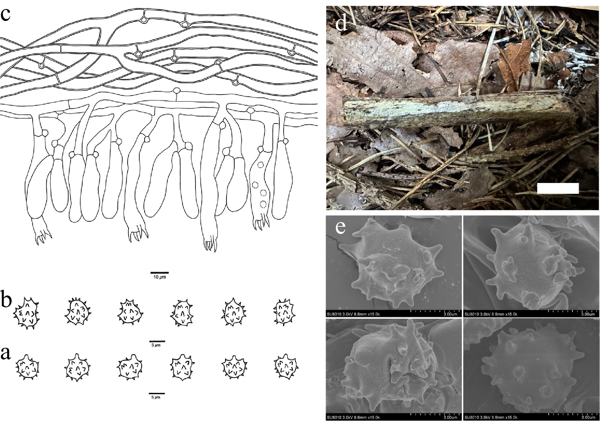
Figure 12.
Basidiomata and microscopic structures of Thelephora nivea (holotype, Dai 37773). (a) Basidiospores in lateral view. (b) Basidiospores in frontal view. (c) A section through a basidioma. (d) Basidiomata on the host substrates. (e) Scanning electron microscope (SEM) of basidiospores. Scale bars: (a), (b) = 5 µm; (c) = 10 µm; (d) = 1 cm.
MycoBank number: MB 864316
Diagnosis – Thelephora nivea is distinguished by resupinate basidiomata, a white hymenophoral surface when fresh and light vinaceous gray to clay-buff when dry, occasionally oil droplets, basidia, broadly ellipsoid to subglobose to bi-, tri-, or quadra-lobed basidiospores.
Type – CHINA. Jilin Province, Jiaohe County, Lafa Mountain, on a fallen branch of Larix olgensis, 11 September 2025, Dai 37773 (holotype BJFC059032).
Etymology – Nivea (Lat.): refers to the species having white basidiomata when fresh.
Basidiomata – Annual, resupinate, separable from the substrate, pelliculose, soft, without odor or taste when fresh, continuous, up to 6 cm long, 1.5 cm wide, and 2 mm thick at the center; hymenophoral surface smooth, white when fresh, and light vinaceous gray to clay-buff and paler than subiculum when dry; sterile margin determinate, farinaceous, white to cream, concolorous to hymenophore.
Rhizomorphs – Absent.
Subicular hyphae – Monomitic; generative hyphae mostly with clamp connections and rarely with simple septa, frequently branched, thick-walled with a wide lumen, without encrustation, more or less interwoven, hyaline in KOH and distilled water, IKI−, CB−, 4.5–7 µm diam; subhymenial hyphae mostly with clamp connections and rarely with simple septa, thin-walled, smooth, 3–5 µm diam; hyphal cells more or less uniform, hyaline in KOH and in distilled water, IKI−, CB−.
Cystidia – Absent.
Basidia – Clavate, more or less constricted in the middle, without transverse septa, hyaline in KOH and distilled water, not stalked, occasionally with oil droplets, with four sterigmata and a clamp connection at the base, 30–65 µm long, 6–9 µm diam at the apex and 4–6 µm at the base; sterigmata 4–6 µm long and 1–3 µm diam at the base.
Basidiospores – Broadly ellipsoid to bi-, tri-, or quadra-lobed in frontal face and broadly ellipsoid to subglobose in lateral face, echinulate, yellowish in KOH and distilled water, IKI−, CB−, (5.5–)5.7–7.6(–7.7) × (4.4–)4.6–6.4(–6.9) µm, L = 6.35 µm, W = 5.29 µm, Q = 1.17–1.21 (n = 30/1); echinuli usually isolated, sometimes grouped in two or more, up to 1.5 µm long.
Habitat – Thelephora nivea was found in northeast China with a temperate continental monsoon climate. Additionally, T. nivea was also known from one eDNA sequence from Japan (Fig. 1).
Notes – In the phylogenetic tree (Fig. 1), T. nivea, T. caryophyllea (Schaeff.) Pers., T. scopiformis Y.Q. Zhu et al., T. scissilis Burt and T. anthocephala (Bull.) Fr. clustered together with high support (100% in ML and 0.96 BPP). Thelephora caryophyllea resembles T. nivea by the absence of cystidia. However, T. caryophyllea is differentiated from T. nivea by its fimbriate sterile margin and longer basidial sterigmata (7–8.5 vs 4–6 µm)[105]. Thelephora scopiformis differs from T. nivea by its erect basidiomata and basidia occasionally covered by dense crystals[5]. Thelephora scissilis is distinguished from T. nivea by its erect basidiomata[105]. Thelephora anthocephala is distinguished from T. nivea by its longer basidiospores (8–10 vs 5.7–7.6 µm)[105].
In morphology, T. nivea shares similar characteristics with T. ceracea by sharing a smooth and vinaceous gray hymenophoral surface. However, phylogenetically, they are distantly related (Fig. 1), and T. ceracea is distinguished from T. nivea by wider basidiospores (6.7–7.8 vs 4.6–6.4 µm).
Thelephora quercicola X. Li, Y.C. Dai & Yuan Yuan, sp. nov. Fig. 13
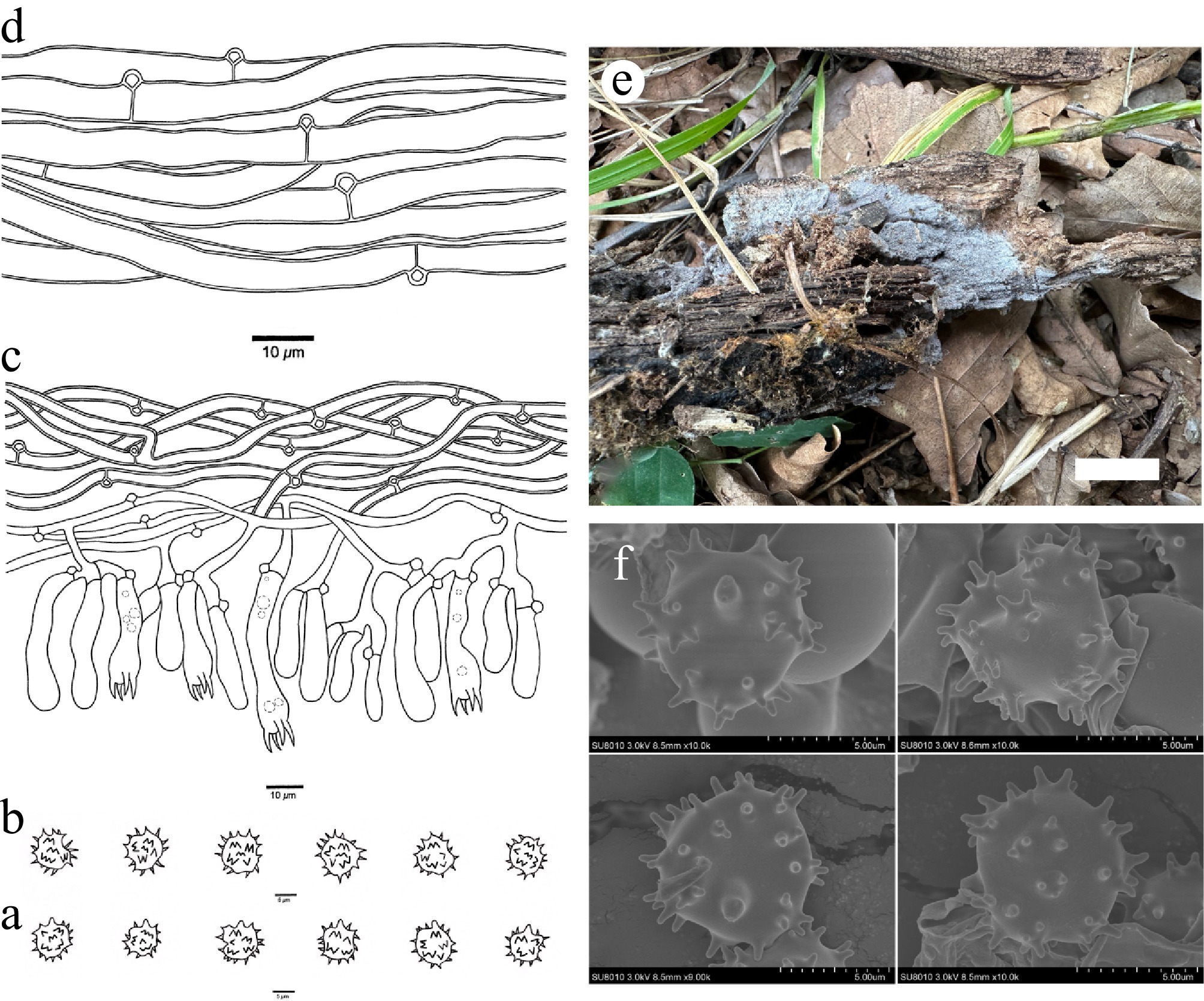
Figure 13.
Basidiomata and microscopic structures of Thelephora quercicola (holotype, Dai 37262). (a) Basidiospores in lateral view. (b) Basidiospores in frontal view. (c) A section through a basidioma. (d) Hyphae from a rhizomorph. (e) Basidiomata on the host substrates. (f) Scanning electron microscope (SEM) of basidiospores. Scale bars: (a), (b) = 5 µm; (c), (d) = 10 µm; (e) = 1 cm.
MycoBank number: MB 864317
Diagnosis – Thelephora quercicola is distinguished by resupinate basidiomata, a bluish-gray hymenophoral surface when fresh and ash gray when dry, presence of rhizomorphs, usually oil droplets, basidia, and broadly ellipsoid to subglobose to bi-, tri-, or quadra-lobed basidiospores.
Type – CHINA. Inner Mongolia Autonomous Region, Horqin Right County, Yongfeng Reservoir, on a fallen branch of Quercus mongolica, 28 August 2025, Dai 37262 (holotype BJFC058521).
Etymology – Quercicola (Lat.): refers to the species growing on Quercus mongolica.
Basidiomata – Annual, resupinate, adherent to the substrate, mucedinoid, soft, without odor or taste when fresh, continuous, up to 5 cm long, 2.5 cm wide, and 1 mm thick at the center; hymenophoral surface granulose, bluish-gray when fresh, and ash gray and concolorous with subiculum when dry; sterile margin determinate, fimbriate, concolorous with hymenophore.
Rhizomorphs – Present in subiculum and margin, 20–45 µm diam; rhizomorphic surface more or less smooth; hyphae in rhizomorph monomitic, undifferentiated, individual hyphae with clamp connections, 4–6.5 µm diam, unbranched, thick-walled with a wide lumen, more or less regularly arranged, golden yellow in KOH and distilled water.
Subicular hyphae – Monomitic; generative hyphae frequently with clamp connections, occasionally branched, thick-walled with a wide lumen, without encrustation, more or less interwoven, hyaline in KOH and distilled water, IKI−, CB+, 2–5 µm diam; subhymenial hyphae frequently with clamp connections, thin-walled, smooth, 2–4 µm diam; hyphal cells more or less uniform, hyaline in KOH and in distilled water, IKI−, CB+.
Cystidia – Absent.
Basidia – Clavate, more or less constricted in the middle, without transverse septa, hyaline in KOH and distilled water, not stalked, usually with oil droplets, with four sterigmata and a clamp connection at the base, 25–45 µm long, 6–8 µm diam at the apex and 3–5 µm at the base; sterigmata 3–5 µm long and 1.5–2.5 µm diam at the base.
Basidiospores – Broadly ellipsoid to subglobose to bi-, tri-, or quadra-lobed in frontal and lateral faces, echinulate, yellowish in KOH and distilled water, IKI−, CB+, (6.4–)6.7–8.7(–8.9) × (5.3–)5.7–7.5(–7.6) µm, L = 7.55 µm, W = 6.80 µm, Q = 1.09–1.14 (n = 60/2); echinuli usually grouped in two or more, up to 1.5 µm long.
Additional specimen (paratype) examined – CHINA. Inner Mongolia Autonomous Region, Horqin Right County, Yongfeng Reservoir, on a fallen branch of Quercus mongolica, 28 August 2025, Dai 37243 (BJFC058502).
Habitat – Thelephora quercicola was found in northeast China with a temperate continental climate.
Notes – Thelephora quercicola is closely related to T. eucoerulea Höhn. & Litsch. (100% in ML and 1.00 BPP) in the phylogenetic tree (Fig. 1) and then clustered with T. mongolica (100% in ML). Thelephora eucoerulea resembles T. quercicola by its continuous basidiomata adherent to the substrate, a granulose hymenophoral surface, and the absence of cystidia. However, T. eucoerulea is distinguished from T. quercicola by its hymenophoral surface that is grayish sepia, rosy-buff, or hazel when dry[26,103,104]. Thelephora mongolica is similar to T. quercicola in its continuous basidiomata and the absence of cystidia. But T. mongolica differs from T. quercicola by the absence of rhizomorphs and by having subicular hyphae with simple septa.
In morphology, T. quercicola shares similar characteristics with T. submurina, including a fimbriate sterile margin, broadly ellipsoid to subglobose basidiospores, and the presence of rhizomorphs. Phylogenetically, T. quercicola and T. submurina are closely related but not a sister lineage; T. submurina differs from T. quercicola by smaller basidiospores (4.6–5.9 × 3.8–5.6 vs 6.7–8.7 × 5.7–7.5 µm).
Thelephora reniformis X. Li, Y.C. Dai & Yuan Yuan, sp. nov. Fig. 14
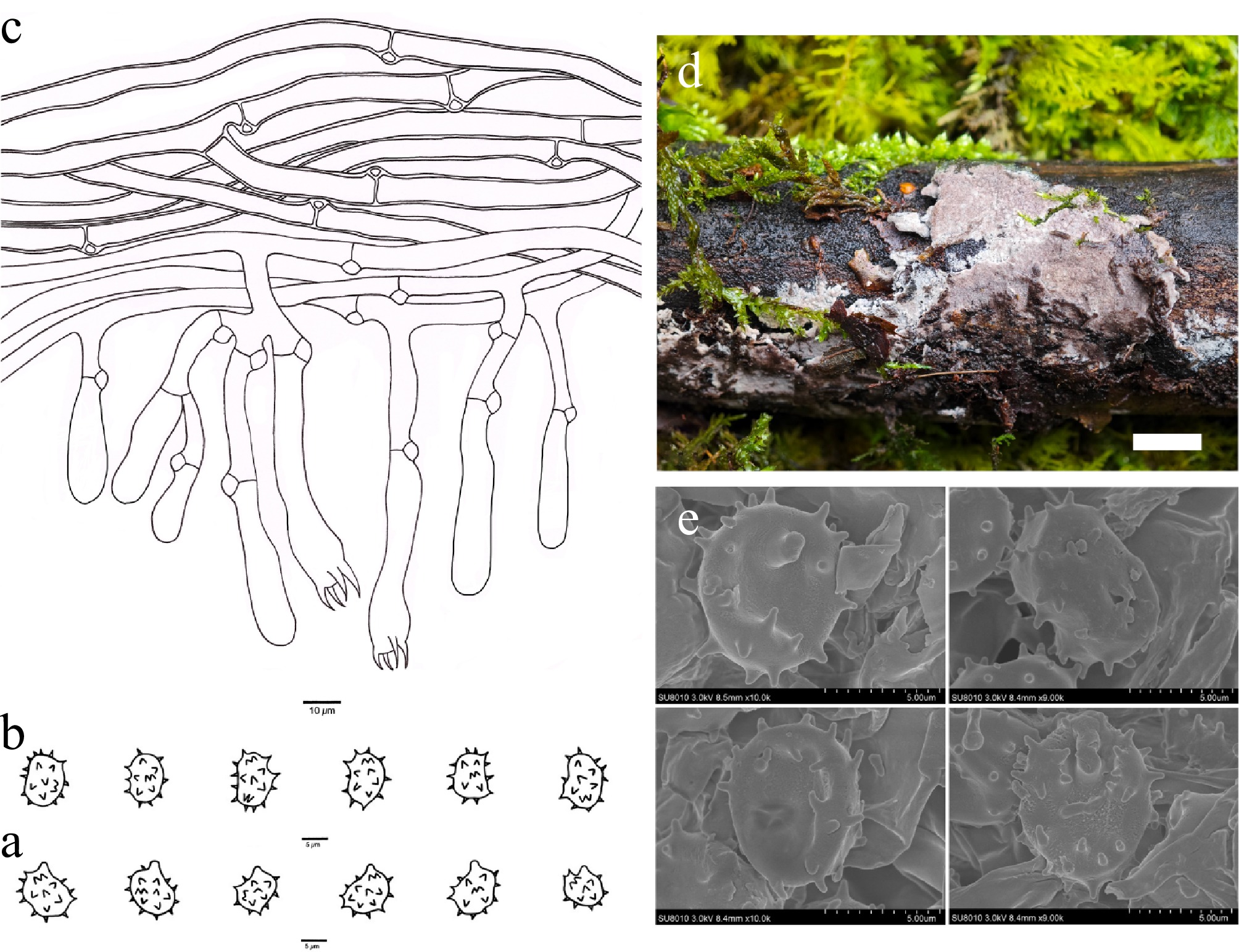
Figure 14.
Basidiomata and microscopic structures of Thelephora reniformis (holotype, Dai 31229). (a) Basidiospores in lateral view. (b) Basidiospores in frontal view. (c) A section through a basidioma. (d) Basidiomata on the host substrates. (e) Scanning electron microscope (SEM) of basidiospores. Scale bars: (a), (b) = 5 µm; (c) = 10 µm; (d) = 1 cm.
MycoBank number: MB 864318
Diagnosis – Thelephora reniformis is distinguished by resupinate basidiomata, a light vinaceous gray hymenophoral surface when fresh and fawn when dry, short sterigmata (2–4 µm), reniform to bi-, tri-, or quadra-lobed basidiospores, and short echinuli (up to 1 µm).
Type – CHINA. Qinghai Province, Xunhua County, Mengdatianchi Nature Reserve, on a fallen angiosperm branch, 12 October 2024, Dai 31229 (holotype BJFC051488).
Etymology – Reniformis (Lat.): refers to the species having reniform basidiospores.
Basidiomata – Annual, resupinate, separable from the substrate, soft, without odor or taste when fresh, continuous, up to 5 cm long, 1.5 cm wide, and 1.5 mm thick at the center; hymenophoral surface smooth, light vinaceous gray when fresh, and fawn and darker than subiculum when dry; sterile margin determinate, fimbriate, white, paler than the hymenophore.
Rhizomorphs – Absent.
Subicular hyphae – Monomitic; generative hyphae frequently with clamp connections, occasionally branched, thick-walled with a wide lumen, without encrustation, more or less interwoven, pale brown in KOH and distilled water, IKI−, CB−, 5–7.5 µm diam; subhymenial hyphae frequently with clamp connections, thin-walled, smooth, 3.5–6.5 µm diam; hyphal cells more or less uniform, pale brown in KOH and in distilled water, IKI−, CB−.
Cystidia – Absent.
Basidia – Clavate, constricted in the middle, without transverse septa, pale brown in KOH and distilled water, not stalked, with four sterigmata and a clamp connection at the base, 30–65 µm long, 5.5–9.5 µm diam at the apex and 4–6.5 µm at the base; sterigmata 2–4 µm long and 1–1.5 µm diam at the base.
Basidiospores – More or less reniform to bi-, tri-, or quadra-lobed in frontal and lateral faces, echinulate, pale brown in KOH and distilled water, IKI−, CB−, (7.0–)7.1–9.6(–9.7) × (5.8–)6.0–7.4(–7.5) µm, L = 8.17 µm, W = 6.76 µm, Q = 1.18–1.22 (n = 60/2); echinuli usually isolated, sometimes grouped in two or more, up to 1 µm long.
Habitat – Thelephora reniformis was found in northwest China with a plateau continental climate.
Notes – Thelephora reniformis is closely related to T. pallidomarginata (H.S. Yuan et al.) Kõljalg et al. (100% in ML and 1.00 BPP) in the phylogenetic tree (Fig. 1) and then clustered with T. ellisii (Sacc.) Zmitr. et al. (70% in ML). Thelephora pallidomarginata resembles T. reniformis by its determinate sterile margin, a smooth hymenophoral surface, and the absence of cystidia. However, T. pallidomarginata is distinguished from T. reniformis by having rhizomorphs and longer sterigmata (4–8 vs 2–4 µm)[48]. Thelephora ellisii shares similar characteristics with T. reniformis by having continuous basidiomata, a smooth hymenophoral surface, and the absence of cystidia. But T. ellisii differs from T. reniformis by basidiomata adherent to the substrate and the presence of rhizomorphs[106].
In morphology, T. reniformis resembles T. subvinosogrisea by sharing a smooth and light vinaceous gray to fawn hymenophoral surface and lacking rhizomorphs. But phylogenetically, they are distantly related (Fig. 1), and T. subvinosogrisea is distinguished from T. reniformis by having basidiomata adherent to the substrate, longer basidia sterigmata (5–9 vs 2–4 µm), and broadly ellipsoid to subglobose basidiospores.
Thelephora rhododendri X. Li, Y.C. Dai & Yuan Yuan, sp. nov. Fig. 15
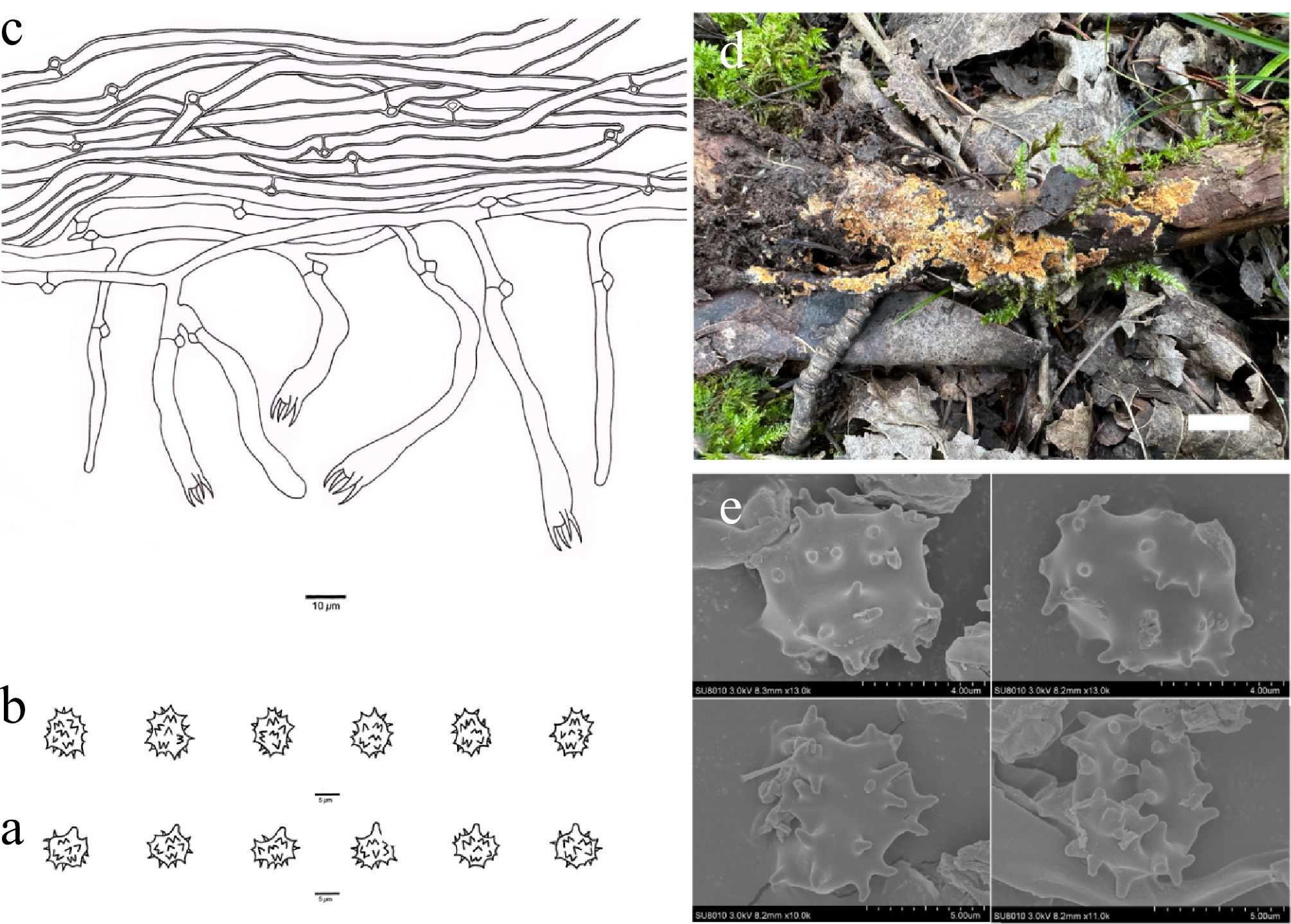
Figure 15.
Basidiomata and microscopic structures of Thelephora rhododendri (holotype, Dai 29574). (a) Basidiospores in lateral view. (b) Basidiospores in frontal view. (c) A section through a basidioma. (d) Basidiomata on the host substrates. (e) Scanning electron microscope (SEM) of basidiospores. Scale bars: (a), (b) = 5 µm; (c) = 10 µm; (d) = 1 cm.
MycoBank number: MB 864319
Diagnosis – Thelephora rhododendri is distinguished by resupinate basidiomata, subulate cystidioles, ellipsoid to bi-, tri-, or quadra-lobed basidiospores, and short echinuli (up to 1 µm).
Type – CHINA. Qinghai Province, Huzhu County, Beishan Forest Park, Langshidang, on a fallen branch of Rhododendron, 28 July 2024, Dai 29574 (holotype BJFC049833).
Etymology – Rhododendri (Lat.): refers to the species growing on Rhododendron.
Basidiomata – Annual, resupinate, separable from the substrate, crustose, soft, without odor or taste when fresh, discontinuous, up to 6 cm long, 3 cm wide, and 1.2 mm thick at the center; hymenophoral surface smooth, curry-yellow to cinnamon-buff when fresh, and honey-yellow and concolorous with subiculum when dry; sterile margin determinate, fimbriate, buff-yellow, paler than hymenophore.
Rhizomorphs – Absent.
Subicular hyphae – Monomitic; generative hyphae frequently with clamp connections, occasionally branched, thick-walled with a wide lumen, without encrustation, more or less interwoven, pale brown in KOH and distilled water, IKI−, CB−, 2.5–4.5 µm diam; subhymenial hyphae frequently with clamp connections, thin-walled, smooth, 2–4.5 µm diam; hyphal cells more or less uniform, pale brown in KOH and in distilled water, IKI−, CB−.
Cystidia – Cystidia absent; subulate cystidioles present, arising from subhymenial hyphae, without encrustation, 20–40 µm long, 1–3 µm diam at the base and 1–2 µm diam at the apex, pale brown in KOH and distilled water, IKI−, CB−.
Basidia – Clavate, flexuous, without transverse septa, pale brown in KOH and distilled water, not stalked, with four sterigmata and a clamp connection at the base, 30–65 µm long, 5–9 µm diam at the apex and 2–4.5 µm at the base; sterigmata 5–7.5 µm long and 1–2 µm diam at the base.
Basidiospores – ellipsoid to bi-, tri-, or quadra-lobed in frontal and lateral face, echinulate, pale brown in KOH and distilled water, IKI−, CB−, (6.2–)6.3–8.2(–8.3) × (4.5–)4.6–6.7(–6.8) µm, L = 7.01 µm, W = 5.54 µm, Q = 1.24–1.28 (n = 60/2); echinuli usually grouped in two or more, up to 1 µm long.
Additional specimen (paratype) examined – CHINA. Qinghai Province, Huzhu County, Beishan Forest Park, Langshidang, on a fallen branch of Betula, 28 July 2024, Dai 29588 (BJFC049847).
Habitat – Thelephora rhododendri was found in northwest China with a plateau continental climate.
Notes – Thelephora rhododendri is closely related to T. angusta Y.Q. Zhu et al. (100% in ML and 1.00 BPP) in the phylogenetic tree (Fig. 1). Thelephora angusta shares similar characteristics with T. rhododendri by being separable from the substrate, the absence of cystidia, and clamped subhymenial hyphae. However, T. angusta is distinguished from T. rhododendri by the presence of rhizomorphs and shorter basidial sterigmata (2.5–3.5 vs 5–7.5 µm)[5].
Thelephora rhododendri shares similar morphological characteristics with the species T. separata by sharing a smooth and honey-yellow hymenophoral surface and lacking cystidia. However, phylogenetically, they are distantly related (Fig. 1), and T. separata is distinguished from T. rhododendri by the presence of rhizomorphs and transverse septa in basidia[48].
Thelephora roseobubalina X. Li, Y.C. Dai & Yuan Yuan, sp. nov. Fig. 16
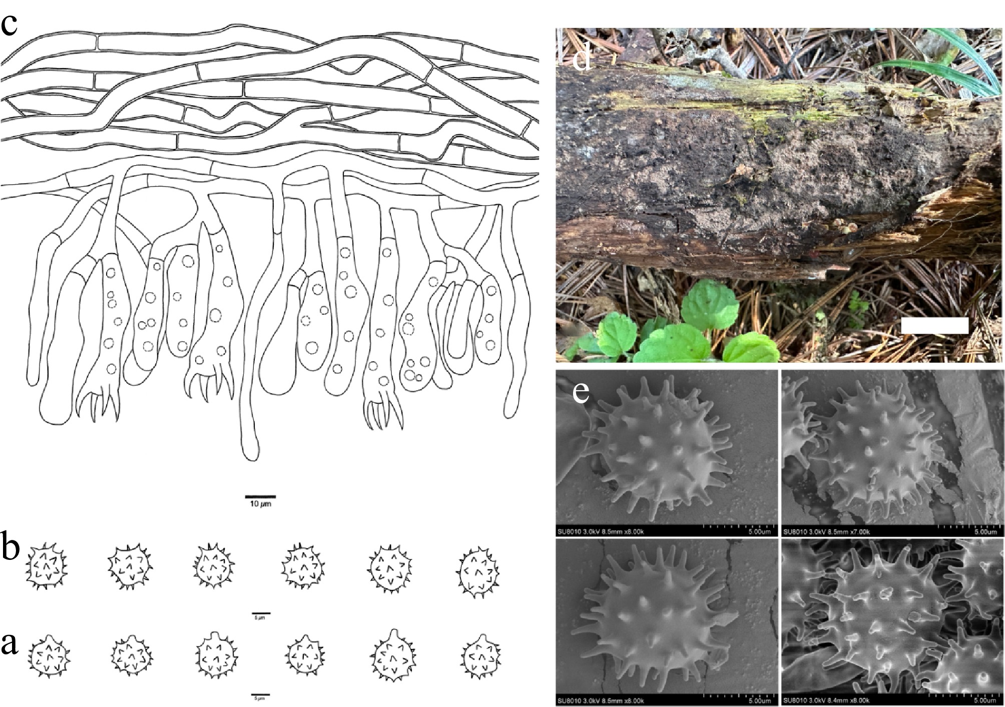
Figure 16.
Basidiomata and microscopic structures of Thelephora roseobubalina (holotype, Dai 37646). (a) Basidiospores in lateral view. (b) Basidiospores in frontal view. (c) A section through a basidioma. (d) Basidiomata on the host substrates. (e) Scanning electron microscope (SEM) of basidiospores. Scale bars: (a), (b) = 5 µm; (c) = 10 µm; (d) = 1 cm.
MycoBank number: MB 864320
Diagnosis – Thelephora roseobubalina is distinguished by resupinate basidiomata, a pinkish-buff hymenophoral surface when fresh and vinaceous gray when dry, clavate cystidioles, long sterigmata (8–11 µm), usually oil-droplet basidia, and subglobose to globose basidiospores.
Type – CHINA. Jilin Province, Jiaohe County, Experimental Forest Reserve, on a rotten trunk of Pinus koraiensis, 11 September 2025, Dai 37646 (holotype BJFC058905).
Etymology – Roseobubalina (Lat.): refers to the species having pinkish-buff basidiomata when fresh.
Basidiomata – Annual, resupinate, separable from the substrate, mucedinoid, soft, without odor or taste when fresh, discontinuous, up to 5 cm long, 2 cm wide, and 0.8 mm thick at the center; hymenophoral surface smooth, pinkish-buff when fresh, and vinaceous gray and concolorous with subiculum when dry; sterile margin determinate, byssoid, concolorous with hymenophore.
Rhizomorphs – Absent.
Subicular hyphae – Monomitic; generative hyphae with simple septa, occasionally branched, slightly thick-walled with a wide lumen, without encrustation, more or less interwoven, pale brown in KOH and distilled water, IKI−, CB−, 5–8 µm diam; subhymenial hyphae with simple septa, thin-walled, smooth, 3–6 µm diam; hyphal cells more or less uniform, pale brown in KOH and in distilled water, IKI−, CB−.
Cystidia – Absent; cystidioles present, arising from subhymenial hyphae, clavate, projecting above the hymenium, aseptate, 40–70 × 3–5 µm, pale brown in KOH and in distilled water.
Basidia – Mostly clavate, without transverse septa, pale brown in KOH and distilled water, not stalked, usually with oil droplets, with four sterigmata and a simple septum at the base, 35–55 µm long, 9–13 µm diam at the apex and 3–6 µm at the base; sterigmata 8–11 µm long and 2–4 µm diam at the base.
Basidiospores – Subglobose to globose in frontal and lateral faces, echinulate, yellowish in KOH and distilled water, IKI−, CB−, (8.3–)8.7–11.4(–11.5) × (7.8–)7.9–9.6(–9.7) µm, L = 9.47 µm, W = 8.79 µm, Q = 1.06–1.09 (n = 30/1); echinuli usually isolated, up to 2 µm long.
Habitat – Thelephora roseobubalina was found in northeast China with a temperate continental climate. Additionally, T. roseobubalina was also known from two eDNA sequences from the United Kingdom (Fig. 1).
Notes – Thelephora roseobubalina clustered with T. longiechinula (X. Lu & H.S. Yuan) Kõljalg et al., T. nanyigouensis Y.Q. Zhu et al., T. badia (Link) Stalpers, and T. cinereoumbrina (Bres.) Kõljalg et al. (52% in ML and 0.99 BPP) in the phylogenetic tree (Fig. 1). Thelephora longiechinula is similar to T. roseobubalina by basidiomata separable from the substrate, a smooth hymenophoral surface, and the absence of rhizomorphs. But T. longiechinula differs from T. roseobubalina by its continuous basidiomata and longer echinuli (4 vs 2 µm)[67]. Thelephora nanyigouensis resembles T. roseobubalina by sharing basidiomata separable from the substrate and the absence of rhizomorphs. But T. nanyigouensis differs from T. roseobubalina by its wider basidiospores (9.6–11.1 vs 7.9–9.6 µm) and shorter basidia sterigmata (3–5 vs 8–11 µm)[5]. Thelephora badia resembles T. roseobubalina by its subicular and subhymenial hyphae with simple septa and the absence of rhizomorphs. However, T. badia is differentiated from T. roseobubalina by its continuous basidiomata adherent to the substrate and a grayish-sepia hymenophoral surface[26]. Thelephora cinereoumbrina is similar to T. roseobubalina by subicular hyphae with simple septa and the absence of rhizomorphs. However, T. cinereoumbrina is distinguished from T. roseobubalina by having continuous and crustose basidiomata adherent to the substrate and basidiospores that are triangular in the frontal face and ellipsoid in the lateral face.
In morphology, T. roseobubalina resembles T. clavigera by sharing mucedinoid basidiomata, a pinkish-buff hymenophoral surface, and lacking rhizomorphs. However, phylogenetically, they are distantly related (Fig. 1), and T. clavigera differs from T. roseobubalina by having cystidia and shorter basidiospores (7.5–8 vs 8.7–11.4 µm)[107].
Thelephora submurina X. Li, Y.C. Dai & Yuan Yuan, sp. nov. Fig. 17
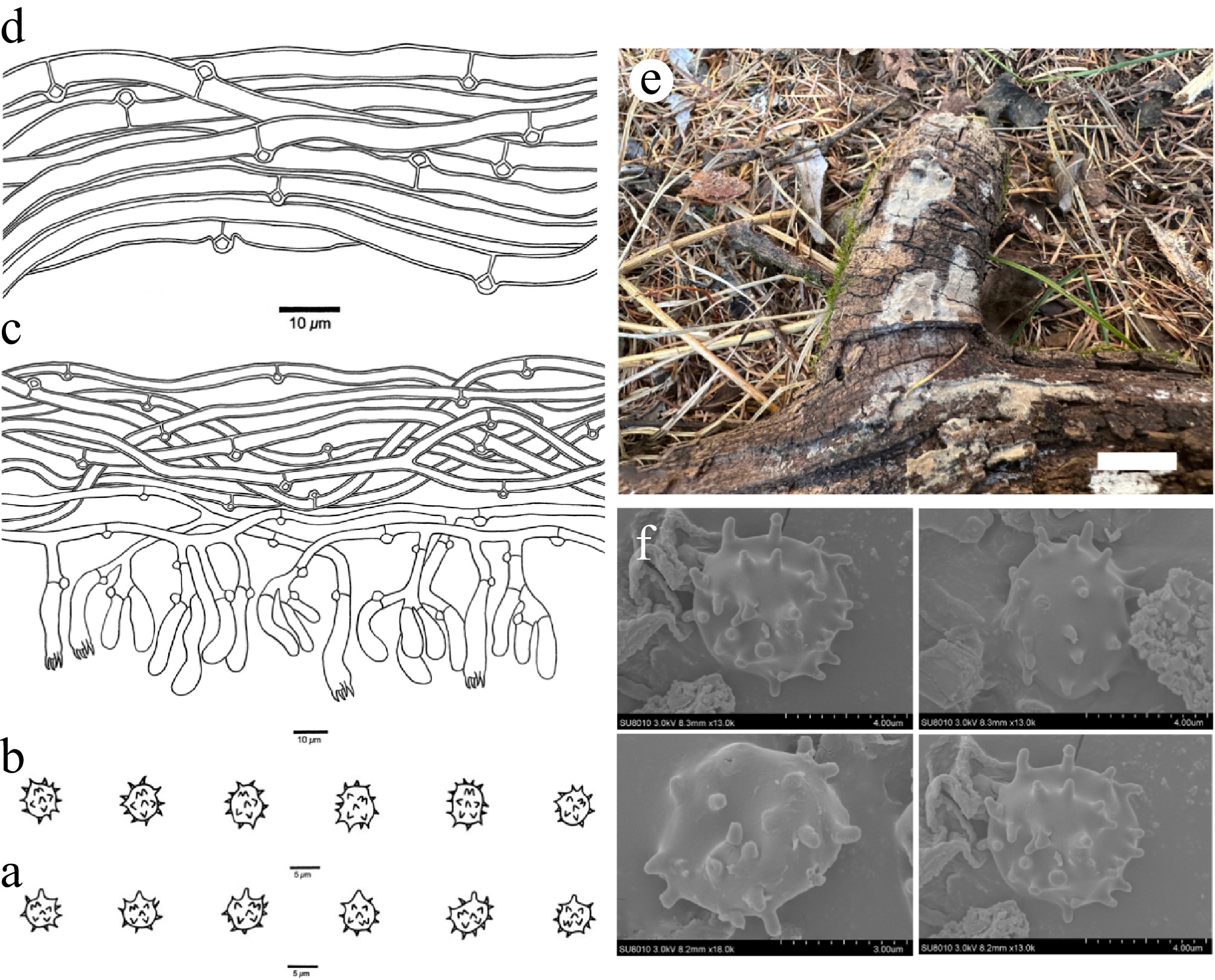
Figure 17.
Basidiomata and microscopic structures of Thelephora submurina (holotype, Dai 38088). (a) Basidiospores in lateral view. (b) Basidiospores in frontal view. (c) A section through a basidioma. (d) Hyphae from a rhizomorph. (e) Basidiomata on the host substrates. (f) Scanning electron microscope (SEM) of basidiospores. Scale bars: (a), (b) = 5 µm; (c), (d) = 10 µm; (e) = 1 cm.
MycoBank number: MB 864321
Diagnosis – Thelephora submurina is distinguished by resupinate basidiomata, a pale mouse-gray hymenophoral surface when fresh and dry, presence of rhizomorphs, broadly ellipsoid to subglobose to bi-, tri-, or quadra-lobed basidiospores, and short echinuli (up to 1 µm).
Type – CHINA. Heilongjiang Province, Tonghe County, Mamading Virgin Forest, on a fallen trunk of Larix olgensis, 10 October 2025, Dai 38088 (holotype BJFC059347).
Etymology – Submurina (Lat.): refers to the species having a pale mouse-gray hymenial surface.
Basidiomata – Annual, resupinate, adherent to the substrate, arachnoid, soft corky, without odor or taste when fresh, continuous, up to 3.5 cm long, 1 cm wide, and 1 mm thick at the center; hymenophoral surface smooth, pale mouse-gray when both fresh and dry, concolorous with subiculum when dry; sterile margin determinate, fimbriate, whitish, paler than hymenophore.
Rhizomorphs – Present in subiculum and margin, 20–50 µm diam; rhizomorphic surface more or less smooth; hyphae in rhizomorph monomitic, undifferentiated, individual hyphae with clamp connections, 3–5 µm diam, unbranched, more or less regularly arranged, thick-walled with a lumen, yellowish in KOH and distilled water.
Subicular hyphae – Monomitic; generative hyphae frequently with clamp connections, occasionally branched, thick-walled with a wide lumen, without encrustation, more or less interwoven, yellowish in KOH and distilled water, IKI−, CB−, 2–5 µm diam; subhymenial hyphae frequently with clamp connections, thin-walled, smooth, 2–4 µm diam; hyphal cells more or less uniform, yellowish in KOH and in distilled water, IKI−, CB−.
Cystidia – Absent.
Basidia – Clavate, slightly winding, without transverse septa, yellowish in KOH and distilled water, not stalked, with four sterigmata and a clamp connection at the base, 20–30 µm long, 4–6 µm diam at the apex and 2–4 µm at the base; sterigmata 1.5–4 µm long and 1–2 µm diam at the base.
Basidiospores – Broadly ellipsoid to subglobose to bi-, tri-, or quadra-lobed in frontal and lateral faces, echinulate, yellowish in KOH and distilled water, IKI−, CB−, (4.5–)4.6–5.9(–6.4) × (3.7–)3.8–5.6(–5.7) µm, L = 5.32 µm, W = 4.63 µm, Q = 1.11–1.17 (n = 60/2); echinuli usually isolated, sometimes grouped in two or more, up to 1 µm long.
Additional specimen (paratype) examined – CHINA. Heilongjiang Province, Tonghe County, Mamading Virgin Forest, on a rotten trunk of Tilia, 10 October 2025, Dai 38089 (BJFC059348).
Habitat – Thelephora submurina was found in northeast China with a temperate continental monsoon climate.
Notes – Thelephora submurina is closely related to T. subtestacea (Bourdot & Galzin) Kõljalg et al. (100% in ML) in the phylogenetic tree (Fig. 1), and both species have basidia with a clamp connection at the base, clamped subhymenial hyphae, and subicular hyphae. However, T. subtestacea differs from T. submurina by having longer basidiospores (35–50 vs 20–30 µm), the absence of rhizomorphs, and the presence of cystidia[29].
In morphology, T. submurina shares similar characteristics with T. quercicola, by sharing a fimbriate sterile margin, broadly ellipsoid to subglobose basidiospores, and the presence of rhizomorphs. Phylogenetically, T. submurina and T. quercicola are closely related, but T. quercicola differs from T. submurina by bigger basidiospores (6.7–8.7 × 5.7–7.5 vs 4.6–5.9 × 3.8–5.6 µm).
Thelephora subvinosogrisea X. Li, Y.C. Dai & Yuan Yuan, sp. nov. Fig. 18
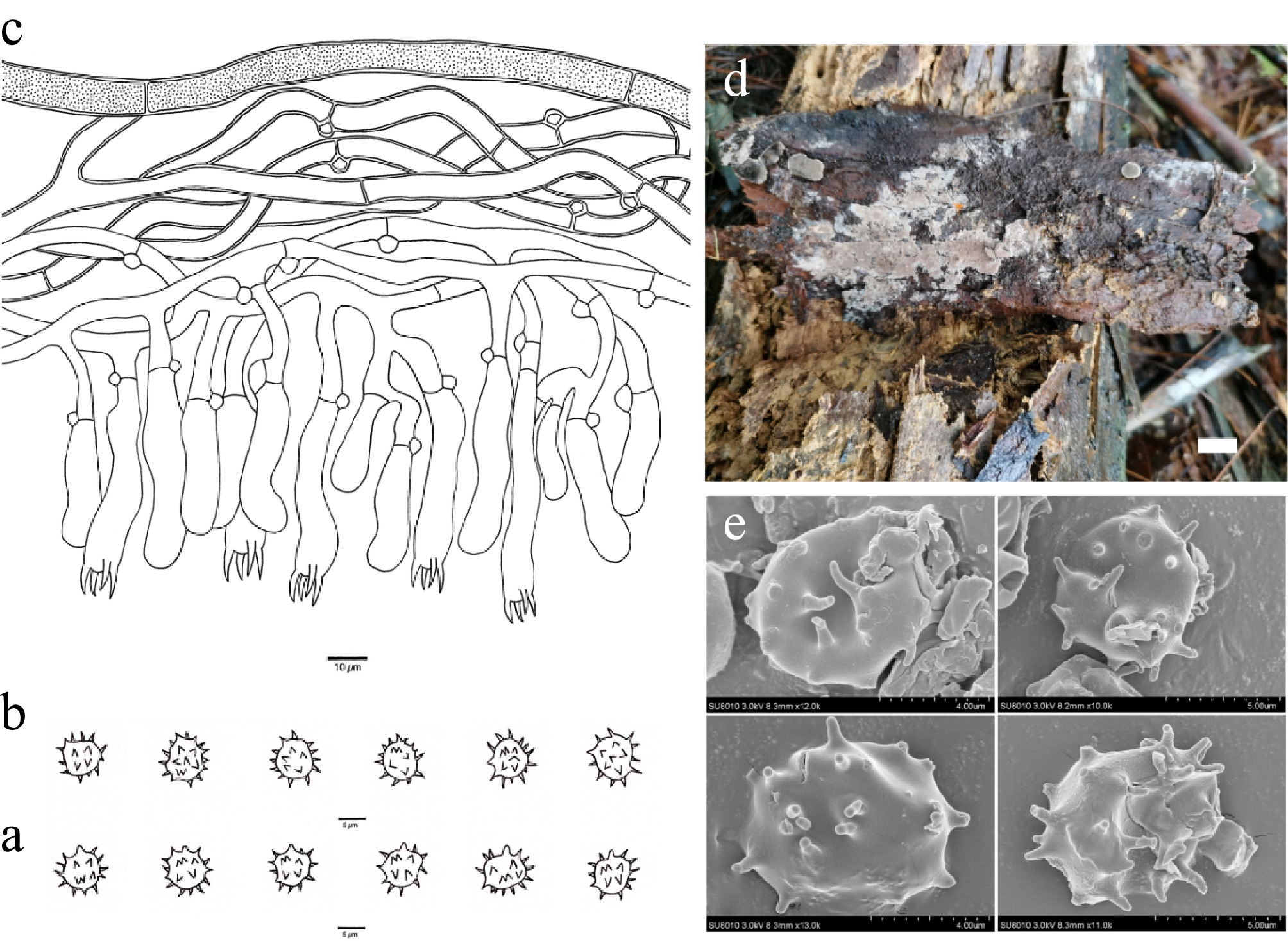
Figure 18.
Basidiomata and microscopic structures of Thelephora subvinosogrisea (holotype, Dai 28754). (a) Basidiospores in lateral view. (b) Basidiospores in frontal view. (c) A section through a basidioma. (d) Basidiomata on the host substrates. (e) Scanning electron microscope (SEM) of basidiospores. Scale bars: (a), (b) = 5 µm; (c) = 10 µm; (d) = 1 cm.
MycoBank number: MB 864322
Diagnosis – Thelephora subvinosogrisea is distinguished by resupinate basidiomata, a light vinaceous gray hymenophoral surface when fresh and fawn when dry, occasionally fine encrustation of subicular hyphae, and broadly ellipsoid to subglobose basidiospores.
Type – CHINA. Jiangxi Province, Nanchang, Shengshuitang Forest Park, on rotten wood of Pinus massoniana, 13 July 2024, Dai 28754 (holotype BJFC049013).
Etymology – Subvinosogrisea (Lat.): refers to the species having a light vinaceous gray hymenophoral surface.
Basidiomata – Annual, resupinate, adherent to the substrate, pelliculose, soft, without odor or taste when fresh, continuous, up to 7 cm long, 2.5 cm wide, and 2 mm thick at the center; hymenophoral surface smooth, light vinaceous gray when fresh, and fawn and paler than subiculum when dry; sterile margin determinate, farinaceous, whitish, distinctly paler than hymenophore.
Rhizomorphs – Absent.
Subicular hyphae – Monomitic; generative hyphae mostly with clamp connections and rarely with simple septa, occasionally branched, thick-walled with a wide lumen, occasionally with fine encrustation, more or less interwoven, pale brown in KOH and distilled water, IKI−, CB−, 5–8.0 µm diam; subhymenial hyphae frequently with clamp connections and rarely with simple septa, thin-walled, smooth, 4–6 µm diam; hyphal cells more or less uniform, pale brown in KOH and in distilled water, IKI−, CB−.
Cystidia – Absent.
Basidia – Clavate, without transverse septa, pale brown in KOH and distilled water, not stalked, with four sterigmata and a clamp connection at the base, 35–65 µm long, 6–9 µm diam at the apex and 4–7 µm at the base; sterigmata 5–9 µm long and 2–4 µm diam at the base.
Basidiospores – Broadly ellipsoid to subglobose in frontal faces and subglobose in lateral faces, echinulate, pale brown in KOH and distilled water, IKI−, CB−, (6.4–)6.5–8.5(–10.2) × (5.2–)5.4–8.0(–8.3) µm, L = 7.54 µm, W = 6.33 µm, Q = 1.17–1.21 (n = 60/2); echinuli usually isolated, sometimes grouped in two or more, up to 1.5 µm long.
Additional specimen (paratype) examined – CHINA. Guangxi Autonomous Region, Leye County, Huangjingdong Forest Park, on a dead tree of Pinus yunnanensis, 8 April 2024, Dai 33210 (BJFC053469).
Habitat – Thelephora subvinosogrisea was found in southern China with a subtropical monsoon climate.
Notes – In the phylogenetic tree (Fig. 1), T. subvinosogrisea, T. tedersooi (Suvi & Kõljalg) Kõljalg et al., T. ventricobasidia T.W. Henkel et al., T. ascendens T.W. Henkel et al., and T. compacta T.W. Henkel & Alvarez-Manj. clustered together with high support (100% in ML and 0.99 BPP). Thelephora tedersooi resembles T. subvinosogrisea by sharing continuous basidiomata and the absence of cystidia. However, T. tedersooi is distinguished from T. subvinosogrisea by its longer basidiospores (9–13 vs 6.5–8.5 µm) and wider basidia at the apex (12–14 vs 6–9 µm)[75]. Thelephora ventricobasidia resembles T. subvinosogrisea by sharing clamped subhymenial hyphae and the absence of cystidia. But T. ventricobasidia differs from T. subvinosogrisea by having rhizomorphs and wider basidia at the apex (10.8–20.3 vs 6–9 µm)[63]. Thelephora ascendens resembles T. subvinosogrisea by having continuous basidiomata separable from the substrate and with clamped subicular and subhymenial hyphae. But T. ascendens is distinguished from T. subvinosogrisea by having utriform basidia and rhizomorphs[63]. Thelephora compacta resembles T. subvinosogrisea by its absence of cystidia. But T. compacta differs from T. subvinosogrisea by having thin-walled subicular hyphae and rhizomorphs[63].
In morphology, T. subvinosogrisea resembles T. reniformis by sharing a smooth and light vinaceous gray to fawn hymenophoral surface and lacking rhizomorphs, but phylogenetically they are distantly related (Fig. 1), and T. reniformis is distinguished from T. subvinosogrisea by being separable from the substrate basidiomata, shorter basidia sterigmata (2–4 vs 5–9 µm), and more or less reniform to bi-, tri-, or quadra-lobed basidiospores.
Thelephora tenuicystidiola X. Li, Y.C. Dai & Yuan Yuan, sp. nov. Fig. 19
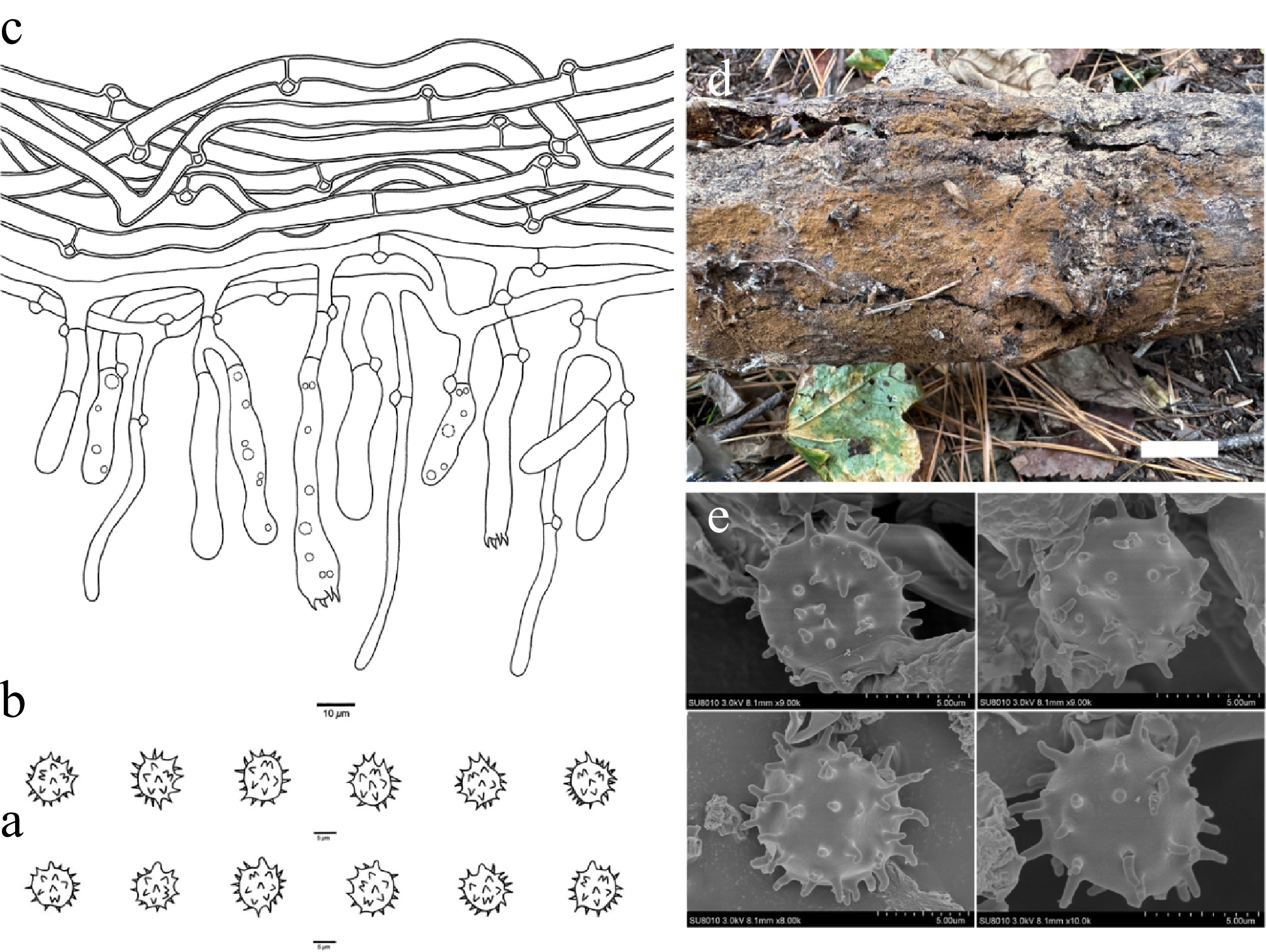
Figure 19.
Basidiomata and microscopic structures of Thelephora tenuicystidiola (holotype, Dai 38254). (a) Basidiospores in lateral view. (b) Basidiospores in frontal view. (c) A section through a basidioma. (d) Basidiomata on the host substrates. (e) Scanning electron microscope (SEM) of basidiospores. Scale bars: (a), (b) = 5 µm; (c) = 10 µm; (d) = 1 cm.
MycoBank number: MB 864323
Diagnosis – Thelephora tenuicystidiola is distinguished by resupinate basidiomata, hyphoid cystidioles, basidia occasionally oil droplets, and subglobose to bi-, tri-, or quadra-lobed basidiospores.
Type – CHINA. Heilongjiang Province, Fangzheng County, Longshan Forest Park, on a rotten trunk of Tilia, 11 October 2025, Dai 38254 (holotype BJFC059513).
Etymology – Tenuicystidiola (Lat.): refers to the species having thin cystidioles.
Basidiomata – Annual, resupinate, adherent to the substrate, mucedinoid, soft, without odor or taste when fresh, continuous, up to 5 cm long, 2.5 cm wide, and 2 mm thick at the center; hymenophoral surface smooth, honey-yellow to clay-buff when both fresh and dry, and concolorous with subiculum when dry; sterile margin determinate, fimbriate, concolorous with hymenophore.
Rhizomorphs – Absent.
Subicular hyphae – Monomitic; generative hyphae frequently with clamp connections, occasionally branched, thick-walled with a wide lumen, without encrustation, more or less interwoven, yellowish in KOH and distilled water, IKI−, CB−, 4.5–7 µm diam; subhymenial hyphae frequently with clamp connections, thin-walled, smooth, 3.5–5.5 µm diam; hyphal cells more or less uniform, yellowish in KOH and in distilled water, IKI−, CB−.
Cystidia – Absent; cystidioles present, arising from subhymenial hyphae, hyphoid, projecting above the hymenium, aseptate, 25–70 × 2–5 µm, yellowish in KOH and in distilled water.
Basidia – Clavate, rarely transverse septa, yellowish in KOH and distilled water, not stalked, occasionally with oil droplets, with four sterigmata and a clamp connection at the base, 35–65 µm long, 5–10 µm diam at the apex and 4–6 µm at the base; sterigmata 1–5 µm long and 1–2 µm diam at the base.
Basidiospores – Subglobose to bi-, tri-, or quadra-lobed in frontal and lateral faces, echinulate, pale brown in KOH and distilled water, IKI−, CB−, (7.7–)7.9–10.1(–10.2) × (6.5–)6.6–8.6(–8.8) µm, L = 8.57 µm, W = 7.61 µm, Q = 1.10–1.14 (n = 60/2); echinuli usually isolated, sometimes grouped in two, up to 2.5 µm long.
Additional specimen (paratype) examined – CHINA. Heilongjiang Province, Fangzheng County, Longshan Forest Park, on a rotten trunk of Tilia, 11 October 2025, Dai 38255 (BJFC059514); Tonghe County, Mamading Virgin Forest, on the bark of Populus, 10 October 2025, Dai 38147 (BJFC059406).
Habitat – Thelephora tenuicystidiola was found in northeast China, with a cold temperate and temperate continental monsoon climate. Additionally, T. tenuicystidiola was also known from one eDNA sequence from Russia (Fig. 1).
Notes – Thelephora tenuicystidiola is related to T. liaoningensis (H.S. Yuan & Y.C. Dai) Kõljalg et al. (96% in ML) in the phylogenetic tree (Fig. 1). Thelephora liaoningensis is similar to T. tenuicystidiola in its continuous and mucedinoid basidiomata, with clamped subhymenial hyphae and the absence of rhizomorphs. However, T. liaoningensis is distinguished from T. tenuicystidiola by having an indeterminate and farinaceous sterile margin and shorter basidiospores (6.3–7.8 vs 7.9–10.1 µm)[48].
In morphology, T. tenuicystidiola resembles T. separata by having a honey-yellow hymenophoral surface and subglobose to bi-, tri-, or quadralobed basidiospores. However, phylogenetically they are distantly related (Fig. 1), and T. separata is distinguished from T. tenuicystidiola by the presence of rhizomorphs and narrower subicular hyphae (1.5–3.5 vs 4.5–7 µm)[48].
Thelephora vinosogrisea X. Li, Y.C. Dai, Y.D. Wu & Yuan Yuan, sp. nov. Fig. 20
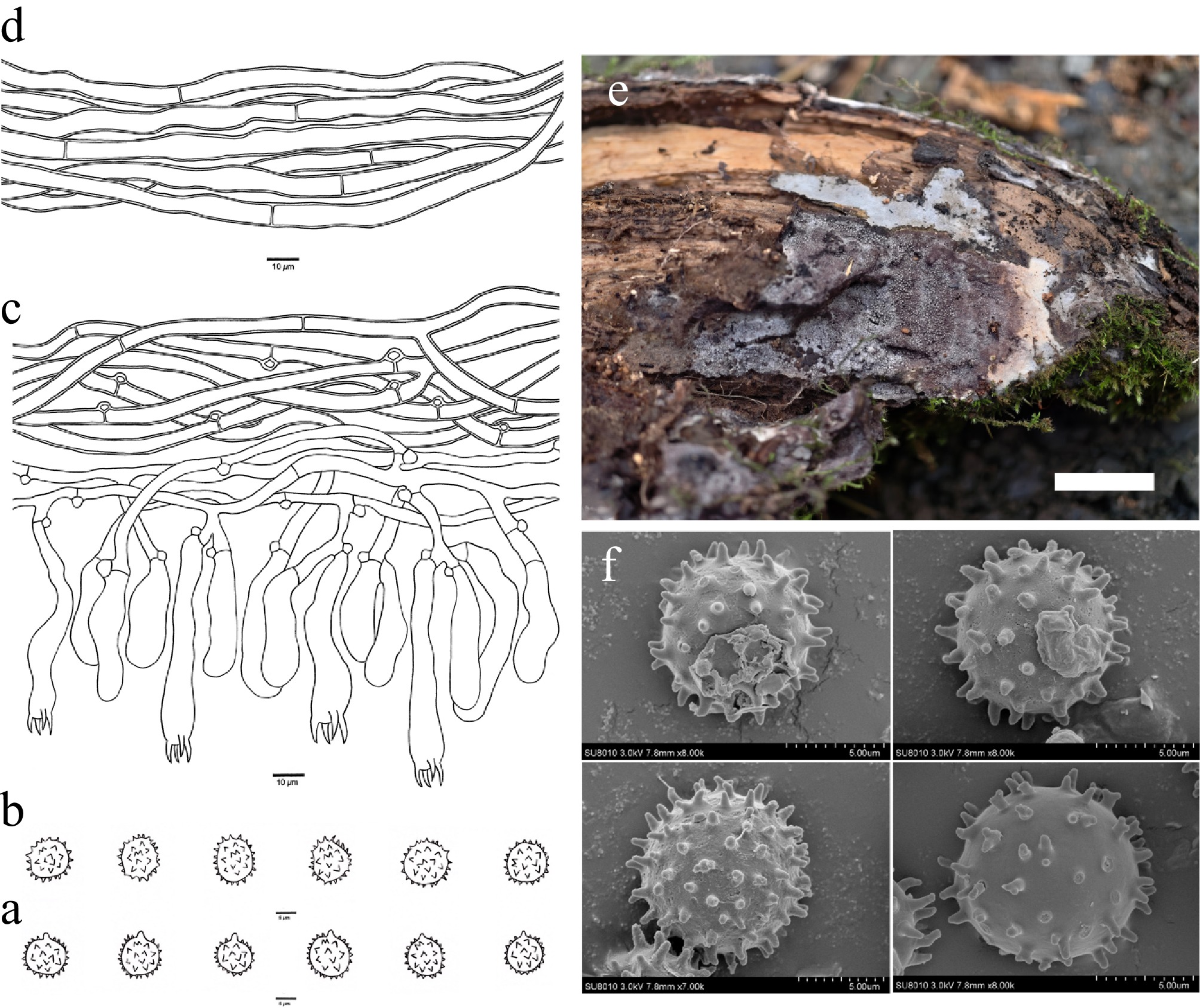
Figure 20.
Basidiomata and microscopic structures of Thelephora vinosogrisea (holotype, Yuan 3514). (a) Basidiospores in lateral view. (b) Basidiospores in frontal view. (c) A section through a basidioma. (d) Hyphae from a rhizomorph. (e) Basidiomata on the host substrates. (f) Scanning electron microscope (SEM) of basidiospores. Scale bars: (a), (b) = 5 µm; (c), (d) = 10 µm; (e) = 1 cm.
MycoBank number: MB 864324
Diagnosis – Thelephora vinosogrisea is distinguished by resupinate basidiomata, a vinaceous-gray hymenophoral surface when fresh and dark gray when dry, presence of rhizomorphs, stalked basidia, subglobose to globose basidiospores.
Type – CHINA. Sichuan Province, Yajiang County, Mixigou, on charred wood of Pinus yunnanensis, 4 October 2025, Yuan 3514 (holotype BJFC063060).
Etymology – Vinosogrisea (Lat.): refers to the species having a vinaceous-gray hymenophoral surface when fresh.
Basidiomata – Annual, resupinate, separable from the substrate, crustose, soft, without odor or taste when fresh, continuous, up to 3 cm long, 2 cm wide, and 1.5 mm thick at the center; hymenophoral surface granulose, vinaceous gray when fresh, and dark gray and darker than subiculum when dry; sterile margin determinate, farinaceous, concolorous with hymenophore.
Rhizomorphs – Present in subiculum and margin, 30–60 µm diam; rhizomorphic surface more or less smooth; hyphae in rhizomorph monomitic, undifferentiated, individual hyphae with simple septa, 4–7 µm diam, unbranched, thick-walled with a wide lumen, more or less regularly arranged, greenish in KOH and distilled water.
Subicular hyphae – Monomitic; generative hyphae mostly with clamp connections and rarely with simple septa, occasionally branched, thick-walled with a wide lumen, without encrustation, more or less interwoven, pale brown in KOH and distilled water, IKI−, CB−, 4–6.5 µm diam; subhymenial hyphae mostly with clamp connections and rarely with simple septa, thin-walled, smooth, 3–5.5 µm diam; hyphal cells more or less uniform, pale brown in KOH and in distilled water, IKI−, CB−.
Cystidia – Absent.
Basidia – Clavate, without transverse septa, hyaline in KOH and distilled water, stalked, with four sterigmata and a clamp connection at the base, 50–80 µm long, 6–12 µm diam at the apex and 2–6 µm at the base; sterigmata 7–10 µm long and 1–3 µm at the base.
Basidiospores – Subglobose to globose in frontal and lateral faces, echinulate, pale brown in KOH and distilled water, IKI−, CB−, (8.5–)8.6–10.9(–11.0) × (7.8–)8.0–10.2(–10.5) µm, L = 9.44 µm, W = 8.92 µm, Q = 1.03–1.06 (n = 60/2); echinuli usually isolated, sometimes grouped in two, up to 1.5 µm long.
Additional specimen (paratype) examined – CHINA. Sichuan Province, Yajiang County, Mixigou, on charred wood of Pinus yunnanensis, 4 October 2025, Yuan 3517 (BJFC063063).
Habitat – Thelephora vinosogrisea was found in southwest China with a continental monsoon plateau climate.
Notes – In the phylogenetic tree (Fig. 1), T. vinosogrisea, T. bidoupensis (X. Lu & H.S. Yuan) Kõljalg & Svantesson, T. aurantiotincta Corner, T. grandinioides C.L. Zhao & X.F. Liu, T. subtropica X.J. Zhang & C.L. Zhao, T. sikkimensis K. Das et al., T. bubalinomarginata B.K. Cui et al., and T. cacao Y.Q. Zhu et al., clustered together with moderate support (55% in ML and 0.99 BPP). Thelephora bidoupensis differs from T. vinosogrisea by having a smooth hymenophoral surface and smaller basidiospores (6–7 × 5.5–6.5 vs 8.6–10.9 × 8.0–10.2 µm)[67]. Thelephora aurantiotincta is distinguished from T. vinosogrisea by its erect basidiomata with a very strong smell of fenugreek[105]. Thelephora grandinioides differs from T. vinosogrisea by having erect basidiomata and smaller basidiospores (5.3–7.4 × 4–6.5 vs 8.6–10.9 × 8.0–10.2 µm)[73]. Thelephora subtropica differs from T. vinosogrisea by having shorter basidia (28–45 vs 50–80 µm) and smaller basidiospores (6–8 × 5–7.5 vs 8.6–10.9 × 8.0–10.2 µm)[49]. Thelephora sikkimensis is distinguished from T. vinosogrisea by having erect basidiomata and densely crystalline subhymenium hyphae[85]. Thelephora bubalinomarginata differs from T. vinosogrisea by having erect basidiomata with a strong fenugreek odor when dry[34]. Thelephora cacao differs from T. vinosogrisea by having erect basidiomata with a pungent odor when dry[5].
In morphology, T. vinosogrisea resembles T. murina by having a vinaceous-gray hymenophoral surface. However, phylogenetically, they are distantly related (Fig. 1), and T. murina is distinguished from T. vinosogrisea by being adherent to the substrate and lacking rhizomorphs.
Thelephora violaceardesiaca X. Li, Y.C. Dai & Yuan Yuan, sp. nov. Fig. 21
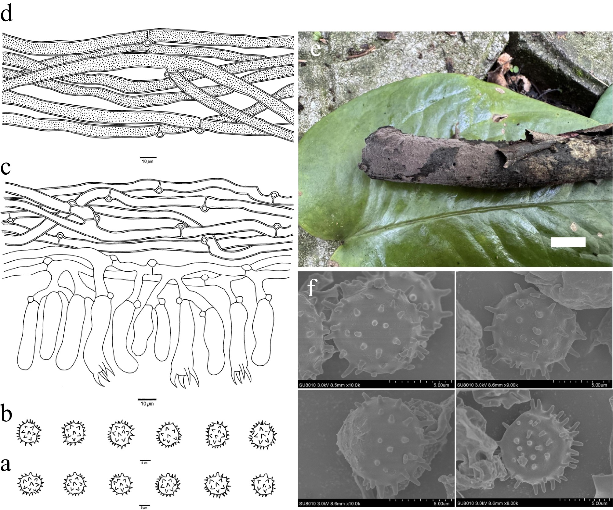
Figure 21.
Basidiomata and microscopic structures of Thelephora violaceardesiaca (holotype, Dai 35184). (a) Basidiospores in lateral view. (b) Basidiospores in frontal view. (c) A section through a basidioma. (d) Hyphae from a rhizomorph. (e) Basidiomata on the host substrates. (f) Scanning electron microscope (SEM) of basidiospores. Scale bars: (a), (b) = 5 µm; (c), (d) = 10 µm; (e) = 1 cm.
MycoBank number: MB 864325
Diagnosis – Thelephora violaceardesiaca is distinguished by resupinate basidiomata, a grayish-violet to vinaceous hymenophoral surface when fresh and dry, fusiform cystidioles, and subglobose to bi-, tri-, or quadra-lobed basidiospores.
Type – CHINA. Qinghai Province, Huzhu County, Beishan Forest Park, Langshidang, on a fallen branch of Picea crassifolia, 10 August 2025, Dai 35184 (holotype BJFC056445).
Etymology – violaceardesiaca (Lat.): refers to the species having a grayish-violet to vinaceous hymenophore when fresh and dry.
Basidiomata – Annual, resupinate, separable from the substrate, mucedinoid, soft, without odor or taste when fresh, continuous, up to 4 cm long, 2 cm wide, and 1 mm thick at the center; hymenophoral surface smooth, grayish violet to vinaceous when both fresh and dry, and concolorous with subiculum when dry; sterile margin determinate, farinaceous, concolorous with hymenophore.
Rhizomorphs – Present in subiculum and margin, 30–50 µm diam; rhizomorphic surface more or less smooth; hyphae in rhizomorph monomitic, undifferentiated, individual hyphae with clamp connections, 6.5–9.5 µm diam, occasionally branched, thick-walled with a wide lumen, usually with fine encrustation, more or less regularly arranged, dark brown in KOH and distilled water.
Subicular hyphae – Monomitic; generative hyphae frequently with clamp connections, frequently branched, thick-walled with a wide lumen, without encrustation, more or less interwoven, hyaline in KOH and distilled water, IKI−, CB+, 4.0–6.0 µm diam; subhymenial hyphae frequently with clamp connections, thin-walled, smooth, 4.0–5.5 µm diam; hyphal cells more or less uniform, hyaline in KOH and in distilled water, IKI−, CB+.
Cystidia – Absent; cystidioles present, arising at a level with the basidia, fusiform, 25–30 × 7–11 µm, hyaline in KOH and distilled water, a clamp connection at the base.
Basidia – Clavate, without transverse septa, hyaline in KOH and distilled water, not stalked, with four sterigmata and a clamp connection at the base, 30–40 µm long, 4–7 µm diam at the apex and 4–7 µm at the base; sterigmata 5–8 µm long and 1–4 µm diam at the base.
Basidiospores – Subglobose to bi-, tri-, or quadra-lobed in frontal and lateral faces, echinulate, pale brown in KOH and distilled water, IKI−, CB−, (8.1–)8.2–10.3(–10.4) × (7.4–)7.8–9.4(–9.7) µm, L = 9.43 µm, W = 8.73 µm, Q = 1.07–1.11 (n = 60/2); echinuli usually isolated, up to 2 µm long.
Habitat – Thelephora violaceardesiaca was found in northwest China, with a plateau continental climate.
Notes – Thelephora violaceardesiaca is more or less related to T. brevisterigmata (X. Lu & H.S. Yuan) Kõljalg & Svantesson and T. stipitobasidia (X. Lu & H.S. Yuan) Kõljalg & Svantesson in the phylogenetic tree (Fig. 1). Thelephora brevisterigmata resembles T. violaceardesiaca by lacking cystidia and basidiospores subglobose to bi-, tri-, or quadra-lobed in frontal and lateral faces. However, T. brevisterigmata is distinguished from T. violaceardesiaca by smaller basidiospores (7–8 × 5.5–7 vs 8.2–10.3 × 7.8–9.4 µm)[67]. Thelephora stipitobasidia resembles T. violaceardesiaca by sharing a smooth hymenophoral surface and the presence of rhizomorphs. However, T. stipitobasidia is distinguished from T. violaceardesiaca by smaller basidiospores (6.5–8 × 4.5–7 vs 8.2–10.3 × 7.8–9.4 µm)[67].
Morphologically, Thelephora violaceardesiaca resembles T. olivaceobrunnea by having mucedinoid basidiomata and subglobose to bi-, tri-, or quadralobed basidiospores. However, phylogenetically they are distantly related (Fig. 1), and T. olivaceobrunnea is distinguished from T. violaceardesiaca by having smaller basidiospores (6.5–7.0 × 5.5–6.0 vs 8.2–10.3 × 7.8–9.4 µm) and lacking rhizomorphs[48].
-
In this study, taxonomic and phylogenetic investigations of Thelephora were conducted based on morphological characteristics and phylogenetic analyses combining ITS and LSU sequences, using specimens collected from multiple regions across China. A total of 20 new species have been identified and described (Fig. 1; Supplementary Table S1). The discovery of these new species significantly enriches available knowledge on the species diversity of Thelephora in China, bringing the total number of species described to 108.
In recent years, phylogenetic studies based on multi-gene sequences have revealed that the traditional genera Thelephora and Tomentella do not represent independent monophyletic groups, but together form a well-supported evolutionary clade[25,26,28,108]. Consequently, Kõljalg et al.[29] formally merged the genus Tomentella into Thelephora. Our study supports the revised framework, with all newly described species with resupinate basidiomata addressed in Thelephora, and our phylogeny demonstrated that all our new species nested in Thelephora. For example, in the phylogenetic tree, the resupinate species T. bidoupensis and the new species T. vinosogrisea are intermingled with the stipitate species T. grandinioides, T. sikkimensis, T. subtropica, and T. bubalinomarginata; the resupinate new species T. nivea is intermingled with the stipitate species T. caryophyllea, T. iqbalii, T. scopiformis, T. scissilis, and T. anthocephala.
The 20 new species in this study were collected from multiple provinces across China, spanning from cold-temperate to tropical vegetation, including Qinghai, Inner Mongolia, Sichuan, Heilongjiang, Jiangxi, Guangxi, Hainan, Xizang, and Jilin. Their habitats are highly diverse, encompassing subalpine coniferous forests, temperate mixed coniferous–broadleaf forests, and tropical monsoon rainforests. Host plants involved belong to Pinaceae (e.g., Pinus, Abies, Larix, Keteleeria), Fagaceae (e.g., Quercus), Tiliaceae (e.g., Tilia), Salicaceae (e.g., Populus), Ericaceae (e.g., Rhododendron), and bamboos (subfamily Bambusoideae, Poaceae). Again, our results confirm the broad host associations and ecological adaptability of Thelephora as ectomycorrhizal fungi[72,109−112]. On the other hand, the species diversity of Thelephora within China remains greatly underexplored, particularly in biodiversity hotspots such as the karst regions of southwestern China, the coniferous forest areas of northeastern China, and the margins of the Qinghai–Xizang Plateau[113−116]. The new species proposed in this study described here are based on only a limited number of specimens; therefore, host specificity, actual distribution ranges, and population status of the species described here require further investigation.
In this study, we compiled a dataset of ITS and LSU sequences that currently provides relatively broad coverage of the Thelephora group. This dataset integrates sequences from global public databases (e.g., NCBI, UNITE) along with newly generated sequences from the present study. Furthermore, high-throughput sequencing technology has revealed a large number of uncultured Thelephora sequences from environmental samples. These unnamed 'thelephoroid' environmental sequences are among the most important groups available in public repositories. Among them, some eDNA sequences showed high similarity to the new species through BLAST searches, and we therefore included these sequences in our phylogenetic analyses. In the phylogenetic analyses, these environmental sequences were consistently recovered together with their corresponding new species in highly supported clades. The successful matching examples demonstrate that accurately linking environmental sequences to morphologically defined species based on vouchered specimens is feasible. However, how to systematically achieve such linking remains a major challenge for future research in this field, as well as an important opportunity for in-depth assessment of the diversity and ecological functions of this group[117,118].
Based on the moderate to strong support (ML ≥ 70%/BPP ≥ 0.90) from maximum likelihood (ML) and Bayesian inference (BI) analyses, we divided the monophyletic groups in the phylogenetic tree into clades 1 to 22. The new species placed in the highly supported clades may share certain common features. For example, Thelephora ceracea and T. reniformis clustered in clade 1, and species in this clade are characterized by basidiospore echinuli less than 1.5 µm long; T. keteleeriae, T. niveomarginata, T. caeruleoalba, and T. murina fell in clade 5, and species in this clade are characterized by resupinate basidiomata and basidia sterigmata 1.5–3 µm at the base; T. subvinosogrisea clustered in clade 6, and species in this clade are characterized by basidiomata adherent to the substrate; T. griseosepiacea clustered in clade 20, and species in this clade are characterized by lacking cystidia and rhizomorphs; T. roseobubalina fell in clade 21, and species in this clade are characterized by lacking rhizomorphs.
Although the genus Thelephora is an indispensable ectomycorrhizal fungal group in forest ecosystems, the rarity of its basidiomata in nature has long constrained traditional morphology-based taxonomic studies. In recent years, molecular data have increasingly revealed extensive cryptic diversity within this genus; however, most of these molecular operational taxonomic units (MOTUs) lack corresponding morphological evidence, leaving the taxonomic framework unstable. To assess the potential species diversity of Thelephora in China, we analysed ITS sequences from China available in GenBank. At a 97% sequence similarity threshold, we recovered 116 OTUs. The Chao1 richness estimator projected a potential species richness of approximately 220 species for Thelephora in China, a value significantly higher than the 88 species currently known from China, strongly indicating that the diversity of Thelephora has been substantially underestimated. Against this background, through systematic field collections and multi-locus phylogenetic analyses, we successfully describe 20 new species, raising the number of known Thelephora species in China to 108. By providing detailed morphological illustrations for each new species, we establish a verifiable bridge between molecular cryptic diversity and morphologically defined taxonomic entities, thereby granting formal taxonomic recognition to a substantial number of previously overlooked 'dark diversity' that existed only as environmental DNA sequences. This study also validates the generic-level reclassification, merging Tomentella into Thelephora, enriches the molecular backbone of this clade, and provides critical voucher materials for future investigations into host specificity, biogeographic history, and mycorrhizal adaptive evolution[77,84]. Nonetheless, these 20 new species represent only the tip of the iceberg. Future efforts integrating broader global sampling and genomic approaches will be essential to fully resolve the species boundaries and ecological functions of this important ectomycorrhizal lineage.
-
The authors confirm contributions to the paper as follows: conception and design of the study: Li X, Zhang X, Cui Y; data collection: Li X, Zhang X, Cui Y, Zhuang L, Zeng G, Wu Y, Yuan Y; analysis and interpretation of results: Li X, Yuan Y; draft manuscript preparation: Li X, Ghobad-Nejhad M. All authors reviewed the results and approved the final version of the manuscript.
-
The data that support the findings of this study are available in the National Center for Biotechnology Information (NCBI) databases and Index Fungorum.
-
The authors declare that they have no competing interests.
-
accompanies this paper online at: https://doi.org/10.48130/mycosphere-0026-0019.
- Supplementary Table S1 A list of species, specimens and GenBank accession numbers of the sequences used in this study.
- Supplementary Table S2 A list of new species ITS sequences blast result in this study.
- Copyright: © 2026 by the author(s). Published by Maximum Academic Press, Fayetteville, GA. This article is an open access article distributed under Creative Commons Attribution License (CC BY 4.0), visit https://creativecommons.org/licenses/by/4.0/.
-
About this article
Cite this article
Li X, Zhang X, Cui Y, Zhuang L, Zeng G, et al. 2026. Twenty new species of resupinate Thelephora (Thelephorales, Basidiomycota) from China. Mycosphere 17: e019 doi: 10.48130/mycosphere-0026-0019 

Twenty new species of resupinate Thelephora (Thelephorales, Basidiomycota) from China
- Received: 25 June 2026
- Revised: 28 July 2026
- Accepted: 30 August 2026
- Published online: 12 October 2026
Abstract: Thelephora is an important genus of macrofungi belonging to the Basidiomycota. Participating in ectomycorrhizal associations, species of the genus play indispensable roles in maintaining forest productivity and long-term ecosystem health. Moreover, a number of Thelephora species are of economic importance for their edibility and medicinal properties. In this study, based on morphological and phylogenetic analyses, 20 new species of Thelephora with resupinate basidiomata are described, namely T. bambusicola, T. caeruleoalba, T. ceracea, T. griseosepiacea, T. keteleeriae, T. koraiensis, T. longissima, T. mongolica, T. murina, T. nivea, T. niveomarginata, T. quercicola, T. reniformis, T. rhododendri, T. roseobubalina, T. submurina, T. subvinosogrisea, T. tenuicystidiola, T. vinosogrisea, and T. violaceardesiaca. Phylogenetic relationships of Thelephora were constructed by integrating DNA sequences of the internal transcribed spacer (ITS) and the large subunit of nuclear ribosomal RNA (LSU). Molecular phylogenetic analyses based on Maximum Likelihood (ML) and Bayesian Inference (BI) confirmed these 20 new species nested within Thelephora with high support. Each new species is presented with an illustrated description and a detailed comparison with its related and morphologically similar species.
-
Key words:
- Ectomycorrhizal fungi /
- Phylogeny /
- Systematics /
- Taxonomy /
- Thelephora