









-
The capabilities of fungi are remarkable in addressing future global demands, encompassing health, sustainable agriculture, and renewable energy[1]. Therefore, it is our responsibility to recognize the presence of concealed mycota worldwide. These newly discovered species will play a significant role in advancing agriculture, industrial biotechnology, medicine, and disease control[1]. To achieve these advancements, we must be diligent in our scientific practices, ensuring robust taxon sampling and accurate referencing when introducing new fungal taxa. This is crucial for achieving better outcomes in fungal research and driving progress in our fields[2]. In fungi, there are two of the two largest classes within the Ascomycota phylum: Dothideomycetes and Sordariomycetes. Members of them occur as pathogens, saprobes, endophytes, or epiphytes, and are widely utilized in economically significant industries[3−5]. Their diverse functions as metabolites and biocontrol agents make them a fascinating area of study[6,7]. Dothideomycetes are primarily distinguished by the presence of bitunicate asci, which possess two wall layers and are often fissitunicate[8−10]. In contrast, Sordariomycetes are characterized by inoperculate, unitunicate asci lacking a hinged cap and with a single wall layer[11]. Traditional identification of fungi, especially within the diverse Ascomycota classes Dothideomycetes and Sordariomycetes, primarily relies on morphological characteristics observed under microscopy, such as the structure of fruiting bodies (ascomata), ascus type, and spore (ascospore or conidia) morphology[9,10]. Although contemporary research incorporates DNA-based molecular techniques, traditional morphological methods continue to serve as the foundation for preliminary classification and the description of new species[2,12,13]. Both classes exhibit a wide range of lifestyles; notably, saprobic fungi, as major decomposers of organic matter, play a crucial ecological role in terrestrial and aquatic environments[9,11,14,15]. Their substantial influence on plant primary production, carbon mineralization, and segregation is a testament to their significant contribution to the environment[14,16]. Moreover, fungi's adaptability to various conditions, such as moisture levels, temperature, substrate availability, and nutrient content, significantly influences their ability to decompose organic matter[17]. Studying fungal distribution worldwide is crucial to a better understanding of the fungal cycle and its changes[18].
The study of fungal diversity has several crucial aspects, including biotechnology research, understanding ecosystem functioning, and protecting human health and agricultural security[2,19,20]. Fungal biodiversity hotspots around the world significantly impact the increasing number and diversity of both micro- and macro-fungi. The magnitude of our discoveries is truly impressive. As of the present, 15,266 new fungal species have been reported in China, underscoring the country's considerable biodiversity[21]. The majority of these species are found in Southern China and account for 8.69% of all globally described fungal species, placing China second only to the United States[2,22,23]. These discoveries have been documented across all 34 provincial-level regions, with Yunnan Province reporting the highest number at 3,869 species, nearly one-fifth of the national total. The distinctive geographical features of Yunnan support both a highland cold-resistant biome in the west and a tropical biome in the south and southwest, making the province a key area for fungal discoveries and a leading hotspot for biotechnology research[23−37].
This study aims to advance understanding of the diversity and distribution of saprobic fungal communities in the KIB using both morphological and molecular approaches. Dead plant material bearing fungal fruiting bodies was collected from the KIB premises in Kunming, Yunnan, China, to facilitate the identification of both novel and previously known saprobic fungal species. Through detailed morphological examination and multi-locus phylogenetic analyses, two new species and seven new records within Dothideomycetes and Sordariomycetes were identified from the KIB garden. Furthermore, this research presents the first comprehensive checklist of fungi collected at KIB, establishing a valuable resource for future studies.
-
Dead and decaying branches, petioles, and culms with fungal fruiting bodies were collected from the garden of the Kunming Institute of Botany, Yunnan Province, China, and taken to the KIB mycology laboratory after important collection details were recorded[38]. The specimens were examined under an Olympus SZ61 (Japan) series stereo- and light microscope, photographed with an OLYMPUS SZ2-ILST and an Industrial Digital Camera 16NP USB3.0 (Panasonic, Japan) microscope imaging system. The morphological characteristics of the fungi were recorded. We followed the method of Senanayake et al.[38] to isolate a single spore and obtain a pure culture of the fungus on potato dextrose agar (PDA). Microstructural observation was performed using scanning electron microscopy (ZEISS GeminiSEM and ZEISS Sigma 300 apparatus). Spores or other relevant structures were carefully isolated with a needle under a stereomicroscope and transferred onto an aluminum specimen stub, pre-coated with conductive adhesive to ensure well-dispersed, non-overlapping placement. The mounted sample was rapidly frozen. In a low-temperature preparation chamber, the sample was coated with a thin layer of gold under vacuum for approximately 30 min to enhance conductivity and minimize charging during observation. After coating, the frozen sample was maintained at a low temperature and directly transferred to the SEM chamber for imaging. Images were acquired at appropriate accelerating voltages and magnifications to examine the surface structure of the samples[39]. The Tarosoft Image Framework program v0.9.0.7. was used to measure all microscopic structures of the fungi, and Adobe Photoshop CS3 Extended v10.0 (Adobe Systems, USA) was used to process and edit the images used in the figures. Herbarium specimens are deposited in the Kunming Institute of Botany, Academia Sinica (KUN-HKAS), while the living cultures are deposited in the Kunming Institute of Botany Culture Collection (KUNCC/KUMCC). Facesoffungi (FoF) number and Index Fungorum (IF) number were obtained as explained in Jayasiri et al.[40] and Index Fungorum[41], respectively. In addition, sexual/asexual morphs sporulation in the culture medium was induced following a slightly modified method of Su et al.[42]. Sterilized toothpicks and pine needles were placed on either side of a PDA plate, and a small piece of mycelium grown on PDA was placed on the baits. The cultures were incubated at 28 °C. For in vitro sporulation, the plates were examined using a stereo microscope. Fungal microstructures were prepared on slides, observed, photographed, measured, and compiled into a photographic plate.
Molecular studies
-
Actively growing mycelium was carefully scraped from a 7–10-day-old PDA culture using a sterile scalpel and transferred into a 1.5 mL centrifuge tube for DNA extraction. The Biospin Fungus Genomic DNA Extraction Kit–BSC14S1 (BioFlux, China) was used to extract DNA, following the manufacturer's instructions, and extracted DNA was preserved at −20 °C. DNA amplifications were performed by polymerase chain reaction (PCR). A portion of the large-subunit nuclear rRNA gene (LSU) was amplified using primer pairs LR0R and LR5[43]. The small 18S subunit nuclear rRNA gene (SSU) was amplified with primer pairs NS1 and NS4[44]. Primer pairs ITS4 and ITS5 were used to amplify the 5.8S rDNA gene and flanking internal transcribed spacers (ITS)[44]. The translation elongation factor 1-alpha gene (tef1) was amplified by using primers EF1-983F and EF1-2218R[45], or the primers EF1-728F and EF1-986R[46]. The RNA polymerase II second-largest subunit (rpb2) gene was amplified with primers fRPB2 and fRPB2-7cR[47]. The beta-tubulin (tub2) gene was amplified using primers Btub2Fd and Btub4Rd[48]. The PCR thermal cycle programs for ITS, LSU, and SSU were as follows: an initialization step of 94 °C for 3 min, followed by 35 cycles of 94 °C for 30 s, an annealing step at 55 °C for 50 s, an elongation step at 72 °C for 1 min, and a final extension step of 72 °C for 10 min. The PCR thermal cycle programs for rpb2, tef1, and tub2 was as follows: an initialization step of 95 °C for 3 min, followed by 40 cycles of 95 °C for 50 s, an annealing step (52 °C, 30 s for tub2; 55 °C, 50 s for rpb2, and tef1), an elongation step at 72 °C for 90 s, and a final extension step of 72 °C for 10 min.
Phylogenetic analyses
-
The ITS, LSU, SSU, and protein-coding genes (rpb2, tef1, and tub2) were used for different fungal groups as needed. All reference sequences were retrieved from GenBank using the latest references for each fungal group. Sequences were aligned with BioEdit v5.0.6[49] and ClustalX v1.83[50], or with MAFFT v6.864b (
https://mafft.cbrc.jp/alignment/server/index.html )[51]. The alignments were visually checked and manually improved where necessary. Phylogenetic analyses were carried out with MrBayes v3.2.7a[52] for Bayesian inference analysis (BI). A Maximum Likelihood analysis (ML) was performed via the CIPRES web portal[53] using RAxML v7.2.8 as part of the 'RAxML-HPC2 on TG' tool[54] or implemented in RAxMLGUI v0.9b2[55]. Other phylogenetic details are outlined in Jeewon et al.[56,57]. Phylogenetic trees were drawn with FigTree v1.4[58]. Maximum likelihood bootstrap support (MLBS) equal or greater than 50%–70% and Bayesian posterior probabilities (BYPP) values equal or greater than 0.95 are indicated on the resulting tree topology in each figure. Newly generated sequences were deposited at the NCBI GenBank. GenBank accession numbers for sequenced genes are given in the descriptions under the materials examined. The same isolate was duplicated and remade as sequences A and B. -
In this paper, we describe two new species and seven new records of saprobic fungal strains collected from the KIB (Yunnan, China). The novelties are morphologically illustrated, and phylogenies based on multi-locus sequence data are provided to accommodate species in their orders, families, and genera where appropriate.
Phylogenetic analyses
-
Analysis 1: Botryosphaeria phylogeny was based on combined ITS, LSU, tef1, and tub2 sequence data.
Forty strains are included in the combined analyses, which comprise 2,223 characters across the ITS, LSU, tef1, and tub2 alignments. Macrophomina phaseolina (CBS 227.33 and CBS 162.25) was used as the outgroup taxon, and related sequences were obtained as described by Tian et al.[59]. Single-loci analyses were performed to compare the topology and clade stability with those of combined-locus analyses. The tree topology of the ML analysis is similar to that of the BI analysis. The best RAxML tree had a final likelihood value of −4756.457710. The matrix contained 267 distinct alignment patterns, with 18.94% of characters undetermined or missing. Estimated base frequencies were as follows: A = 0.226896, C = 0.273860, G = 0.274223, T = 0.225021; substitution rates AC = 0.835737, AG = 2.668379, AT = 1.055693, CG = 0.817208, CT = 6.080596, GT = 1.000000; gamma distribution shape parameter α = 0.084375. The collection (KUMCC 21-0103A, KUMCC 21-0103B) in the present study phylogenetically clustered with B. dolichospermatii strains (CGMCC 3.19098, CGMCC 3.19097, CGMCC 3.19096).
Analysis 2: Helminthosporium phylogeny was based on combined LSU, SSU, ITS, and tef1 sequence data.
Fifty-six strains were included in the analyses, comprising 3,776 characters including gaps. The tree is rooted with Periconia pseudodigitata (KT1395), and related sequences were obtained from Hyde et al.[60]. The matrix contained 1,263 distinct alignment patterns, with 36.99% of characters undetermined or missing. Estimated base frequencies were as follows; A = 0.240622, C = 0.242963, G = 0.270350, T = 0.246065; substitution rates AC = 1.991544, AG = 3.170223, AT = 2.023982, CG = 0.880620, CT = 7.722965, GT = 1.000000; gamma distribution shape parameter α = 0.171719. The trees from the two analyses (ML and BI) were identical. Our collection (KKMCC 21-0076) in the present study phylogenetically clustered between H. velutinum strains (MFLUCC 16-1360, MFLUCC 16-1096, CPC 26297, L131, CGMCC 3.23572) with statistical support (96% MLBS/1.00BYPP).
Analysis 3: Neohendersonia phylogeny was based on combined SSU, LSU, and ITS sequence data.
Nineteen strains were included in the combined analyses, which comprise 2,681 characters for SSU, LSU, and ITS alignment. Boeremia exigua (CBS 431.74) was used as the outgroup taxon, and related sequences were obtained following Sun et al.[61]. Single-locus analyses were performed to compare the topology and clade stability with those of combined-locus analyses. The tree topology of the ML analysis is similar to the BI analysis. The best RAxML tree had a final likelihood value of −7346.622368. The matrix contained 461 distinct alignment patterns, with 24.34% of characters undetermined or missing. Estimated base frequencies were as follows: A = 0.250407, C = 0.221818, G = 0.276438, T = 0.251338; substitution rates AC = 1.578736, AG = 3.664305, AT = 1.349205, CG = 0.450256, CT = 6.079060, GT = 1.000000; gamma distribution shape parameter α = 0.079251. Multi-locus phylogenetic analyses showed that our strains (KUMCC 21-0056, KUMCC 21-0107) belong to Neohendersonia and form a well-separated lineage from N. kickxii and N. tongrenensis.
Analysis 4: Magnibotryascoma phylogeny was based on combined LSU, SSU, ITS, tef1, and rpb2 sequence data.
Fifty-three strains were included in the analyses, comprising 4,359 characters, including gaps. The tree was rooted with Lophiostoma arundinis (CBS 621.86), and related sequences were obtained from Ren et al.[62]. The matrix contained 1,143 distinct alignment patterns, with 46.56% of characters undetermined or missing. Estimated base frequencies were as follows; A = 0.242454, C = 0.254882, G = 0.276848, T = 0.225816; substitution rates AC = 1.235534, AG = 2.891989, AT = 1.669777, CG = 1.089129, CT = 8.990044, GT = 1.000000; gamma distribution shape parameter α = 0.178080. The trees from the two analyses (ML and BI) were identical. Multi-locus phylogenetic analyses show that our new isolate (KUMCC 21-0112) clusters with M. mali (KUMCC 21-0516) but receives low statistical support.
Analysis 5: Apiospora phylogeny was based on ITS sequence data.
Seventy-nine strains were included in the analyses, comprising 913 characters for the ITS alignment. Seiridium phylicae (CBS 133587) in Sporocadaceae (Amphisphaeriales) is used as the outgroup taxon, and related sequences are taken from Pintos & Alvarado[63]. The tree topology of the ML analysis is similar to the Bayesian analysis. The best RaxML tree with a final likelihood value of −5256.291374 is presented. The matrix contained 439 distinct alignment patterns, with 25.11% of characters undetermined or missing. Estimated base frequencies were as follows: A = 0.239919, C = 0.248556, G = 0.233761, T = 0.277764; substitution rates AC = 0.826486, AG = 1.770838, AT = 0.788607, CG = 0.944632, CT = 2.720760, GT = 1.000000; gamma distribution shape parameter α = 0.301517. Multi-locus phylogenetic analyses show that our new isolate (KUMCC 21-0047) clusters with A. yunnana strains, with high statistical support (100% MLBS/1.00 BYPP).
Analysis 6: Pseudolachnella phylogeny was based on combined LSU, ITS, and tef1 sequence data.
Thirty strains were included in the analyses, comprising 2,869 characters in the LSU, ITS, and tef1 alignments. Dinemasporium strigosum (MAFF 244355) and D. cruciferum (MAFF 244327) are used as outgroup taxa, and related sequences are taken from Wu & Diao[64]. Single-locus analyses were performed to compare the topology and clade stability with those of combined-locus analyses. The tree topology of the ML analysis is similar to the Bayesian analysis. The best RaxML tree with a final likelihood value of −3089.572070 is presented. Estimated base frequencies were as follows: A = 0.259453, C = 0.230435, G = 0.223495, T = 0.286617; substitution rates AC = 0.775950, AG = 1.961909, AT = 0.980782, CG = 0.452485, CT = 3.571483, GT = 1.000000; gamma distribution shape parameter α = 0.150142. Multi-locus phylogenetic analyses show our new isolate (KUMCC 21-0048) clustered with P. tengii (NN047882) with high statistical support (100% MLBS/1.00 BYPP).
Analysis 7: Distoseptispora phylogeny was based on combined ITS, LSU, tef1, and rpb2 sequence data.
Eighty-six strains were included in the combined analyses, which comprise 3,466 characters for ITS LSU, tef1, and rpb2 after alignment. Pseudostanjehughesia aquitropica (MFLUCC 16-0569) and P. lignicola (MFLUCC 15-0352) were used as the outgroup taxa, and related sequences were obtained following Liao et al.[65]. Single-locus analyses were performed to compare the topology and clade stability with those of combined-gene analyses. The tree topology of the ML analysis is similar to the Bayesian analysis. The best RaxML tree with a final likelihood value of −35748.256699 is presented. The matrix contained 1,775 distinct alignment patterns, with 28.49% of characters undetermined or missing. Estimated base frequencies were as follows: A = 0.240683, C = 0.265261, G = 0.282208, T = 0.211848; substitution rates AC = 1.367285, AG = 3.313867, AT = 1.258963, CG = 0.898119, CT = 6.927241, GT = 1.000000; gamma distribution shape parameter α = 0.254970. Multi-locus phylogenetic analyses showed that Distoseptispora and our strain (KUMCC 21-0039) clustered with D. guizhouensis (GZCC 21-0666) with high statistical support (100% MLBS/1.00 BYPP).
Analysis 8: Peroneutypa phylogeny was based on combined ITS and tub2 sequence data.
Forty-three strains were included in the combined analyses, which comprised 1,182 characters in the ITS and tub2 alignments. Allocryptovalsa polyspora (MFLU 17-1218) and A. elaeidis (MFLUCC 15-0707) were used as the outgroup taxa, and related sequences were obtained following Mao et al.[66]. Single-locus analyses were performed to compare the topology and clade stability with those of combined-locus analyses. The tree topology of the ML analysis is similar to the Bayesian analysis. The best RAxML tree had a final likelihood value of −9820.994449. The matrix contained 748 distinct alignment patterns, with 35.42% of characters undetermined or missing. Estimated base frequencies were as follows: A = 0.214321, C = 0.280618, G = 0.243945, T = 0.261117; substitution rates AC = 0.864463, AG = 2.289966, AT = 1.381503, CG = 1.060632, CT = 3.181007, GT = 1.000000; gamma distribution shape parameter α = 0.513331. Multi-locus phylogenetic analyses show that our new isolates (KUMCC 21-0049, KUMCC 21-0100) are sister lineages to P. scoparia (MFLUCC 11-0478), with 70% MLBS statistical support.
Analysis 9: Melanographium phylogeny was based on combined LSU, ITS, SSU, tef1, and rpb2 sequence data.
Fourteen strains were included in the analyses, which comprised 4,428 characters for LSU, ITS, SSU, tef1, and rpb2 alignment. Related sequences are taken from Samarakoon et al.[67]. Two strains from Zygosporium oscheoides (MFLUCC 14-0402 and CBS 195.79) were used as the outgroup. The tree topology of the ML analysis is similar to the Bayesian analysis. The best RaxML tree with a final likelihood value of −12077.017651 is presented. The matrix contained 622 distinct alignment patterns, with 26.22% of characters undetermined or missing. Estimated base frequencies were as follows: A = 0.243063, C = 0.249875, G = 0.264718, T = 0.242345; substitution rates AC = 1.293241, AG = 2.428649, AT = 1.255505, CG = 0.928677, CT = 6.755147, GT = 1.000000; gamma distribution shape parameter α = 0.133168. Multi-locus phylogenetic analyses show that our new isolates (KUMCC 21-0101A, KUMCC 21-0101B) are sister lineages to M. citri (GZCC21-0208, GZCC21-0212), with 99% MLBS/ 1.00 BYPP statistical support.
Taxonomy
-
Dothideomycetes O.E. Erikss. & Winka
Botryosphaeriales C.L. Schoch, Crous & Shoemaker
Botryosphaeriaceae Theiss. & Syd.
Botryosphaeria Ces. & De Not.
Generally, Botryosphaeria is regarded as a species complex, owing to the high level of intraspecific variation among its isolates, which overlap with those of other related species, while morphological characteristics are no longer routinely employed for the identification[59,68,69]. Botryosphaeria species are known as plant saprobes, pathogens, and endophytes, with a global distribution across a wide variety of mainly woody hosts[68,70]. Diseases caused by pathogens belonging to the genus Botryosphaeria have resulted in significant losses in various economically important crops. To identify Botryosphaeria sp., it is recommended to provide ITS sequence data, combined with tef1 and tub2, which can effectively distinguish closely related species[71]. In our study, we introduced a new host record of B. dolichospermatii as a saprobe from Acer sp.
Botryosphaeria dolichospermatii Z.P. Dou, W. He & Y. Zhang, in Dou, Zhao, He & Zhang, Mycosystema 40(3): 479 (2021)
Index Fungorum number: IF 826937, Figs 1, 2
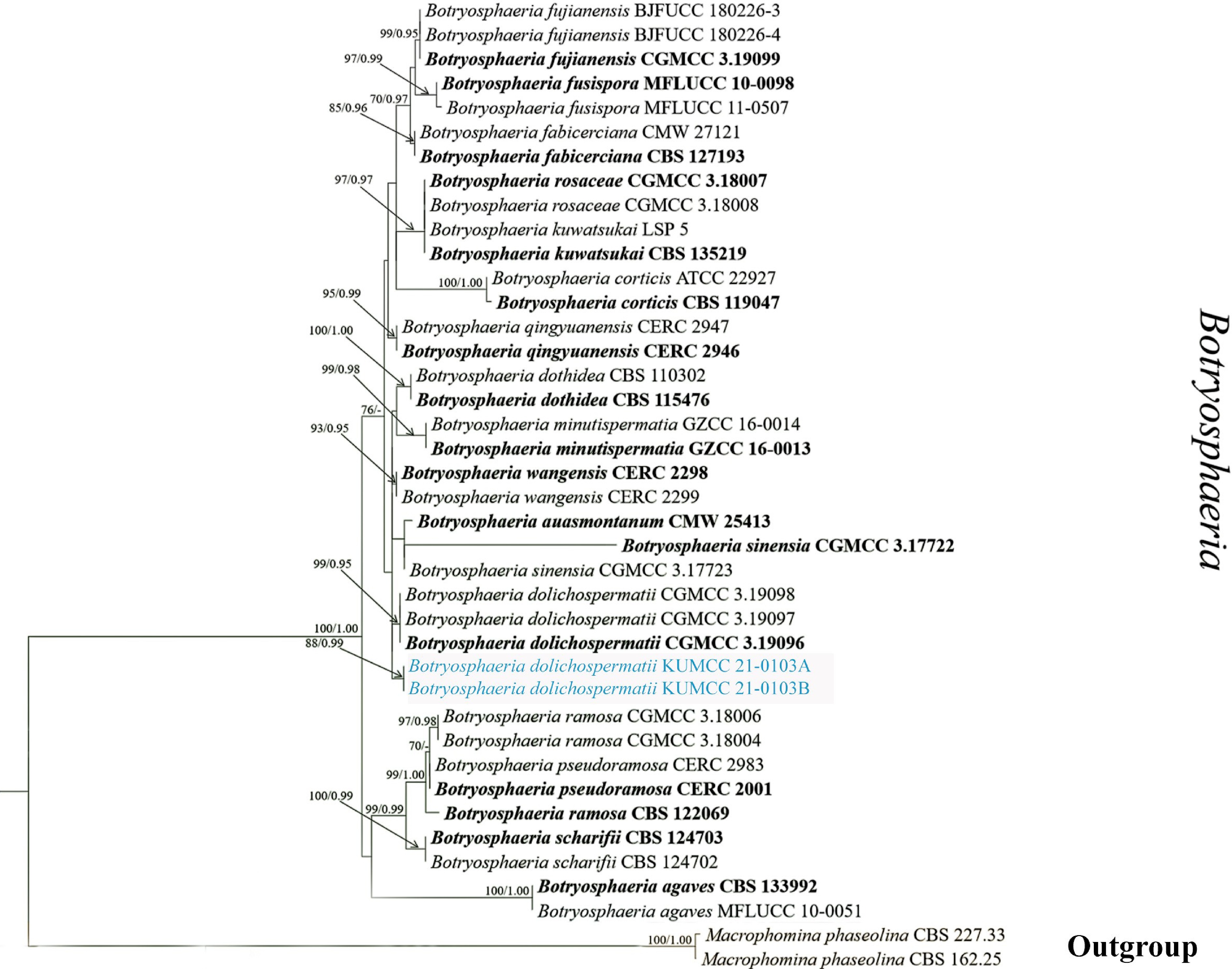
Figure 1.
Phylogram generated from Maximum Likelihood analysis based on ITS, LSU, tef1, and tub2 sequence data representing Botryosphaeria species. Bootstrap values for ML equal to or greater than 60% and BYPP values greater than 0.95 (the rounding of values to two decimal proportions) from BI analysis are labelled on the nodes. A newly generated sequence is in blue, and type species are bold.
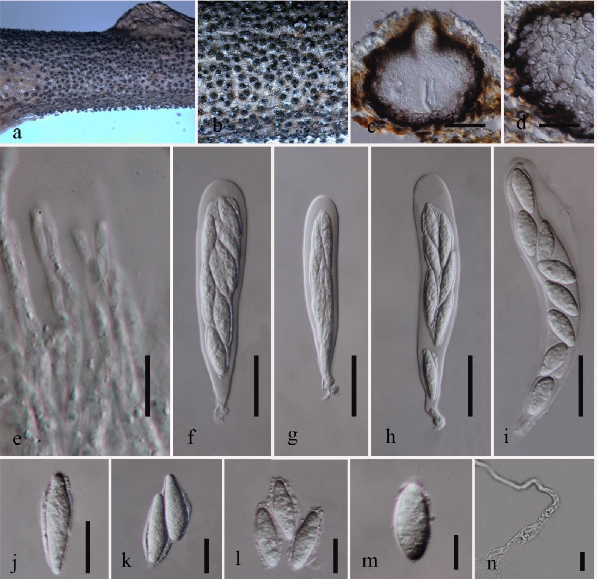
Figure 2.
Botryosphaeria dolichospermatii (KUN-HKAS 115560). (a), (b) Ascomata on host surface. (c) Vertical section of ascoma. (d) Peridium. (e) Pseudoparaphyses. (f)–(i) Asci. (j)–(m) Ascospores. (n) Germinated ascospore. Scale bars: (c) = 50 μm; (d), (f)–(i) = 20 μm; (e), (j)–(n) = 10 μm.
Saprobic on dead branches of Acer sp. Sexual morph: Ascomata 125–165 μm high × 189 190–225 μm diam. (
$\overline x $ $\overline x $ $\overline x $ $\overline x $ Culture characteristics: Ascospores germinate on PDA within 24 h, and germ tubes are produced from both sides. Colonies grow fast on PDA, effuse, velvety to hairy, circular, white in the first week, and brown to dark brown after 1 week from above and below.
Known host and distribution: On the cankered branch of Vaccinium uliginosum in Fujian, China[59]; on dead branches of Acer sp. in Yunnan, China (this study).
Material examined: China, Yunnan, Kunming, Kunming Institute of Botany, Chinese Academy of Science, on dead branches of Acer sp. (Sapindaceae), 15 May 2020, S. Tibpromma, ST 48 (KUN-HKAS 115560, new host record), living culture KUMCC 21-0103.
GenBank accession numbers: KUMCC 21-0103: SSU: PV942129; ITS: PV873194; LSU: PV942116; tub2: PX055888.
Notes: Our new collection morphologically resembles the type collection of Botryosphaeria described in Phillips et al.[72]. The new strain makes a weak independent clade and is closely related to B. dolichospermatii (CGMCC 3.19096T) in multigene phylogenetic analysis (Fig. 1). Botryosphaeria dolichospermatii was initially introduced as an asexual morph[59], while our new isolate is a sexual morph, and it fits well with the generic description of the sexual morph of Botryosphaeria[73]. The sexual morph of our sample is characterized by black, uniloculate, erumpent ascomata, resembling those of other Botryosphaeria species such as B. dothidea and B. rosaceae[71]. A comparison of LSU, ITS, tef1, and tub2 gene sequences between the new isolate and B. dolichospermatii (CGMCC 3.19096T) revealed no differences, except for two gaps in the ITS region within the areas analyzed in this study. Consequently, the new isolate is presented as the first recorded sexual morph and a new host record of B. dolichospermatii, supported by both morphological and phylogenetic similarities.
Pleosporales Luttr. ex M.E. Barr
Massarinaceae Munk
Helminthosporium Link
The type species of Helminthosporium, H. velutinum, is characterized by hyphomycetous with solitary, cylindrical, unbranched, brown conidiophores that produce obclavate, distoseptate conidia in integrated, terminal or intercalary, polytretic conidiogenous cells[74−77]. Most taxa in the Helminthosporium complex have later been placed in other genera, initially based on morphological characters and later based on molecular data, although the status of some species remained unresolved[78]. While a comprehensive study of the genus has been revised by Voglmayr & Jaklitsch[79], more than 225 species were listed in Helminthosporium[34,80,81]. In the present study, we collected H. velutinum from a branch of an unknown plant at the KIB, Yunnan, China, and this is the second strain that was reported from Yunnan Province, China.
Helminthosporium velutinum Link [as 'Helmisporium'], Mag. Gesell. naturf. Freunde, Berlin 3(1–2): 10, tab. 1:9 (1809)
Index Fungorum number: IF 250075, Figs 3, 4
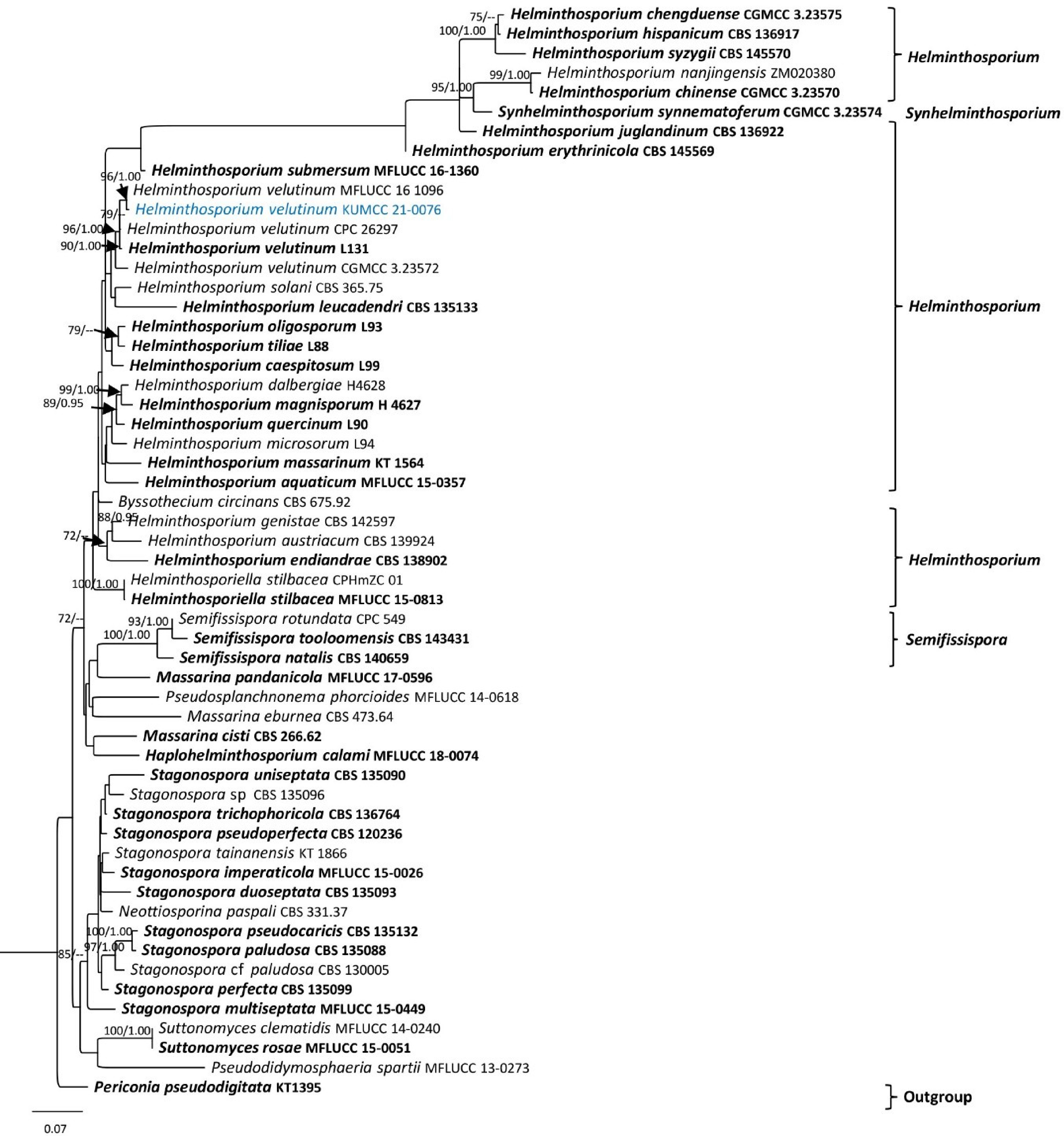
Figure 3.
Phylogram generated from Maximum Likelihood analysis based on LSU, SSU, ITS, and tef1 sequence data. Bootstrap values for ML equal to or greater than 70% and BYPP values greater than 0.95 (rounded to two decimal places) from the BI analysis are labeled on the nodes. A newly generated sequence is in blue, and type species are bold.
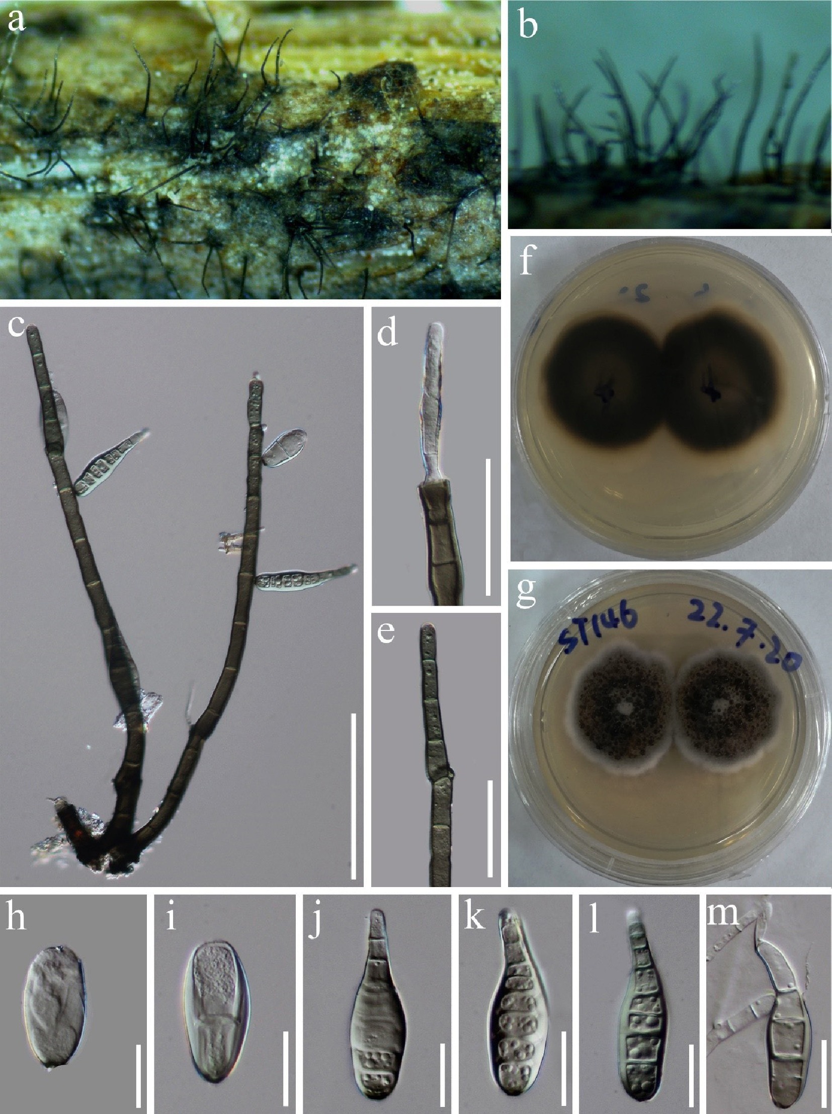
Figure 4.
Helminthosporium velutinum (KUN-HKAS 115606). (a), (b) Colonies on the substrate. (c) Conidiophores and conidia. (d), (e) Conidiogenesis. (f) Reverse view of culture on PDA. (g) Forward a few of the cultures in PDA. (h)–(l) Conidia. (m) Germinating conidium. Scale bars: (c)= 100 μm; (d), (e) = 50 μm; (h)–(m) = 20 μm.
Saprobic on dead branches of an unknown plant species. Sexual morph: Undetermined. Asexual morph: Hyphomycetes. Conidiophores mononematous, macronematous, mostly unbranched, percurrently proliferating from cut ends, dark brown, smooth, 321–340 μm long (
$\overline x $ $\overline x $ $\overline x $ $\overline x $ Culture characteristics: Colonies on PDA, after 7 d at 25 ˚C in dark grey to brown with watery substrate, effuse, velvety, margin grey to white, reverse dark brown with pale-brown margin.
Known hosts and distribution: On the cankered branch of Acer campestre in Italy[79], on the leaves of Canna generalis in Taiwan, China[82], on the stem of Passiflora edulis in Australia[83], on the twigs of Ulex europaeus in Scotland (UK)[84], on dead fruit of Musa acuminata in Thailand[85], on decaying branches of an unidentified host in Sichuan, China[78], on submerged wood in streams in Yunnan, China[86], and on a branch of an unknown plant in Yunnan, China (this study).
Material examined: China, Yunnan, Kunming, Kunming Institute of Botany, Chinese Academy of Science, on dead branches of an unknown plant species, 17 July 2020, S. Tibpromma, ST 146 (KUN-HKAS 115606), living culture KUMCC 21-0076.
GenBank accession numbers: KUMCC 21-0076: SSU: PV942134; ITS: PV873202; LSU: PV942124; tef1: PX055884.
Notes: New strain (KUMCC 21-0076) clusters with the strains of Helminthosporium velutinum (MFLUCC 16-1096, CPC 26297, CGMCC 3.23572, and L131[epitype]) and makes sister lineage to MFLUCC 16-1096 with 96% MLBS/1.00 BYPP statistic support (Fig. 3). In a BLASTn search of the NCBI GenBank, the closest match of the ITS sequence of the new isolate (KUMCC 21-0076) with 100% similarity was H. velutinum strains (voucher 202107001, MFLUCC 16-1096, and MFLUCC 16-1300).
Helminthosporium velutinum is found on a wide range of dead plant material, and it is cosmopolitan in distribution[78]. Our new isolate shares a similar morphology with the type strain of H. velutinum (L131) in having solitary, cylindrical, unbranched, brown conidiophores that produce obclavate, distoseptate conidia[74−77]. Based on morphology and the similarity of ITS sequences from various collections worldwide, we identified our isolates as H. velutinum. The species is primarily associated with dead plant material in terrestrial ecosystems worldwide[77,82−85]. Our new isolate is also from a branch of an unidentified plant species in Yunnan, China.
Neohendersoniaceae A. Giraldo & Crous
Neohendersonia Petr.
Neohendersonia was introduced by Petrak[87] with N. pyriformis as the type species. However, Giraldo et al.[88] suggested that N. pyriformis should be considered synonymous with N. kickxii, a proposition later supported by subsequent literature; N. kickxii was proposed as the type species, being the older name[88−90]. Currently, there are five species accepted in the genus; only N. kickxii and N. tongrenensis have sequence data, and all species were reported only as coelomycetes[61,88]. In this study, we introduced a new species of this genus and its sexual morph.
Neohendersonia camelliae Jayasiri, Tibpromma & Karun., sp. nov.
Index Fungorum number: IF905408; Facesoffungi number: FoF 18867, Figs 5, 6
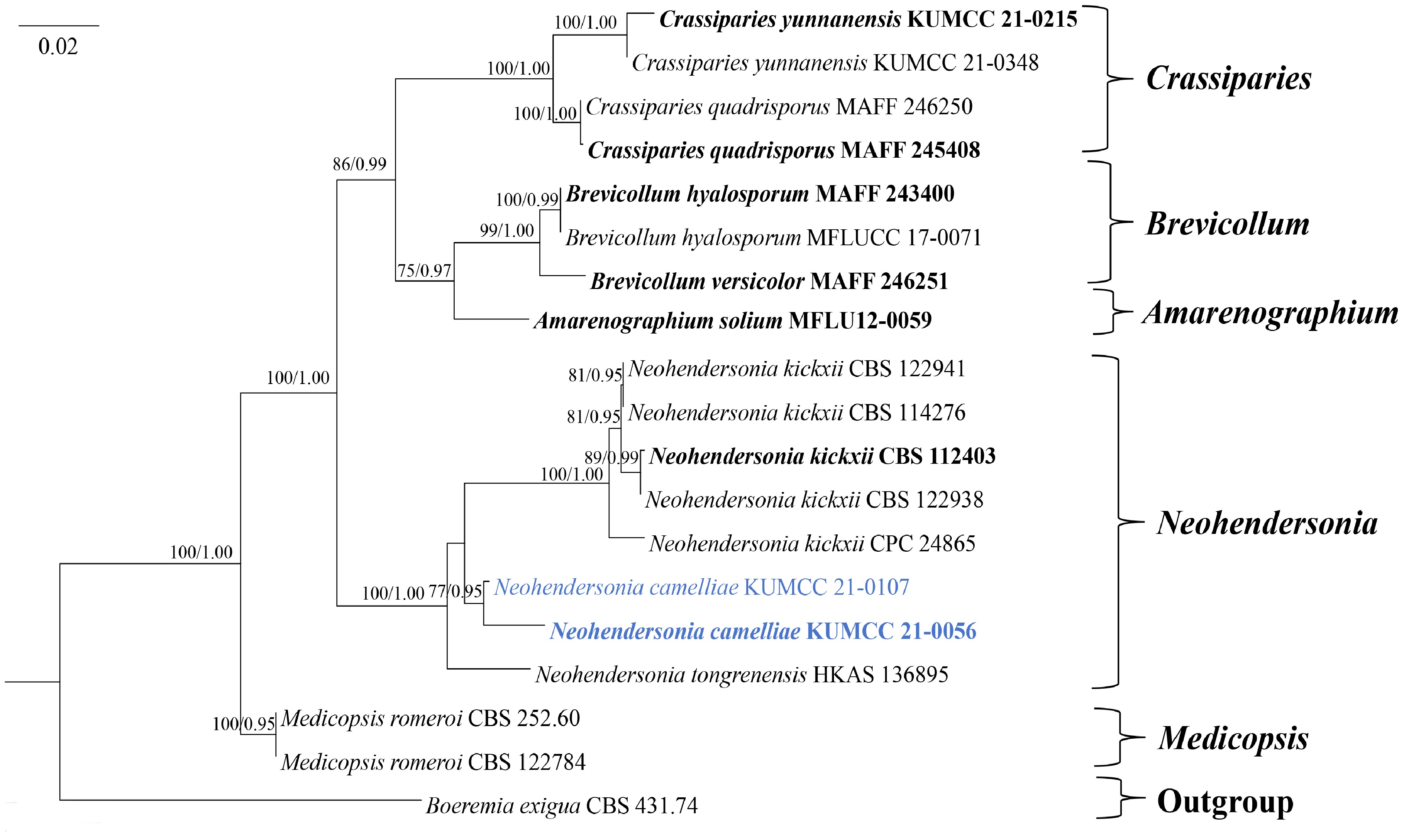
Figure 5.
Phylogram generated from Maximum Likelihood analysis based on SSU, LSU, and ITS sequence data representing Neohendersonia species. Bootstrap values for ML equal to or greater than 70% and BYPP greater than 0.95 (rounded to two decimal places) from the BI analysis are labeled on the nodes. Strains of the newly described species are in blue, while type strains are in bold.
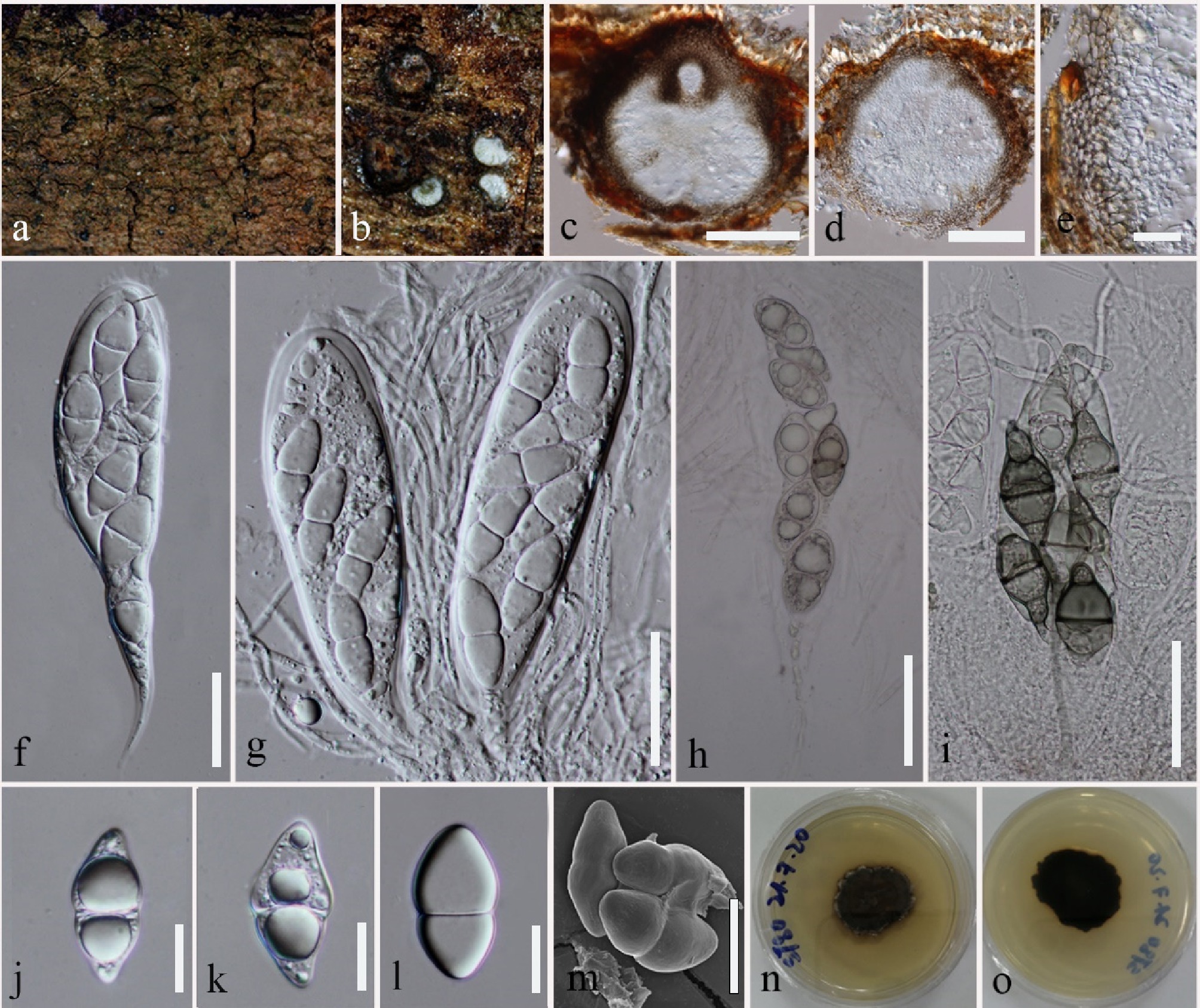
Figure 6.
Neohendersonia camelliae (KUN-HKAS 115578, holotype). (a), (b) Ascomata on host surface. (c), (d) Vertical section of ascomata. (e) Peridium. (f) Ascus. (g) Asci embedded in narrowly cellular pseudoparaphyses. (h) Ascus with mature ascospores. (i) Germinated ascospores. (j)–(m) Ascospores (m): SEM. (n) Upper view on PDA. (o) Reverse view on PDA. Scale bars: (c), (d) = 100 μm; (e), (j)–(m) = 20 μm; (f)–(i) = 50 μm.
Etymology: The specific epithet reflects the host genus, Camellia.
Holotype: KUN-HKAS 115578
Saprobic on dead branches of Camellia sp. and unknown plant species. Sexual morph: Ascomata 233–308 high × 210–307 μm diam. (
$\overline x $ $\overline x $ $\overline x $ Culture characteristics: Ascospores germinated within 12 h on PDA, colonies grown on PDA at room temperature (20–25 °C). Colonies are circular to irregular, dull, with wrinkled, yellow-green to brown aerial hyphae at the edge, dark green on the reverse, and produce a yellow-orange pigment on PDA.
Material examined: China, Yunnan, Kunming, Kunming Institute of Botany, Chinese Academy of Science, on dead branches of Camellia sp. (Theaceae), 08 June 2020, S. Tibpromma, ST 80 (KUN-HKAS 115578, holotype), ex-type KUMCC 21-0056; ibid., on dead branches of an unidentified plant, 25 May 2020, S. Tibpromma, ST 58 (KUN-HKAS 115566, paratype), ex-paratype KUMCC 21-0107.
GenBank accession numbers: KUMCC 21-0056: SSU: PV942132; ITS: PV873199; LSU: PV942121; tef1: PX055881; rpb2: PX055870. KUMCC 21-0107: SSU: PV948970; ITS: PV873197; LSU: PV942119; tef1: PX055879; rpb2: PX055868.
Notes: Neohendersonia camelliae is introduced as a new species, mainly based on phylogenetic analyses of a combined SSU, ITS, LSU, rpb2, and tef1 dataset and nucleotide comparison. Neohendersonia camelliae clusters sister lineage with the strains of N. kickxii with low statistical support (Fig. 5). In a BLASTn search of NCBI GenBank, the closest match of the ITS sequence of the new isolates (KUMCC 21-0056 and KUMCC 21-0107) with 92.91% similarity was the type strain of N. kickxii (CBS 112403). The comparison of ITS sequence between N. camelliae (KUMCC 21-0056) and N. kickxii (CBS 112403) shows a 47 bp (including gaps) difference (9.1%). Thus, we introduced our isolate as a new species in accordance with the guidelines of Jeewon & Hyde[91]. In addition, we did not obtain the asexual morph in the culture. Therefore, a morphological comparison between our new species and Neohendersonia kickxii was not possible, as the latter has so far only been reported in its asexual morph[88]. Currently, five other species are known within the genus Neohendersonia[41,63,89,92]. Among other genera in the family Neohendersoniaceae, Neohendersonia camelliae shares a more similar morphology with the genus Crassiparies[93,94] than with Brevicollum and Medicopsis. The sexual morph of Crassiparies (C. quadrisporus) is characterized by scattered, immersed, subglobose ascomata with an ostiolar neck, thin ascomata wall with polygonal to rectangular cells, septate, branched, and anastomosed pseudoparaphyses; 4-spored, cylindrical to clavate, pedicellate asci, and hyaline, broadly fusiform, l-septate ascospores[93,94]. However, they are difficult to distinguish morphologically but can be well separated in phylogeny.
Notably, all strains of Neohendersonia kickxii identified in Austria (CBS 122938, CBS 122941), Italy (CBS 112403), and Sweden (CBS 114276) were from Fagus spp. whereas our two new strains were isolated from Camellia sp. and dead branches of an unknown species in Yunnan, China.
Teichosporaceae M.E. Barr
Magnibotryascoma Thambug. & K.D. Hyde
Thambugala et al.[95] introduced Magnibotryascoma to accommodate M. uniseriata (≡Misturatosphaeria uniseriata). Magnibotryascoma species have been reported as woody-based saprobes on Acer cappadocicum, Clematis vitalba, Machilus yunnanensis, Malus halliana, Ribes sanguineum, Robinia pseudoacacia, Salix sp., Shorea assamica, and Vaccinium myrtillus from Belgium, China, Germany, Norway, and the United Kingdom[62,96,97]. The sexual morph of this genus is characterized by solitary or aggregated ascomata, fissitunicate, cylindrical to cylindrical-clavate asci, and partially overlapping, brown to dark brown, fusiform to elliptical, 1–3-septate ascospores[95], while the asexual morph is characterized by pycnidial, uniloculate, semi-erumpent to superficial, subglobose to globose, dark brown to black conidiomata, multi-layered conidiomatal walls, enteroblastic, phialidic, integrated, truncate to cylindrical, hyaline conidiogenous cells, and hyaline to reddish-brown, aseptate conidia[34,97,98]. In a previous study, Jaklitsch et al.[96] synonymized this genus with Teichospora, but the morphological differences between the two genera are notable. Later, phylogenetic analyses by Hongsanan et al.[9] positioned Magnibotryascoma as a distinct lineage within Teichosporaceae, separate from Teichospora, thereby supporting its recognition as a distinct genus. Currently, eight species are accepted in this genus[41]. Morphological characteristics and phylogenetic analyses of combined ITS, LSU, SSU, and tef1-α sequence data revealed their taxonomic positions within Magnibotryascoma[34,98]. In this study, we found other M. mali from unidentified tree species in Yunnan, China.
Magnibotryascoma mali Phukhams., Wanas. & K.D. Hyde 2017
Index Fungorum number: IF553255; Facesoffungi number: FoF03387, Figs 7, 8
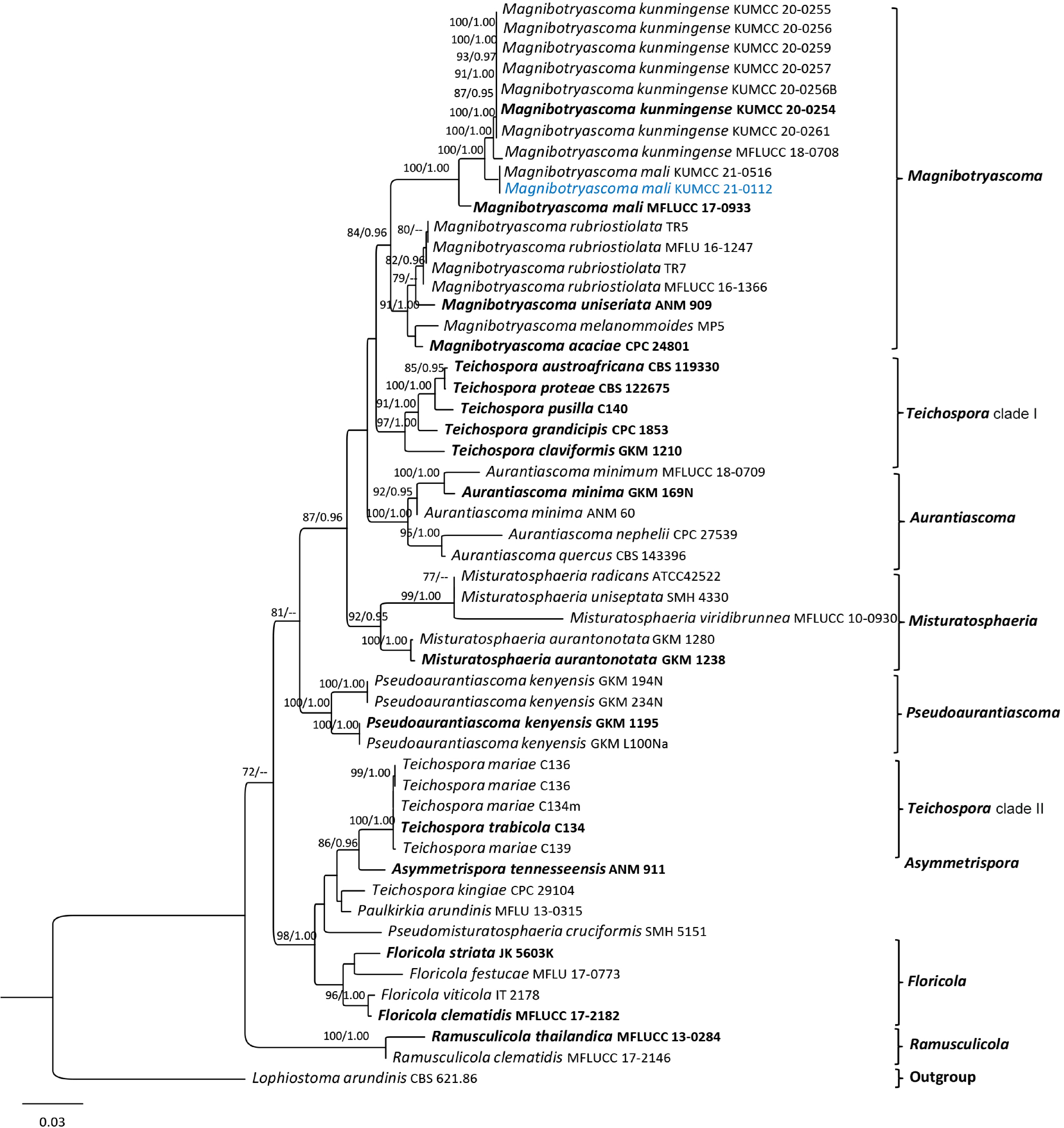
Figure 7.
Phylogram generated from maximum likelihood analysis based on LSU, SSU, ITS, tef1, and rpb2 sequence data. Bootstrap values for ML equal to or greater than 70% and BYPP equal to or greater than 0.95 (rounded to two decimal places) from the BI analysis are labeled on the nodes. A newly generated sequence is in blue, and type strains are bold.
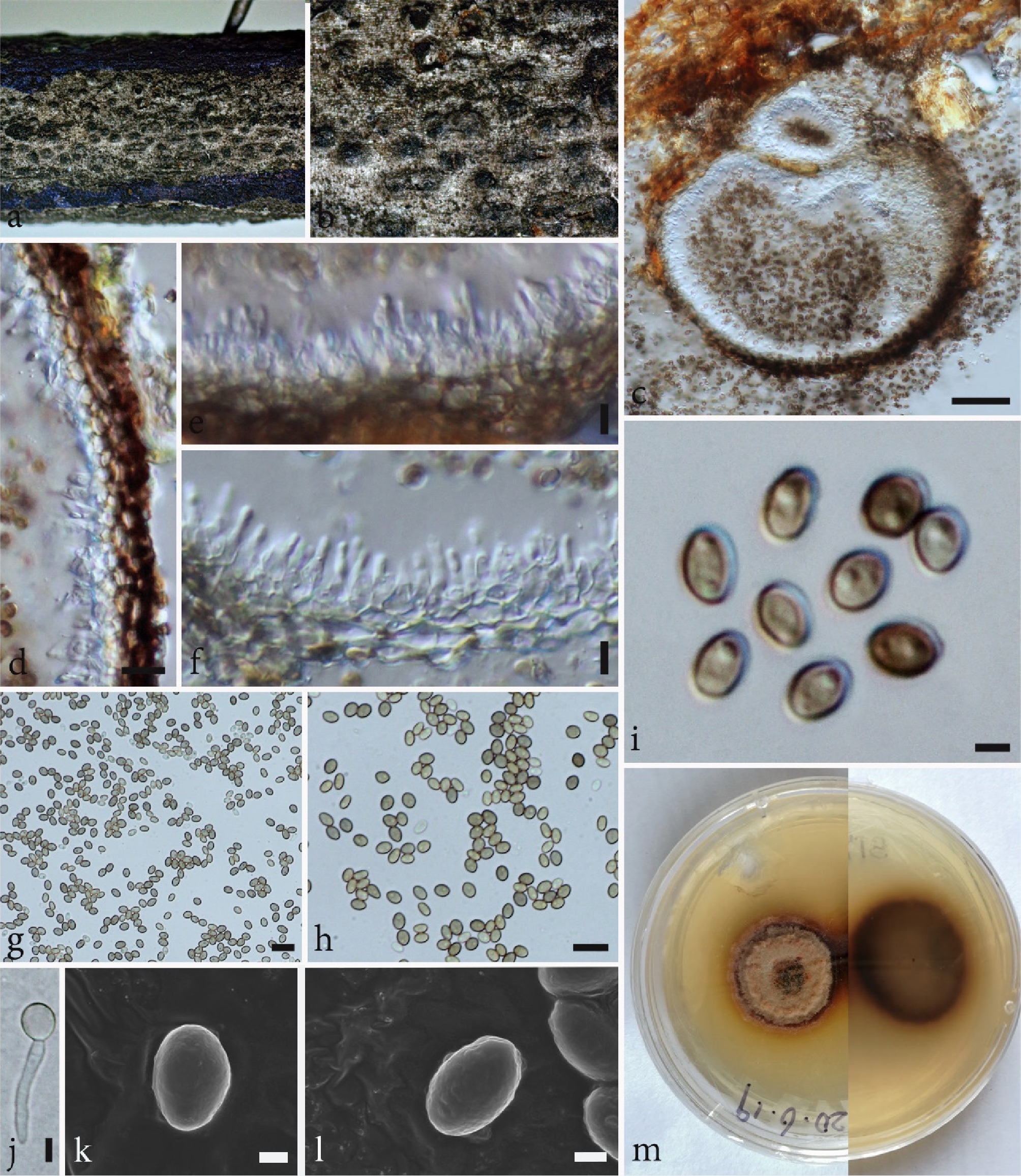
Figure 8.
Magnibotryascoma mali (KUN-HKAS 115583). (a), (b) Conidiomata observed on the host surface. (c) Horizontal section of a conidioma. (d) Pycnidial wall. (e), (f) Conidiogenous cells. (g)–(i), (k), (l) Conidia; (k), (l) SEM photos. (j) Germinated conidium. Scale bars: (c) = 50 µm; (d) = 20 µm; (e)–(h) = 10 µm; (i), (k), (l) = 1 µm; (j) = 2 µm.
Saprobic on dead branches of an unidentified tree species. Sexual morph: Undetermined. Asexual morph: Coelomycetous. Conidiomata 147–182 μm high, 203–280 μm diam. (
$\overline x $ $\overline x $ $\overline x $ Culture characteristics: Colonies grow on PDA at 20 °C. Colonies after 14 d dense, circular, slightly raised, rough surface, with curled or scalloped entire margin, radiated, with four layers in surface view (Fig. 8m), pale yellowish to brown at the margin, paler towards the center, separated by pale-grey to dark-grey concentric ring near the margin, with yellow pigment diffusing in the agars, and pale to dark brown in reverse.
Known host and distribution: On decayed twigs of Malus halliana in Yunnan, China[99], on a dead branch of Metrosideros sp. in New Zealand[100], on dead branches of Osmanthus fragrans in Sichuan, China[101], on dead branches of unknown species in Yunnan, China (this study).
Material examined: China, Yunnan, Kunming, Kunming Institute of Botany, Chinese Academy of Science, on dead branches of unknown species, 09 June 2020, S. Tibpromma, ST 93 (KUN-HKAS 115583), living culture KUMCC 21-0112.
GenBank accession numbers: KUMCC 21-0112: SSU: PV942133; ITS: PV873201; LSU: PV942123; tef1: PX055883; rpb2: PX055872.
Notes: In multigene phylogenetic analyses, our new strain clustered with the strain of Magnibotryascoma mali (KUMCC 21-0516) with 100% MLBS/1.00 BYPP statistical support (Fig. 7). In the NCBI BLASTn search based on ITS sequence, the closest match of our new strain is M. mali (KUMCC 21-0516) with 100% similarity; tef1 sequence closest match with M. mali (GZAAS 23-0648) with 99.70% similarity; rpb2 sequence closest match with M. mali (MFLUCC 17-0933) with 98.58% similarity. Based on the phylogenetic tree, our strain clustered with M. mali (KUMCC 21-0516) while sister to the extype of M. mali (MFLUCC 17-0933) (Fig. 7). The morphology of our new strain matched the description by Ren et al.[62], and also matches with the holotype of M. mali (MFLU 17-0559), which was introduced from decayed twigs of Malus halliana (Rosaceae) in Yunnan, China[99].
According to the phylogenetic tree of Ren et al.[62] and Chethana et al.[101], their isolates of M. mali are related to M. mali (MFLUCC 17-0933, ex-type) but do not group together with the results of our phylogenetic analyses. Our M. mali does not group with the ex-type of M. mali (MFLUCC 17-0933) (Fig. 7). Therefore, with this conspecific status, further taxonomic work is needed to resolve identification, phylogenetic position, and relationships between M. mali members.
Sordariomycetes O.E. Erikss. & Winka
Sordariomycetidae O.E. Erikss. & Winka
Amphisphaeriales D. Hawksw. & O.E. Erikss.
Apiosporaceae K.D. Hyde, J. Fröhl., Joanne E. Taylor & M.E. Barr
Apiospora Sacc.
The genus Apiospora was introduced by Saccardo[102], with A. montagnei as the type species. Members of this genus can survive as endophytes, saprobes, or pathogens, which have a wide range of distribution in different habitats such as animal tissues, seaweeds, and even oligotrophic environments (lichens, air, and soil)[103−105]. The genus was reported with both its sexual and asexual morphs. Sexual morphs are characterized by multi-locular stromata and 1-septate (near the lower cell) ascospores[106]. Asexual morphs were characterized by dark-brown conidia, with a longitudinal, transparent slit or hyaline conidiophores, basauxic conidiogenous cells, and pale-brown to brown conidia[104−107]. In this study, we collected known species A. yunnana as saprobic on dead bamboo culms in the KIB garden, and this is the third time that this species has been found on bamboo in China.
Apiospora yunnana (D.Q. Dai & K.D. Hyde) Pintos & P. Alvarado, Fungal Systematics and Evolution 7: 207 (2021)
Index Fungorum number: IF837739, Figs 9, 10
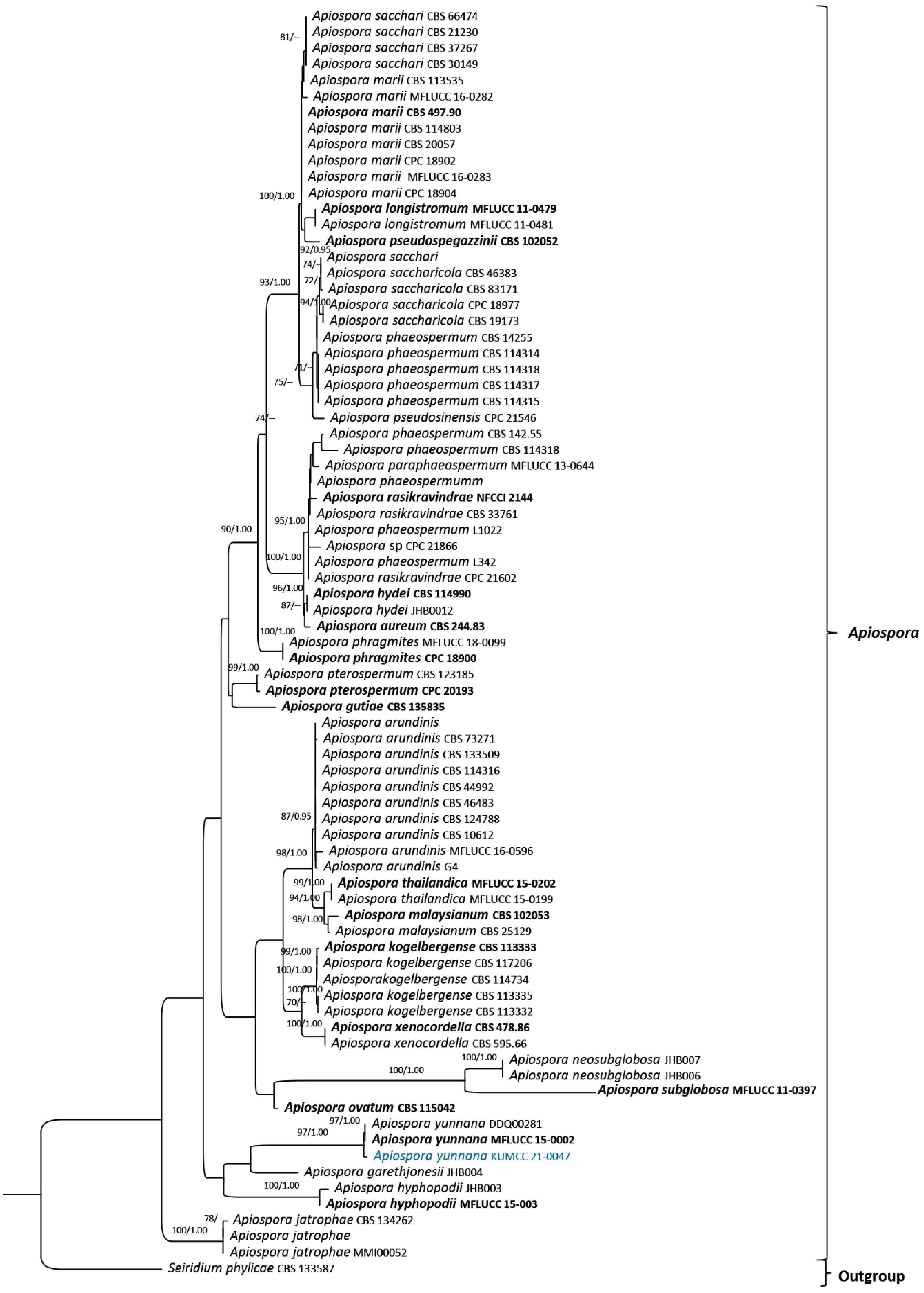
Figure 9.
Phylogram generated from Maximum Likelihood analysis based on ITS sequence data representing Apiospora. Bootstrap values for ML equal to or greater than 70% and BYPP values greater than 0.95 (rounded to two decimal places) from the BI analysis labelled on the nodes. A newly generated sequence is in blue, and ex-type strains are in bold.
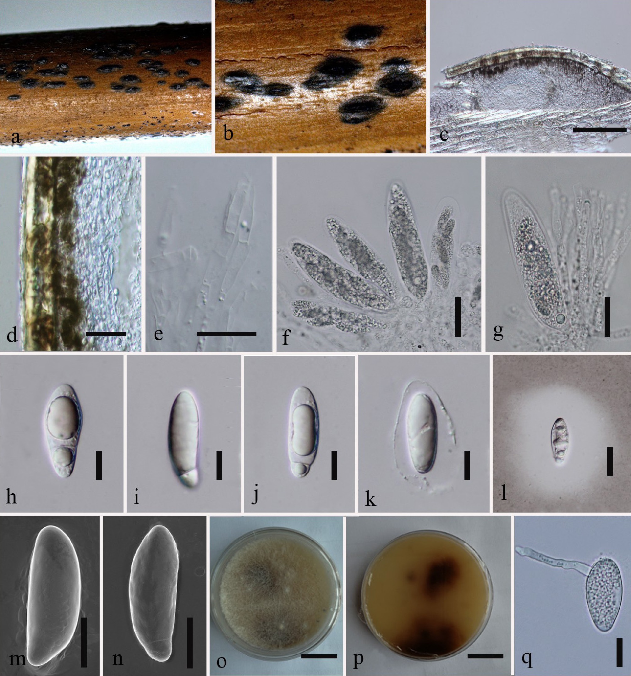
Figure 10.
Apiospora yunnana (HKAS 115564). (a), (b) Ascomata on host surface. (c) Vertical section of ascomata. (d) Peridium. (e) Paraphyses. (f), (g) Asci. (h)–(n) Ascospores; (l) ascospore with mucilaginous sheath in India ink; (m), (n) SEM photos. (o) Upper view on PDA. (p) Reverse view on PDA. (q) Germinated ascospore. Scale bars: (c) = 100 µm; (d), (l) = 20 cm; (e), (h)–(k), (m), (n), (q) = 10 µm; (f), (g) = 30 µm; (o), (p) = 2 cm.
≡ Arthrinium yunnanum D.Q. Dai & K.D. Hyde, in Dai, Phookamsak, Wijayawardene, Li, Bhat, Xu, Taylor, Hyde & Chukeatirote, Fungal Diversity 82: 69 (2016)
Saprobic on dead bamboo culms. Sexual morph: Ascostromata 0.6–1.2 mm long, 0.4–0.6 mm wide, 0.2–0.3 mm high, solitary, or occasionally with two, gregarious, immersed, subepidermal, fusiform to ellipsoid, black, with a long-slit opening at the top. Ascomata 126–150 μm diam. × 123–183 μm high in section, peritheciod, subglobose, light brown to dark brown, with a distinct ostiole at the centre, crowned by a black clypeus. Peridium laterally 30–46 μm thick, composed of brown and small light brown to reddish brown to hyaline elongated cells of textura angularis, surrounded by 100–300 μm stromatic tissues, composed of small cells of textura angularis. Hamathecium composed of dense, 3–5 μm broad, septate, unbranched, paraphyses. Asci 85–92 × 17–25 μm (
$\overline x $ $\overline x $ Culture characteristics: Ascospores germinate on PDA within 24 h, and germ tubes develop from the upper part. Colonies fast growing on PDA at 25 °C, sparse, circular, with irregular edges, flattened, hairy to feathery, white from above, brown to dark brown in the centre from below. Mycelium is superficial to immersed in medium, with branched, septate, smooth hyphae.
Known host and distribution: On dead culms of Phyllostachys nigra in Yunnan, China[106], on decaying culms of Phyllostachys heteroclada in Sichuan, China[108], on dead culms of bamboo (Poaceae) in Yunnan, China (this study).
Material examined: China, Yunnan, Kunming, Kunming Institute of Botany, Chinese Academy of Science, on dead culms of bamboo (Poaceae), 15 May 2020, S. Tibpromma, ST 55 (KUN-HKAS 115564), living culture KUMCC 21-0047.
GenBank accession numbers: KUMCC 21-0047: SSU: PV942130; ITS: PV873195; LSU: PV942117; tub2: PX055889.
Notes: Apiospora yunnana was described by Dai et al.[106] based on a specimen collected in Yunnan Province, China. This species occurred on dead culms of Phyllostachys nigra (black bamboo, Poaceae)[106]. Our new strain (KUMCC 21-0047) groups within the type strain of A. yunnana, with statistical support 97% MLBS/1.00 BYPP (Fig. 9). The new collection has similar ascospore, ascus, and ascocarp morphology to the type strain. Therefore, the strain collected from the same place and host group (bamboo) is assigned to A. yunnana.
Chaetosphaeriales Huhndorf, A.N. Mill. & F.A. Fernández
Chaetosphaeriaceae Réblová, M.E. Barr & Samuels
Pseudolachnella Teng
Pseudolachnella, a genus segregated from Pseudolachnea, was established to accommodate species with multiseptate conidia, with P. scolecospora as the type species[109]. Under the genus Pseudolachnella, 23 species are accepted, and for many of them, ITS, LSU, and tef1 sequences are available[42,65]. Members of this genus have been reported only as asexual morphs and characterized by stromatic, acervular, setose conidiomata with marginal setae, phialidic conidiogenous cells, and conidia with single to multiple appendages at each end[110,111]. Dead bamboo substrates from China, India, and Japan are common hosts for this genus[111,112]. In this study, we introduce the sexual morph of P. tengii, the first for this genus.
Pseudolachnella tengii W.P. Wu & Y.Z. Diao, Fungal Diversity 116: 501 (2022)
Index Fungorum Number: IF841674, Figs 11, 12
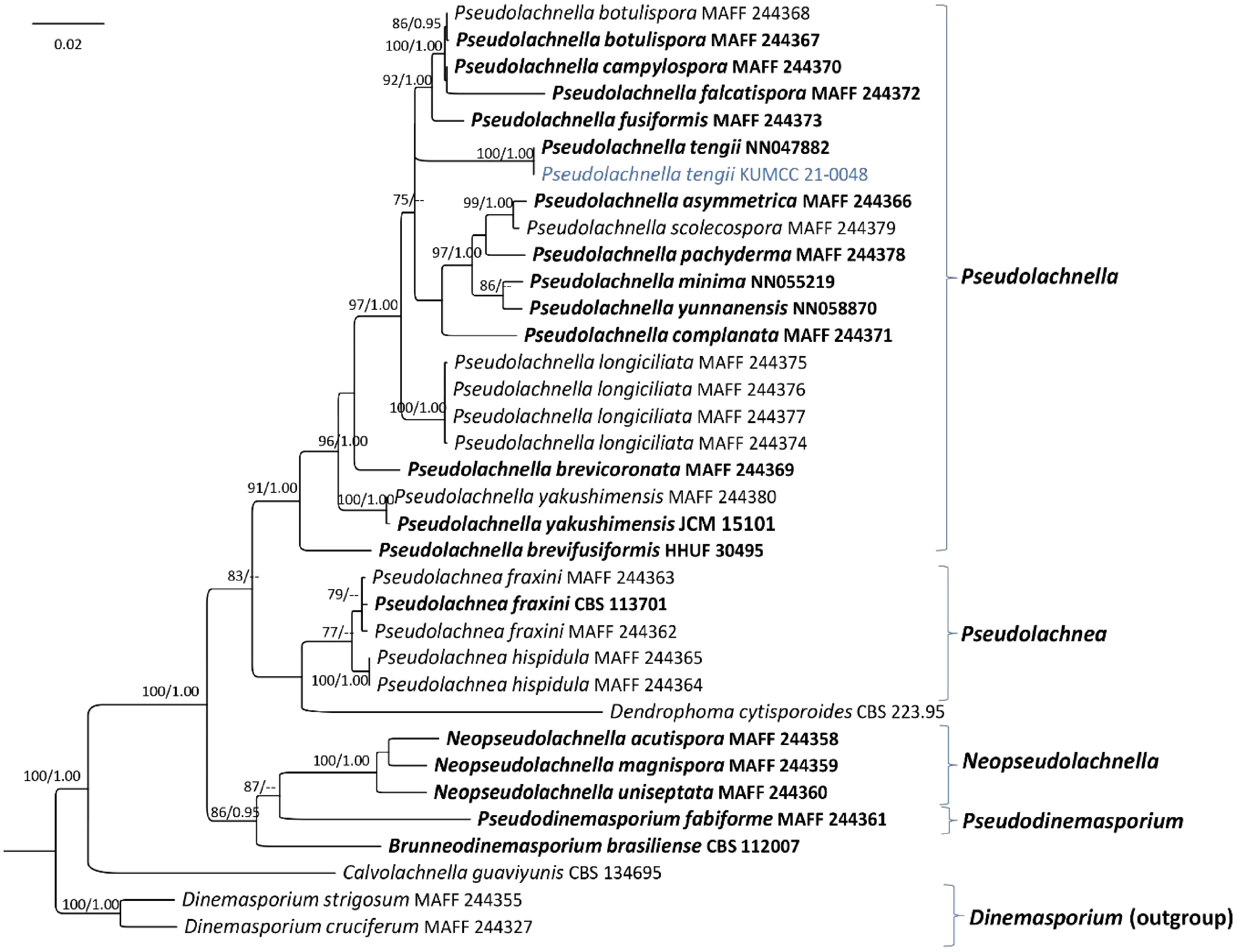
Figure 11.
Phylogram generated from Maximum Likelihood analysis based on LSU, ITS, and tef1 sequence data representing Pseudolachnella and its relatives. Bootstrap values for ML equal to or greater than 70% and BYPP values equal to or greater than 0.95 (rounded to two decimal places) from the BI analysis are labelled on the nodes. A newly generated sequence is in blue, and ex-type strains are in bold.
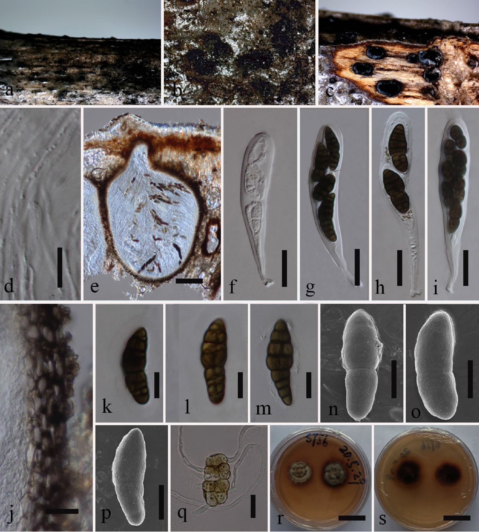
Figure 12.
Pseudolachnella tengii (KUN-HKAS 115565). (a)–(c) Ascomata on host surface. (d) Paraphyses. (e) Vertical section of ascoma. (f)–(i) Asci. (j) Peridium. (k)–(p) Ascospores; (n)–(p) SEM photos. (q) Germinated ascospore. (r) Upper view on PDA. (s) Reverse view on PDA. Scale bars: (d) = 10 µm; (e) = 100 cm; (f)–(j) = 20 µm; (k)–(q) = 10 µm; (r), (s) = 2 cm.
Saprobic on a dead branch of a liana. Sexual morph: Ascomata 432–441 × 420–445 μm, scattered, solitary, immersed, irregular in shape, globose to subglobose, uniloculate, with a short central ostiole. Peridium 15–35 μm thick, thin-walled, of equal thickness, composed of 5–7 layers of flattened, dark brown, pseudoparenchymatous cells of textura angularis, with paler brown to hyaline towards the inner layer. Hamathecium 1.2–2.0 μm wide, filamentous, septate, branched, long, hyaline paraphyses. Asci 150–210 × 20–37 μm (
$\overline x $ $\overline x $ Culture characteristics: Ascospores germinated within 12 h on PDA, colonies grown on PDA at room temperature (20–25 °C). Colonies circular to irregular, dull surface and wrinkled, yellow to dark brown, dark brown on the reverse, producing a yellow-orange pigment on PDA.
Known host and distribution: On the dead culm of Phyllostachys sp. in Yunnan, China[64], on a dead branch of a liana in Yunnan, China (this study).
Material examined: China, Yunnan, Kunming, Kunming Institute of Botany, Chinese Academy of Science, on a dead branch of a liana, 26 May 2020, S. Tibpromma, ST 56 (KUN-HKAS 115565), living culture KUMCC 21-0048.
GenBank accession numbers: KUMCC 21-0048: ITS: PV873196; LSU: PV942118; tef1: PX055878; rpb2: PX055867.
Notes: In the phylogenetic analysis, the new isolate formed a sister group with Pseudolachnella tengii (NN047882T), with 100% MLBS and 1.00 BYPP statistical support (Fig. 11). Pseudolachnella tengii was introduced as an asexual morph, and the genus Pseudolachnella comprised only asexual morph species[64,110]. Therefore, morphological comparison is impossible as our new isolate is a sexual morph. As well, we are unsuccessful in getting the asexual morph from the culture. Based on nucleotide comparisons between our isolates and P. tengii (NN047882T), the ITS and LSU sequences are identical, while tef1 and rpb2 are available for P. tengii (NN047882T). In this study, we introduced the new isolate as the sexual morph of P. tengii, and both the holotype (HMAS 352045)[64] and our new isolate (HKAS 115565) were collected from the KIB in Kunming, Yunnan, China.
Distoseptisporales Z.L. Luo, H.Y. Su & K.D. Hyde
Distoseptisporaceae K.D. Hyde & McKenzie
Distoseptispora K.D. Hyde, McKenzie & Maharachch
The genus Distoseptispora is one of the Sporidesmium-like genera introduced by Su et al.[113], with the type species, D. fluminicola, isolated from decaying wood in Yunnan, China. Distoseptispora, as a single genus within Distoseptisporaceae, occurs mainly as asexual morphs, while only two species have been reported as sexual morphs, viz. D. hyalina and D. licualae[114,115]. Species of this genus were found forming effuse, hairy colonies on decaying wood, plant stems, bamboo culms, and fallen leaves and shafts in terrestrial and freshwater habitats, mostly reported from Asia (mainly in China and Thailand)[31,116−119]. Furthermore, members of this genus show macronematous, mononematous, septate, unbranched, cylindrical, olivaceous to brown conidiophores; mono- or polyblastic, integrated, terminal, cylindrical, determinate, or percurrently extending conidiogenous cells; and acrogenous, solitary, smooth or verruculose, euseptate or distoseptate conidia[31]. In this study, we found known species of Distoseptispora (D. guizhouensis) from an unidentified plant in the KIB garden.
Distoseptispora guizhouensis X. Tang, Jayaward., J.C. Kang & K.D. Hyde, in Hyde et al., Mycosphere 12(1): 1126 (2021)
Index Fungorum number: IF558532, Figs 13, 14
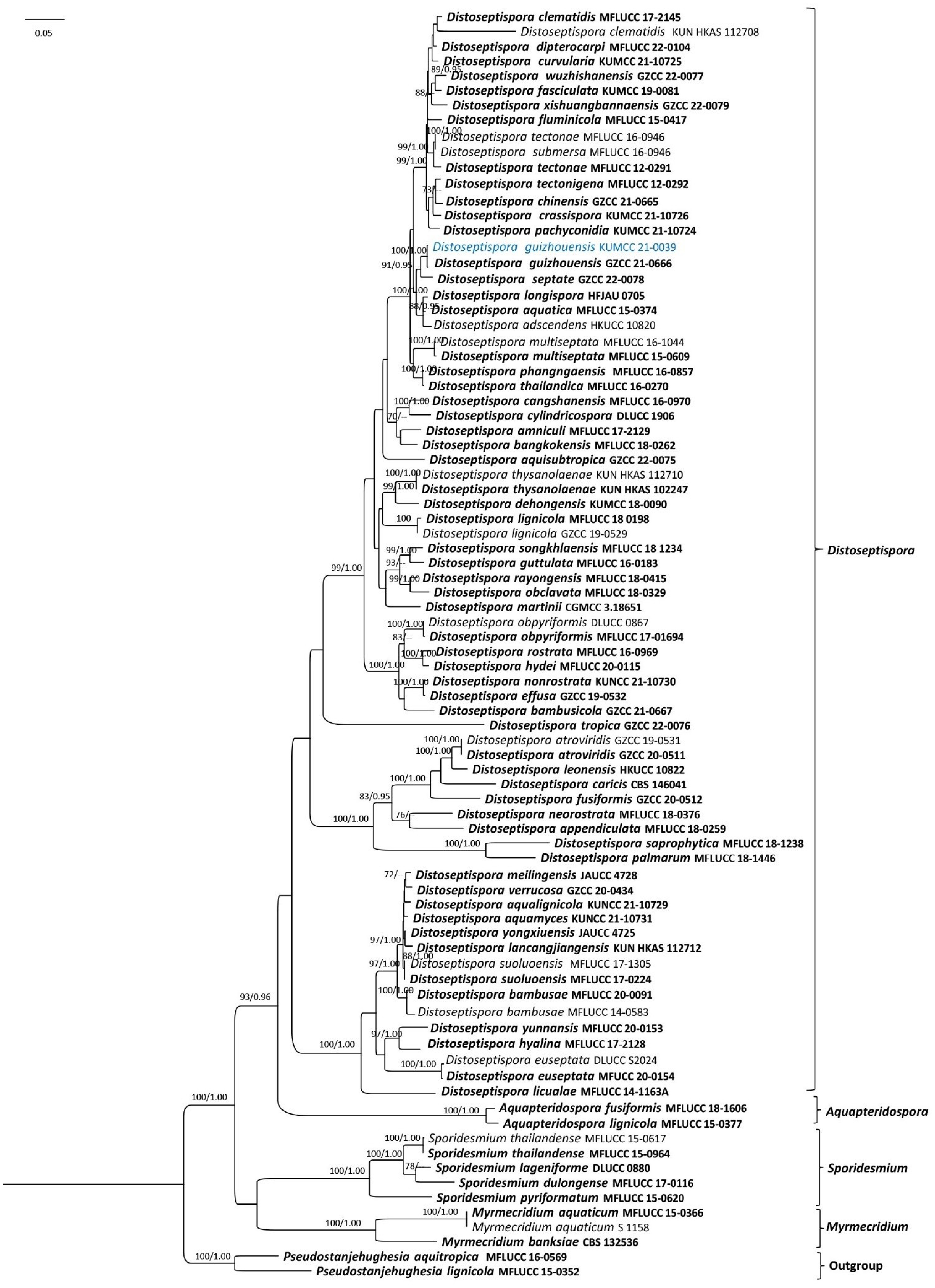
Figure 13.
Phylogram generated from Maximum Likelihood analysis based on ITS, LSU, tef1, and rpb2 sequence data representing Distoseptispora species. Bootstrap values for ML equal to or greater than 70% and BYPP values equal to or greater than 0.95 (rounded to two decimal places) from the BI analysis are labelled on the nodes. A newly generated sequence is in blue, and type species are in bold.
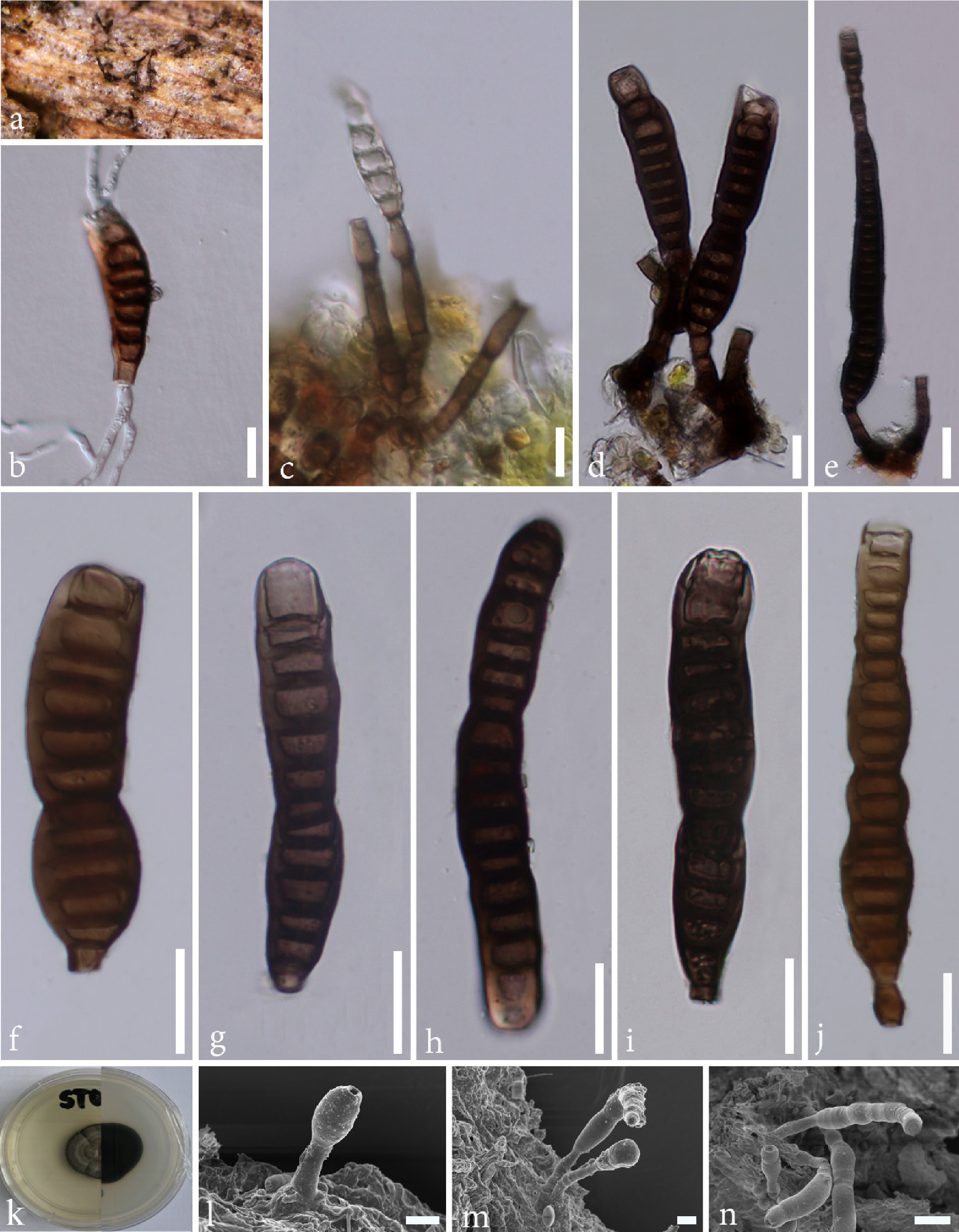
Figure 14.
Distoseptispora guizhouensis (KUN-HKAS 115537). (a) Colonies on host surface. (b) Germinated conidium. (c)–(e) Conidiophores, conidiogenous, and developing conidia. (f)–(j) Conidia. (k) Colony on PDA. (l)–(n) SEM photographs of conidiogenous cells and conidia. Scale bars: (b)–(e), (l), (m) = 10 μm; (f)–(j), (n) = 20 μm.
Saprobic on decaying branch of an unidentified plant. Sexual morph: Undetermined. Asexual morph: Hyphomycetous. Colonies on wood effuse, hairy, dark brown, scattered or in small groups, glistening, usually setiform. Mycelium is partly immersed, partly superficial, composed of septate, smooth-walled, pale-brown to hyaline hyphae. Conidiophores 21–44 × 3.8–4.9 μm (
$\overline x $ $\overline x $ Culture characteristics: Conidia germinate on PDA within 24 h, and germ tubes are produced from both ends. Colonies growing on PDA, slow growing after 1 month at 25 °C in natural light; circular, with dense, three-layered (forward grey, white, and grey, respectively), and a dark-brown margin. In reverse, dark brown to black with a smooth margin.
Known host and distribution: Decaying wood in Guizhou[61] and Yunnan, China (this study).
Material examined: China, Yunnan, Kunming, Kunming Institute of Botany, Chinese Academy of Science, decaying branch of unknown plant, 6 May 2020, S. Tibpromma, ST03 (KUN-HKAS 115537); living culture KUMCC 21-0039.
GenBank accession numbers: KUMCC 21-0039: SSU: PV948969; ITS: PV873190; LSU: PV951539; tef1: PX055874; rpb2: PX055863.
Notes: Our new isolate is well supported together with Distoseptispora guizhouensis (GZCC 21-0666T) (100% MLBS/ 1.00 BYPP, Fig. 13). Also, it shares similar morphology with the holotype of D. guizhouensis (GZAAS21-0381) in having macronematous, mononematous, erect, cylindrical conidiophores, monoblastic, integrated, terminal, determinate, cylindrical, brown conidiogenous cells, and acrogenous, obclavate, distoseptate, brown to dark-brown conidia[61]. Conidiophore and conidia sizes of our new isolate (KUN-HKAS 115537) and holotype (GZAAS21-0381[61]) are shown with the conidiophore overlap size (21–44 × 3.8–4.9 μm vs 21–50 × 4–9 μm), but the holotype conidia are bigger than our isolate (73–184 × 10–19 μm vs 90–273 × 15–21). Based on nucleotide comparisons, two base pairs differ in the ITS region between our isolates and D. guizhouensis (GZCC 21-0666, ex-type), whereas the LSU and tef1 sequences are identical. Here, we report the new collection of D. guizhouensis in Yunnan, China, accompanied by SEM photographs of its conidia for the first time. However, the species name was corrected in the original as D. guizhouensis, but in Index Fungorum and MycoBank, it was recorded as D. guizhounesis. Therefore, it will need to be corrected in the future.
Xylariales Nannf.
Diatrypaceae Nitschke
Peroneutypa Berl.
Peroneutypa is characterized by long, prominent necks, with small, truncate apices, urn-shaped asci, and allantoid ascospores[120−122]. Li et al.[123] accepted Peroneutypa with 27 species, but approximately 76 epithets are listed in Index Fungorum[41]. Members of Peroneutypa are saprobes or pathogens and are widely distributed in terrestrial and marine habitats[124−128]. In our study, we identified the known species Peroneutypa (P. scoparia) using morphological and phylogenetic data. In addition, this is the first time P. scoparia has been found on Trachycarpus sp. in Yunnan Province, China.
Peroneutypa scoparia (Schwein.) Carmarán & A.I. Romero
Index Fungorum number: IF500713; Facesoffungi number: FoF 01998, Figs 15, 16
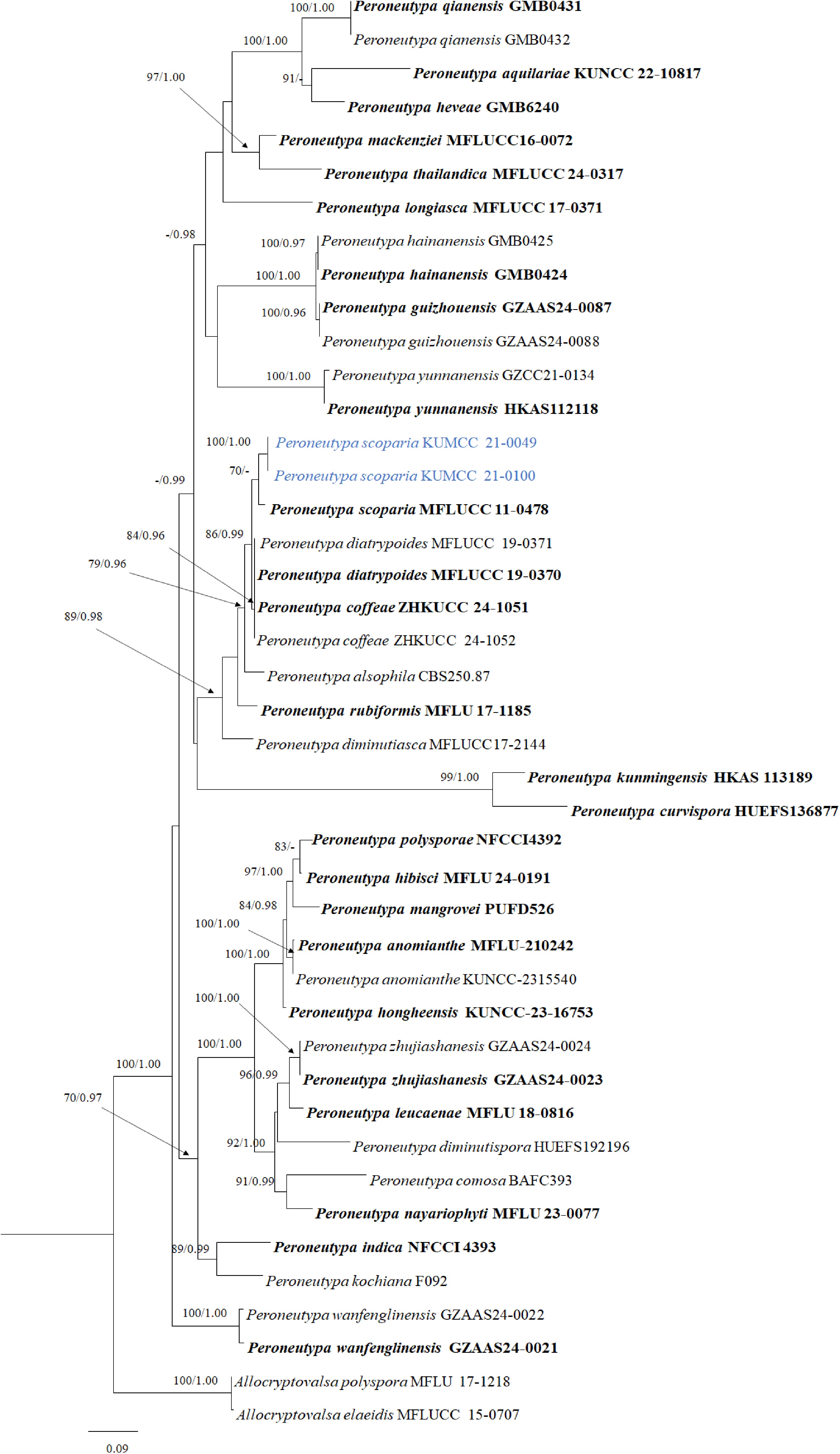
Figure 15.
Phylogram generated from Maximum Likelihood analysis based on ITS and tub2 sequence data representing Peroneutypa. Bootstrap values for ML equal to or greater than 70% and BYPP values equal to or greater than 0.95 (rounded to two decimal places) from the BI analysis are labeled on the nodes. The newly generated sequences are in blue, and the type species and the reference specimens are in bold.
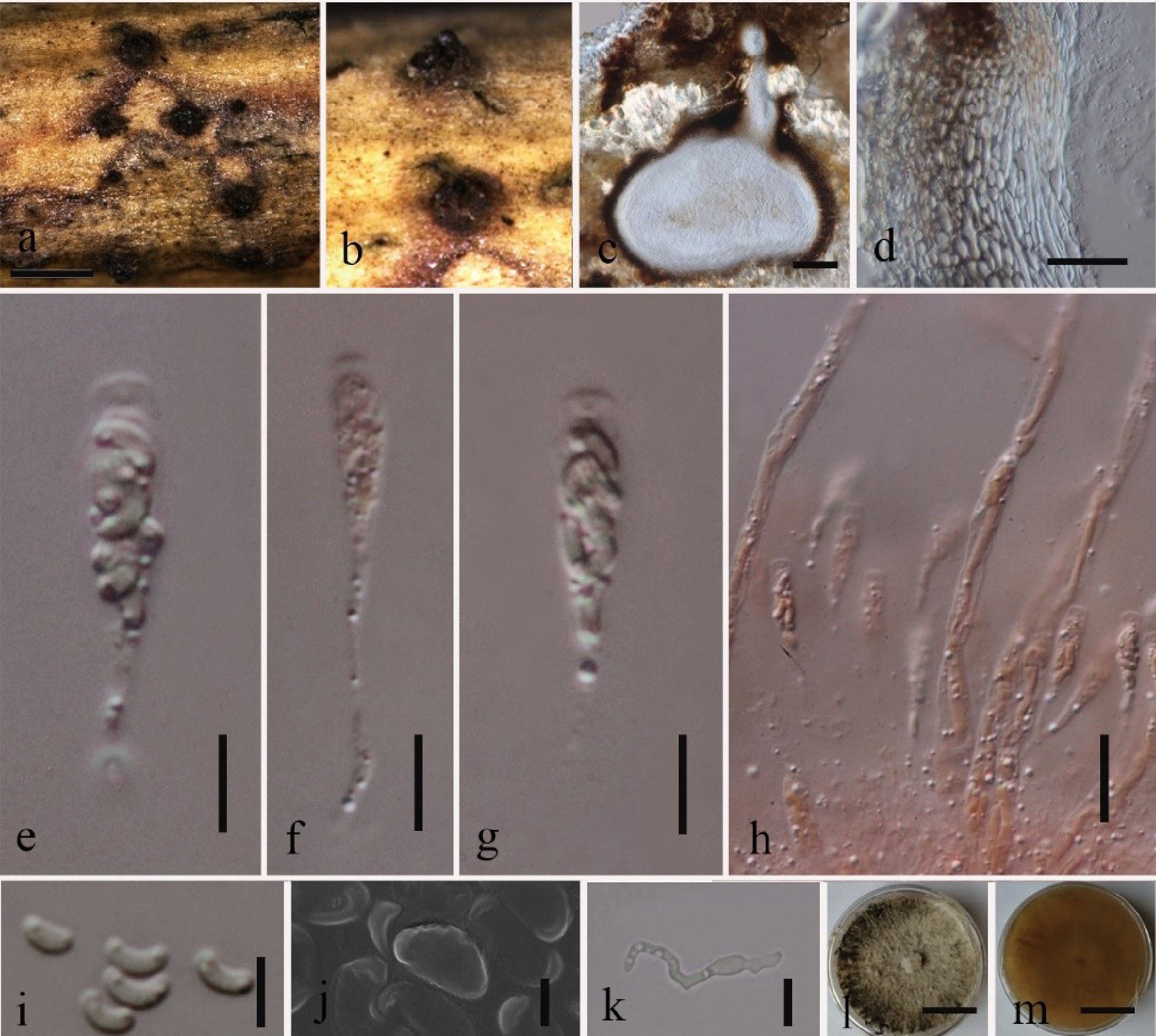
Figure 16.
Peroneutypa scoparia (HKAS 115568). (a), (b) Ascomata on host surface. (c) Vertical section of ascoma. (d) Peridium. (e)–(g) Asci (stained with Congo red). (h) Paraphyses (stained with Congo red). (i), (j) Ascospores; (j) SEM photo. (k) Germinated ascospore. (l) Upper view on PDA. (m) Reverse view on PDA. Scale bars: (a) = 500 µm; (c) = 50 µm; (d) = 10 cm; (e)–(i), (k) = 5 µm, (j) = 2 µm; (l), (m) = 2 cm.
Synonyms:
=Sphaeria scoparia Schwein.
=Valsa scoparia (Schwein.) M.A. Curtis.
=Eutypella scoparia (Schwein.) Ellis & Everh.
=Peroneutypella scoparia (Schwein.) Berl.
=Eutypa scoparia (Schwein.) L.C. Tiffany & J.C. Gilman.
=Valsa ceratophora sensu Cooke.
=Valsa heteracantha Sacc.
=Eutypa heteracantha (Sacc.) Sacc.
=Engizostoma heteracanthum (Sacc.) Kuntze.
=Peroneutypa heteracantha (Sacc.) Berl.
=Peroneutypella heteracantha (Sacc.) Rehm.
=Peroneutypella heteracantha f. berberidis Rehm.
=Peroneutypa heteracantha f. arundinis-donacis Speg.
=Peroneutypa heteracantha f. ricini-communis Speg.
=Peroneutypa heteracantha f. casuarinae-strictae Speg.
=Peroneutypa heteracantha f. annonae-cherimoliae Speg.
Saprobic on dead petioles of Trachycarpus sp. Sexual morph: Ascomata perithecial, 350–450 μm diam., 150–170 μm high (
$\overline x $ $\overline x $ $\overline x $ $\overline x $ Culture characteristics: Ascospores germinate on PDA within 24 h, and germ tubes are produced from both ends. Colonies after 1 week white, cottony, with ropy hyphal strands near the centre with diffuse margins; reverse coloration pale yellow; after 1 month, grey to dark-brown radial strands of aerial hyphae, reverse yellow to brown.
Known host and distribution with molecular data: on leaves of Garcinia dulcis from Songkhla, Thailand[105], on dead culms of bamboo and on dead wood of Dalbergia cana, Microcos paniculate, and Afzelia xylocarpa from Chiang Rai, Thailand[130,131], on living branches of Broussonetia papyrifera from Argentina[7], on unknown bark from Chiang Mai, Thailand[129], on diseased dieback marine wood from Phetchaburi, Thailand[132], and on dead petioles of Trachycarpus sp. and unknown plants from Yunnan, China (this study).
Material examined: China, Yunnan, Kunming, Kunming Institute of Botany, Chinese Academy of Science, on dead petioles of Trachycarpus sp. (Arecaceae), 28 June 2020, Tibpromma, ST 63 (KUN-HKAS 115568), living culture KUMCC 21-0049; ibid., on dead branches of an unidentified plant, 1 May 2020, S. Tibpromma, ST 40 (KUN-HKAS 115766), living culture KUMCC 21-0100.
GenBank accession numbers: KUMCC 21-0049: SSU: PV942131; ITS: PV873198; LSU: PV942120; tef1: PX055880; tub2: PX055890; rpb2: PX055869. KUMCC 21-0100: SSU: PV942127; ITS: PV873192; LSU: PV942114; tef1: PX055876; tub2: PX055886; rpb2: PX055865.
Notes: Phylogenetic analyses of a combined ITS and tub2 sequence dataset showed that our strains (KUMCC 21-0049 and KUMCC 21-0100) cluster with Peroneutypa scoparia (MFLUCC 11-0478) with 70% MLBS statistical support (Fig. 15). Our sample differs slightly from P. scoparia (MFLU 15–1183, reference specimen) by having a relatively small ostiolar canal (130–180 × 48–60 μm vs 195–530 × 330–720 μm). Therefore, the combined ITS-tub2 phylogeny and morphological features support our isolates as P. scoparia. In addition, this is the first time P. scoparia has been found in China, and the first time SEM images of its ascospores have been provided.
Sordariomycetes genera incertae sedis
Melanographium Sacc.
This genus, typified by M. selenioides[133], is characterized by dark and unbranched conidiophores generally arising in a group from an immersed stroma with polyblastic conidiogenous cells and sympodial proliferation[134−136]. Conidia are pigmented, single-celled, frequently reniform in shape, and often with a hyaline germ slit[134−136]. Species are distinguished by the length of the conidiophores, the pattern of conidiophore grouping (single, synnemata, loosely or dense fascicles), conidial shape (straight, curved, or strongly curved), and conidial size[136]. In the Index Fungorum[41], 19 species have been recorded, but only a few species have molecular data. In this study, we introduce a new species based on both molecular and morphological data.
Melanographium trachycarpi Jayasiri, Tibpromma & Karun., sp. nov.
Index Fungorum number: IF905409; Facesoffungi number: FoF 18869, Figs 17, 18
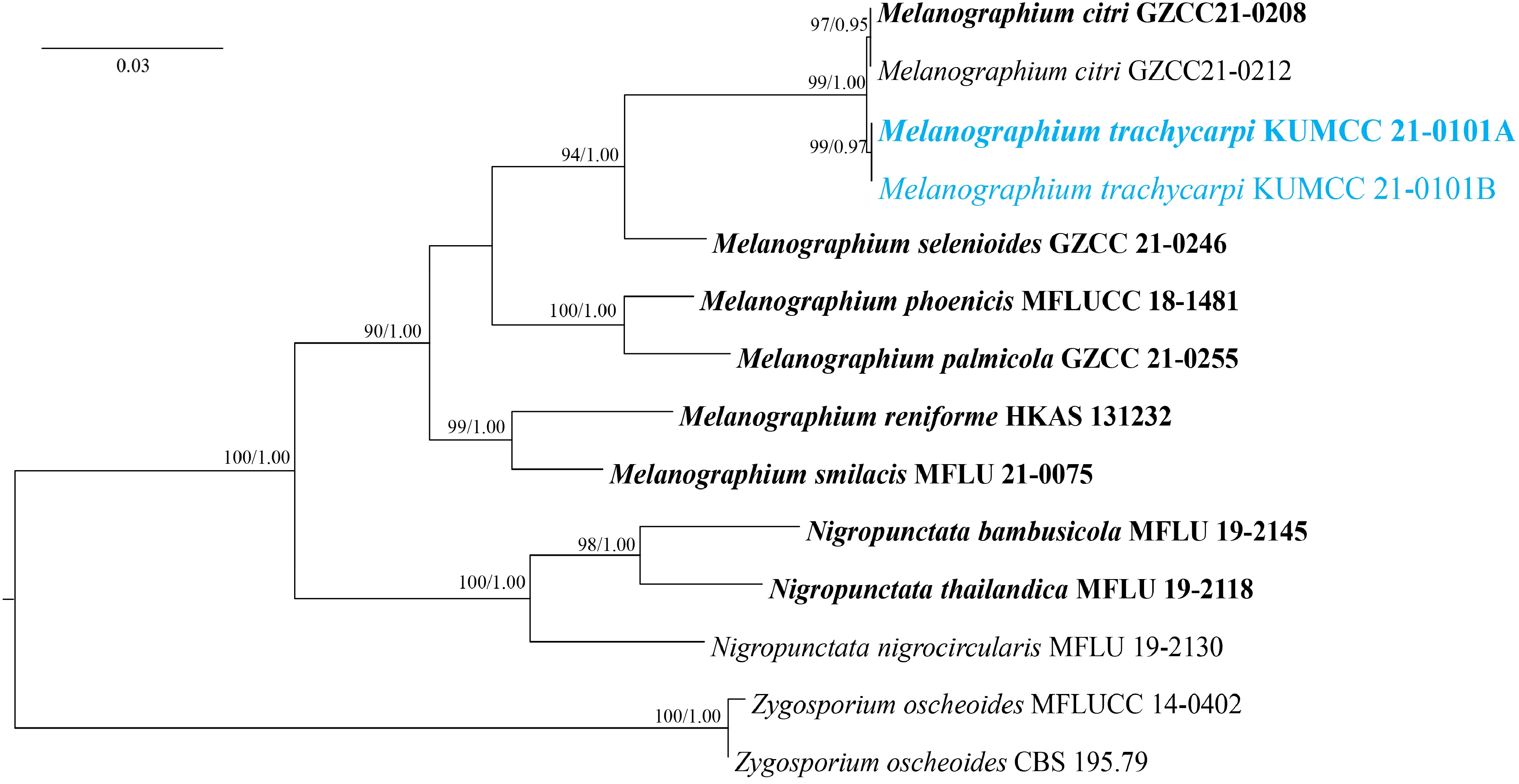
Figure 17.
Phylogram generated from Maximum Likelihood analysis based on LSU, ITS, and rpb2 sequence data representing Melanographium and closely related genera. Bootstrap values for ML equal to or greater than 70% and BYPP values equal to or greater than 0.95 (rounded to two decimal places) from the BI analysis are labelled on the nodes. The newly generated sequences are in blue, and type species are in bold.
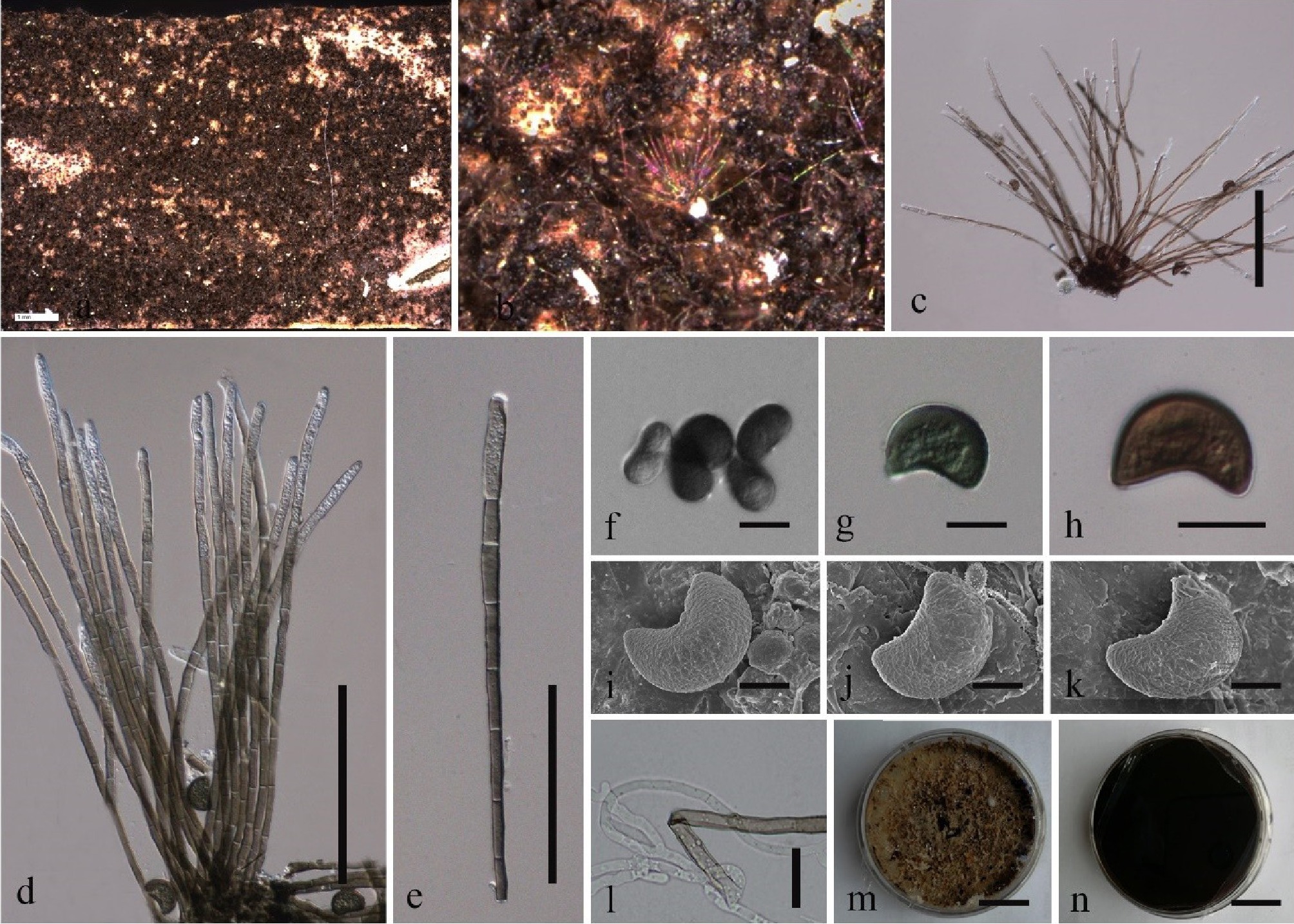
Figure 18.
Melanographium trachycarpi (KUN-HKAS 115558, holotype). (a), (b) Colonies on the host surface. (c)–(e) Conidiophores. (f)–(k) Conidia; (i)–(k) SEM photos. (l) Germinated conidiophore. (m) Upper view on PDA. (n) Reverse view on PDA. Scale bars: (c), (d), (e) = 100 µm; (f)–(h), (l) = 10 cm; (i)–(k) = 5 µm; (m), (n) = 2 cm.
Etymology: The specific epithet 'trachycarpi' refers to the host plant genus, Trachycarpus.
Holotype: KUN-HKAS 115558
Saprobic on dead petioles of Trachycarpus sp. Sexual morph: Undetermined. Asexual morph: Colonies on natural substrate superficial, effuse, velvety, tufted, dark brown. Mycelium immersed, composed of branched, septate, smooth, brown hyphae. Conidiophores 115–268 × 2.7–5.6 μm (
$\overline x $ $\overline x $ Culture characteristics: Conidiophore germinated on PDA, colony circular, flossy, velvety. Colonies after 2 weeks yellow green to green brown, flossy, velvety, entire edge, flat; reverse coloration brown to black.
Material examined: China, Yunnan, Kunming, Kunming Institute of Botany, Chinese Academy of Science, on dead petioles of Trachycarpus sp. (Arecaceae), 15 May 2020, S. Tibpromma, ST 45 (KUN-HKAS 115558, holotype), ex-type living culture KUMCC 21-0101.
GenBank accession numbers: KUMCC 21-0101: SSU: PV942128; ITS: PV873193; LSU: PV942115; tef1: PX055877; tub2: PX055887; rpb2: PX055866.
Notes: Melanographium trachycarpi was found on dead branches of Trachycarpus sp. Melanographium trachycarpi shares common characteristics with other Melanographium species, including effused and tufted colonies, loosely or densely fasciculate conidiophores, and reniform, aseptate, darkly pigmented conidia[96−99,101,102]. Phylogenetically, M. trachycarpi is a sister lineage with two strains of M. citri (GZCC21-0208T, GZCC21-0212) with 99% MLBS/1.00 BYPP statistical support (Fig. 17). However, M. trachycarpi (KUN-HKAS 115558) differs from M. citri (HKAS 115665, reference specimen) by having short conidiophores (115–268 μm long, 2.7–5.6 μm) with olivaceous-brown to dark-brown conidia, reniform or broad obovoid to semicircular and lacking a germ-slit, while M. citri has long conidiophores (130–880 μm long, 20–140 μm) with pale to dark-brown conidia, which are curved, reniform or elliptical, sometimes obovoid, with a subhyaline longitudinal germ-slit[137]. We therefore recognize our collection as a new species, namely M. trachycarpi.
-
At the KIB, there are over 8,000 plant species in the garden, showcasing rich biodiversity[138]. Due to the high diversity of plant species, soil types, and often varied organic inputs, these environments support a wide range of fungi. In a previous study, several fungi were identified and introduced as new or known species, mostly found as saprobic fungi (Table 1[139]). As KIB's garden is a managed garden, containing diverse plant species often harbors beneficial saprobic fungi. Saprobic fungi are well known as decomposers of diverse plant litter, wood, and organic debris. Several hosts and fungi exhibit patterns that are generally non-random, influenced by evolutionary history, ecological specialization, and specific environmental factors. While some fungi are generalists, many form highly specialized relationships with specific host plants, especially within mycorrhizal networks (beneficial) and pathogenic (disease-causing) groups[140−142]. This research study makes a significant contribution to the understanding of fungal diversity by describing two new species previously unreported and seven new host records, highlighting the rich fungal diversity at the KIB, Yunnan, China. These findings are the result of detailed morphological observations combined with multigene phylogenetic analyses (SSU, ITS, LSU, rpb2, tef1, and tub2), confirming the novelty and taxonomic placement of these taxa. Besides introducing them based on morphology and DNA sequences, this study presents SEM photographs of some species for the first time and provides a checklist of fungi from KIB for the years 2010–2025, which may help other researchers interested in fungi at the KIB (Table 1).
Table 1. Checklist of fungi collected from KIB (from 2010–2025).
Species name New taxa/
known taxaHost/substrate Life mode Ref. Ascomycota Alternaria alternata Known Magnolia grandiflora Saprobic Jayasiri et al.[149] Arthrinium yunnanum New Phyllostachys nigra Saprobic Dai et al.[106] Austropleospora keteleeriae New Keteleeria fortunei Saprobic Jayasiri et al.[149] Bartalinia kunmingensis New Zea mays Saprobic Phookamsak et al.[150] Brunneofusispora sinensis Known Magnolia denudata Saprobic Wanasinghe et al.[139] Brunneomurispora lonicerae New Lonicera maackii Saprobic Phookamsak et al.[150] Camporesiomyces mali New Malus halliana Saprobic Hyde et al.[144] Ceratosphaeria yunnanensis New Heteropanox fregans Saprobic Manawasinghe et al.[151] Cladosporium cladosporioides Known Magnolia grandiflora Saprobic Jayasiri et al.[149] Diatrypella yunnanensis New Unidentified plant Saprobic Hyde et al.[144] Diatrypella yunnanensis New Unidentified plant Saprobic Hyde et al.[144] Didymella coffeae-arabicae Known Leucaena sp. Saprobic Jayasiri et al.[149] Didymella magnoliae New Magnolia grandiflora Saprobic Jayasiri et al.[149] Diplodia magnoliigena New Magnolia grandiflora Saprobic Jayasiri et al.[149] Hysterobrevium constrictum Known Unidentified plant Saprobic Hyde et al.[144] Keissleriella caraganae New Caragana arborescens Saprobic Phookamsak et al.[150] Liua muriformis New Lonicera maackii Saprobic Phookamsak et al.[150] Lonicericola hyaloseptispora New Lonicera maackii Saprobic Phookamsak et al.[150] Montagnula aquilariae Known Unidentified plant Saprobic Wanasinghe et al.[152] Montagnula thevetiae New Thevetia peruviana Saprobic Wanasinghe et al.[152] Murilentithecium lonicerae New Lonicera maackii Saprobic Phookamsak et al.[150] Neofusicoccum parvum Known Magnolia grandiflora Saprobic Jayasiri et al.[149] Neoleptosporella camporesiana New Heteropanax fragrans Saprobic Manawasinghe et al.[151] Neosetophoma lonicerae New Lonicera maackii Saprobic Phookamsak et al.[150] Nigrograna magnoliae New Magnolia denudata Saprobic Wanasinghe et al.[152] Nothophoma quercina Known Keteleeria fortunei Saprobic Jayasiri et al.[149] Paratrimmatostroma kunmingensis New Fern Saprobic Phookamsak et al.[150] Paucispora kunmingense New Malus halliana (Rosaceae) Saprobic Hyde et al.[153] Periconia byssoides Known Prunus armeniaca Saprobic Yang et al.[154] Periconia byssoides Known Magnolia grandiflora Saprobic Jayasiri et al.[149] Periconia chimonanthi New Chimonanthi praecox Saprobic Yang et al.[154] Periconia cortaderiae Known Caragana arborescens Saprobic Phookamsak et al.[150] Periconia kunmingensis New Fern Saprobic Phookamsak et al.[155] Phaeopoacea muriformis New unidentified grass species Saprobic Hyde et al.[153] Phaeosphaeria chinensis New Wisteria sp. Saprobic Jayasiri et al.[149] Phaeoseptum mali New Malus halliana Saprobic Phookamsak et al.[156] Phaeosphaeria cycadis New Cycas sp. Leaf spots Phookamsak et al.[150] Phaeosphaeriopsis aloes Known Yucca elephantipes Saprobic Boonmee et al.[157] Phragmocamarosporium hederae Known Cycas Leaf spots Phookamsak et al.[150] Plectosphaerella kunmingensis New Water agar as a contaminated fungus Unknown Phookamsak et al.[150] Plenodomus artemisiae New Artemisia sp. Saprobic Phookamsak et al.[150] Pseudolachnella tengii New Phyllostachys sp. Saprobic Wu & Diao[64] Pleurotheciella dimorphospora New Malus sp. Saprobic Boonmee et al.[157] Pseudochaetosphaeronema kunmingense New Cerasus pseudocerasus Saprobic Hyde et al.[144] Pseudopithomyces chartarum Known Magnolia grandiflora Saprobic Jayasiri et al.[149] Pseudopithomyces kunmingensis New unidentified grass species Saprobic Abreu et al.[153] Rhytidhysteron camporesii New Unidentified plant Saprobic Hyde et al.[144] Sclerostagonospora rosae New Rosa sp. Saprobic Wanasinghe et al.[158] Septoriella neomuriformis New unidentified grass Saprobic Wanasinghe & Mharachchikumbura[159] Shearia formosa Known Magnolia denudata Saprobic Wanasinghe et al.[139] Sparticola muriformis New unidentified grass Saprobic Karunarathna et al.[160] Sphaerellopsis paraphysata Known Liriope spicata Rust on living leaves Phookamsak et al.[150] Stagonosporopsis pini New Pinus sp. Saprobic Jayasiri et al.[149] Stomiopeltis chinensis New Harpephyllum sp. Saprobic Jayasiri et al.[149] Tetraploa bambusae New Bamboo Saprobic Phookamsak et al.[161] Thyronectria kunmingensis New unidentified plant Saprobic Tun et al.[162] Torula ficus Known Magnolia grandiflora Saprobic Jayasiri et al.[149] Torula gaodangensis Known Malus sp. Saprobic Hyde et al.[144] Torula mackenziei Known Unidentified plant Saprobic Boonmee et al.[157] Vaginatispora fuckelii Known Rosa sp. Saprobic Hyde et al.[153] Wojnowiciella kunmingensis New Lonicera maackii Saprobic Phookamsak et al.[150] Basidiomycota Amanita cingulata New - Liu et al.[163] Amanita exitialis New Soil in forests dominated by Fagaceae Cai et al.[164] Amanita flavipes Known On soil in woodland Cai et al.[165] Amanita fuliginea Known On soil in forests dominated by Fagaceae Cai et al.[164] Amanita fuligineoides Known On soil in forests dominated by Fagaceae Cai et al.[164] Amanita griseorosea New Forests of Fagaceae Cai et al.[164] Amanita manginiana Known - Cai et al.[165] Amanita molliuscula New On soil under trees of Pinus and Quercus Cai et al.[164] Amanita pallidorosea Known On soil in forests of Pinaceae and Fagaceae Cai et al.[164] Amanita parviexitialis New Forests dominated by Fagaceae Cai et al.[164] Amanita pseudoporphyria Known In woodland of Coniferae Cai et al.[165] Amanita rimosa Known Soil in forests dominated by Fagaceae Cai et al.[164] Amanita sp. (Phalloideae) - Cai et al.[165] Amanita subfuliginea New On soil in forests of Fagaceae and Pinus Cai et al.[164] Amanita subpallidorosea Known On soil in forests dominated by Quercus and Cyclobalanopsis Cai et al.[164] Amanita virosa Known On soil in forests of Pinaceae and Fagaceae Cai et al.[164] Apiculospora spartii Known delete (new host and geographical record) Dead leaves of Yucca gigantea Saprobic Wijayawardene et al.[166] Cantharellus phloginus New On soil, associated with Castanopsis and Pinus Shao et al.[167] Cantharellus vaginatus New Under mixed forest dominated by Castanopsis and Pinus Shao et al.[168] Cantharellus zangii New On ground in subalpine mixed forests dominated by Larix potaninii var. macrocarpa, and Picea likiangensis Tian et al.[169] Chiua angusticystidiata New On soil Wu et al.[170] Chiua olivaceoreticulata New On soil Wu et al.[170] Chiua virens New combinations - Wu et al.[170] Chiua viridula New On soil Wu et al.[170] Cibaomyces glutinis New On buried rotten wood in broad-leaved forests dominated by Fagaceae Hao et al.[171] Cyptotrama glabra New On rotten wood of broad-leaved trees Qin & Yang[172] Flammulina velutipes var. filiformis New On base of dead trunk Saprobic Ge et al.[173] Flammulina velutipes var. himalayana New On base of dead trunk of Betulaceae Saprobic Ge et al.[173] Flammulina yunnanensis New On dead trunk of Lithocarpus or Quercus Ge et al.[174] Hymenoboletus filiformis New On soil, associated with Lithocarpus and Castanopsis Li & Yang[175] Hymenoboletus griseoviridis New On soil, associated with Fagaceae Li & Yang[175] Hymenoboletus jiangxiensis New On soil, associated with Fagaceae Li & Yang[175] Hymenoboletus luteopurpureus New On soil Wu et al.[170] Paraxerula ellipsospora New In forests dominated by Pinus Qin et al.[176] Physalacria corneri New On rotten trunk of broad-leaved trees Qin & Yang[172] Physalacria lacrymispora New On dead culm of Chimonobambusa Saprobic Qin & Yang[172] Physalacria sinensis New On rotten wood of Quercus variabilis Qin & Yang[172] Pseudolepiota zangmui New In mixed broadleaved forest of Fagaceae Ge & Yang[177] Rhodotus asperior New On dead trunk in forest dominated by Castanopsis and Lithocarpus Tang et al.[178] Suillus alpinus New On ground under Larix potaninii Shi et al.[179] Tuber baoshanense New - Wan et al.[180] Tuber calosporum New In soil under mixed forest with Pinus yunnanensis Wan et al.[181] Tuber polymorphosporum New In soil Wan et al.[180] Tuber shidianense New In humus soil under mixed forest with Pinus yunnanensis Wan et al.[181] Tuber sinoniveum New In humus soil under Pinus armandii Xu et al.[182] Tylocinum griseolum New On soil Wu et al.[170] Zangia chlorinosma New combinations - Li &Yang[175] Zangia erythrocephala New In mixed forests of Picea, Pinus, and Quercus aquifolioides Li &Yang[175] Zangia olivacea New In mixed forests of Picea, Pinus densata, and Quercus semicarpifolia Li &Yang[175] Zangia olivaceobrunnea New In mixed forests of Keteleeria, Pinus yunnanensis, and Quercus variabilis Li &Yang[175] Zangia roseola New combinations Pinus yunnanensis Li &Yang[175] Remark: all taxa were kept the same name when they were introduced. In this study, we introduced Melanographium trachycarpi as a new species; our new species shares common features with other Melanographium species, including effused and tufted colonies, loosely or densely fasciculate conidiophores, and reniform, aseptate, darkly pigmented conidia[133−136,143,144]. Since no sequence data are available for several Melanographium species, we used the keys to Melanographium species from previous studies to identify our species, together with species with molecular data[135−137,144,145]. We found that our collection resembles M. palmicola and M. spinulosum (loose conidiophore with length < 1,000 μm)[133,135]. Conidia of M. palmicola are ellipsoidal, obovoid or subcymbiform, dark olivaceous-brown, and smaller (15–18 × 8–10 μm); while M. spinulosum has 17–23 × 11–15 μm, obovate, dark reddish-brown conidia[133,135]. Conidia in our collection measure 14–19 × 7.6–10.5 μm and are reniform or broad obovoid to semicircular, unlike those of M. palmicola and M. spinulosum. Also, phylogeny supported in our sample is well separated from other species in Melanographium, with a close relationship to M. citri (GZCC21-0208T, GZCC21-0212), with 99% MLBS/1.00 BYPP statistical support (Fig. 17)
Phylogenetically comparable to the genus Neohendersonia, N. camelliae is introduced here as a new species with sexual morph, based on phylogenetic analyses of a combined SSU, ITS, LSU, rpb2, and tef1 dataset and nucleotide comparisons. Splanchnonema-loricatum was considered the sexual morph of N. kickxii, which is characterized by large ascomata with a thick-walled peridium, but there is no unequivocal evidence for this sexual-asexual connection[146]. The sexual morph of N. camelliae fits well with the generic description of the sexual morph of Neohendersoniaceae and has many more similar characteristics to members of Crassiparies[93,94]. However, they are similar in morphology but can be well separated in phylogeny, and this is the first sexual morph in the genus (Fig. 5). In Fig. 7, there are two points discussed. First, two strains of Magnibotryascoma mali are not grouped with the type strain, as discussed in the notes of M. mali section. Second, Teichospora members are also shown not to group together, instead forming two distinct clades. These results are similar to other publications that Teichospora did not group together but still clustered within Teichosporaceae[137,147,148]. With this problem, the natural generic delimitation of this genus needs to be resolved in the future through taxonomic and phylogenetic studies. Additionally, in this study, we identified another sample of Apiospora yunnana collected from bamboo in Yunnan, China. However, two additional collections, including the holotype, were also found on bamboo in China[106,108]. Therefore, future research should examine whether this species exhibits host specificity, referring to a fungus's ability to infect, colonize, or establish relationships with particular host organisms (Poaceae). In this study, we also identified known species (Botryosphaeria dolichospermatii and Pseudolachnella tengii) that have been reported only in asexual morphs in their life cycles. This study reported, for the first time, another advantage of their sexual morphology, with the potential to further enrich our knowledge.
Our study provides important insights into fungal diversity at the KIB, Yunnan, China, and highlights the need for further research. Expanding the sampling scope in the KIB area may reveal additional species or shed light on the distribution patterns of the taxa identified here. Additionally, future research should focus on the genetic diversity of these species across populations to explore potential intraspecific variation. Advanced molecular techniques such as whole-genome sequencing could provide deeper insights into their evolutionary history and functional capabilities.
In conclusion, this study significantly expands the known diversity of the classes Dothideomycetes and Sordariomycetes, enriching our understanding of fungal diversity in Yunnan Province, China (especially the KIB), by describing two new species and seven new records. These findings not only enhance our knowledge of the fungal taxonomy and phylogeny of these groups but also highlight the biogeographical importance of KIB as a biodiversity hotspot. Therefore, the continuous exploration of the fungal diversity of this area is crucial for uncovering undiscovered taxa, and our study underscores the need for ongoing exploration and conservation of this unique area.
This study was supported by the National Natural Science Foun-rhhzhyphendation of China (32260004). Samantha C. Karunarathna and Saowaluck Tibpromma thank the Yunnan Revitalization Talents Support Plan (High-End Foreign Experts Program), and the Key Laboratory of Yunnan Provincial Department of Education of the Deep-Time Evolution on Biodiversity from the Origin of the Pearl River. The authors extend their appreciation to King Saud University, the Researchers Supporting Project Number (RSP2024R197), King Saud University, Riyadh, Saudi Arabia. Hongbo Jiang appreciates the Postdoctoral Directional Training Foundation of Yunnan Province (Grant no. E33O38E261) under Yunnan Provincial Department of Human Resources and Social Security, Yunnan, China, and the 1st batch of national (Chinese) postdoctoral overseas attraction program in 2023 from the Ministry of Education of China (MOE) and the Department of Science and Technology of Yunnan, China (Grant no. 202504BI090009). Jaturong Kumla thanks Chiang Mai University for its support.
-
The authors confirm their contribution to the paper as follows: study conception and design, data collection, draft manuscript preparation: Tibpromma S, Jayasiri SC, Xu RF, Liu R, Lu L, Karunarathna SC; analysis and interpretation of results: Tibpromma S, Jayasiri SC, Xu RF, Liu R, Karunarathna SC; review and editing: Jiang HB, Bhat J, Dawoud TM, Dai DQ, Tarafder E, Kumla J, Xu J; draft manuscript preparation: Wariss HM. All authors reviewed the results and approved the final version of the manuscript.
-
The data generated for this study are available in NCBI, Index Fungorum, and Facesoffungi databases.
-
The authors declare that they have no conflict of interest.
-
Authors contributed equally: Saowaluck Tibpromma, Subashini C. Jayasiri
- Copyright: © 2026 by the author(s). Published by Maximum Academic Press, Fayetteville, GA. This article is an open access article distributed under Creative Commons Attribution License (CC BY 4.0), visit https://creativecommons.org/licenses/by/4.0/.
-
About this article
Cite this article
Tibpromma S, Jayasiri SC, Xu RF, Liu R, Jiang HB, et al. 2026. Additions to saprobic Dothideomycetes and Sordariomycetes from Kunming Institute of Botany, Yunnan, China. Studies in Fungi 11: e024 doi: 10.48130/sif-0026-0024 

Additions to saprobic Dothideomycetes and Sordariomycetes from Kunming Institute of Botany, Yunnan, China
- Received: 23 October 2024
- Revised: 23 April 2026
- Accepted: 12 May 2026
- Published online: 09 September 2026
Abstract: Our efforts to investigate microfungi in the Kunming Institute of Botany (KIB) garden have led to the discovery of several fungi on dead plant parts. Through comprehensive morphological studies, illustrations, and combined multi-locus phylogenies, we identified nine taxa from our collections, all belonging to the classes Dothideomycetes and Sordariomycetes. In particular, we introduce seven new host records and two new species, each with unique characteristics. The discovery of these new species, Melanographium trachycarpi and Neohendersonia camelliae, greatly enhances our understanding of fungal diversity and paves the way for further research. Meanwhile, Botryosphaeria dolichospermatii and Pseudolachnella tengii are reported here for the first time in their sexual form, which has the potential to further expand our knowledge. Additionally, a checklist of fungi collected from KIB between 2010 and 2025 is included for maintenance and institutional record-keeping, for those interested in fungi at KIB.
-
Key words:
- Botanical garden /
- DNA /
- Microfungi /
- Phylogeny /
- Taxonomy