ARTICLE   Open Access    

Cytogenetics, ploidy, and genome sizes of rose (Rosa spp.) cultivars and breeding lines

More Information
  • Roses (Rosa spp. L.) are valuable horticultural crops with global production, markets, and utilization. Cytogenetics of roses can be complicated with variation in ploidy among species and hybrids and occurrence of unreduced gametes, unbalanced (canina) meiosis, and aneuploidy. Most modern rose cultivars are complex, interspecific hybrids with unknown ploidy. Despite most breeding efforts being focused on crossing cultivated varieties, the genome size information is often only available at the generalized species level. The goal of this study was to survey cultivars and breeding lines to determine relative genome sizes and ploidy levels. Flow cytometry was used to determine relative genome size and ploidy levels of 174 accessions of shrub, hybrid tea, grandiflora, floribunda, polyantha, R. chinensis, and R. rugosa cultivars and breeding lines. Chromosome counts were performed to calibrate relative genome size to ploidy level and confirm previously published ploidy reports. 1Cx relative genome size ranged from 0.46 to 0.64 pg and the 2C relative genome size ranged from 0.96 pg to 1.28 pg for diploids, 1.38 to 1.86 pg for triploids, and 1.87 to 2.50 pg for tetraploids when using DAPI fluorochrome and Pisum sativum 'Ctirad' as the internal standard. Chromosome counts further substantiated these ranges and confirmed ploidy of cultivars that were in disagreement with earlier reports for three cultivars. These results provide an extensive database of genome sizes and ploidy for diverse cultivars and breeding lines of rose and establish/validate flow cytometry methods for future applications.
  • The Lonicera Linn. genus is a constituent member of the Caprifoliaceae family[1]. It is the largest genus in this family and comprises at least 200 species with a notable presence in North Africa, North America, Asia, and Europe[1]. Members of the Lonicera genus possess a wide range of economic benefits from their use as ornamental plants to food and as plants credited with numerous health benefits. Conspicuous among the numerous members of this genus with known medicinal uses are L. japonica, L. macranthoides, L. hypoglauca, L. confusa, and L. fulvotomentosa[2]. Though these species feature prominently in the Chinese Pharmacopoeia, other species such as L. acuminata, L. buchananii, and L. similis are recognized as medicinal resources in certain parts of China[1]. Among the aforementioned species, L. japonica takes precedence over the rest due to its high medicinal and nutritional value[3,4]. For instance, the microRNA MIR2911, an isolate from L. japonica, has been reported to inhibit the replication of viruses[57]. Also, the water extract of L. japonica has been used to produce various beverages and health products[8]. The Lonicera genus therefore possesses huge prospects in the pharmaceutical, food, and cosmetic industries as an invaluable raw material[9].

    The main active constituents of the Lonicera genus include organic acids, flavonoids, iridoids, and triterpene saponins. Chlorogenic acids, iridoids, and flavones are mainly credited with the anti-inflammatory, antiviral, anticancer, and antioxidant effects of the various Lonicera species[1013]. Their hepatoprotective, immune modulatory, anti-tumor and anti-Alzheimer’s effects are for the most part ascribed to the triterpene saponins[1416]. As stated in the Chinese Pharmacopoeia and backed by the findings of diverse research groups, the plants of the Lonicera genus are known to possess high amounts of organic acids (specifically chlorogenic acid) and pentacyclic triterpenoid saponins[2,1719]. The flower and flower bud have traditionally served as the main medicinal parts of the Lonicera genus even though there is ample evidence that the leaves possess the same chemical composition[20]. A perusal of the current scientific literature reveals the fact that little attention has been devoted to exploring the biosynthesis of the chemical constituents of the Lonicera genus with the view to finding alternative means of obtaining higher yields. It is therefore imperative that priority is given to the exploration of the biosynthesis of these bioactive compounds as a possible means of resource protection. There is also the need for further research on ways to fully tap the medicinal benefits of other plant parts in the Lonicera genus.

    Here, we provide a comprehensive review of relevant scientific literature covering the structure, pharmacology, multi-omics analyses, phylogenetic analysis, biosynthesis, and metabolic engineering of the main bioactive constituents of the Lonicera genus. Finally, we proffer suggestions on the prospects of fully exploiting and utilizing plants of the Lonicera genus as useful medicinal plant resources.

    A total of at least 400 secondary metabolites have been reported for the Lonicera genus. These metabolites are categorized into four main groups (Fig. 1a), including not less than 50 organic acids, 80 flavonoids, 80 iridoids, and 80 triterpene saponins[2123]. Organic acids are mainly derivatives of p-hydroxycinnamic acid and quinic acid. Among the organic acids, chlorogenic acids are reported to be the main bioactive compounds in L. japonica[2426]. The organic acids are most abundant in the leaves, while the least amounts are found in the stem of L. japonica. The flowers of the plant are known to contain moderately high amounts of organic acids[27]. The basic core structure of the flavonoids is 2-phenylchromogen. Luteolin and its glycoside which are characteristic flavonoids of the Lonicera genus are most abundant in L. japonica[28]. On the whole, the flavonoid contents in L. japonica are also highest in the leaves, available in moderate amounts in the flowers, and in lowest amounts in the stem[21]. The core structures of the iridoids are iridoid alcohols, the chemical properties of which are similar to hemiacetal. The iridoids often exist in the form of iridoid glycosides in plants. Secoiridoids glycosides are predominant in the Lonicera genus[25]. In L. japonica, the contents of the iridoids are most abundant in the flowers, moderate in leaves, and lowest in the stem[21]. The characteristic saponins of the Lonicera genus are mainly pentacyclic triterpenoids, including the hederin-, oleanane-, lupane-, ursulane- and fernane-types, etc[22]. The hederin-type saponins are reported in the highest amounts in L. macranthoides[17] (Fig. 1b).

    Figure 1.  Core structures of main secondary metabolites and their distribution in five species of Lonicera. (a) 1 and 2, the main core structures of organic acids; 3, the main core structures of flavonoids; 4, the main core structures of iridoids; 5, the main core structures of triterpene saponins. (b) Comparison of dry weight of four kinds of substances in five species of Lonicera[17,28].

    The similarities between chlorogenic acid (CGA) and flavonoids can be traced back to their biosynthesis since p-coumaroyl CoA serves as the common precursor for these compounds[29]. p-coumaroyl CoA is obtained through sequential catalysis of phenylalanine and its biosynthetic intermediates by phenylalanine-ammonia-lyase (PAL), cinnamate 4-hydroxylase (C4H) and 4-coumarate CoA ligase (4CL)[3033].

    CGA is a phenolic acid composed of caffeic acid and quinic acid and is the most important bioactive compound among the organic acids. Its biosynthesis has been relatively well-established; three main biosynthetic routes have been propounded (Fig. 2a). One route relates to the catalysis of caffeoyl-CoA and quinic acid by hydroxycinnamoyl-CoA quinate transferase (HQT)/hydroxycinnamoyl CoA shikimate/quinate hydroxycinnamoyl transferase (HCT) to produce CGA[3437]. The HQT-mediated pathway has been deemed the major route for CGA synthesis in in different plant species[38,39]. The second biosynthetic route stems from the biosynthesis of p-coumaroyl quinate through the catalytic effect of HCT/HQT and subsequent hydroxylation of p-coumaroyl quinate under the catalysis of p-coumarate 3'-hydroxylase (C3’H)[34,36,37]. For the third route, caffeoyl glucoside serves as the intermediate to form CGA, a process that is catalyzed by hydroxycinnamyl D-glucose: quinic acid hydroxycinnamyl transferase (HCGQT)[40,41].

    Figure 2.  Biosynthetic pathways of main bioactive constituents of Lonicera. (a) Biosynthetic pathways of chlorogenic acid. (b) Biosynthetic pathways of luteoloside. (c) Biosynthetic pathways of secologanin. (d) Biosynthetic pathways of hederin-type triterpene saponins. PAL, phenylalanine ammonia-lyase; C4H, cinnamate 4-hydroxylase; 4CL, 4-hydroxycinnamoyl CoA ligase; HCT, hydroxycinnamoyl CoA shikimate/quinate hydroxycinnamoyl transferase; C3’H, p-coumaroyl 3-hydroxylase; HQT, hydroxycinnamoyl-CoA quinate transferase; UGCT, UDP glucose: cinnamate glucosyl transferase; CGH, p-coumaroyl-D-glucose hydroxylase; HCGQT, hydroxycinnamoyl D-glucose: quinate hydroxycinnamoyl transferase; CHS, Chalcone synthase; CHI, Chalcone isomerase; FNS, Flavone synthase; F3H, flavonoid 30-monooxygenase/flavonoid 30-hydroxylase; UF7GT, flavone 7-O-β-glucosyltransferase; GPS, Geranyl pyrophosphatase; GES, geraniol synthase; G8O, geraniol 10-hydroxylase/8-oxidase; 8HO, 8-hydroxygeraniol oxidoreductase; IS, iridoid synthase; IO, iridoid oxidase; 7DLGT, 7-deoxyloganetic acid glucosyltransferase; 7DLH, 7-deoxyloganic acid hydroxylase; LAMT, loganic acid O-methyltransferase; SLS, secologanin synthase; FPS, farnesyl pyrophosphate synthase; SS, squalene synthase; SE, squalene epoxidase; β-AS, β-amyrin synthase; OAS, oleanolic acid synthetase.

    The key enzymes in the biosynthesis of p-coumaroyl CoA, and invariably CGA, thus, PAL, C4H, and 4CL have been established in diverse studies such as enzyme gene overexpression/knockdown[42], enzyme activity analysis[33] and transcriptomics[18]. However, the centrality of HQT in the biosynthesis of CGA remains disputable. While some studies have reported a strong correlation between HQT expression level with CGA content and distribution[18,35,39,43,44], others found no such link[45], bringing into question the role of HQT as a key enzyme in CGA biosynthesis.

    Few studies have been conducted on the regulation of CGA biosynthesis in the Lonicera genus. It was found that overexpression of the transcription factor, LmMYB15 in Nicotiana benthamiana can promote CGA accumulation by directly activating 4CL or indirectly binding to MYB3 and MYB4 promoters[46]. LjbZIP8 can specifically bind to PAL2 and act as a transcriptional repressor to reduce PAL2 expression levels and CGA content[47]. Under NaCl stress, increased PAL expression promoted the accumulation of phenolic substances in leaves without oxidative damage, a condition that was conducive to the accumulation of the bioactive compounds in leaves[48].

    Luteolin and its derivative luteolin 7-O- glucoside (luteoloside) are representative flavonoids of the Lonicera genus. Similar to CGA, luteolin is biosynthesized from p-coumaroyl CoA but via a different route. The transition from p-coumaroyl CoA to luteolin is underpinned by sequential catalysis by chalcone synthetase (CHS), chalcone isomerase (CHI), flavonoid synthetase (FNS), and flavonoid 3'-monooxygenase/flavonoid 3'-hydroxylase (F3'H)[45,49,50] (Fig. 2b). Luteoloside is synthesized from luteolin by UDP glucose-flavonoid 7-O-β-glucosyltransferase (UF7GT)[51]. Similar to CGA biosynthesis, the key enzymes of luteolin synthesis include PAL, C4H, and 4CL in addition to FNS[33,45,52]. The content of luteoloside was found to be highly abundant in senescing leaves relative to other tissues such as stem, flowers, and even young leaves[52]. Through transcriptomic analysis, luteoloside biosynthesis-related differentially expressed unigenes (DEGs) such as PAL2, C4H2, flavone 7-O-β-glucosyltransferase (UFGT), 4CL, C4H, chalcone synthase 2 and flavonoid 3'-monooxygenase (F3'H) genes were found to be upregulated in the senescing leaves. Biosynthesis-related transcription factors such as MYB, bHLH, and WD40 were also differentially expressed during leaf senescence[52], while bHLH, ERF, MYB, bZIP, and NAC were differentially expressed during flower growth[53]. Further analysis of the transcription factors revealed that MYB12, MYB44, MYB75, MYB114, MYC12, bHLH113, and TTG1 are crucial in luteoloside biosynthesis[52,53].

    The biosynthesis of terpenoids mainly involves three stages; formation of intermediates, formation of basic structural skeleton, and modification of basic skeleton[54]. The intermediates of terpenoids are mainly formed through the mevalonate (MVA) and methylerythritol phosphate (MEP) pathways, and eventually converted to the universal isoprenoid precursors, isopentenyl pyrophosphate (IPP) and its isomer dimethylallyl pyrophosphate (DMAPP) through a series of enzyme-catalyzed reactions. Under the catalysis of geranyl pyrophosphatase (GPS), IPP is then converted to geranyl pyrophosphate (GPP). Different terpenoids are subsequently derived from GPP as the intermediate product. For instance, in the formation of secoiridoid, GPP first removes the phosphoric acid group to obtain geraniol, second through a series of reactions such as oxidation and cyclization, the skeleton of iridoid, namely iridodial, can be obtained. Finally, through a series of reactions, the basic carbon skeleton of the secoiridoid, namely secologanin, is obtained[5561] (Fig. 2c). In the formation of triterpene saponins, the key step lies in the formation of the precursor, 2,3-oxidosqualene, a reaction that is catalyzed by squalene epoxidase (SE). There are many pentacyclic triterpenes in the Lonicera genus, the most important type being the hederin-type saponins with hederagenin as aglycones. Hederin-type saponins are produced after the synthesis of oleanolic acid from β-amyrin and catalyzed by β-starch synthetase (β-AS) and Oleanolic acid synthase (OAS)[62,63]. The skeletal modification of the triterpenoid saponins is mainly achieved via the activities of the CYP450 enzymes and UDP-glycosyltransferase (UGT). Hence, the corresponding aglycones are first obtained via oxidation by the CYP450 enzymes (e.g., CYP72A), and further subjected to glycosylation by the appropriate UGT enzyme[6365] (Fig. 2d). Skeletal formations of the iridoids and triterpene saponins in general have been well researched, but the same cannot be said about the enzymes involved in biosynthesis of these groups of compounds in the Lonicera genus. To fully utilize the iridoids and triterpene saponins in the Lonicera genus, it is necessary to further explore their biosyntheses with the view to enhancing and optimizing the process.

    Given the importance of the bioactive compounds in the Lonicera genus, continual isolation of these compounds using the traditional methods are not only tedious and time-consuming, but also unsustainable. With the development and application of microbial metabolic engineering, different strategies have been introduced to produce these bioactive compounds by heterologous synthesis (Table 1).

    Table 1.  Biosynthesis of Lonicera-specialized metabolites using metabolic engineering.
    Engineering bacteriaOperational methodsProductsYieldRefs
    S. cerevisiaeEliminate the tyrosine-induced feedback inhibition, delete genes involved in competing pathways and overexpress rate-limiting enzymesCaffeic acid569.0 mg/L[69]
    S. cerevisiaeEmploye a heterologous tyrosine ammonia lyase and a 4HPA3H complex composed of HpaB and HpaC derived from different speciesCaffeic acid289.4 mg/L[73]
    S. cerevisiaeSupply and recycle of three cofactors: FADH2, S-adenosyl-L-methion, NADPHCaffeic acid
    Ferulic acid
    Caffeic acid: 5.5 g/L;
    Ferulic acid: 3.8 g/L
    [117]
    E. coliKnocking out competing pathwaysCaffeic acid7,922 mg/L[118]
    E. coliArtificial microbial community, a polyculture of three recombinant Escherichia coli strainsChlorogenic acid250 μM[68]
    Cell-free biosynthesisExtract and purify spy-cyclized enzymes (CFBS-mixture)Chlorogenic acid711.26 mg/L[70]
    S. cerevisiaeThree metabolic engineering modules were systematically optimized: shikimate pathway and carbon distribution, branch pathways, CGA pathway genesChlorogenic acidFlask fermentation: 234.8 mg/L;
    Fed-batch fermentation:
    806.8 mg/L
    [119]
    E. coliUsing modular coculture engineering: construction of the defective strain improves the production and utilization of precursor substancesChlorogenic acid131.31 mg/L[122]
    E. coliIntroduce heterologous UDP-glucose biosynthetic genesLuteolin34 mg/L[120]
    Y. lipolyticaOverexpression of the key genes involved in the mevalonate pathway, the gene encoding cytochrome P450 (CYP716A12) to that encoding NADPH-P450 reductaseOleanolic acid129.9 mg/L[85]
    S. cerevisiaeImprove the pairing efficiency between Cytochrome P450 monooxygenase and reductase and the expression level of key genesOleanolic acid606.9 mg/L[121]
    S. cerevisiaeHeterologous expression and optimization of CrAS, CrAO, and AtCPR1, and regulation of ERG1 and NADPH regeneration systemOleanolic acid433.9 mg/L[123]
     | Show Table
    DownLoad: CSV

    Due to the demand for CGA in the food, pharmaceutical, chemical, and cosmetic industries, the traditional means of obtaining the same requires a relatively longer period for plant maturation to obtain low yields of the desired product. This therefore brings into question the sustainability and efficiency of this approach. The alternative and sustainable approach has been to produce CGA using synthetic biology and metabolic engineering.

    Current research has sought to utilize Escherichia coli (and its mutant strain) and Saccharomyces cerevisiae to synthetically generate CGA and other flavonoids[6673]. For instance, Cha et al. employed two strains of E. coli to produce a relatively good yield of CGA (78 mg/L). Their approach was based on the ability of one strain to generate caffeic acid from glucose and the other strain to use the caffeic acid produced and quinic acid as starting materials to synthesize CGA[66]. Using a bioengineered mutant of E. coli (aroD mutant), Kim et al. increased the yield of CGA to as high as 450 mg/L[67]. Others have sought to increase the yield of CGA by employing a polyculture of three E. coli strains that act as specific modules for the de novo biosynthesis of caffeic acid, quinic acid and CGA. This strategy eliminates the competition posed by the precursor of CGA (i.e., caffeic acid and quinic acid) and generally results in improved production of CGA[68]. Saccharomyces cerevisiae is a chassis widely used for the production of natural substances from plants with an intimal structure that can be used for the expression of cytochrome P450 enzymes that cannot be expressed in E. coli. Researchers have used yeast to increase the production of organic acids[69]. A de novo biosynthetic pathway for the construction of CGA in yeast has been reported new cell-free biosynthetic system based on a mixture of chassis cell extracts and purified Spy cyclized enzymes were adopted by Niu et al. to a produce the highest yield of CGA reported so far up to 711.26 ± 15.63 mg/L[70].

    There are many studies on the metabolic engineering for the synthesis of flavonoids, but few on luteolin and its glycosides. Strains of E. coli have been engineered with specific uridine diphosphate (UDP)-dependent glycosyltransferase (UGT) to synthesize three novel flavonoid glycosides. These glycosides were quercetin 3-O-(N-acetyl) quinovosamine (158.3 mg/L), luteolin 7-O-(N-acetyl) glucosaminuronic acid (172.5 mg/L) and quercetin 3-O-(N-acetyl)-xylosamine (160.8 mg/L)[71]. Since most of the flavonoid glycosides synthesized in E. coli are glucosylated, Kim et al. in their bid to synthesize luteolin-7-O-glucuronide, deleted the araA gene that encodes UDP-4-deoxy-4-formamido-L-arabinose formyltransferase/UDP-glucuronic acid C-4'' decarboxylase in E.coli and were able to obtain a yield of 300 mg/L of the desired product[72].

    Terpenoidal saponins are mostly derived from slow-growing plants and usually possess multiple chiral centers[74]. Traditional isolation and even chemical synthesis of the terpenoidal saponins are both tedious and uneconomical for large-scale production. Therefore, it is necessary to find other ways to synthesize these compounds known to have diverse pharmacological functions.

    Heterologous synthesis has become an important way to improve the target products. With the development of synthetic biology, heterologous synthesis of triterpene saponins involves chassis of both plant and microbial origin. In this regard, Nicotiana benthamiana is a model plant species for the reconstruction of the biosynthetic pathways of different bioactive compounds including monoterpenes, hemiterpenes, and diterpenes[59,7577]. Aside from Nicotiana benthamiana, other plants have also been used as heterologous hosts[78]. Heterologous synthesis using microbial hosts mainly involves Saccharomyces cerevisiae and Escherichia coli[7981], and other microorganisms[82,83]. Comparatively, plants as biosynthetic hosts have the advantages of an established photosynthetic system, abundant supply of relevant enzymes, and presence of cell compartments, etc. They are however not as fast growing as the microorganisms, and it is also difficult to extract and separate the desired synthesized compounds from them as hosts.

    Although heterologous synthesis has many advantages, the premise of successful construction of synthetic pathway in host is to elucidate the unique structure of the compound and the key enzyme reaction mechanism in the biosynthetic pathway. There is little research on metabolic engineering of the hederin-type pentacyclic triterpene saponins in Lonicera, but there are studies on the heterologous synthesis of its aglycone precursor, oleanolic acid[84,85]. There is a dearth of scientific literature on key enzymes in the biosynthesis of pentacyclic triterpenoid saponins in the Lonicera genus.

    Scientific evidence by diverse research groups has linked members of the Lonicera genus to a wide range of pharmacological effects (Fig. 3). These pharmacological effects are elicited by different chemical constituents, much of the underlying mechanisms of which have been elucidated by the omics techniques. Here, we summarize the pharmacological effects and pharmacodynamics of the Lonicera genus in the last 6 years.

    Figure 3.  Schematic summary of four main pharmacological effects (anti-inflammatory, antimicrobial, anti-oxidative and hepatoprotective effects) of the Lonicera genus and the underlying mechanisms of actions.

    Bioactive compounds of plants in the Lonicera genus have demonstrated varying degrees of anti-inflammatory actions. In a recent study, Lv et al. showed that lonicerin inhibits the activation of NOD-like receptor thermal protein domain associated protein 3 (NLRP3) through regulating EZH2/AtG5-mediated autophagy in bone marrow-derived macrophages of C57BL/6 mice[86]. The polysaccharide extract of L. japonica reduces atopic dermatitis in mice by promoting Nrf2 activation and NLRP3 degradation through p62[87]. Several products of Lonicera have been reported to have ameliorative effects on DSS-induced colitis. Among them, flavonoids of L. rupicola can improve the ulcerative colitis of C57BL/6 mice by inhibiting PI3K/AKT, and pomace of L. japonica can improve the ulcerative colitis of C57BL/6 mice by improving the intestinal barrier and intestinal flora[88,89]. The flavonoids can also ameliorate ulcerative colitis induced by local enema of 2,4,6-trinitrobenzene sulfonic acid (TNBS) in Wistar rats by inhibiting NF-κB pathway[90]. Ethanol extract from L. Japonica has demonstrated the potential to inhibit the expressions of inflammatory cytokines in serum and macrophages of LPS-induced ICR mice[91]. The water extract of L. japonica and luteolin were found to exhibit their anti-inflammatory effects via the inhibition of the JAK/STAT1/3-dependent NF-κB pathway and induction of HO-1 expression in RAW263.7 cells induced by pseudorabies virus (PRV)[92].

    Existing scientific evidence indicates that the extracts of plants in the Lonicera genus exhibit strong inhibition against different pathogenic microorganisms. Phenolic compounds from L. japonica demonstrated a particularly significant inhibitory effect against Staphylococcus aureus and Escherichia coli, in vitro, making these compounds potential food preservatives[93]. Influenza A virus is a serious threat to human health. Recent research has found the ethanol extract of L. japonica to possess a strong inhibitory effect against H1N1 influenza virus-infected MDCK cells and ICR mice[94]. The incidence of the COVID-19 pandemic called to action various scientists in a bid to find safe and efficacious treatment[95]. Traditional Chinese medicines became an attractive alternative in this search. The water extract of the flower bud of L. japonica which has traditionally served as a good antipyretic and antitussive agent attracted the attention of researchers. Scientific evidences have confirmed that the water extract of L. japonica can induce let-7a expression in human rhabdomyosarcoma cells or neuronal cells and blood of lactating mice, inhibiting the entry and replication of the virus in vitro and in vivo[96]. In addition, the water extract of L. japonica also inhibits the fusion of human lung cancer cells Calu-3 expressing ACE2 receptor and BGK-21 cells transfected with SARS-CoV-2 spike protein, and up-regulates the expression of miR-148b and miR-146a[97].

    Oxidative stress has been implicated in the pathophysiology of many diseases, hence, amelioration of the same could be a good therapeutic approach[98,99]. In keeping with this therapeutic strategy, various compounds from the Lonicera genus have demonstrated the ability to relieve oxidative stress due to their pronounced antioxidant effects. For instance, the polyphenolic extract of L. caerulea berry was found to activate the expression of AMPK-PGC1α-NRF1-TFAM proteins in the skeletal muscle mitochondria, improve the activity of SOD, CAT and GSH-Px enzymes in blood and skeletal muscle, relieve exercise fatigue in mice by reducing oxidative stress in skeletal muscle, and enhance mitochondrial biosynthesis and cell proliferation[100]. The diverse health benefits of the anthocyanins from L. japonica have been mainly credited to their antioxidant and anti-inflammatory effects. The anthocyanin and cyanidin-3-o-glucoside have been reported to possess the potential to prolong life and delay senescence of Drosophila through the activation of the KEAP1/NRF2 signaling pathway[101].

    The liver is an essential organ that contributes to food digestion and detoxification of the body. These functions expose the liver to diverse toxins and metabolites. The Lonicera genus is rich in phytochemicals that confer protection on the liver against various toxins. The phenolic compound, 4, 5-di-O-Caffeoylquinic acid methyl ester was shown to be able to improve H2O2-induced liver oxidative damage in HepG2 cells by targeting the Keap1/Nrf2 pathway[102]. Hepatic fibrosis is a complex dynamic process, with the propensity to progress to liver cancer in severe cases. The L. japonicae flos water extract solution increased the cell viability of FL83B cells treated with thioacetamide (TAA), decreased the levels of serum alanine aminotransferase (ALT) and alkaline phosphatase (ALP), inhibited the transformation growth factor β1 (TGF-β1) and liver collagen deposition[103]. Sweroside, a secoiridoid glucoside isolate of L. japonica is known to protect the C57BL/6 mice liver from hepatic fibrosis by up-regulating miR-29a and inhibiting COL1 and TIMP1[104].

    Aside from the aforementioned, other pharmacological effects have been ascribed to the Lonicera genus. The ethanolic extract of L. caerulea has been reported to inhibit the proliferation of SMMC-7721 and H22 hepatoma cells, while its anthocyanins induced the apoptosis of tumor cells via the release of cytochrome C and activation of caspase[105]. AMPK/PPARα axes play an important role in lipid metabolism. A chlorogenic acid-rich extract of L. Japonica was found to significantly decrease the early onset of high-fat diet-induced diabetes in Sprague-Dawley rats via the CTRPs-AdipoRs-AMPK/PPARα axes[106]. In a high-fat diet-induced non-alcoholic fatty liver disease in C57BL/6 mice, treatment with L. caerulea polyphenol extract decreased serum inflammatory factors and endotoxin levels and the Firmicutes/Bacteroidetes ratio, an indication of its modulatory effect on the gut microbiota[107]. The iridoid-anthocyanin extract from L. caerulea berry contributed to alleviating the symptoms of intestinal infection with spirochaeta in mice[108].

    The traditional classification of the Lonicera genus based on the morphology of member plants is further categorized into two subgenera, Chamaecerasus and Periclymenum. The Chamaecerasus includes four categories, Coeloxylosteum, Isika, Isoxylosteum and Nintooa. The Periclymenum includes two categories, Subsect. Lonicera and Subsect. Phenianthi (Supplemental Table S1).

    High-throughput chloroplast genome sequencing of L. japonica found its length to be 155078 bp, which is similar to the structure of the typical angiosperm chloroplast genome. It contains a pair of inverted repeat regions (IRa and IRb, 23774 bp), a large single copy region (LSC, 88858 bp) and a small single copy area (SSC, 18672 bp)[109,110]. However, compared with chloroplast genomes of other plants, the chloroplast genome of L. japonica has a unique rearrangement between trnI-CAU and trnN-GUU[110]. Based on the phylogenetic analysis of the plastid genomes of seven plants in the Lonicera genus, 16 diverging hot spots were identified as potential molecular markers for the development of the Lonicera plants[111]. The phylogeny of Lonicera is rarely researched at the molecular level and the pattern of repetitive variation and adaptive evolution of the genome sequence is still unknown. Chloroplast genome sequences are highly conserved, but insertions and deletions, inversions, substitutions, genome rearrangements, and translocations also occur and have become powerful tools for studying plant phylogeny[112,113].

    We present here the phylogenetic tree of the Lonicera genus based on the published complete chloroplast genome sequences downloaded from the National Center for Biotechnology Information (NCBI) database using the Maximum likelihood method (Fig. 4). Based on our chloroplast phylogenies, we propose to merge L. harae into Sect. Isika and L. insularis into Chamaecerasus, but whether L. insularis belongs to Sect. Isika or Sect. Coeloxylosteum is uncertain. Based on protein-coding regions (CDS) of the chloroplast genome or complete chloroplast genomes, Liu et al. and Chen et al. supported the classification of the two subgenera in Lonicera[111,114]. Sun et al. and Srivastav et al. demonstrated a classification between the two subgenera with more species by using sequences of nuclear loci generated, chloroplast genome, and restriction site-associated DNA sequencing (RADSeq)[115,116]. However, our phylogenetic analysis and that of Sun et al. show relations within the subgenus Chamaecerasus are tanglesome in some respects[116]. Plant traits are affected by the environment to varying degrees. Since evidence of plant speciation is implicit in its genome sequence, comparative analysis at the molecular level provides a relatively accurate depiction of inherent changes that might have occurred over time. These findings suggest the need for more species of the Lonicera genus to be sequenced to provide a more accurate theoretical basis for the evolution of the Lonicera plants and a more effective revision in the classification of the Lonicera genus.

    Figure 4.  Phylogenetic tree of 42 species of the Lonicera genus based on complete chloroplast genome sequence data. The phylogenetic tree was constructed by the maximum likelihood method. Coeloxylosteum, Isika, Isoxylosteum, and Nintooa belong to Chamaecerasus and Subsect. Lonicera belongs to the Periclymenum. Chamaecerasus and Periclymenum are the two subgenera of Lonicera. 'Not retrieved' indicates that the species failed to retrieve a subordinate taxon in the Lonicera.

    The Lonicera genus is rich in diverse bioactive compounds with immeasurable prospects in many fields. Members of this genus have been used for thousands of years in traditional Chinese medicine for heat-clearing and detoxification. These plants generally have a good taste and form part of the ingredients of various fruit juices. In cosmetics, they are known to possess anti-aging and moisturizing functions. Plants of the Lonicera genus are also known for their good ecological adaptability and can be used to improve soil and ecological environment. Based on the value of the Lonicera genus, besides researching their use through molecular biological means, their efficient utilization can also be promoted in the following ways: (1) The stems and leaves of the plants could be developed for consumption and use since the chemical profiles of these parts do not differ significantly from the flowers. This way, the wastage of this scarce resource could be minimized or avoided. (2) Most of the Lonicera plants are vines or shrubs and their natural regeneration speed is slow, so the introduction and domestication of species could be strengthened to avoid overexploitation of wild resources.

    At present, only the research on the biosynthesis and efficacy of chlorogenic acid is quite comprehensive and has been used widely in various fields. There is limited research on various aspects of other bioactive compounds and should therefore be given priority in future research goals. Currently, the multi-omics analytical approach has gradually evolved as a reliable and helpful analytical platform. Hence, multi-omics research on the Lonicera genus could lead to discoveries in drug discovery and human health.

    The authors confirm contribution to the paper as follows: study conception and design, draft manuscript preparation: Yin X, Chen X, Li W, Tran LSP, Lu X; manuscript revision: Yin X, Chen X, Li W, Tran LSP, Lu X, Chen X, Yin X, Alolga RN; data/literature collection: Chen X, Yin X; figure preparation: Chen X, Yin X; figure revision: Alolga RN, Yin X, Chen X, Li W, Tran LSP, Lu X. All authors reviewed the results and approved the final version of the manuscript.

    All data generated or analyzed during this study are included in this published article and its supplementary information file.

    This work was partially supported by the National Natural Science Foundation of China (NSFC, Nos 82173918 and 82373983).

  • The authors declare that they have no conflict of interest. Xiaojian Yin is the Editorial Board member of Medicinal Plant Biology who was blinded from reviewing or making decisions on the manuscript. The article was subject to the journal's standard procedures, with peer-review handled independently of this Editorial Board member and the research groups.

  • [1]

    Austin D. 1999. Botanica's roses: the encyclopedia of roses. New York: Welcome Rain Publishers. 704 pp. https://catalog.lib.ncsu.edu/catalog/NCSU1366015

    [2]

    Cairns T. 2003. Horticultural Classification Schemes. In Reference Module in Life Sciences, Encyclopedia of Rose Science, eds. Roberts AV, Debener T, Gudin S. London: Academic Press. pp 117−24. http://doi.org/10.1016/B0-12-227620-5/00022-7

    [3]

    Rehder A. 1940. Manual of cultivated trees and shrubs. New York: The Macmillan Company. 996 pp.

    [4]

    Wissemann V. 2003. Conventional Taxonomy (Wild Roses). In Encyclopedia of Rose Science, eds. Roberts AV, Debener T, Gudin S. London: Academic Press. pp. 111−17. https://doi.org/10.1016/B0-12-227620-5/00019-7

    [5]

    Darlington CD, Wylie AP. 1955. Chromosome atlas of flowering plants. London: Allen & Unwin. 519 pp. https://catalog.lib.ncsu.edu/catalog/NCSU449130

    [6]

    Gudin S. 2000. Rose: genetics and breeding. In Plant Breeding Reviews, ed. Janick J. 17:viii,338. USA: John Wiley & Sons. pp. 159−90. https://doi.org/10.1002/9780470650134.ch3

    [7]

    Koopman WJM, Wissemann V, de Cock K, van Huylenbroeck J, de Riek J, et al. 2008. AFLP markers as a tool to reconstruct complex relationships: a case study in Rosa (Rosaceae). American Journal of Botany 95:353−66

    doi: 10.3732/ajb.95.3.353

    CrossRef   Google Scholar

    [8]

    Crane YM, Byrne DH. 2003. Karyology. In Reference Module in Life Sciences, Encyclopedia of Rose Science, eds. Roberts AV, Debener T, Gudin S. London: Academic Press. pp. 267−73. https://doi.org/10.1016/B0-12-227620-5/00026-4

    [9]

    Wylie AP. 1954. Chromosomes of garden roses. In The American Rose Annual. vol. 39. Harrisburg, Pa: American Rose Society. pp. 36−66. https://catalog.lib.ncsu.edu/catalog/NCSU279108

    [10]

    Shahare ML, Shastry SVS. 1963. Meiosis in garden roses. Chromosoma 13:702−24

    doi: 10.1007/BF00325984

    CrossRef   Google Scholar

    [11]

    Rowley G. 1960. Aneuploidy in the genus Rosa. Journal of Genetics 57:253−68

    doi: 10.1007/BF02987232

    CrossRef   Google Scholar

    [12]

    Dickson EE, Arumuganathan K, Kresovich S, Doyle JJ. 1992. Nuclear DNA content variation within the Rosaceae. American Journal of Botany 79:1081−86

    doi: 10.1002/j.1537-2197.1992.tb13697.x

    CrossRef   Google Scholar

    [13]

    Roberts AV, Gladis T, Brumme H. 2009. DNA amounts of roses (Rosa L.) and their use in attributing ploidy levels. Plant Cell Reports 28:61−71

    doi: 10.1007/s00299-008-0615-9

    CrossRef   Google Scholar

    [14]

    Zlesak DC. 2006. Rose. In Flower Breeding and Genetics, ed. Anderson NO. Netherlands: Springer. pp. 695−738. https://doi.org/10.1007/978-1-4020-4428-1_26

    [15]

    Gudin S. 2003. Breeding overview. In Encyclopedia of Rose Science, eds. Roberts AV, Debener T, Gudin S. London: Academic Press. pp. 25−30. https://doi.org/10.1016/B0-12-227620-5/00175-0

    [16]

    van Huylenbroeck J, Leus L, van Bockstaele E. 2005. Interploidy crosses in roses: use of triploids. Acta Horticulturae 690:109−12

    doi: 10.17660/ActaHortic.2005.690.15

    CrossRef   Google Scholar

    [17]

    Crespel L, Gudin S. 2003. Evidence for the production of unreduced gametes by tetraploid Rosa hybrida L. Euphytica 133:65−69

    doi: 10.1023/A:1025640405827

    CrossRef   Google Scholar

    [18]

    Ueckert J, Byrne D, Crosby K, Hodnett G, Stelly D. 2013. The utilization of the polyploid nature of roses. Acta Horticulturae 1064:73−78

    doi: 10.17660/ActaHortic.2015.1064.9

    CrossRef   Google Scholar

    [19]

    Mokadem HE, Crespel L, Meynet J, Gudin S. 2002. The occurrence of 2n-pollen and the origin of sexual polyploids in dihaploid roses (Rosa hybrida L.). Euphytica 125:169−77

    doi: 10.1023/A:1015830803459

    CrossRef   Google Scholar

    [20]

    Crespel L, Gudin S, Meynet J, Zhang D. 2002. AFLP-based estimation of 2n gametophytic heterozygosity in two parthenogenetically derived dihaploids of Rosa hybrida L. Theoretical and Applied Genetics 104:451−56

    doi: 10.1007/s001220100695

    CrossRef   Google Scholar

    [21]

    Werlemark G. 2000. Evidence of apomixis in hemisexual dogroses, Rosa section Caninae. Sexual Plant Reproduction 12:353−59

    doi: 10.1007/s004970000028

    CrossRef   Google Scholar

    [22]

    Werlemark G, Uggla M, Nybom H. 1999. Morphological and RAPD markers show a highly skewed distribution in a pair of reciprocal crosses between hemisexual dogrose species, Rosa sect Caninae. Theoretical and Applied Genetics 98:557−63

    doi: 10.1007/s001220051104

    CrossRef   Google Scholar

    [23]

    Klášterská I, Natarajan AT. 1974. Cytological studies of the genus Rosa with special reference to the section Caninae. Hereditas 76:97−108

    doi: 10.1111/j.1601-5223.1974.tb01181.x

    CrossRef   Google Scholar

    [24]

    Greilhuber J. 1998. Intraspecific variation in genome size: a critical reassessment. Annals of Botany 82:27−35

    doi: 10.1006/anbo.1998.0725

    CrossRef   Google Scholar

    [25]

    Rounsaville TJ, Ranney TG. 2010. Ploidy levels and genome sizes of Berberis Land Mahonia Nutt. species, hybrids, and cultivars. HortScience 45:1029−33

    doi: 10.21273/HORTSCI.45.7.1029

    CrossRef   Google Scholar

    [26]

    Soltis PS, Marchant DB, Van de Peer Y, Soltis DE. 2015. Polyploidy and genome evolution in plants. Current Opinion in Genetics & Development 35:119−25

    doi: 10.1016/j.gde.2015.11.003

    CrossRef   Google Scholar

    [27]

    Laport RG, Ng J. 2017. Out of one, many: The biodiversity considerations of polyploidy. American Journal of Botany 104:1119−21

    doi: 10.3732/ajb.1700190

    CrossRef   Google Scholar

    [28]

    Ranney TG, Ryan CF, Deans LE, Lynch NP. 2018. Cytogenetics and genome size evolution in Illicium L. HortScience 53:620−23

    doi: 10.21273/HORTSCI12922-18

    CrossRef   Google Scholar

    [29]

    Shearer K, Ranney TG. 2013. Ploidy levels and relative genome sizes of species, hybrids, and cultivars of dogwood (Cornus spp.). HortScience 48:825−30

    doi: 10.21273/HORTSCI.48.7.825

    CrossRef   Google Scholar

    [30]

    Jones JR, Ranney TG, Lynch NP, Krebs SL. 2007. Ploidy levels and relative genome sizes of diverse species, hybrids, and cultivars of Rhododendron. Journal American Rhododendron Society 61:220−27

    Google Scholar

    [31]

    Lattier JD, Ranney TG, Fantz, PR, Avent T. 2014. Identification, nomenclature, genome sizes, and ploidy levels of Liriope and Ophiopogon Taxa. HortScience 49:145−51

    doi: 10.21273/HORTSCI.49.2.145

    CrossRef   Google Scholar

    [32]

    Hembree WG, Ranney TG, Lynch NP, Jackson, BE. 2020. Identification, genome sizes, and ploidy of Deutzia. Journal of the American Society for Horticultural Science 145:88−94

    doi: 10.21273/JASHS04779-19

    CrossRef   Google Scholar

    [33]

    Parris JK, Ranney, TG, Knap HT, Baird WV. 2010. Ploidy levels, relative genome sizes, and base pair composition in Magnolia. Journal of the American Society for Horticultural Science 135:533−47

    doi: 10.21273/JASHS.135.6.533

    CrossRef   Google Scholar

    [34]

    Doležel J, Greilhuber J, Suda J. 2007. Estimation of nuclear DNA content in plants using flow cytometry. Nature Protocols 2:2233−44

    doi: 10.1038/nprot.2007.310

    CrossRef   Google Scholar

    [35]

    Moyne AL, Souq F, Yean LH, Brown SC, Boulay M, et al. 1993. Relationship between cell ploidy and regeneration capacity of long term Rosa hybrida cultures. Plant Science 93:159−68

    doi: 10.1016/0168-9452(93)90045-2

    CrossRef   Google Scholar

    [36]

    Kermani MJ, Jowkar A, Hoseini ZS, Koobaz P. 2017. Chromosome measurements of wild roses of Iran. ISHS Acta Horticulturae 1240:27−32

    doi: 10.17660/ActaHortic.2019.1240.4

    CrossRef   Google Scholar

    [37]

    Yokoya K, Roberts AV, Mottley J, Lewis R, Brandham PE. 2000. Nuclear DNA amounts in roses. Annals of Botany 85:557−61

    doi: 10.1006/anbo.1999.1102

    CrossRef   Google Scholar

    [38]

    Zlesak DC, Whitaker VM, George S, Hokanson SC. 2010. Evaluation of roses from the Earth-Kind® trials: black spot (Diplocarpon rosae Wolf) resistance and ploidy. HortScience 45:1779−87

    doi: 10.21273/HORTSCI.45.12.1779

    CrossRef   Google Scholar

    [39]

    Jacob Y, Teyssier C, Reynders-Aloisi S, Brown SC. 1996. Use of flow cytometry for the rapid determination of ploidy level in the genus Rosa. Acta Horticulturae 424:273−78

    doi: 10.17660/ActaHortic.1996.424.49

    CrossRef   Google Scholar

    [40]

    Zlesak DC. 2009. Pollen diameter and guard cell length as predictors of ploidy in diverse rose cultivars, species, and breeding lines. Floriculture & Ornamental Biotechnology 3:53−70

    Google Scholar

    [41]

    Ranney TG. 2006. Polyploidy: From evolution to new plant development. Combined Proceedings International Plant Propagators' Society 56:383−89

    Google Scholar

    [42]

    Lattier JD, Chen H, Contreras RN. 2019. Variation in genome size, ploidy, stomata, and rDNA signals in Althea. Journal of the American Society for Horticultural Science 144:130−40

    doi: 10.21273/JASHS04618-18

    CrossRef   Google Scholar

    [43]

    Greilhuber J, Temsch EM, Loureiro JC. 2007. Nuclear DNA content measurement. In Flow Cytometry with Plant Cells: Analysis of Genes, Chromosomes and Genomes, eds. Jaroslav Dolezel J, Greilhuber J, Suda J. Weinheim: Wiley-VCH Verlag GmbH & Co. KGaA. pp. 67−101. https://doi.org/10.1002/9783527610921.ch4

    [44]

    Chen H, Chung MC, Tsai YC, Wei FJ, Hsieh JS, et al. 2015. Distribution of new satellites and simple sequence repeats in annual and perennial Glycine species. Botanical Studies 56:22

    doi: 10.1186/s40529-015-0103-9

    CrossRef   Google Scholar

  • Cite this article

    Harmon DD, Chen H, Byrne D, Liu W, Ranney TG. 2023. Cytogenetics, ploidy, and genome sizes of rose (Rosa spp.) cultivars and breeding lines. Ornamental Plant Research 3:10 doi: 10.48130/OPR-2023-0010
    Harmon DD, Chen H, Byrne D, Liu W, Ranney TG. 2023. Cytogenetics, ploidy, and genome sizes of rose (Rosa spp.) cultivars and breeding lines. Ornamental Plant Research 3:10 doi: 10.48130/OPR-2023-0010

Figures(2)  /  Tables(2)

Article Metrics

Article views(7023) PDF downloads(1408)

ARTICLE   Open Access    

Cytogenetics, ploidy, and genome sizes of rose (Rosa spp.) cultivars and breeding lines

Ornamental Plant Research  3 Article number: 10  (2023)  |  Cite this article

Abstract: Roses (Rosa spp. L.) are valuable horticultural crops with global production, markets, and utilization. Cytogenetics of roses can be complicated with variation in ploidy among species and hybrids and occurrence of unreduced gametes, unbalanced (canina) meiosis, and aneuploidy. Most modern rose cultivars are complex, interspecific hybrids with unknown ploidy. Despite most breeding efforts being focused on crossing cultivated varieties, the genome size information is often only available at the generalized species level. The goal of this study was to survey cultivars and breeding lines to determine relative genome sizes and ploidy levels. Flow cytometry was used to determine relative genome size and ploidy levels of 174 accessions of shrub, hybrid tea, grandiflora, floribunda, polyantha, R. chinensis, and R. rugosa cultivars and breeding lines. Chromosome counts were performed to calibrate relative genome size to ploidy level and confirm previously published ploidy reports. 1Cx relative genome size ranged from 0.46 to 0.64 pg and the 2C relative genome size ranged from 0.96 pg to 1.28 pg for diploids, 1.38 to 1.86 pg for triploids, and 1.87 to 2.50 pg for tetraploids when using DAPI fluorochrome and Pisum sativum 'Ctirad' as the internal standard. Chromosome counts further substantiated these ranges and confirmed ploidy of cultivars that were in disagreement with earlier reports for three cultivars. These results provide an extensive database of genome sizes and ploidy for diverse cultivars and breeding lines of rose and establish/validate flow cytometry methods for future applications.

    • Rosa is one of the most economically important genera in ornamental horticulture. The genus' natural distribution is widespread throughout the northern hemisphere[1]. While taxonomic classification within the genus is considered difficult with past efforts recognizing more than 300 species, Rosa is now generally considered to contain around 140 to 195 species[2]. The currently accepted taxonomic system was devised by Rehder[3] and revised by Wissemann[4]. The system contains four subgenera: Hesperhodos (two species), Platyrhodon (one species), Hulthemia (one species), and Eurosa (also named Rosa; 180 species). Subgenus Eurosa is further divided into 10 sections. Despite the large number and wide distribution of species, the origin of modern garden roses (R. hybrida) is believed to have originated primarily from 10 species in the subgenus Eurosa (Table 1)[5, 6]. Three more taxa believed to be involved in the origin of modern cultivars are likely hybrids, including R. damascena (R. gallica × R. moschata or R. phoenicea), R. alba (R. canina × R. gallica), and R. centifolia (R. ruba × R. moschata) × R. alba[6]. Because of the long history of hybridization and interbreeding cultivated varieties, the cultivated rose gene pool is relatively isolated from the wild gene pool[7].

      Table 1.  Previously published values for relative genome size and ploidy levels of Rosa species that contributed to modern cultivated R. hybrida.

      SpeciesSectionPloidyRelative genome size (pg)1Cx genome size (pg)Reference
      R. caninaCaninae52.84−3.070.57−0.61[1]
      R. chinensisIndicae21.270.64[1]
      R. foetidaPimpinellifoliae41.950.49[2]
      R. gallicaGallicae42.200.55[2]
      R. giganteaIndicae
      R. moschataSynstylae
      R. multifloraSynstylae21.150.58[1]
      R. phoeniceaSynstylae
      R. rugosaCinnamomeae21.140.57[1]
      R. wichurainaSynstylae21.130.57[2]
      Mean0.57 ± 0.05
      Genome size data from (1) Roberts et al.[13] and (2) Yokoya et al.[37] No published genome size data for species with blank categories.

      The base monoploid chromosome number in rose is seven[8]. Most R. hybrida-derived species are diploid (2n = 2x = 14) while R. gallica and R. foetida are tetraploid (2n = 4x = 28), and R. canina is pentaploid (2n = 5x = 35; Table 1). Although aneuploidy among garden roses may not be as rare as previously thought[9, 10], roses in the section Caninae provide the most instances of aneuploidy[11]. The nuclear DNA 1Cx-value in subfamily Rosoideae is approximately 0.66 pg[12], while the reported nuclear DNA 1Cx-values within Rosa range from 0.39 to 0.65 pg[13] and most species involved in the origin of modern roses contain 1Cx-values around 0.5 to 0.6 pg (Table 1).

      Meiosis in rose varies based on the ploidy as well as homology between chromosomes[14]. Heteromorphic bivalents are often observed in cultivated roses, especially in tetraploid varieties[10]. Both interstitial and terminal deletions alter chromosome structures, which may play a substantial role in the genetic diversity among cultivated varieties. The presence of both heteromorphic bivalents and univalents significantly affect fertility in tetraploid cultivated roses, with a higher frequency of univalents or heteromorphic bivalents being positively correlated with pollen sterility[10].

      While some modern rose cultivars are triploids or diploids, most elite cultivars are tetraploids. Ploidy differences between parents can affect both the cross-success rate and the resulting offspring's fertility. Unreduced gamete production has been observed at multiple ploidy levels in rose. Unreduced gametes in triploid genotypes led to the development of the first tetraploid Bourbon and Hybrid Tea roses[15]. Pentaploid and hexaploid progeny has resulted from crosses between triploids and tetraploids, indicating unreduced gamete production in the triploid and tetraploid parent, respectively[16]. Pollen size observations revealed likely unreduced gamete production in tetraploid cultivars 'Anna' and 'Sweet Promise'[17]. Pollen observation in a different study revealed possible 2n gamete production in 20% of diploids and 4% of tetraploids[18]. 2n pollen has also been observed in studies of dihaploid roses, potentially allowing breeders to more efficiently introgress wild, diploid germplasm into cultivated tetraploid gene pools[19, 20].

      Species in the section Caninae are either tetraploid, pentaploid, or hexaploid and can exhibit an unusual form of meiosis as well as apomixis[21]. During their unique meiosis, the chromosomes form seven bivalents, with the remainder forming univalents. During microsporogenesis, the univalents are eliminated, causing the pollen to only carry seven chromosomes originating from the bivalents (n = 7). In megasporogenesis, a combination of bivalents and univalents form the egg cell (n = 2n − 7). This results in matrilineal inheritance among offspring[22]. Successful hybridization between Caninae species and species in the other sections depends on the direction of the cross as well as the number of chromosomes in each species[23].

      Genome size can give insights into genome evolution and taxonomic relationships[2428]. Also, genome size data can be used to estimate ploidy levels among closely related taxa when calibrated with cytogenetic work[2933]. Flow cytometry provides an accurate and efficient method to determine relative genome size and ploidy level of plant tissues[34]. Previous reports on genome size in roses have focused primarily on species with little information for specific cultivars and breeding lines[13, 3537].

      Rosa hybrida is a diverse cultivated taxon with thousands of registered cultivars across dozens of horticultural classes[1]. However, limited cytogenetic information and lack of ploidy level knowledge can hinder breeding efforts. The objectives of this study were to determine genome sizes and estimated ploidy for a broad collection of rose cultivars and breeding lines and evaluate flow cytometry as a reliable method for determining ploidy in rose.

    • Flow cytometry was performed on 154 rose cultivars and 20 rose breeding lines to determine relative 2C holoploid genome size, 1Cx monoploid genome size, and ploidy estimates (Table 2). Of the accessions screened, we found that 11 were diploids (2n = 2x = 14), 24 triploids (2n = 3x = 21), and 139 tetraploids (2n = 4x = 28) (Table 2 and Fig. 1).

      Table 2.  Relative genome size and estimated ploidy determined using flower cytometry and chromosome counts for rose cultivars and breeding lines.

      Taxa/cultivarSource/accessionRelative 2C genome
      size (pg ± SEM)
      Estimated ploidy
      level (x)
      1Cx value
      (pg)
      10037N017TAMU1.62 ± 0.0530.54
      10038N001TAMU0.98 ± 0.0120.49
      16015N005TAMU2.30 ±0.0140.58
      16502N012TAMU1.87 ± 0.0640.47
      17005N171TAMU1.52 ± 0.0330.51
      17010N049TAMU1.43 ± 0.0530.48
      17020N143TAMU1.41 ± 0.0330.47
      17035N035TAMU1.55 ± 0.0230.52
      17037N063TAMU1.38 ± 0.0430.46
      170400N082TAMU1.42 ± 0.0530.47
      17300HT9R5P093TAMU0.98 ± 0.0220.49
      18032HT9R2P82TAMU2.28 ± 0.0340.57
      19006HT9R5P018TAMU1.88 ± 0.0540.47
      19006HT9R5P019TAMU1.99 ± 0.0640.50
      19007HT9R2P080TAMU2.25 ± 0.0940.56
      19007HT9R5P050TAMU2.14 ± 0.0740.54
      19007HT9R5P112TAMU2.17 ± 0.0940.54
      19012HT9R5P137TAMU1.41 ± 0.0330.47
      19025HT9R8P166TAMU1.54 ± 0.0330.51
      1907HT9R5P069TAMU2.19 ± 0.0440.55
      About Face™ 'Wekosupalz'RRG2.23 ± 0.0140.56
      Apricot Drift® 'Meimirrote'JCRA2.20 ± 0.0340.53
      Artistry™ 'Jacirst'RRG2.12 ± 0.0540.53
      At Last® 'Horcogjil'MHCREC2.40 ± 0.1040.60
      'Belinda's Dream'Biltmore Estate1.86 ± 0.0230.62
      Blushing Knock Out® 'Radyod'Biltmore Estate2.28 ± 0.0940.57
      'Blushing Queen'Biltmore Estate2.44 ± 0.0340.61
      Blush™ Veranda® 'Korfloci04'Biltmore Estate2.45 ± 0.0340.61
      Brass Band™ 'Jaccofl'RRG1.95 ± 0.00440.49
      Brigadoon™ 'Jacpal'RRG2.16 ± 0.0240.54
      Carefree Beauty™ 'Bucbi'MHCREC2.31 ± 0.0440.58
      Carefree Delight® 'Meipotal'MHCREC1.72 ± 0.0330.57
      Carefree Spirit 'Maizmea'Biltmore Estate2.42 ± 0.0840.60
      'Cecile Brunner'Biltmore Estate1.16 ± 0.2420.58
      Cherry Parfait™ 'Meisponge'RRG2.23 ± 0.0540.56
      'Chrysler Imperial'RRG2.30 ± 0.0740.58
      Cinderella™ Fairy Tale 'Korfobalt'Biltmore Estate2.42 ± 0.0640.60
      Claire Austin 'Ausprior'JCRA1.98 ± 0.0340.49
      Crimson Bouquet™ 'Korbeteilich'RRG2.18 ± 0.0540.55
      Crocus Rose 'Ausquest'JCRA1.93 ± 0.0340.48
      Crown Princess Margareta® 'Auswinter'JCRA1.90 ± 0.0340.47
      Darcey Bussell 'Ausdecorum'Biltmore Estate2.38 ± 0.0140.60
      Day Breaker™ 'Frycentury'RRG2.16 ± 0.0540.54
      Dick Clark 'Wekfunk'RRG2.19 ± 0.0540.55
      Doris Day® 'Wekmajuchi'Biltmore Estate2.35 ± 0.0840.59
      Double Delight® 'Andeli'RRG2.03 ± 0.0640.51
      Double Knock Out® 'Radtko'MHCREC2.40 ± 0.0240.60
      'Dr. Huey'MHCREC1.86 ± 0.033*0.62
      Dream Come True™ 'Wekdocpot'RRG2.13 ± 0.0240.53
      'Dublin'RRG2.06 ± 0.0340.52
      Easy Does It® 'Harpageant'RRG2.05 ± 0.0340.51
      Easy Elegance® All the Rage 'Bairage'JCRA1.93 ± 0.0140.48
      Easy Elegance® Calypso 'Baiypso'JCRA2.05 ± 0.0340.51
      Easy Elegance® Champagne Wishes 'Baicham'JCRA1.96 ± 0.0440.49
      Easy Elegance® Chi™ 'Bailim'JCRA1.61 ± 0.0630.54
      Easy Elegance® Como Park 'Baiark'JCRA2.20 ± 0.0840.55
      Easy Elegance® Head over Heels 'Baieels'JCRA1.53 ± 0.0730.51
      Easy Elegance® Kashmir 'Baimir'JCRA2.03 ± 0.0540.51
      Easy Elegance® Music Box 'Baibox'JCRA2.12 ± 0.0340.53
      Easy Elegance® My Girl 'Baigirl'JCRA1.98 ± 0.0240.50
      Easy Elegance® Mystic Fairy® 'Baifairy'JCRA1.59 ± 0.0830.53
      Easy Elegance® Paint the Town 'Baitown'JCRA1.62 ± 0.0130.54
      Easy Elegance® Screaming Neon Red™ 'Baineon'JCRA2.09 ± 0.0840.52
      Easy Elegance® Super Hero 'Baisuhe'JCRA2.22 ± 0.0440.56
      Easy Elegance® Yellow Submarine 'Baiine'JCRA2.09 ± 0.0340.52
      'Electron'RRG2.15 ± 0.0340.54
      Elle® 'Meibderos'RRG2.33 ± 0.0840.58
      Eureka™ 'Korsuflabe'RRG2.22 ± 0.0940.56
      Fame!™ 'Jaczor'RRG2.31 ± 0.0540.58
      Firefighter® 'Oradal'RRG2.10 ± 0.0340.53
      First Editions® Campfire 'Ca 29'JCRA2.11 ± 0.0740.53
      First Kiss 'Jacling'RRG2.14 ± 0.00340.54
      Francis Meilland® 'Meitroni'RRG2.26 ± 0.0340.56
      Funny Face™ 'Baiface'JCRA2.16 ± 0.0740.54
      Gemini™ 'Jacnepal'RRG1.99 ± 0.0340.50
      'Gene Boerner'RRG2.17 ± 0.0440.54
      Gold Medal® 'Aroyqueli'Biltmore Estate2.44 ± 0.0140.61
      Grand Champion™ 'Meimacota'JCRA2.11 ± 0.0740.53
      'Hansa Rugosa'Biltmore Estate1.10 ± 0.0120.55
      Home Run™ 'Wekcisbako'TAMU2.21 ± 0.084*0.55
      Honey Dijon™ 'Weksproulses'RRG2.22 ± 0.00440.55
      Honey Perfume™ 'Jacarque'RRG2.10 ± 0.0840.53
      Hot Cocoa™ 'Wekpaltlez'RRG2.20 ± 0.0140.55
      J06-20-14-03TAMU0.96 ± 0.0220.48
      Julia Child™ 'Wekvossutono'RRG2.12 ± 0.0240.53
      Knock Out® 'Radrazz'MHCREC2.23 ± 0.044*0.56
      'Kortersen'JCRA2.22 ± 0.0340.55
      Lavener Veranda® 'Korfloci67'Biltmore Estate2.47 ± 0.0140.62
      Lemon Fizz® 'Korfizzlem'TAMU2.19 ± 0.0540.55
      Lichfield Angel 'Ausrelate'JCRA2.09 ± 0.0240.52
      Limoncello™ 'Meijecycka'TAMU1.74 ± 0.0230.58
      Lion's™ Fairy Tale 'Korvanaber'Biltmore Estate2.31 ± 0.0540.58
      Look-A-Likes® Apple Dapple™ 'Meiplumty'JCRA1.60 ± 0.0330.53
      'Louis Philippe' (China, Guérin 1834)JCRA1.52 ± 0.0230.51
      Love and Peace™ 'Baipeace'RRG2.07 ± 0.0440.52
      Lucetta 'Ausemi'Biltmore Estate2.41 ± 0.0440.60
      Malvern Hills® 'Auscanary'JCRA1.04 ± 0.042*0.52
      Marmalade Skies™ 'Meimonblan'RRG2.29 ± 0.0340.57
      Mayor of Casterbridge 'Ausbrid'JCRA2.02 ± 0.0240.50
      Melody Parfumee™ 'Dorient'JCRA1.90 ± 0.0540.48
      Memorial Day™ 'Wekblunez'RRG2.27 ± 0.0740.57
      Midas Touch™ 'Jactou'RRG2.12 ± 0.0440.53
      Milestone 'Jacles'RRG2.24 ± 0.0740.56
      Miracle on the Hudson® 'LGHR1'JCRA1.98 ± 0.014*0.49
      'Mister Lincoln'RRG2.14 ± 0.0140.54
      Moondance 'Meinivoz'RRG2.09 ± 0.0440.52
      Mother of Pearl® 'Meiludere'Biltmore Estate2.43 ± 0.0240.61
      Munstead Wood 'Ausbernard'JCRA2.25 ± 0.0640.56
      'Mutabilis'MHCREC1.27 ± 0.0220.62
      Neon Lights 'Jacout'RRG2.31 ± 0.0440.58
      New Zealand 'Macgenev'RRG2.38 ± 0.064*0.60
      Olympiad™ 'Macauck'RRG2.23 ± 0.0240.56
      Opening Night™ 'Jacolber'RRG2.28 ± 0.0940.57
      Oso Easy Double Red® 'Meipeporia'MHCREC2.40 ±0.0440.60
      Oso Easy Hot Paprika® 'Farrowrsp'SMN2.13 ± 0.0540.53
      Oso Easy Italian Ice® 'Chewnicebell'MHCREC 2019-0212.32 ± 0.0140.58
      Oso Easy Lemon Zest®MHCREC2.35 ± 0.0240.59
      Oso Easy Paprika® 'Chewmaytime'JCRA2.19 ± 0.0640.55
      Oso Easy Peasy® 'Phyllis Sherman'MHCREC1.28 ± 0.00320.62
      Oso Easy® Petit Pink 'Zlemarianneyoshida'MHCREC 2019-0222.32 ± 0.0740.58
      Oso Easy® Double Pink 'Meiriftday'SMN1.63 ± 0.0630.54
      Oso Easy® Mango Salsa 'Chewperadventure'MHCREC 2019-0302.49 ± 0.0840.62
      Oso Easy® Urban Legend® 'Chewpatout'MHCREC 2019-0202.31 ± 0.0540.58
      Oso Happy® Smoothie 'Zlecharlie'TAMU0.96 ± 0.0120.48
      Pascali 'Lenip'RRG2.15 ± 0.0640.54
      Peach Drift® 'Meijocos'JCRA1.50 ± 0.0430.50
      Petite Knock Out® 'Meibenbino'JCRA2.38 ± 0.0640.59
      Pink Double Knock Out® 'Radtkopink'MHCREC2.28 ± 0.0640.57
      Pink Peace 'Meibil'RRG2.01 ±0.0940.50
      'Pink Promise'RRG2.23 ± 0.0440.56
      Pomponella™ Fairy Tale 'Korpompan'Biltmore Estate2.41 ± 0.0240.60
      'Queen Elizabeth'RRG2.15 ± 0.0540.54
      Queen of Hearts™ 'Korliolow'Biltmore Estate2.42 ± 0.00340.60
      Queen of Sweden® 'Austiger'Biltmore Estate2.50 ± 0.0240.63
      Rainbow Sorbet™ 'Baiprez'RRG2.18 ± 0.0140.55
      Reminiscent™ Coral 'Bozfra221'SMN2.29 ± 0.0740.57
      Reminiscent™ Crema 'Bozfra121'SMN2.27 ± 0.0240.57
      Reminiscent™ Pink 'Bozfra021'SMN2.23 ± 0.0440.56
      Ringo All-Star™ 'Cheweyesup'MHCREC2.34 ±0.0440.58
      Ringo® Double Pink 'Chewdelight'SMN2.17 ± 0.00340.54
      Rise Up Amberness™ 'Chewamberness'SMN2.23 ± 0.0440.56
      Rise Up Lilac Days™ 'Chewlilacdays'SMN1.53 ± 0.0230.51
      Rise Up™ Ringo® 'Chewgateway'SMN2.12 ± 0.0740.53
      Rosa ×fortunianaMHCREC1.26 ± 0.0120.61
      Royal Amethyst™ 'Devmorada'RRG2.09 ± 0.0240.53
      Saint Patrick™ 'Wekamanda'RRG2.33 ±0.0240.58
      Scentimental™ 'Wekplapep'RRG2.07 ± 0.0740.52
      Scepter'd Isle 'AuslandJCRA2.04 ± 0.0440.51
      Secret™ 'Hilaroma'RRG2.07 ± 0.0540.52
      'Setina'JCRA1.98 ± 0.0440.50
      Sexy Rexy® 'Macrexy'RRG2.09 ± 0.0640.52
      Singin' in the Rain 'Macivy'RRG2.26 ± 0.0340.56
      'Soaring to Glory'MHCREC2.45 ± 0.00340.61
      'Spice' China RoseJCRA1.10 ± 0.0120.55
      Strike it Rich™ 'Wekbepmey'RRG2.26 ± 0.0440.57
      Sunny Knock Out® 'Radsunny'TAMU1.76 ± 0.0230.59
      Sunorita® 'Chewgewest'SMN2.21 ± 0.0340.55
      Sunrosa Red™ 'Zarsbjoh'JCRA2.25 ± 0.0240.56
      Sunset Celebration™ 'Fryxotic'RRG2.14 ± 0.0440.54
      Sunshine Daydream 'Meikanaro'RRG1.91 ± 0.054*0.48
      Tahitian Sunset™ 'Jacgodde'RRG2.24 ± 0.0640.56
      Tahitian Treasure™ 'Radtreasure'JCRA2.09 ± 0.0640.52
      'Talisman'JCRA1.94 ± 0.0240.48
      The Charlatan 'Meiguimov'JCRA1.67 ± 0.0430.56
      'Tiffany'RRG2.32 ± 0.1140.58
      Top Gun™ 'Wekmoridahor'TAMU2.15 ± 0.0240.54
      Touch of Class™ 'Kricarlo'RRG2.24 ± 0.0740.56
      Twilight Zone 'Wekebtidere'Biltmore Estate2.41 ± 0.0740.60
      Voodoo 'Aromiclea'RRG2.10 ± 0.0240.52
      Whisper™ 'Dicwisp'RRG2.23 ± 0.0440.56
      White Drift® 'Meizorland'MHCREC1.83 ± 0.0130.61
      White Knock Out® 'Radwhite'JCRA1.98 ± 0.0340.50
      'White Queen'Biltmore Estate2.26 ± 0.0840.57
      Wild Blue Yonder™ 'Wekisoblip'RRG2.01 ± 0.0440.50
      Zaide™ 'Korparofe'Biltmore Estate2.40 ±0.00240.60
      MHCREC: Mountain Horticulture Crops Research and Extension Center, Mills River, NC, USA. Biltmore Estate: Biltmore Estate Rose Garden, Asheville, NC, USA. JCRA: JC Raulston Arboretum, Raleigh, NC, USA. RRG: Raleigh Rose Garden, Raleigh, NC, USA. TAMU: Texas A&M University, College Station, TX, USA. SMN: Spring Meadow Nursery, Grand Haven, MI, USA. * Indicates ploidy level was confirmed with microscopy.
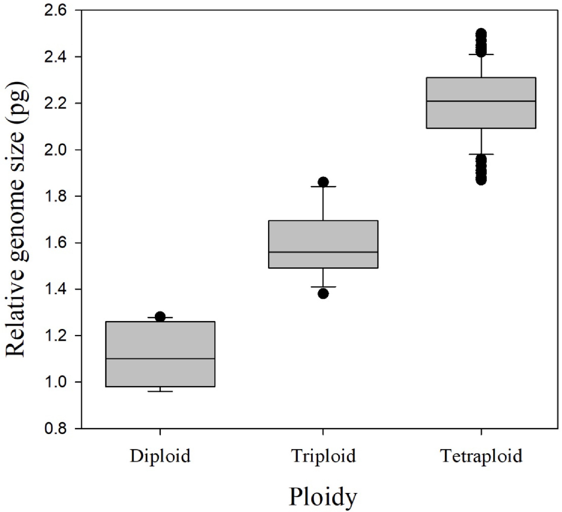
      Figure 1. 

      Relative genome size for each of the three observed ploidy levels, for 174 rose species, cultivars, and breeding lines, determined by flow cytometry. Boxes display the lower and upper quartiles of the population with a horizontal line within the box showing the median. Vertical lines outside the boxes extend 1.5 times the interquartile range and data points outside of this range are considered potential outliers.

      The mean 1Cx genome size for all cultivars was 0.55 ± 0.04 (SEM) pg with a range from 0.46 to 0.64 pg (Table 2). The 2C relative genome size range was 0.96–1.28 pg for diploids, 1.38–1.86 pg for triploids, and 1.87–2.50 pg for tetraploids, providing clear distinction between the genome size ranges of different euploid levels (Fig. 1). The mean relative genome sizes were 1.10 ± 0.13 pg, 1.59 ± 0.14 pg, and 2.20 ± 0.15 pg for diploids, triploids, and tetraploids, respectively.

      Sixteen out of the 174 cultivars and breeding lines have had ploidy previously documented[38, 40]. Of these 16 accessions, our data confirm the previously reported ploidy of 10 accessions and dispute the ploidy of the other six accessions, i.e., Knock Out®, Home Run®, Miracle on the Hudson®, Blushing Knock Out®, Double Knock Out®, and Oso Easy Paprika®. Each of these six cultivars was previously reported as triploid (D. Zlesak, Per. Comm.)[38, 40]. However, the relative genome sizes determined by flow cytometry indicate that each cultivar is tetraploid (Table 2).

      Chromosome counts were performed on seven of the accessions used for genome size determination including Knock Out®, Home Run®, Miracle on the Hudson®, Malvern Hills®, 'New Zealand', 'Dr. Huey', and Sunshine Daydream. Knock Out®[38, 40], Home Run®[38, 40], and Miracle on the Hudson® (D. Zlesak, Per. Communication) were chosen because of contradicting reports of ploidy between our flow cytometry estimates and previous reports. Malvern Hills® and New Zealand were chosen to calibrate relative genome size to ploidy for diploids and tetraploids, respectively (Table 2). 'Dr. Huey' and Sunshine Daydream were chosen to calibrate relative genome size to ploidy and help discern the transition point for relative genome size values for the largest triploid and the smallest tetraploid, respectively (Table 2). We found that Malvern Hills® was diploid, 'Dr. Huey' was triploid, and Knock Out®, Home Run®, Miracle on the Hudson®, 'New Zealand', and Sunshine Daydream were tetraploid (Fig. 2).
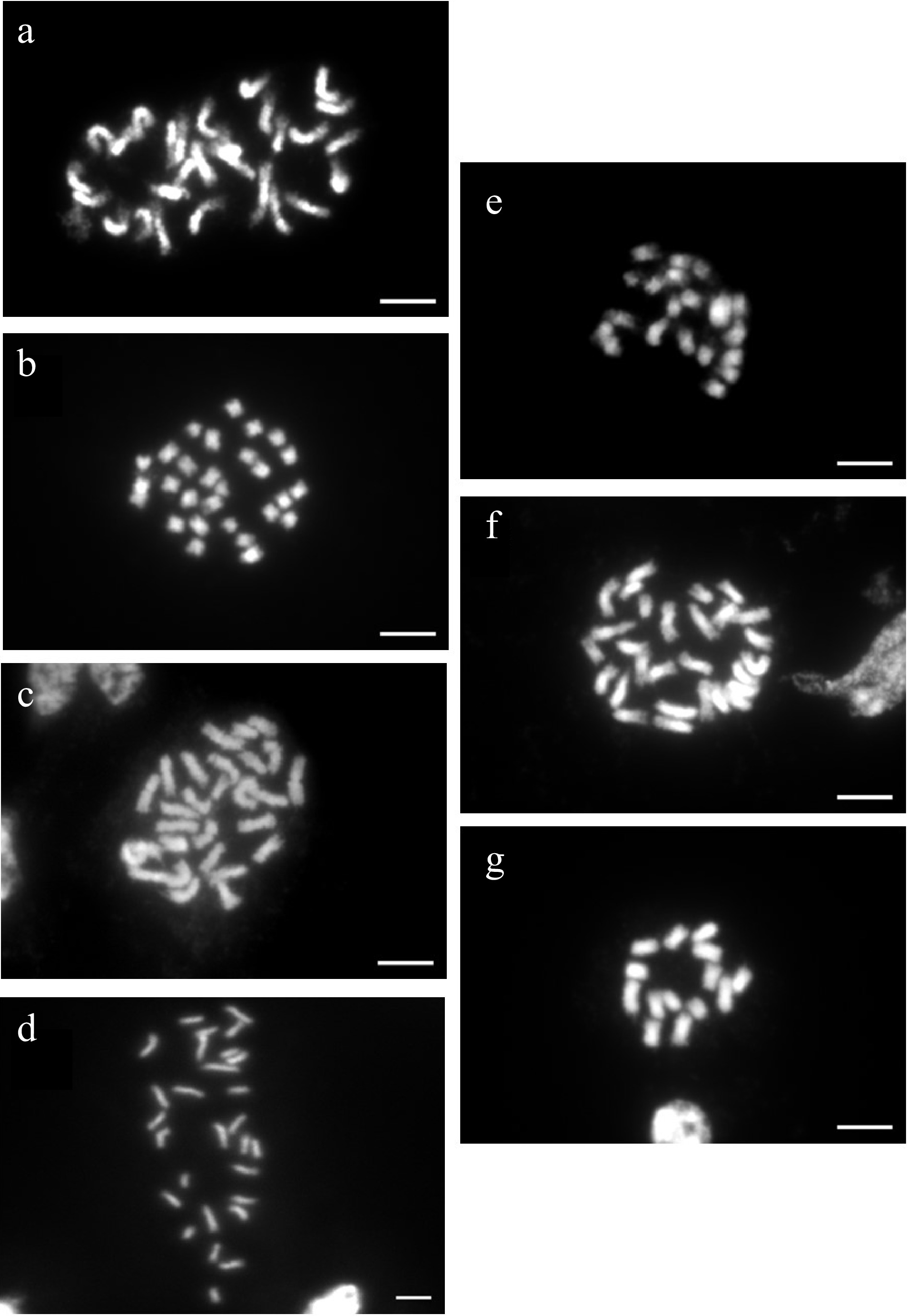
      Figure 2. 

      Ploidy determination by counting chromosomes of metaphase cells of seven rose cultivars. Chromosome images of (a) tetraploid Miracle on the Hudson® (2n = 4x = 28), (b) tetraploid Home Run™ (2n = 4x = 28), (c) tetraploid New Zealand (2n = 4x = 28), (d) tetraploid Sunshine Daydream (2n = 4x = 28), (e) triploid 'Dr. Huey' (2n = 3x = 21), (f) tetraploid Knockout® (2n = 4x = 28), and (g) diploid Malvern Hills® (2n = 2x = 14). Bars = 10 µM.

    • Determination of ploidy using microscopy methods is tedious, challenging, and often difficult to obtain clear resolution of all individual chromosomes and accurate counts. Other indirect methods, such as pollen diameter and guard cell length[18, 40], can be correlated with ploidy but often lack the resolution to clearly differentiate between isoploids and anisoploids[40]. Furthermore, guard cell length only determines the ploidy level of the L-I histogenic layer, but gives no information about the L-II layer, from which pollen and egg cells are derived[41]. Although flow cytometry can be an effective method for estimating genome size and associated ploidy, this approach needs to be validated and calibrated among closely related species. Furthermore, variations in methodology including extraction buffers, fluorochrome stains, and internal standards can skew results from one study to another. For example, different fluorochrome stains may give slightly different estimates of genome size. Both propidium iodide (PI) and 4', 6-diamidino-2-phenylindole (DAPI) are effective and consistent for determining relative genome size and ploidy among closely related taxa[33] when methods are standardized.

      This study found that the base genome sizes (1Cx) were relatively conserved in the roses tested and that 2C genome sizes could be used to distinguish among diploid, triploid, and tetraploid cytotypes. The mean 1Cx genome size for all cultivars included in this study was 0.55 ± 0.04 pg with a range from 0.46 to 0.64 pg (Table 2). The average 1Cx genome size for these cultivars aligned well with the mean 1Cx genome size for the 10 rose species that contributed to modern cultivars estimated to be 0.57 ± 0.05 pg (Table 1) even though both Roberts et al.[13] and Yokoya et al.[37] used Petroselinum crispum as a calibration standard and PI as a fluorochrome stain.

      The gap in genomes sizes between triploids and tetraploids was relatively small with 1.38–1.86 pg for triploids and 1.87–2.50 pg for tetraploids. Because of this small gap between genome sizes for triploids and tetraploids, chromosome counts were completed to accurately identify ploidy for cultivars near this transition point. Using microscopy, we found 'Dr. Huey' (2C = 1.86 pg) to be triploid, and Sunshine Daydream (2C = 1.91 pg) to be tetraploid. For roses with genome sizes near this transition point, chromosome counts may be required to confirm ploidy.

      In cases where our ploidy estimates differed from prior reports, our reassessment of chromosome numbers using microscopy (e.g., Knock Out®, Home Run®, and Miracle on the Hudson®) substantiated our estimates derived from flow cytometry. These discrepancies may be the result of mislabeling or incorrect identification of specific clones, cytochimeras, or the inherent difficulty in clearly visualizing and counting plant chromosomes.

      Accurate ploidy-level assessment is essential to breeding species with a variety of ploidy levels. A cultivar's ploidy level can affect its fertility and potential usefulness in a breeding program. For example, Hibiscus syriacus cultivars 'Minerva', 'Diane', and 'Helene' were initially reported to be sterile, triploid cultivars (allo-hexaploid) when they were released by the US National Arboretum[42]. However, a recent flow cytometry analysis reported these cultivars found in nurseries to be fertile tetraploids[42]. The origin of the ploidy discrepancy, whether the result of initial misidentification, propagation of a mislabeled stock plant with various ploidy levels, or the reversion of a cytochimera, is unclear[42]. Flow cytometry is a useful, rapid tool for the determination of ploidy levels for the basis of breeding, ploidy manipulation, and uncovering past classification errors.

      In addition to validating the application of flow cytometry for ploidy determination in roses, the results of this study provide an expanded database on the genome sizes and ploidy of 174 diverse rose cultivars and breeding lines. This research further contributes to the cytogenetic understanding of roses and a broader foundation for future breeding and improvement.

    • One hundred and fifty four rose cultivars and 20 rose breeding lines were included in this study (Table 2). Sixteen accessions with previously reported ploidy levels were included in this study to help calibrate genome size to ploidy level and to determine validity of previous reports[3840]. Emerging flower bud tissue of these cultivars and breeding lines were collected from rose gardens, public breeding programs, and nurseries (Table 2). If emerging floral bud tissue was not available, emerging new leaf tissue was collected. Leaf tissue was collected from P. sativum 'Ctirad' grown in the greenhouse and used as the internal standard (2C DNA content = 8.76 pg[43]).

    • Relative genome size was determined using flow cytometry. Tissue samples were chopped using a double-sided razor blade in a 50 mm petri dish with 200 μL of extraction buffer (CyStain PI Absolute P; Sysmex Partec, Munster, Germany). P. sativum 'Ctirad' was chopped with Rosa tissue. The finely chopped tissues and buffer were poured through a 50 μm nylon-mesh filter into a test tube and stained with 800 μL 4',6-diamidino-2-phenylindole (DAPI) fluorochrome (CyStain ultraviolet Precise P Staining Buffer; Sysmex Partec). DAPI was used due to its speed, low toxicity, precision (low sample coefficient of variance), and repeatable results for determining ploidy[37]. Samples were tested in a completely randomized design using a flow cytometer (Quantum P; Quantum Analysis, Munster, Germany). 2C DNA content was calculated using the formula: DNA content of the internal standard × (mean fluorescence of sample / mean fluorescence of the internal standard). At least two replicates were used for each cultivar. The relationship between ploidy and genomic size was calibrated using roses in the same section with known ploidy as a reference[13].

    • Chromosome counts were conducted to confirm the relationship between genome size and ploidy[44]. Roots or young shoot tips of 0.5 cm in length were collected and placed in the pre-fixative solution containing 2 mM 8-hydroxyquinoline and 70 mg·L−1 cycloheximide in microcentrifuge vials. Vials were stored in the dark for 3 h at ambient room temperature and subsequently placed in a dark refrigerator at approximately 4 °C for an additional hour. Following the pre-fixative treatment, tissue was removed and rinsed thoroughly in deionized water. The tissue was then placed in Farmer's fixative solution containing 95% ethanol and glacial acetic acid (3:1 v/v) and left at 4 °C overnight or until tissues could be further processed.

      Fixed tissues were removed from the fixative solution and rinsed with deionized water for 1 min per time in triplicate. After washing, the youngest, most tender tissues were placed on a microscope slide with 30 µL of a cell wall digestive enzyme mixture of 6% pectinase (Sigma Chemical; St. Louis, MO, USA) and 6% cellulase (Onozuka R-10; Yakult Honsha, Tokyo, Japan) in 75 mM KCl buffer (pH 4.0). Prepared slides with the cell wall digestive enzyme buffer were placed in an enclosed container with moistened paper towels and incubated at 37 °C between 3 h and overnight depending on the cultivar. The next day, the buffer was diluted with 1−3 drops deionized distilled water and any excess buffer was removed with a Kimwipe®. Then, two drops of a modified fixative buffer containing 95% methanol and glacial acetic acid (3:1, v/v) were added to the sample on each slide. Tissue was broken up using a small spatula, spreading the macerated tissue evenly on the slides in a circular pattern. Additional drops of the modified fixative buffer were added when needed.

      Once the tissue was spread out, the extra fixative buffer was burned off and the slides were incubated at 37 °C overnight to be fully dried, then stored at room temperature. Dried slides were stained using Vectashield® Antifade Mounting Medium with (Vector Laboratories; Burlingame, CA, USA) with DAPI. Chromosomes of metaphase cells were counted using a compound microscope (Axio Imager.A2, Zeiss; Oberkochen, Germany).

      • The authors thank Raleigh Rose Garden, JC Raulston Arboretum, Spring Meadow Nursery, Biltmore Estate, and the Texas A&M Rose Breeding Program for sharing tissue for use in this study. The authors also thank Nathan Lynch for his technical assistance. This work was funded, in part, by the North Carolina Agriculture Research Service (NCARS), Raleigh, NC, the North Carolina Biotechnology Center, Research Triangle Park, NC, the Hatch project 02685 from the US Department of Agriculture National Institute of Food and Agriculture, and Spring Meadow Nursery, Grand Haven, MI, USA.

      • The authors declare that they have no conflict of interest.

      • Copyright: © 2023 by the author(s). Published by Maximum Academic Press, Fayetteville, GA. This article is an open access article distributed under Creative Commons Attribution License (CC BY 4.0), visit https://creativecommons.org/licenses/by/4.0/.
    Figure (2)  Table (2) References (44)
  • About this article
    Cite this article
    Harmon DD, Chen H, Byrne D, Liu W, Ranney TG. 2023. Cytogenetics, ploidy, and genome sizes of rose (Rosa spp.) cultivars and breeding lines. Ornamental Plant Research 3:10 doi: 10.48130/OPR-2023-0010
    Harmon DD, Chen H, Byrne D, Liu W, Ranney TG. 2023. Cytogenetics, ploidy, and genome sizes of rose (Rosa spp.) cultivars and breeding lines. Ornamental Plant Research 3:10 doi: 10.48130/OPR-2023-0010

Catalog

  • About this article

/

DownLoad:  Full-Size Img  PowerPoint
Return
Return