ARTICLE   Open Access    

Variation in plant traits and nutrient uptake among banana varieties in shaded agroecology under areca nut canopy

More Information
  • Banana is an excellent companion crop cultivated with coconut, areca nut, coffee, or cacao. The yield performance of bananas however differs due to cultivar response to light, water, and nutrient availability in the intercropping system. The current study aims to understand the performance of different banana varieties under the areca nut shaded system by trait variation in growth, phenology, fresh bunch mass (yield), and soil nutrient balance patterns. Five banana varieties were screened in a field experiment in a high-density areca garden for vegetative traits, phenology, yield components, and nutrient budgets. Variety Amti recorded wider leaves and greater leaf area and also recorded higher bunch yield in plant and first ratoon crop than other varieties. The Velchi variety recorded the highest leaf emergence rate during winter and recorded the highest percentage of plants with a bunch in both the plant and first ratoon crops. The study revealed nutrient mining of nitrogen, phosphorous, and potassium in the areca–banana system. The effective balance of the available soil phosphorus was found to be highly negative for Grand Nain (−50.3 kg·ha−1·year−1) and negative (−23.6 kg·ha−1·year−1) for Amti. The results reveal the role of choice variety, indicator traits, and nutrient management strategies in enhancing banana productivity in agroforestry systems.
  • Scale insects (Hemiptera: Coccomorpha) with hypogeic habits are considered of high phytosanitary relevance for coffee crops (Rubiaceae: Coffea spp.) in Colombia[1]. A total of 65 species of scale insects associated with coffee roots have been recorded in Colombia[24]. The most species-rich family is the Pseudococcidae with 28 species distributed in nine genera: Dysmicoccus Ferris, 1950 (13 spp.), followed by Pseudococcus Westwood, 1840 (four spp.), Phenacoccus Cockerell, 1893 (three spp.), Planococcus Ferris, 1950, and Spilococcus Ferris, 1950 (two spp. each), and Chorizococcus McKenzie, 1960, Distichlicoccus Ferris, 1950, Ferrisia Fullaway, 1923, and Paraputo Laing, 1929 (one sp. each). For the family Rhizoecidae, 19 species have been recorded in six genera, namely, Rhizoecus Kunckel d'Herculais, 1878 (13 spp.), Pseudorhizoecus Green, 1933 (two spp.), and Capitisetella Hambleton, 1977, Coccidella Hambleton, 1946, Geococcus Green, 1902, and Ripersiella Tinsley, 1899 (one sp. each). Other minor families include Coccidae and Ortheziidae (five spp. in each family), Xenococcidae (three spp.), and Putoidae and Diaspididae (two spp. in each family) and Margarodidae (one sp.). For this study all previous records were re-analysed with the purpose of providing an accurate list of species

    The taxonomic identification of scale insects by a morphological approach is particularly difficult, mainly for two reasons. First, they are small insects (usually < 5 mm) that require the preparation of slide-mounted specimens. Second, the taxonomic keys needed for morphological identifications are primarily designed for adult female specimens[5]. Differing from other insect orders (e.g., Coleoptera, Diptera and Hymenoptera), female scale insects lack well-defined tagmata, as well as sclerites, sutures, or discernible areas. Characters of taxonomic value in scale insects include cuticular processes, such as pores, ducts and, setae[5]. Recognizing these cuticular structures on such small bodies poses a difficult task for non-expert entomologists. To facilitate accessible identification, this manuscript offers an illustrated taxonomic key to scale insect species associated with coffee roots in Colombia and is aimed at users with basic knowledge of scale insect morphology.

    A careful revision of the specimens studied by Caballero et al.[2], preserved in the Scale Insect Collection at the Entomological Museum 'Universidad Nacional Agronomía Bogota' UNAB (Bogotá, Colombia), was carried out to exclude species that are doubtfully recorded from coffee roots in Colombia. This re-assessment allowed the compilation of an accurate list of species that could be included in the taxonomic key. Additional species and information from Caballero[3] and Caballero et al.[4] also were used for construction of the key. List of species recorded for Colombia and c ollection data of specimens analized are in Supplemmental Table S1 and S2 respectivately.

    The illustrated taxonomic key (Table 1) is based on the external morphology of the adult female with a dichotomous structure. Each couplet after the first one is numbered followed by the number of the preceding couplet in parenthesis, e.g. 12(7) means that couplet 12 is derived from couplet 7; the numbers at the end of the couplet indicate the next couplet in order to arrive at the species name that best matches the character states selected by the user. It is illustrated in most of the steps using microphotographs. Acquisition and analysis of images were done with a Lumenera 1-5C camera and the software Image Pro Insight 8.0. Designs were performed with Affinity Photo V 2.1 and Affinity Designer V 2.1 software. The taxonomic keys were structured with some adaptations of published taxonomic keys[615]. The general morphological terminology follows Kondo & Watson[5] with specific terminology for Coccidae[6,16], Margarodidae[17], Ortheziidae[7], Diaspididae[18], Pseudococcidae, Putoidae[8,9], and Rhizoecidae[10,11]. The abdominal segmentation is given as SabdI for abdominal segment 1 to SabdVIII for abdominal segment 8. All microphotographs are of adult female scale insects or their taxonomically important morphological structures.

    Table 1.  Illustrated taxonomic key.
    No.DetailsRef.
    1Abdominal spiracles present (Fig. 1a)2
    Abdominal spiracles absent (Fig. 1b)7
    Fig. 1 Abdominal spiracles (sp) on margin (a) present on Eurhizococcus colombianus, (b) absent on Distichlicoccus takumasai.
    2(1)Anal aperture without pores and setae (Fig. 2a); legs shorter than half of the transversal diameter of body (Fig. 2b); eyespots and mouthparts absentEurhizococcus colombianus

    Jakubski, 1965
    Anal aperture forming a well-developed anal ring with pores and setae (Fig. 2c); legs longer than transversal diameter of body; eyespots and mouthparts present (Fig. 2d)
    3
    Fig. 2 Eurhizococcus colombianus: (a) Anal aperture without pores and setae in the border, (b) section of mid body showing the length of hind leg (lel) and transversal body line (btl). Insignorthezia insignis: (c) Anal aperture with pores (po) and setae (st), (d) section of head with protruding eyespot (es) and labium (lb).
    3(2)Antennae each with eight segments (Fig. 3a)4
    Antennae each with fewer than five segments (Fig. 3b)5
    Fig. 3 (a) Eight-segmented antenna. (b) Four-segmented antenna.
    4(3)Transversal bands of spines absent in ventral region surrounded by an ovisac band (Fig. 4a); dorsal interantennal area without sclerosis (Fig. 4b)Insignorthezia insignis (Browne, 1887)
    Transversal bands of spine plates present in ventral region surrounded by an ovisac band (Fig. 4c); longitudinal sclerosis on dorsum in interantennal area (Fig. 4d)Praelongorthezia praelonga (Douglas, 1891)
    Fig. 4 Insignorthezia insignis: (a) Abdomen without transversal clusters of wax plates, (b) Dorsal interantennal area without sclerosis. Praelongorthezia praelonga: (c) Abdomen with transversal clusters of wax plates marked by dash lines, (d) dorsal interantennal area with a longitudinal sclerotic plate (ep).
    5(3)Antennae each with three segments (Fig. 5a)Newsteadia andreae Caballero, 2021
    Antennae each with four segments (Fig. 5b)6
    Fig. 5 (a) Three-segmented antenna of Newsteadia andreae. Note the presence of pseudosegmentation which gives the appearance of additional segments in the last antennal segment. (b) Four-segmented antenna of Mixorthezia minima.
    6(5)Dorsal area anterior to anal ring with simple pores on protuberances (Fig. 6a); ventral areas surrounding each coxa with a row of wax plate spines (Fig. 6b)Mixorthezia minima Koczné Benedicty & Kozár, 2004
    Dorsal area anterior to anal ring without simple pores or protuberances (Fig. 6c); ventral areas posterior to each coxa without wax plate spines (Fig. 6d)Mixorthezia neotropicalis (Silvestri, 1924)
    Fig. 6 Mixorthezia minima: (a) Dorsum of area anterior to anal ring with close-up of simple pores on protuberances (dash box); (b) ventral area posterior to each coxa with a row of wax plate spines (dash box). Mixorthezia neotropicalis: (c) Close-up of dorsum of area anterior to anal ring lacking simple pores on protuberances (dash box); (d) ventral area posterior to each coxa without associated wax plate spines.
    7(1)Anal plates present (Fig. 7a)8
    Anal plates absent (Fig. 7b)12
    Fig. 7 (a) Anal apparatus of Saissetia coffeae with anal plates (ap) covering the anal aperture (aa). (b) Anal apparatus of Pseudococcus sp. with anal aperture lacking anal plates.
    8(7)Antennae and legs with length similar to or shorter than spiracles (Fig. 8a)9
    Antennae and legs with length at least twice as long as spiracles (Fig. 8b)11
    Fig. 8 (a) Antenna (an) and foreleg (lg) (green lines), and anterior spiracle (sp) (yellow line) of Toumeyella coffeae showing their relative length. Note the similar size of the limbs and spiracle. (b) Antenna (an) and leg (lg) (green lines), and anterior spiracle (sp) (yellow line) of Coccus viridis showing their relative length. Note the relatively smaller size of the spiracle.
    9(8)Ventral tubular macroducts present (Fig. 9)Toumeyella coffeae
    Kondo, 2013
    Ventral tubular macroducts absent10
    Fig. 9 Ventral tubular macroducts (dash box) and close-up of macroducts (photo on right side).
    10(9)Orbicular pores (Fig. 10a) and cribriform platelets present (Fig. 10b); dorsal setae absent; opercular pores absentCryptostigma urichi (Cockerell, 1894)
    Orbicular pores and cribriform platelets absent; dorsal setae present (Fig. 10c); numerous opercular pores present throughout mid areas of dorsum (Fig. 10d)Akermes colombiensis Kondo & Williams, 2004
    Fig. 10 Cryptostigma urichi: (a) Orbicular pore and (b) close-up of a cribriform platelet. Akermes colombiensis: (c) Close-up of a dorsal body setae (dash box) and (d) close-up of opercular pores (arrows).
    11(8)Band of ventral tubular ducts in lateral and submarginal regions absent, ventral tubular ducts of one type; anal plates without discal setae (Fig. 11a); dorsal body setae capitate or clavate (Fig. 11b); perivulvar pores with seven or eight loculi, rarely with 10 loculi (Fig. 11c)Coccus viridis
    (Green, 1889)
    Band of ventral tubular ducts in lateral and submarginal regions present, submarginal region with two types of tubular ducts (Fig. 11d); anal plates with discal setae (Fig. 11e); dorsal body setae spine-like, apically pointed (Fig. 11f); perivulvar pores mostly with 10 loculi (Fig. 11g)Saissetia coffeae
    (Walker, 1852)
    Fig. 11 Coccus viridis: (a) Anal plates without discal setae; (b) dorsal body setae capitate (top) or clavate (below); (c) multilocular disc pores mostly with eight loculi. Saissetia coffeae: (d) Ventral submarginal region with two types of tubular ducts; (e) each anal plate with a discal seta; (f) dorsal body setae acute; (g) multilocular disc pores with mostly 10 loculi.
    12(7)Cerarii present on body margin, at least a pair on each anal lobe (Fig. 12a)13
    Cerarii absent on body margin (Fig. 12b)38
    Fig. 12 Abdominal body margin of (a) Pseudococcus sp. with three cerarii (dash box) and (b) Rhizoecus sp. (dash box) without cerarii.
    13(12)Enlarged oral collar tubular ducts composed of a sclerotized area surrounding the border and a set of flagellated setae (Ferrisia-type oral collar tubular ducts) (Fig. 13a)Ferrisia uzinuri
    Kaydan & Gullan, 2012
    Oral collar tubular ducts simple, not as above (Fig. 13b) or absent14
    Fig. 13 (a) Ferrisia-type oral collar tubular ducts with aperture of tubular duct (ad) surrounded by a sclerotized area (sa) and associated flagellate setae (fs). (b) Oral collar tubular ducts simple (arrows).
    14(12)Antenna with nine segments (Fig. 14a)15
    Antenna with eight segments (Fig. 14b) or fewer (Fig. 14c)19
    Fig. 14 Antenna with (a) nine segments, (b) eight segments and (c) seven segments.
    15(14)Cerarii with more than five conical setae (Fig. 15a); hind trochanter with six sensilla, three on each surface (Fig. 15b)16
    Cerarii with two lanceolate setae (Fig. 15c); hind trochanter with four sensilla, two on each surface (Fig. 15d)17
    Fig. 15 Puto barberi: (a) upper and lateral view of a cerarius, (b) close-up of the surface of trochanter with three sensilla (arrows). Phenacoccus sisalanus: (c) cerarius, (d) trochanter with two sensilla (arrows) on single surface.
    16(15)Cerarii with tubular ducts (Fig. 16a)Puto antioquensis
    (Murillo, 1931)
    Cerarii without tubular ducts (Fig. 16b)Puto barberi
    (Cockerell, 1895)
    Fig. 16 (a) Cerarius associated with tubular ducts (arrows). (b) Cerarius without tubular ducts.
    17(15)Oral collar tubular ducts absentPhenacoccus sisalanus Granara de Willink, 2007
    Oral collar tubular ducts present, at least on venter (Fig. 17)18
    Fig. 17 Ventral surface with oral collar tubular ducts (dash circles).
    18(17)Oral collar tubular ducts restricted to venterPhenacoccus solani
    Ferris, 1918
    Oral collar tubular ducts present on dorsum and venterPhenacoccus parvus Morrison, 1924
    19(14)Oral rim tubular ducts present (Fig. 18)20
    Oral rim tubular ducts absent26
    Fig. 18 Oral rim tubular ducts in upper view (dash circles) and close-up of lateral view.
    20(19)Oral rim tubular ducts present on venter onlyPseudococcus landoi (Balachowsky, 1959)
    Oral rim tubular ducts present on both dorsum and venter21
    21(20)Cerarii restricted to anal lobes (Fig. 19a)Chorizococcus caribaeus Williams & Granara de Willink, 1992
    Cerarii present, at least on the last five abdominal segments (Fig. 19b)22
    Fig. 19 Location of cerarii (dash boxes) on abdominal margin with close-up of cerarius (a) restricted to anal lobes (dash boxes) and (b) cerarii present on the last five abdominal segments.
    22(21)Circulus absent (Fig. 20a)23
    Circulus present (Fig. 20b)24
    Fig. 20 Ventral mid area of abdominal segments III and IV (dash box) of (a) Distichlicoccus takumasai without circulus and (b) Pseudococcus jackbeardsleyi with circulus.
    23(22)Multilocular disc pores present on venter of SabdIV and posterior segments (Fig. 21a); hind coxa with translucent pores and hind femur without translucent pores (Fig. 21b)Spilococcus pressus
    Ferris, 1950
    Multilocular disc pores absent, if some present, not more than three around vulvar opening (i.e. venter of SabdVII or SabdVIII); hind coxa without translucent pores (Fig. 21c) and hind femur with translucent pores (Fig. 21d)Distichlicoccus takumasai Caballero, 2021
    Fig. 21 Spilococcus pressus: (a) Ventral section of abdomen with multilocular disc pores (arrows); (b) hind leg with close-up of coxa with translucent pores (arrows). Distichlicoccus takumasai: (c) Hind coxa without translucent pores; (d) hind femur with translucent pores (arrows).
    24(22)Eyes without discoidal pores nor sclerotized surrounding area (Fig. 22a); circulus with transversal diameter 40 to
    60 µm (Fig. 22b)
    Pseudococcus luciae Caballero, 2021
    Eyes with discoidal pores and sclerotized surrounding area (Fig. 22c); circulus diameter 100 to 200 µm (Fig. 22d)26
    25(24)Oral rim tubular ducts on dorsal abdominal segments numbering three to eight; area between posterior ostiole and cerarius of SabdVII without oral rim tubular ducts (Fig. 23a)Pseudococcus elisae Borchsenius, 1947
    Oral rim tubular ducts on dorsal abdominal segments numbering 14 to 27; area between posterior ostiole and cerarius of SabdVII with an oral rim tubular duct (Fig. 23b)Pseudococcus jackbeardsleyi Gimpel & Miller, 1996
    Fig. 22 Pseudococcus luciae: (a) Eyespot without surrounding sclerotized area nor associated pores; (b) circulus ca. 58 µm wide. Pseudococcus jackbeardsleyi: (a) Eyespot with sclerotized area (sa) and associated pores (po); (d) circulus ca. 154 µm wide.
    Fig. 23 (a) Dorsal margin of abdominal segments VI to VIII, between cerarius of anal lobe (C1), cerarius of SabdVII (C2) and posterior ostiole (os) without oral rim tubular ducts. (b) Dorsal margin of abdominal segments VI to VIII, between cerarius of anal lobe (C1), cerarius of SabdVII (C2) and posterior ostiole (os) with an oral rim tubular duct and/or cerarius adjacent to SabdVII.
    26(19)Oral collar tubular ducts (Fig. 24) on both dorsum and venter27
    Oral collar tubular ducts restricted to venter28
    Fig. 24 Oral collar tubular duct in lateral view.
    27(26)Hind coxa with translucent pores (Fig. 25a); anal lobe with sclerotized bar, not on a sclerotized area (Fig. 25b); multilocular disc pores present posterior to fore coxaPlanococcus citri-minor complex
    Hind coxa without translucent pores (Fig. 25c); anal lobe without sclerotized bar, on a sclerotized area (Fig. 25d); multilocular disc pores absent posterior to fore coxaDysmicoccus quercicolus (Ferris, 1918)
    28(27)Oral collar tubular ducts absent on venter of both head and thorax.29
    Oral collar tubular ducts present on either head or thorax, but not on both areas (Fig. 26)30
    Fig. 25 Planococcus citri-minor complex: (a) Hind coxa with translucent pores (dash box) and (b) anal lobe with a sclerotization forming a bar (ab). Dysmicoccus quercicolus: (c) Hind coxa without translucent pores and (d) anal lobe with irregular broad sclerotized area (sa).
    Fig. 26 Marginal area of Dysmicoccus grassii, lateral to posterior spiracle (ps), with close-up of oral collar tubular ducts (oc) (left side).
    29(28)Translucent pores present on hind coxa, trochanter, femur and tibia (Fig. 27a); marginal clusters of oral collar tubular ducts on venter of SabdVI and SabdVIIDysmicoccus caribensis Granara de Willink, 2009
    Translucent pores restricted to hind femur and tibia (Fig. 27b); marginal clusters of oral collar tubular ducts present on venter of SabdIV to SabdVIIParaputo nasai
    Caballero, 2021
    Fig. 27 (a) Hind leg of Dysmicoccus caribensis with translucent pores on coxa (cx), trochanter (tr) and femur (fm), and tibia (tb). (b) Hind leg of Paraputo nasai with translucent pores restricted to femur (fm) and tibia (tb).
    30(28)Hind coxa with translucent pores (Fig. 28a)Dysmicoccus sylvarum
    Williams & Granara de Willink, 1992
    Hind coxa without translucent pores (Fig. 28b)31
    Fig. 28 (a) Translucent pores on hind coxa. (b) Translucent pores absent on hind coxa.
    31(30)Hind trochanter with translucent pores (Fig. 29a)Dysmicoccus varius
    Granara de Willink, 2009
    Hind trochanter without translucent pores (Fig. 29b)32
    Fig. 29 Translucent pores (a) on hind trochanter, (b) absent from hind trochanter.
    32(31)Oral collar tubular ducts present on margin of thorax (Fig. 30)33
    Oral collar tubular ducts absent from margin of thorax34
    Fig. 30 Prothorax margin of Dysmicoccus grassii with close-up of oral collar tubular ducts.
    33(32)Multilocular disc pores absent on SabdV; dorsal area immediately anterior to anal ring with tuft of flagellate setae; longest flagellate seta as long as diameter of anal ring (Fig. 31a), and discoidal pores larger than trilocular pores (Fig. 31b)Dysmicoccus radicis
    (Green, 1933)
    Multilocular disc pores present on SabdV; dorsal area immediately anterior to anal ring without a tuft of flagellate setae; flagellate setae much shorter than diameter of anal ring (Fig. 31c) and discoidal pores smaller than trilocular pores (Fig. 31d)Dysmicoccus grassii (Leonardi, 1913)
    34(32)Oral collar tubular ducts absent in interantennal area35
    Oral collar tubular ducts present in interantennal area (Fig. 32)36
    35(34)Translucent pores on hind leg restricted to tibia (Fig. 33a)Dysmicoccus perotensis
    Granara de Willink, 2009
    Translucent pores on hind leg present on tibia and femur (Fig. 33b)Dysmicoccus joannesiae-neobrevipes complex
    Fig. 31 Dysmicoccus radicis: (a) Area anterior to anal ring with a cluster of flagellate setae (fs) and anal ring (ar) showing the diameter of the different pores (dash box); (b) discoidal pores (dp) and trilocular pores (tp). Dysmicoccus grassii: (c) Area anterior to anal ring with scattered short flagellate setae (fs) contrasted with anal ring (ar) diameter (dash box); (d) discoidal pores (dp) and trilocular pores (tp) with similar diameter.
    Fig. 32 Interantennal area (dash box) of Dysmicoccus brevipes with close-up of oral collar tubular ducts.
    Fig. 33 (a) Hind leg of Dysmicoccus perotensis with close-up of femur and tibia with translucent pores on tibia only (arrows). (b) Hind leg of Dysmicoccus joannesiae-neobrevipes complex with close-up of femur and tibia with translucent pores (arrows).
    36(34)Hind coxa with translucent pores (see Fig. 28a)Dysmicoccus mackenziei
    Beardsleyi, 1965
    Hind coxa without translucent pores (see Fig. 28b)37
    37(36)Dorsal SabdVIII setae forming a tuft-like group, each seta conspicuously longer than remaining dorsal abdominal setae (Fig. 34a) and setal length similar to anal ring diameter (60–80 µm long)Dysmicoccus brevipes (Cockerell, 1893)
    Dorsal SabdVIII setae evenly distributed, each setae as long as remaining dorsal abdominal setae (Fig. 34b) and length less than half diameter of anal ringDysmicoccus texensis-neobrevipes complex
    38(12)Tritubular ducts absent39
    Tritubular ducts present (Fig. 35a-b)46
    Fig. 34 (a) Abdomen of Dysmicoccus brevipes with dorsal setae on SabdVIII (lfs) longer than setae on anterior segments (sfs). (b) Abdomen of Dysmicoccus texensis-neobrevipes complex with dorsal setae (ufs) along the abdominal segments of uniform length and scattered distribution.
    Fig. 35 (a) Tritubular duct in upper (left) and lateral view (right) with the border of the cuticular ring attached to tubules. (b) Tritubular duct with the border of the cuticular ring widely separated from tubules (arrows).
    39(38)Anal lobes strongly protruded, bulbiform (Fig. 36a) jutting out from margin for a distance equivalent to diameter of anal ring40
    Anal lobes shallow, if protruded, their length never more than half of diameter of anal ring (Fig. 36b)42
    Fig. 36 (a) Abdomen of Neochavesia caldasiae with anal lobes (al) protruding beyond the anal aperture (aa). (b) Abdomen of Ripersiella sp. with anal lobes (al) at the same level as the anal aperture (aa).
    40(39)Anal aperture located at the same level as the base of anal lobes (Fig. 37a); antennae located on ventral margin of headNeochavesia caldasiae (Balachowsky, 1957)
    Anal aperture located anterior to bases of anal lobes (Fig. 37b); antennae located on dorsum of head41
    Fig. 37 (a) Abdomen of Neochavesia caldasiae with anal aperture (aa) positioned between the anal lobes (al), at the same level as the bases of anal lobes (dash line). (b) Abdomen of Neochavesia eversi with anal aperture (aa) situated anterior to the bases of the anal lobes (al) (dash line).
    41(40)Antennae each with five segments, situated on a membranous base (Fig. 38a); length of hind claw less than length of hind tarsus (Fig. 38b)Neochavesia trinidadensis (Beardsley, 1970)
    Antennae each with four segments, situated on a sclerotized base (Fig. 38c); hind claw longer than hind tarsus (Fig. 38d)Neochavesia eversi (Beardsley, 1970)
    Fig. 38 (a) Antenna with four segments and a membranous base (mb). (b) Hind tarsus (green line) longer than the hind claw (red line). (c) Antenna with four segments and a sclerotized base (sb). (d) Hind tarsus (green line) shorter than hind claw (red line).
    42(39)Body setae capitate, at least on one surface (Fig. 39a)43
    Body setae never capitate (Fig. 39b)44
    Fig. 39 (a) Capitate setae. (b) Flagellate setae.
    43(42)Anal aperture without associated cells (Fig. 40a); three-segmented antennae (Fig. 40b); ventral setae in median
    and submedian regions capitate
    Capitisitella migrans
    (Green, 1933)
    Anal aperture surrounded by cells (Fig. 40c); six-segmented antennae (Fig. 40d); ventral setae in medial and submedial regions flagellateWilliamsrhizoecus coffeae
    Caballero & Ramos, 2018
    44(42)Three-segmented antennae (Fig. 41a); circulus present (Fig. 41b)Pseudorhizoecus bari
    Caballero & Ramos, 2018
    Five-segmented antennae (Fig. 41c); circulus absent45
    Fig. 40 Capitisitella migrans: (a) Anal aperture of surrounded only by setae; (b) antenna composed of three segments. Williamsrhizoecus coffeae: (c) Anal aperture of surrounded by setae and cells (flesh); (d) antenna composed of six segments.
    Fig. 41 Pseudorhizoecus bari: (a) Antenna composed of three segments and (b) circulus. (c) Antenna of Pseudorhizoecus proximus composed of five segments.
    45(44)Multilocular disc pores absent; anal aperture ornamented with small protuberances and two to five short setae, each seta never longer than 1/3 diameter of anal aperture, without cells (Fig. 42a)Pseudorhizoecus proximus
    Green, 1933
    Multilocular disc pores present (Fig. 42b); anal aperture not ornamented with small protruberances, ring with well-developed cells and six long setae, each seta as long as diameter of anal ring (Fig. 42c)Ripersiella andensis (Hambleton,
    1946)
    Fig. 42 (a) Anal aperture of Pseudorhizoecus proximus surrounded by protuberances (pr) and a few short setae (st). Ripersiella andensis: (b) Ventral section of abdomen with multilocular disc pores (mp); (c) anal aperture with a ring of cells and six long setae (se).
    46(38)Anal lobes strongly protruded, conical, each one with a stout spine at apex (Fig. 43a)Geococcus coffeae
    Green, 1933
    Anal lobes flat or barely protruded, without spines at apex (Fig. 43b)47
    47(46)Venter of abdomen with clusters of trilocular pores in medial region (Fig. 44a)Coccidella ecuadorina Konczné Benedicty & Foldi, 2004
    Venter of abdomen with trilocular pores evenly dispersed, never forming clusters in medial region (Fig. 44b)48
    Fig. 43 (a) Abdomen of Geococcus coffeae with protruding anal lobe (al) with a stout spine at the apex (sp). (b) Abdomen of Rhizoecus sp. with anal lobe (al) flat, with numerous flagellate setae (fs) at the apex.
    Fig. 44 (a) Ventral surface of Coccidella ecuadorina with clusters of trilocular pores (tc) (dash box) on medial region of abdomen. (b) Ventral surface of Rhizoecus sp. with trilocular pores (tr) scattered on venter of abdomen.
    48(47)Antennae with six well-developed segments (Fig. 45a)51
    Antennae with five well-developed segments (Fig. 45b), apical segment sometimes partially divided (Fig. 45c)49
    Fig. 45 (a) Six-segmented antenna. (b) Five-segmented antenna. (c) Five-segmented antenna with partially divided apical segment (pd). Note: antennal segments numbered in Roman numerals.
    49(48)Antennae length more than 140 µm (Fig. 46a); tritubular ducts of similar diameter to trilocular pores (± 2 µm variation) (Fig. 46b); tritubular ducts with space between ductules and edge as wide as the ductules (Fig. 46c); slender ductule, width/length ratio 1:6Rhizoecus coffeae
    Laing, 1925
    Antennae length less than 130 µm (Fig. 46d); tritubular ducts of diameter nearly twice diameter of trilocular pores (Fig. 46e); tritubular ducts with reduced space or without space between ductules and edge (Fig. 46f); stout ductule, width/length ratio 1:350
    50(49)Tubular ducts present (Fig. 47a); each anal lobe with around 28 dorsal setae of similar length, greater than 30 µm (Fig. 47b, al); and dorsal marginal clusters of setae on SabdVII 20–30 µm long (Fig. 47b, SabdVII)Rhizoecus setosus (Hambleton, 1946)
    Tubular ducts absent; each anal lobe with around 14 dorsal setae, with length less than 15 µm (Fig. 47c, al); dorsal marginal clusters of setae on SabdVII with length less 15 µm (Fig. 47c, SabdVII)Rhizoecus compotor
    Williams & Granara de Willink, 1992
    Fig. 46 (a) Antenna ca. 207 µm long. (b) Tritubular ducts (td) and trilocular pores (tp) with similar diameter. (c) Close-up of a tritubular duct indicating the space between the cuticular ring (mg) and the ductule (dt). (d) Antenna ca. 105 µm long. (e) Each tritubular duct (td) twice the diameter of a trilocular pore (tp). (f) Close-up of tritubular duct without a space between the cuticular ring (mg) and the ductule (dt).
    Fig. 47 Rhizoecus setosus: (a) Tubular ducts (td); (b) anal lobe (al) and abdominal segment (SabdVII) with marginal clusters of setae longer than 30 µm. (c) Abdomen of Rhizoecus compotor with marginal cluster of setae shorter than 20 µm on anal lobe (al) and abdominal segment (SabdVII).
    51(48)Fore tibia with at least one of two internal preapical setae spine-like (Fig. 48a-b)52
    Fore tibia with both internal preapical setae flagellate (Fig. 48c)56
    Fig. 48 Fore legs with preapical setae on tibia (ft): (a) one flagellate (fs) and one spine seta (ss), (b) with a pair of spine setae (ss), (c) with a pair of flagellate setae (fs).
    52(51)Fore tibia with one internal preapical spine-like setae and other seta flagellate (Fig. 48a); anal ring composed of spine-like setae (Fig. 49a); circulus absentRhizoecus spinipes (Hambleton, 1946)
    Fore tibia with both internal preapical setae spine-like (Fig. 48b); anal ring composed of flagellate-like setae (Fig. 49b); at least, one circulus present (Fig. 49c)53
    Fig. 49 (a) Anal ring (ar) of Rhizoecus spinipes with spine-like setae (ss). (b) Anal ring (ar) of Rhizoecus arabicus with flagellate setae (fs). (c) Circulus of Rhizoecus cacticans.
    53(52)Claw digitules setose and short, length less than half length of claw (Fig. 50a)54
    Claw digitules capitate and long, as long as claw (Fig. 50b)55
    Fig. 50 Claw with claw digitule: (a) setose (sd), (b) flagellate (fd).
    54(53)Anal ring with external row composed of 35 cells or more (Fig. 51a, ext); anal ring with external and internal rows separated by a space as wide as a cell of the external row (Fig. 51a, spc); anal ring cells without spicules (Fig. 51a, sp)Rhizoecus variabilis Hambleton, 1978
    Anal ring with external row composed of less than 30 cells (Fig. 51b, ext); anal ring with external and internal rows separated by a narrow space, as wide as half (or less) a cell of the external row (Fig. 51b, spc); anal ring cells with spicules (Fig. 51b, sp)Rhizoecus arabicus Hambleton, 1976
    Fig. 51 (a) Anal ring of Rhizoecus variabilis with external row (ext) of anal ring consisting of over 35 cells; external row separated from the internal row (int) by a similar width as the diameter of a cell (spc). (b) Anal ring of Rhizoecus arabicus with external row (ext) of anal ring with less than 30 cells; external row separated from the internal row (int) by a width less than half the diameter of a cell (spc); cells of the external row with spicules (sp).
    55(53)More than 80 tritubular ducts; circulus with basal diameter at least five times greater than apical diameter (Fig. 52a); stick-like genital chamber, parallel borders and all of similar width and structure, length across about two abdominal segments (169–175 µm long) (Fig. 52b)Rhizoecus atlanticus (Hambleton, 1946)
    Less than 50 tritubular ducts; circulus with basal diameter less than three times the apical diameter (Fig. 52c); genital chamber with basal third two times wider than anterior two-thirds, length across one abdominal segment (43–52 µm long) (Fig. 52d)Rhizoecus cacticans (Hambleton, 1946)
    Fig. 52 Rhizoecus atlanticus: (a) Circulus with diameter at base five times the apical diameter, (b) genital chamber tubular shape, length ca. 150 µm long. Rhizoecus cacticans: (c) Circulus with diameter at base about two times the apical diameter, (d) genital chamber with proximal section basiform and distal section tubular, with arms, length ca. 45 µm long.
    56(51)Multilocular disc pores absent on dorsumRhizoecus mayanus (Hambleton, 1946)
    Multilocular disc pores present on dorsum57
    57(56)Marginal prothoracic setae length greater than 50 µm (Fig. 53a); marginal SabdVII setae length greater than 45 µm (Fig 53b)Rhizoecus colombiensis Ramos-Portilla & Caballero, 2016
    Marginal prothoracic setae length less than 25 µm (Fig. 53c); marginal SabdVII setae length less than 30 µm (Fig. 53d)58
    Fig. 53 Rhizoecus colombiensis: (a) Body margin with a long seta (pts) (> 40 µm), longer than remaining setae in prothorax; (b) margin of abdominal segment VII (SabdVII) (st). with a long seta (pts) (> 40 µm), longer than remaining setae in abdomen. Rhizoecus americanus: (c) Margin of prothorax (pts) with setae of uniform length, shorter than 30 µm; (d) margin of abdominal segment VII (SabdVII) with setae (st) shorter than 30 µm.
    58(57)Tritubular ducts of two sizesRhizoecus caladii
    Green, 1933
    Tritubular ducts of three sizesRhizoecus americanus (Hambleton, 1946)
     | Show Table
    DownLoad: CSV

    The following illustrated taxonomic key (Table 1) is a tool for the identification of adult female scale insects (Hemiptera : Sternorrhyncha : Coccomorpha) associated with coffee roots in Colombia, which includes 59 species from seven families (see Supplemental Table S1).

    The taxonomic key includes 59 species associated with coffee roots. Hemiberlesia sp., Odonaspis sp., Rhizoecus stangei McKenzie, 1962, Spilococcus mamillariae (Bouche, 1844), Planococcus citri (Risso, 1813) and Planococcus minor (Maskell, 1897) were excluded from the key. In the case of the two armoured scale insects, the specimens were found in Berlese funnel samples associated with coffee roots[2], however, there is no evidence of these species feeding on the roots and there are no previous records of association of Hemiberlesia nor Odonaspis species with coffee roots.

    Previous records of single specimens of R. stangei and S. mamillariae by Caballero et al.[2] were determined as misidentifications of Rhizoecus caladii Green, 1933 and Spilococcus pressus Ferris, 1950, respectively. Spilococcus mamillariae is considered as an oligophagous species, but mainly associated with Cactaceae plants and feeding on the aerial parts of plants[19,20]. There are no records of S. mamillariae being found on any plant species of the family Rubiaceae, and hence we have removed this species from the list of species associated with coffee roots. The species R. stangei, which has been recorded only from Mexico and lacks information on its host plant[21] has apparently not been found since its original description[8].

    Planococcus citri and Pl. minor were listed also by Caballero et al.[2] as literature records. the morphological identification of P. citri and P. minor needs to be complemented with molecular and geographical analysis to be more accurate[22]. Therefore, the present key considers only identification to the Planococcus citri-minor complex.

    Furthermore, many specimens of Dysmicoccus collected from coffee roots in Colombia have morphological character states that overlap with Dysmicoccus neobrevipes Beardsley, 1959, Dysmicoccus joannesiae (Costa Lima, 1939) and Dysmicoccus texensis (Tinsley, 1900). The first case is a mix of character states of D. texensis and D. neobrevipes. The number of setae in the abdominal cerarii and the size of oral collar tubular ducts are the most important characters used to differentiate the adult females of Dysmicoccus species[8,23]. Adult females of D. texensis have a consistent pattern of only two setae in all thoracic and abdominal cerarii, along with a uniform size of oral collar tubular ducts (OC). On the other hand, D. neobrevipes varies in the number of setae in the cerarii, ranging from two to seven, accompanied by two distinct sizes of OC. These character states are generally constant among specimens found on the aerial parts of plants. However, among the specimens examined here, while the anal lobe cerarii consistently have two setae on the specimens of D. texensis found on the roots, the remaining cerarii display a variable number of setae, notably ranging from two to five, particularly within the abdominal cerarii. Furthermore, the OC of these specimens all are the same size. Regarding the differences in number of setae in the cerarii, Granara de Willink[23] underlined the need of more comprehensive studies to definitively separate these species.

    The second case involves D. joannesiae and D. neobrevipes. These species exhibit similarities in the number of setae on each cerarius (ranging from two to seven setae per cerarius) and differences in the number of clusters of OC along the abdominal margin; D. joannesiae has more than 25 clusters of OC and D. neobrevipes has fewer than 10 clusters of OC[8]. Granara de Willink also separated these two species by the presence of OC on the thorax and head[23] (present in D. neobrevipes and absent in D. joannesiae). Within the specimens of putative D. neobrevipes studied here, a few had clusters of OC numbering 15 to 20 along the abdominal margin and OC on the thorax and head. The primary challenge with addressing this dilemma lies in the fact that D. joannesiae has only been reported on Joannesia princeps Vell., 1798 (Euphorbiaceae) in Brazil and on Annona muricata (Annonaceae) intercepted in London from Saint Lucia[8,24]. Moreover, there has been no additional morphological variations recorded in the new records of D. joannesiae since its initial description in 1932 by Costa Lima. Therefore, the character states defining D. joannesiae are based on six type specimens. Based on these arguments, the following taxonomic key considers two species complex groups, namely the Dysmicoccus texensis-neobrevipes complex and the D. joannesiae-neobrevipes complex.

    Following article 31.1.2 of the International Commission of Nomenclature (ICZN), herein we make a change in nomenclature for Distichlicoccus takumasae Caballero, 2021. The ending -ae for takumasae is incorrect because the species was dedicated to Dr. Takumasa Kondo (a male coccidologist), and thus the correct ending is -i, hence the species epithet is herein amended to 'takumasai'. The corrected name is Distichlicoccus takumasai Caballero, 2021.

    After reviewing the species of scale insects associated with coffee roots in Colombia, we have compiled a list of 59 species (Supplemental Table S1). Although this study did not focus on the effect of habit (aerial vs underground) or host plant on the morphology of scale insects, we detected significant morphological variation within facultative hypogeal species. Until further studies allow an understanding of the overlap of character states between D. texensisD. neobrevipes and D. joannesiaeD. neobrevipes, we suggest considering these species as a morphological complex for hypogeal specimens. Further ecomorphological studies should be conducted to determine whether the morphology of a species may differ when feeding on the aerial parts compared when feeding on the underground parts of a host and to try to elucidate what factors trigger those changes, especially in species associated with coffee plants. As for the species complex, further collecting, morphological, and molecular studies should help elucidate these taxonomic problems.

    During the literature review performed for this study, we realized that most of the records of species are limited to mentioning the host but not the plant part on which collections were made, however, it is suspected that most species are normally collected from the aerial parts of the plant host. Although this taxonomic key is limited to root-associated species recorded in Colombia, this key could be useful for identifying scale insects associated with coffee in other tropical regions, extending also to species collected from the aerial parts of the hosts.

    The authors confirm contributions to the paper as follows: study conception and design: Caballero A, Kondo T; data collection: Caballero A, Kondo T; analysis and interpretation of results: Caballero A, Kondo T; draft manuscript preparation: Caballero A, Kondo T. Both authors reviewed the results and approved the final version of the manuscript.

    The data (microscopy slides of specimens) that support the findings of this study are available in the Scale insect repository of the entomological museum Universidad Nacional Agronomia Bogota – UNAB, Facultad de Ciencias Agrarias, Colombia. All data generated or analyzed during this study are included in this published article and its supplementary information files.

    The authors thank Dr. Andrea Ramos-Portilla for clarifying some aspects of the morphological variations of Rhizoecus species and Dr. Penny Gullan (Australian National University, Canberra, Australia) for reviewing an earlier version of the manuscript. Many thanks to Erika Valentina Vergara (AGROSAVIA) and Dr. Francisco Serna (Universidad Nacional de Colombia) for their help to access the Museum UNAB. Special thanks to Dr Giuseppina Pellizzari (University of Padova, Italy) for advice on scientific nomenclature. This study was financed by Colciencias (Programa Nacional de Ciencias Básicas [National Program on Basic Sciences]), code 110165843233, contract FP44842-004-2015), by the entomological museum UNAB (Facultad Ciencias Agrarias, Universidad Nacional de Colombia, sede Bogotá) and by Federación Nacional de Cafeteros.

  • The authors declare that they have no conflict of interest.

  • Supplemental Table S1 Varieties used in the study and their international name/group and genome type.
    Supplemental Table S2 Duration and growing degree days of banana varieties to flowering and harvest.
    Supplemental Table S3 Final soil available nutrient contents of banana varieties under Arecanut-banana system.
  • [1]

    Dold C, Staver C, Pocasangre L, Heller J. 2008. Musa in shaded perennial crops-response to light interception. In Conference on Int. Res. on food security natural resource management and rural development, Hohenheim, Germany, 7−9 October 2008. University of Hohenheim, Centre for Agriculture in the Tropics and Subtropics, Germany.

    [2]

    Muralidharan A. 1980. Biomass productivity, plant interactions and economics of intercropping in areca nut garden. PhD Thesis. University of Agricultural Sciences, Bangalore, Karnataka, India. pp. 271

    [3]

    Dauzat J, Eroy MN. 1997. Simulating light regime and intercrop yields in coconut based farming systems. European Journal of Agronomy 7:63−74

    doi: 10.1016/S1161-0301(97)00029-4

    CrossRef   Google Scholar

    [4]

    Food and Agricultural Organisation (FAO). 2018. www.fao.org/faostat/en/#data (Accessed on 26.11.2020)

    [5]

    Sujatha S, Bhat R, Chowdappa P. 2016. Cropping systems approach for improving resource use in areca nut (Areca catechu L.) plantation. Indian Journal of Agricultural Sciences 86:1113−20

    doi: 10.56093/ijas.v86i9.61349

    CrossRef   Google Scholar

    [6]

    Norgrove L. 1998. Musa in multistrata systems: focus on shade. Info musa (International magazine on banana and plantain) 7:17−22

    Google Scholar

    [7]

    Marimuthu R. 2005. Multi species cropping system in coconut garden. Madras Agricultural Journal 92:404−6

    doi: 10.29321/MAJ.10.A01334

    CrossRef   Google Scholar

    [8]

    Rodrigo VHL, Stirling CM, Naranpanawa RMAKB, Herath PHMU. 2001. Intercropping of immature rubber in Sri Lanka: present status and financial analysis of intercrops planted at three densities of banana. Agroforestry Systems 51:35−48

    doi: 10.1023/A:1006449230436

    CrossRef   Google Scholar

    [9]

    Sarmiento-Soler A, Vaast P, Hoffmann MP, Jassogne L, van Asten P, et al. 2020. Effect of cropping system, shade cover and altitudinal gradient on coffee yield components at Mt. Elgon, Uganda. Agriculture, Ecosystems and Environment 295:106887

    doi: 10.1016/j.agee.2020.106887

    CrossRef   Google Scholar

    [10]

    Israeli Y, Plaut Z, Schwartz A. 1995. Effect of shade on banana morphology, growth and production. Scientia Horticulturae 62:45−56

    doi: 10.1016/0304-4238(95)00763-J

    CrossRef   Google Scholar

    [11]

    Ganeshamurthy AN, Satisha GC, Patil P. 2011. Potassium nutrition on yield and quality of fruit crops with special emphasis on banana and grapes. Karnataka Journal of Agriculural Sciences 24(1):29−38

    Google Scholar

    [12]

    Ndabamenye T, Vanlauwe B, Van Asten PJA, Blomme G, Swennen R, et al. 2013. Influence of plant density on variability of soil fertility and nutrient budgets in low input East African highland banana (Musa spp. AAA-EA) cropping systems. Nutrient Cycling Agroecosystems 95(2):187−202

    doi: 10.1007/s10705-013-9557-x

    CrossRef   Google Scholar

    [13]

    Jeyabaskaran KJ, Pitchaimuthu R, Uma S. 2018. Assessing nutrient uptake pattern with respect to dry matter accumulation in Ney Poovan (AB) banana at critical growth stages. Indian Journal of Horticulture 75(3):405−10

    doi: 10.5958/0974-0112.2018.00069.5

    CrossRef   Google Scholar

    [14]

    Lahav E, Turner DW. 1983. Bananas Nutrition. International Potash Institute (IPIN) Bulletin. 7:33

    [15]

    Hauser S. 2010. Growth and yield response of the plantain (Musa spp. ) hybrid 'FHIA 21' to shading and rooting by Inga edulis on a Southern Cameroonian ultisol. Acta Horticulturae 879:487−94

    doi: 10.17660/ActaHortic.2010.879.53

    CrossRef   Google Scholar

    [16]

    Subbaiah VV, Asija GK. 1956. A rapid procedure for utilization of available nitrogen in soil. Current Science 26:258−60

    Google Scholar

    [17]

    Bray RH, Kurtz LT. 1945. Determination of total, organic and available forms of phosphorus in soils. Soil Science 59:39−45

    doi: 10.1097/00010694-194501000-00006

    CrossRef   Google Scholar

    [18]

    Hanway JJ, Heidal H. 1952. Soil analysis methods used in Iowa State College Soil Testing Lab. Iowa Agriculture 57:1−31

    Google Scholar

    [19]

    Alves LA, de Oliveira Denardin LG, Martins AP, Anghinoni I, de Faccio Carvalho PC, et al. 2019. Soil acidification and P, K, Ca and Mg budget as affected by sheep grazing and crop rotation in a long-term integrated crop-livestock system in southern Brazil. Geoderma 351:197−208

    doi: 10.1016/j.geoderma.2019.04.036

    CrossRef   Google Scholar

    [20]

    Rethinam P. 1990. Potassium nutrition in plantation crops. Proceedings of Seminar on Potassium for Plantation Crops. Nov. 6-8, 1990. Gurugram, Haryana Indiapp: Potash Research Institute of India. pp. 7−15.

    [21]

    Arunachalam V, Reddy DSV. 2007. Foliar traits of jasmine plants intercropped in coconut. Agroforestry Systems 71:19−23

    doi: 10.1007/s10457-007-9084-3

    CrossRef   Google Scholar

    [22]

    Rodrigo VHL, Stirling CM, Teklehaimanot Z, Nugawela A. 1997. The effect of planting density on growth and development of component crops in rubber/banana intercropping systems. Field Crops Research 52:95−108

    doi: 10.1016/S0378-4290(96)01069-6

    CrossRef   Google Scholar

    [23]

    Khayat E, Regev S, Amira M, Duvdevani A. 1999. Proposed mechanism for the occurrence of the long narrow leaf (LNL) Cavendish mutant in micropropagated Cavendish bananas. Info musa (International magazine on banana and plantain) 8(2):8−9

    Google Scholar

    [24]

    Israeli Y, Reuveni O, Lahav E. 1991. Qualitative aspects of somaclonal variations in banana propagated by in vitro techniques. Scientia Horticulturae 48(1-2):71−88

    doi: 10.1016/0304-4238(91)90154-Q

    CrossRef   Google Scholar

    [25]

    Murray DB. 1961. Shade and fertilizer relations in the banana. Tropical Agriculture 38:123−32

    Google Scholar

    [26]

    Turner DW, Lahav E. 1983. The growth of banana plants in relation to temperature. Australian Journal of Plant Physiology 10(1):43−53

    doi: 10.1071/PP9830043

    CrossRef   Google Scholar

    [27]

    Turner DW, Fortescue JA, Ocimati W, Blomme G. 2016. Plantain cultivars (Musa spp. AAB) grown at different altitudes demonstrate cool temperature and photoperiod responses relevant to genetic improvement. Field Crops Research 194:103−11

    doi: 10.1016/j.fcr.2016.02.006

    CrossRef   Google Scholar

    [28]

    Norgrove L, Hauser S. 2002. Yield of plantain grown under different tree densities and 'slash and mulch' versus 'slash and burn' management in an agrisilvicultural system in southern Cameroon. Field Crops Research 78:185−95

    doi: 10.1016/S0378-4290(02)00134-X

    CrossRef   Google Scholar

    [29]

    Ashokan PK, Wahid PA Sreedharan C. 1988. Relative uptake of 32P by cassava, banana, elephant foot yam and groundnut in intercropping systems. Plant and Soil 109:23−30

    doi: 10.1007/BF02197576

    CrossRef   Google Scholar

    [30]

    Ndabamenye T, Van Asten PJA, Blomme G, Vanlauwe B, Swennen R, et al. 2013. Ecological characteristics and cultivar influence optimal plant density of East African highland bananas (Musa spp. AAA-EA) in low input cropping systems. Scientia Horticulturae 150:299−311

    doi: 10.1016/j.scienta.2012.11.037

    CrossRef   Google Scholar

    [31]

    Arunachalam V, Fernandes CM, Salgaonkar DC. 2020. Quick method to quantify the potassium and sodium content variation in leaves of banana varieties. Analytical Sciences 36(10):1255−59

    doi: 10.2116/analsci.20P096

    CrossRef   Google Scholar

    [32]

    Paramesh V, Arunachalam V, Nath AJ. 2019. Enhancing ecosystem services and energy use efficiency under organic and conventional nutrient management system to a sustainable arecanut based cropping system. Energy 187:115902

    doi: 10.1016/j.energy.2019.115902

    CrossRef   Google Scholar

    [33]

    Rajan KC, Geethakumari VL, Mohammed Kunju U, Elizabeth Syriac K. 1988. Intercropping banana varieties in coconut gardens. Agricultural Research Journal of Kerala, 26(1):79−84

    Google Scholar

    [34]

    Assuncao Neto JF, Farias WR, Mareli JP, Amorim EP, Cordeiro ZJ, et al. 2015. Superior banana varieties in a Cacao-Cabruca agroforestry system in South of Bahia. 3rd World Congress on integrated crop-livestock-forest systems. International symposium on integrated crop-livestock systems - Towards sustainable intensification, Brasília, Brizil, 2015. Cruz das Almas, Brazil: Embrapa Mandioca e Fruticultura. www.alice.cnptia.embrapa.br/alice/bitstream/doc/1037666/1/APDJ1SuperiorbananavarietiesinaCacaoCabrucaagrofo.pdf

    [35]

    Malia IE. 2021. Intercropping of several cultivars of banana and plantain under coconut based in North Sulawesi, Indonesia. E3S Web of Conferences 232:03007

    doi: 10.1051/e3sconf/202123203007

    CrossRef   Google Scholar

  • Cite this article

    Arunachalam V, Vanjari SS, Paramesh V, Viswakarma S, Prabhu DC, et al. 2023. Variation in plant traits and nutrient uptake among banana varieties in shaded agroecology under areca nut canopy. Technology in Agronomy 3:15 doi: 10.48130/TIA-2023-0015
    Arunachalam V, Vanjari SS, Paramesh V, Viswakarma S, Prabhu DC, et al. 2023. Variation in plant traits and nutrient uptake among banana varieties in shaded agroecology under areca nut canopy. Technology in Agronomy 3:15 doi: 10.48130/TIA-2023-0015

Figures(2)  /  Tables(5)

Article Metrics

Article views(2736) PDF downloads(461)

ARTICLE   Open Access    

Variation in plant traits and nutrient uptake among banana varieties in shaded agroecology under areca nut canopy

Technology in Agronomy  3 Article number: 15  (2023)  |  Cite this article

Abstract: Banana is an excellent companion crop cultivated with coconut, areca nut, coffee, or cacao. The yield performance of bananas however differs due to cultivar response to light, water, and nutrient availability in the intercropping system. The current study aims to understand the performance of different banana varieties under the areca nut shaded system by trait variation in growth, phenology, fresh bunch mass (yield), and soil nutrient balance patterns. Five banana varieties were screened in a field experiment in a high-density areca garden for vegetative traits, phenology, yield components, and nutrient budgets. Variety Amti recorded wider leaves and greater leaf area and also recorded higher bunch yield in plant and first ratoon crop than other varieties. The Velchi variety recorded the highest leaf emergence rate during winter and recorded the highest percentage of plants with a bunch in both the plant and first ratoon crops. The study revealed nutrient mining of nitrogen, phosphorous, and potassium in the areca–banana system. The effective balance of the available soil phosphorus was found to be highly negative for Grand Nain (−50.3 kg·ha−1·year−1) and negative (−23.6 kg·ha−1·year−1) for Amti. The results reveal the role of choice variety, indicator traits, and nutrient management strategies in enhancing banana productivity in agroforestry systems.

    • The tree and understory crops grown at high density in agroforestry compete for light, water, nutrients, and space. Low interception of light is a major reason for the low productivity of banana in shaded perennial cropping systems[1]. Light interception in the canopy of an adult areca nut (Areca catechu) garden was 48%[2]. Intercrops under the coconut (Cocos nucifera L.) based cropping systems received only 25%–33% of radiation[3]. Areca nut a chewing /masticatory crop is grown on 1.03 million hectares[4]. Areca nut plantations offer scope for intercropping[5] in the alley spaces. Banana is a choice component crop in several agroforestry systems[6] and is grown as a companion crop with coconut[7], immature rubber[8], coffee[9], or cacao[1]. Banana grown with areca nut enhances profitability and resource use efficiency[5].

      Shade affects the growth rate and productivity[10] of banana. Areca-based cropping systems including banana require recycling organic matter to reach the target yields[5]. Nutrient balance investigations compare the reflux of nutrients in and out of the agroecosystems and help understand the competition for nutrients and identify the accumulation (positive balance) or mining (negative balance) pattern of plants. Nutrient use efficiency and yield of most intercrops are reduced in the areca agroforestry system[2]. Banana is a nutrient-exhaustive crop for nitrogen (N), phosphorus (P), and especially potassium (K)[1113]. Each ton of banana bunch removes 5.6, 1.3, 20.3 kg[11], or 6.1, 0.61, 17.8 kg[14] of available NPK from the soil, respectively. The K balance turns increasingly negative in densely planted banana[12] orchards due to mutual shade.

      Most shade tolerance studies on banana are restricted to either the Cavendish (AAA)[10] or French Plantains (AAB)[15]. The response of banana plants, either to shade stress in agroforestry systems[1,6] or to the nutrient balance in mono-cropped orchards[12,13], was studied independently. Response of banana varieties to shade under areca garden including soil nutrient balance analysis was performed to address this gap. The objective of the study was to investigate the performance of different banana varieties for cultivation in an arecanut-shaded system by trait variation in growth, phenology, fresh bunch mass (yield), and soil nutrient balance patterns.

    • The experiment was conducted from 2015 to 2018 at farm B at ICAR-Central Coastal Agricultural Research Institute Old Goa (North Goa District, Goa state, India) (15.5° N, 73.91° E, 16 meters above sea level). The temperature varies from 24 to 37 °C at the location with an average annual rainfall of 3,500 mm. The soil is lateritic in texture with an acidic pH of 5.53 to 5.75 organic carbon content of 0.62 to 0.88%, available N content of 292 to 330 kg·ha−1, available P content of 11.3 to 14 kg·ha−1, and available K content of 124 to 193 Kg·ha−1.

      The sprinkler irrigation method is used to irrigate the plants at twice-a-week intervals from September to May. The main crop areca cultivar Mangala was planted at a spacing of 2 m × 2 m (2,500 plants per hectare) in 1989. The canopy height of areca nut palms during the study ranged from 16−18 m. About 92% of palms were alive during the study period. One banana was planted in the middle of the interspaces of four areca plants on 19 July 2015 at 2 m × 2 m spacing.

    • The experiment was laid out with seven banana (Supplemental Table S1) varieties with four replications in a randomized block design (RBD) with a plot size of six plants. Yield and reproductive traits were recorded only in five or six varieties in the later stages due to mortality or non-bearing of Myndoli and Red Banana. During the reproductive stage, 23 plants in Myndoli and 12 plants in the Red banana variety died out of the 24 plants in each variety. Weather data from the Meteorological Observatory of the ICAR-CCARI were used. Heat units or cumulative growing degree days (GDD) were computed from planting to the date of recording observation using a base temperature of 14 °C.

    • Observations were recorded on the banana plants including the height of pseudostem (cm) and the number of leaves at five-time intervals i.e. Aug 2015, Sep 2015, Feb 2016, Jun 2016, and Aug 2016 approximately corresponding to 500, 1,000, 3,000, 5,000, and 5,500 growing degree days from the day of planting respectively. The height (cm) of the pseudostem was measured from the base of the pseudostem till the emerging point of a new leaf. The number of leaves that emerged on the plant were counted, summed, and recorded. Internodal length (cm) was calculated by dividing the height (cm) by the number of leaves.

      Height increment per thousand degree days was calculated by subtracting the pseudostem height at two different time intervals and dividing the value by the number of thousand accumulated growing degree days (GDD) between the given two-time intervals. The leaf emergence rate per month (ERM) was calculated from the difference between the number of leaves recorded at two given time intervals and by dividing the value by the number of months between the time intervals. The four differential time intervals used for height increment and EMR in the study were Aug 2015 to Sep 2015, Sep 2015 to Feb 2016 (winter) and Feb 2016 to Jun 2016 (summer), Jun to Aug 2016 (rainy).

      At the time of harvest, lamina/leaf length (cm) excluding the petiole was measured on the entire leaf. Lamina/leaf width (cm) was measured at the widest point on the leaf. Leaf area (m2) was calculated using the formula length (m) × width (m) × 0.83[10]. Bunch yield (kg/plant) in the plant and first ratoon crop was recorded by measuring the fresh weight of the whole bunch in each plant. All banana plants do not form a harvestable bunch under shade or any other stress. Hence the percentage of plants in each variety is counted where harvestable bunch was formed and recorded as percent harvest to express the percentage of plants with harvestable bunch. The percent harvest was calculated by the ratio of the number of bearing plants to the number of plants planted in each plot. Bunch yield (fresh weight in kg per plot) per plant crop was calculated by summing the yield obtained in a plot of six plants. The yield obtained from the bearing plants only is summed to work out the yield per plot (25 m2). Hence, bunch yield (fresh weight in kg per plot) was multiplied by 400 to get the yield in kg per hectare assuming 400 plots per hectare. The ratoon crop yield per hectare was calculated in a similar way using the first ratoon crop till May 2018 and expressed without statistical analysis. The number of hands per bunch and the number of fruits per plant were counted in the plant crop.

    • The soil samples were collected at 0–60 cm depth in the middle of each plot of banana intercrop. The initial soil sample was collected one month before planting the intercrops and the final sample was collected after harvest. The samples were air-dried, powdered using a wooden roller, and sieved through a 2 mm sieve. The alkaline potassium permanganate (KMnO4) methodology was used to assess available nitrogen content using a Kjeldahl Semi-Auto Nitrogen Analyzer (Kjelteck 1026) by distillation with KMnO4 followed by titration against 0.02 N sulphuric acid[16]. The available phosphorus concentration was evaluated using the Bray I solution comprising ammonium fluoride and hydrochloric acid, followed by measuring the degree of blue color formed when treated with Molybdate–ascorbic acid, as described by Bray & Kurtz[17]. Following Hanway & Heidal[18], the available potassium content was calculated by extracting the soil with ammonium acetate solution, filtering, and measuring with a Flame photometer (Analab Scientific Instruments Private Ltd, India).

      Soil nutrient availability means the amounts of soil nutrients in chemical forms accessible to plant roots or compounds likely to be convertible to such forms during the growing season. As there is not much variation in the bulk density of the soil due to different treatments from initiation to after completion of the study, the mean value of 1.41 kg·m−3 was considered for all the varietal treatments, and the soil nutrient availability was expressed in kg ·ha−1. A similar methodology of soil nutrient budgeting was followed by Alves et al.[19] in an earlier report.

    • The recommended dose of manures and fertilizers was applied to both areca and banana every year during the study. The chemical fertilizers included per hectare include urea (885.7 kg), rock phosphate (81.5 kg), and muriate of potash (925.9 kg). The vermicompost containing 1.5% nitrogen, 0.9% P2O5, and 1.2% K2O was applied at the rate of 2 kg per plant during October of each year and it supplied 93 kg N, 56 kg P2O5, and 74 kg K2O per ha.To calculate the nutrient budget, all inputs of nutrients such as NPK via fertilizers and vermicompost, and the crop uptakes were quantified. Crop nutrient uptake of NPK was considered by following the work of Rethinam[20] and Lahav & Turner[14]. Nutrient uptake was considered per year basis for calculation. Again a mean bulk density of 1.41 kg·m−3 was considered for all the varietal treatments.

      The soil nutrient budget (SNB) was computed following Alves et al.[19] considering initial and final available soil nutrientsusing the available NPK for the 0–60 cm soil layer, as presented in Eqn. (1):

      (1)

      Where, SNB = soil nutrient budget, FSN = final soil nutrient content (kg ha−1), ISN = initial soil nutrient content (kg ha−1).

      The effective budget of nutrients in the soil was calculated by taking into account the initial and final levels in the 0–60 cm soil layer, in addition to all inputs, via fertilizer, organic manures, and the exits, via crop uptake, as presented in Eqn. (2):

      (2)

      Where, EB = effective budget, FSN = final soil nutrient content (kg·ha−1), ISN = initial soil nutrient content (kg·ha−1), NI = nutrient input via fertilizer and FYM, NO = nutrient output via crop nutrient uptake.

    • The trait mean of six or existing plants in each plot was calculated and subjected to statistical analysis using analysis of variance (ANOVA) using the F test (ratio of treatment mean square to error mean square) and comparing with table values to test the significant differences among the treatments. The LSD method (Least Significant Difference) test was used for mean separations using the CD (Critical Difference) at a 5% level of significance. SAS (Statistical Analysis Software) version 13.0 at web portal http://stat.iasri.res.in/sscnarsportal/ of ICAR-IASRI (Indian Council of Agricultural Research-Indian Agricultural Statistics Research Institute) was used.

    • Plants of the Velchi variety grew fast to a height of nearly 2 m and were significantly taller than other varieties after 3000 GDD of planting. Significant differences were found between banana varieties for pseudostem height and height increment at all five-time intervals (Table 1). Grand Nain recorded the lowest internodal length during all five-time intervals (Table 2).

      Table 1.  Height of pseudostem and height increment of banana varieties at different time intervals.

      VarietyHeight of pesudostem (cm)Height increment (per thousand degree days)
      Aug 2015Sept 2015Feb 2016Jun 2016Aug 2016Aug 15 – Sept 15Sep t15 – Feb 16Feb 16 – Jun 16Jun 16 - Aug 16
      MeanMeanMeanMeanMeanMeanMeanMeanMean
      Velchi58.0 ± 16.5b72.4 ± 14.0 ab191.5 ± 30.1c286.5 ± 30.2bc302.6 ± 21.8bc31.4 ± 6.261.3 ± 21.5a49.1 ± 7.036.7 ± 21.1
      Amti59.5 ± 11.1b65.8 ± 12.6 ab138.6 ± 23.4ab217.8 ± 51.7ab258.0 ± 44.1 b14.0 ± 15.437.4 ± 14.3ab45.6 ± 19.370.8 ± 22.5
      Rasbali56.5 ± 11.1b72.9 ± 14.6 ab135.8 ± 40.7ab209.4 ± 78.9ab233.3 ± 75.9 b35.8 ± 12.032.3 ± 14.3 b38.0 ± 27.340.5 ± 19.9
      Myndoli61.0 ± 11.3ab77.0 ± 18.2 bc142.1 ± 38.2ab168.8 ± 46.8a169.9 ± 55.7 a34.9 ± 27.836.6 ± 10.9ab23.5 ± 10.346.8 ± 22.5
      Robusta38.5 ± 14.4a47.6 ± 14.0 a93.3 ± 23.2 a148.1 ± 30.7 a164.5 ± 34.4 a19.9 ± 4.923.5 ± 8.3 b28.3 ± 7.137.3 ± 27.4
      Grand Nain25.9 ± 10.6 a39.3 ± 8.2 a91.3 ± 6.4 a144.3 ± 31.3 a157.3 ± 34.3 a29.2 ± 6.026.7 ± 3.4 b27.4 ± 15.629.5 ± 13.8
      Red Banana41.2 ± 10.0 a56.9 ± 9.6 a116.9 ± 7.6 a184.3 ± 23.1 a198.9 ± 29.1a34.3 ± 14.430.8 ± 2.2 b34.8 ± 12.633.3 ± 16.5
      Significance**********NS*NSNS
      Statistical significance at * 5% level of significance, ** at 1% level of significance, and at *** 0.1% level of significance. Different lowercase letters within the same column represent significant differences among treatment means at 5% level of significance.

      Table 2.  Number of leaves produced and leaf emergence rate by banana varieties at different time intervals.

      VarietyNumber of leavesLeaf emergence rateInternodal length (cm)
      Aug 2015Sep 2015Feb 2016Jun 2016Aug 2016Aug 15 – Sept 15Sept 15 – Feb 16Feb 16 –Jun 16Jun 16 – Aug 16Aug 2015Sep 2015Feb 2016Jun 2016Aug 2016
      MeanMeanMeanMeanMeanMeanMeanMeanMeanMeanMeanMeanMeanMean
      Velchi5 ± 1.2ab9 ± 0.5 b19 ± 1.0 c28 ± 0.3c29 ± 0.4c0.09 ± 0.0190.08 ± 0.005 a0.07 ± 0.007 a0.04 ± 0.01412.54 ± 6.6 ab8.30 ± 1.9 ab10.28 ± 1.5 a10.40 ± 1.1 ab10.45 ± ab
      Amti4 ± 0.4 a6 ± 0.9a13 ± 1.9 a18 ± 3.3 a21 ± 3.7 ab0.06 ± 0.0190.05 ± 0.010 b0.04 ± 0.012 bcd0.09 ± 0.05916.73 ± 2.6 a11.15 ± 2.5 a10.75 ± 1.1 a12.68 ± 1.1 a12.15 ± 2.2 a
      Rasbali5 ± 0.9ab9 ± 1.6b15 ± 0.6 a22 ± 3.1 b24 ± 2.7 b0.09 ± 0.0320.05 ± 0.008 b0.06 ± 0.021 ab0.05 ± 0.01511.56 ± 2.7 ab8.23 ± 0.3 ab9.03 ± 2.3 ab9.17 ± 2.3 bc9.22 ± 4.5 ab
      Myndoli5 ± 1.0 ab9 ± 0.9 b14 ± 1.7 a17 ± 1.1 a17 ± 1.7 a0.10 ± 0.0120.04 ± 0.018 b0.02 ± 0.008 d0.01 ± 0.05411.89 ± 1.6 ab8.38 ± 1.7 ab10.41 ± 2.9a11.37 ± 2.4 ab12.60 ± 1.0 a
      Robusta6 ± 0.6bc9 ± 0.9 b16 ± 1.1 ab22 ± 2.3 b24 ± 1.8 b0.08 ± 0.0130.05 ± 0.007 b0.04 ± 0.019 bc0.06 ± 0.0286.66 ± 2.3 bc5.40 ± 1.6 bc5.88 ± 1.2 b6.83 ± 0.7 c6.83 ± 1.4 b
      Grand Nain6 ± 0.4 bc9 ± 1.1 b16 ± 0.8 ab22 ± 1.0 b23 ± 0.5 b0.09 ± 0.0290.05 ± 0.007 b0.05 ± 0.011 abc0.03 ± 0.0134.69 ± 2.2 c4.19 ± 0.7 c5.83 ± 0.5 b6.59 ± 1.2 c6.79 ± 1.8 b
      Red Banana4 ± 0.4 a7 ± 0.8a14 ± 1.4 a18 ± 1.0 a20 ± 1.0 a0.07 ± 0.0110.05 ± 0.005 b0.03 ± 0.007 cd0.05 ± 0.0209.46 ± 2.5 bc8.02 ± 2.1 ab8.40 ± 1.2 ab10.60 ± 1.8 ab10.25 ± 0.5 ab
      Significance*********NS******NS***************
      Different lowercase letters within the same column represent significant differences among treatment means at 5% level of significance. Statistical significance at * 5% level of significance, ** at 1% level of significance, SD -Standard deviation.
    • Velchi plants recorded a large number of leaves during 3000, 5000, and 5500 GDD (Table 2). Leaf and petiole dimensions including leaf area at harvest differed significantly among varieties (Table 3). Amti and Red banana had fewer leaves after 1000 GDD from planting than other varieties (Table 2). Amti plants had the widest leaves and Rasbali plants recorded the longest petiole (Table 3).

      Table 3.  Dimensions of leaf and length petiole of banana plants at harvest and yield components of banana varieties during plant crop.

      VarietyLeaf length
      (cm)
      Leaf width
      (cm)
      Leaf area
      (m2)
      Petiole length
      (cm)
      No. of handsNo. of fruits
      per bunch
      Bunch weight
      (kg)
      Fruit weight per
      plant (kg)
      MeanMeanMeanMeanMeanMeanMeanMean
      Velchi171.3 ± 23.1 bc60.6 ± 3.7 b0.8 ± 0.16 b47.6 ± 2.90 ab8.2 ± 0.55 b119.7 ± 11.96 b4.7 ± 1.00 c3.5 ± 1.06
      Amti201.5 ± 12.1 ab79.9 ± 11.1 a1.3 ± 0.26 a42.7 ± 1.63 b10.9 ± 1.74 a186.2 ± 25.13 a11.9 ± 0.90 a10.1 ± 0.72
      Rasbali219.8 ± 36.2a59.1 ± 6.1 b1.0 ± 0.28 ab52.9 ± 11.64 a6.2 ± 1.10c83.4 ± 22.79 c7.7 ± 2.49 bc6.7 ± 1.07
      Robusta158.4 ± 12.8 c63.1 ± 4.0 b0.8 ± 0.11 b26.2 ± 2.06 c7.0 ± 0.62 bc84.3 ± 13.16 c8.8 ± 3.90 ab7.7 ± 2.83
      Grand Nain150.1 ± 5.4 c67.6 ± 5.7 b0.8 ± 0.08 b25.2 ± 1.49 c7.4 ± 0.79bc90.4 ± 25.91 bc9.9 ± 3.89ab18.8 ± 2.92
      Significance***************NS
      Different lowercase letters within the same column represent significant differences among treatment means at p < 0.05. Statistical significance at *** 0.1% level of significance, Statistical significance at * 5% level of significance, NS-Non Significant.
    • Height increment per thousand GDD was significantly different between varieties only from 1000 to 3000 GDD. Leaf emergence rate (EMR) differed significantly among the banana varieties especially from Sep 2015 to Feb 2016 (winter) and Feb 2016 to Jun 2016 (summer). All banana varieties except Velchi recorded lower leaf emergence rates during winter under the areca nut canopy. The Velchi variety produced several leaves during winter and took only 399 days to flower while others flowered after 430 d. The leaf emergence rate was significantly different among varieties only from 1000 to 3000 GDD and 3000 to 5000 GDD. Velchi had a high leaf emergence rate from 1000 to 3000 GDD periods (Table 2). Time from planting to flowering and harvesting did not differ significantly among varieties (Supplemental Table S2). Velchi variety recorded the highest leaf emergence rate during winter (Table 2) and the highest percentage of plants with a harvestable bunch in plant and first ratoon crops (Table 4).

      Table 4.  Yield and percent harvest of banana varieties during plant crop and including first ratoon.

      VarietyPlant
      crop harvest
      (% plants)
      Bunch yield (fresh mass) plant crop (kg·ha−1)Ratoon harvest
      (% plants)
      Ratoon yield (fresh mass)
      (kg·ha−1)
      MeanMeanMeanMean
      Velchi91.6 ± 10 a10237.3 ±
      2079 bc
      62.5 ± 215032.0 ±
      809
      Amti75.0 ± 29 ab21320.1 ±
      6511 a
      45.8 ± 1613458.5 ±
      4359
      Rasbali45.8 ± 28 bc7984.6 ±
      2739 c
      4.2 ± 8176.0 ± 37
      Robusta62.5 ± 16 abc12918.20 ±
      3329 bc
      29.2 ± 84481.4 ± 835
      Grand Nain70.8 ± 21 ab18012.20 ±
      7193 ab
      20.8 ± 83970.7 ±
      1169
      Red Banana33.3 ± 19 c6067.40 ±
      1415 c
      0.0 ± 160.0 ± 0
      Significance***NSNS
      Different lowercase letters within the same column represent significant differences among treatment means at 5% level of significance. Statistical significance at ** 1% level of significance, Statistical significance at * 5% level of significance, NS-Non Significant.
    • Amti had (Fig. 1) the highest number of hands and fruits (Table 3), followed by Grand Nain. Velchi bunches matured early but some of the middle hands did not develop (Fig. 1) properly. Red banana remained a poor yielder in terms of the percent of plants with harvestable bunch and bunch yield and did not yield the first ratoon crop (Table 4).

      Figure 1. 

      Bunches of banana varieties (Robusta, Velchi, Grand Nain, Rasbali, Amti from left to right in order) grown in areca as intercrops.

    • The soil nutrient balance and effective balance of nitrogen, phosphorous, and potassium of five banana varieties are depicted in Fig. 2. An effective balance of the available phosphorus (kg·ha−1·year−1) was found to be highly negative for Grand Nain (−50.3) almost double that of (−23.6) Amti. An effective balance of the available nitrogen (kg·ha−1·year−1) was more negative for Grand Nain (−284.9) than other varieties (−227 to −256.6). Velchi recorded a higher effective negative balance (−185.8) of available potassium (kg·ha−1·year−1) than other varieties (−125 to −141.3) while grown in areca shade (Table 5).
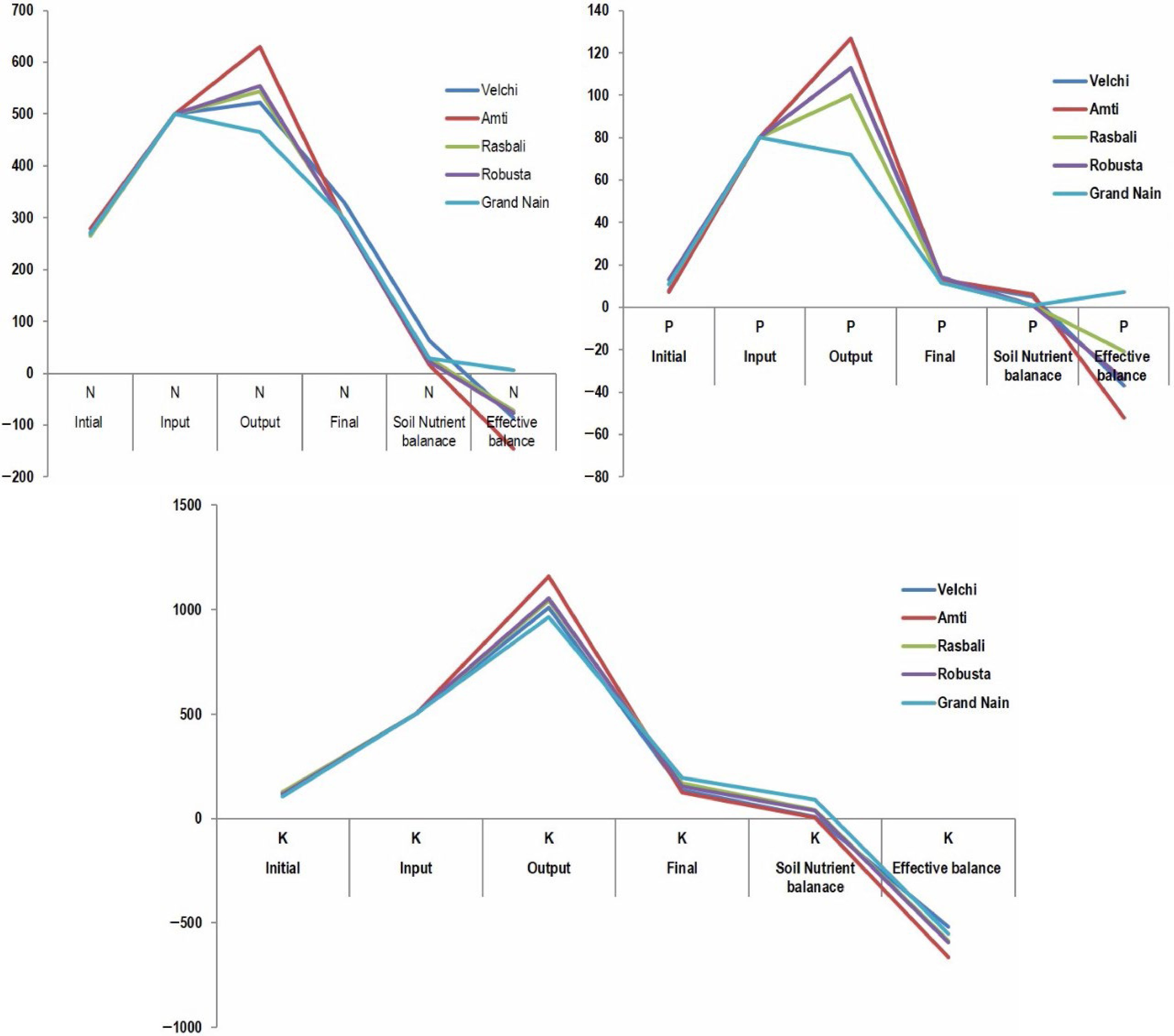
      Figure 2. 

      Soil nutrient budgeting.

      Table 5.  Nutrient budget of banana varieties under arecanut banana system (kg·ha−1·year−1).

      IntialInputOutputFinalSoil nutrient balanceEffective balance
      NPKNPKNPKNPKNPKNPK
      MeanMeanMeanMeanMeanMeanMeanMeanMeanMeanMeanMean
      Velchi266 ±
      46.34
      8 ±
      2.53
      124 ±
      45.11
      500805005221131010330.0 ±
      9.37
      12.8 ±
      4.11
      134.1 ±
      47.04
      63.94.510.0−227.1−30.4−185.8
      Amti278 ±
      78.92
      7 ±
      0.97
      120 ±
      42.10
      500805006301271160293.5 ±
      7.41
      12.9 ±
      2.09
      124.1 ±
      62.16
      15.75.84.5−232.3−23.6−131.5
      Rasbali265 ±
      22.14
      11 ±
      0.45
      127 ±
      5.56
      500805005431001044294.5 ±
      2.44
      11.4 ±
      3.18
      169.8 ±
      51.77
      29.20.842.9−253.5−39.2−139.5
      Robusta271 ±
      59.61
      13 ±
      5.16
      118 ±
      39.58
      500805005541131055292.6 ±
      19.56
      14.1 ±
      3.63
      154.7 ±
      48.74
      21.80.836.7−256.6−34.0−141.3
      Grande
      Nain
      269 ±
      42.79
      11 ±
      2.15
      105 ±
      11.64
      5008050046572965298.3 ±
      62.14
      11.6 ±
      2.78
      193.7 ±
      51.27
      29.10.989.0−284.9−50.3−125.0
    • Banana varieties differed significantly for height increment during winter only. Grand Nain recorded the lowest internodal length during all five time intervals of the study. Shade-sensitive plant species tend to grow tall with elongated internodes under shade in search of light whereas shade-tolerant ones develop short internodes and remain short in stature[21]. Arunachalam & Reddy[21] observed longer shoots of jasmine plants under coconut canopy shade with more nodes during winter than during the rainy season. Similarly, Rodrigo et al.[22]observed an increase in the plant height of both rubber and banana (Kolikuttu AAB group Silk) under the rubber-banana intercropping system compared to their respective sole crops.

    • Amti, the high-yielding cultivar in the areca intercropping system, recorded the widest leaves and high leaf area. Excessive shading reduces the leaf area of banana plants[1, 10], this might be the possible reason for the reduction in leaf width in other varieties. Moreover, the long and narrow leaves are due to genetic mutation in banana[23]. Leaf width and pseudostem height varied widely among the somaclonal variants of banana due to management practices[24]. Arunachalam & Reddy[21] reported a significant difference in leaf width under the shade of coconut for jasmine. Large leaf area leads to high radiation use efficiency[6] thereby increasing dry matter production and high yield[25].

    • All banana varieties recorded significantly lower leaf emergence rates during winter under the areca canopy except Velchi. Neither fertilizers nor shading affects the rate of leaf production during the first six months of planting[25]. Reduction in leaf emergence rate[1, 10] was also observed earlier in the shade-grown banana plants. The Velchi variety produced several leaves during winter and took only 399 d to flower while others took more than 430 d. Banana plants producing more leaves during cool temperatures tend to flower early[26, 27]. A higher leaf emergence rate is observed under trees at low density compared to higher density[28].

    • Amti and Grand Nain recorded a significantly higher individual plant yield and total yield per hectare. Amti variety recorded more hands and fruits than other varieties under the areca nut shade. Marimuthu[7] noticed higher bunch yield and higher yield per hectare in Poovan a cultivar similar to the Amti group of Mysore AAB banana under the coconut-based multi-storey cropping system. The percentage harvest and the fresh bunch yield were higher in the less shaded locations[15]. The number of hands, fruits, and bunch yield per unit area diminished when the planting density of banana increased from 1,400 to > 5,000 plants per hectare[12].

    • The effective balance (−185.8 kg·ha−1·year−1) of available soil potassium of Velchi was more negative than all other varieties (−125 to −141.3 kg·ha−1·year−1). The middle hands of Velchi did not develop, perhaps due to a deficiency of potassium. Neypoovan variety (similar to Velchi) required 2.98:0.64:12.9 kg each of available N, P, and K respectively to produce a one-ton yield[13]. The effective balance of the available soil P was highly negative for Grand Nain. Ashokan et al.[29] observed higher P uptake and good yield in the Mysore (similar to Amti) variety in the cassava-banana-elephant foot yam system. Similarly, highly negative nutrient balances were reported for N and P in mutually shaded dense banana orchards of Rwanda, Africa[30]. Velchi recorded a very high negative balance (−185.8 kg·ha−1·year−1) of available soil K than all other varieties. This negative balance of all the major nutrients can be mainly due to lower nutrient use efficiency, higher nutrient leaching due to heavy rainfall, and high crop requirements. Banana plants grown in the rubber-banana system[8] are not supplied with sufficient fertilizers which can lead to low yields of banana. The application of nutrients as per the requirements of both main and intercrops leads to higher production and improved soil quality[31,32].

      Banana is grown as a preferred component crop in tropical agroforestry systems across the world. Although the current study is conducted at a single location, the results of suitable cultivar type, traits, and nutrient budgeting are applicable after validation at other geographical conditions. The current study was conducted at the Goa Konkan coast on mid west coast of India and found the Mysore Poovan AAB group banana variety Amti as suitable for an arecanut-based agroforestry system. Marimuthu[7] noticed higher bunch yield and higher yield per hectare in Poovan a cultivar similar to the Amti group of Mysore AAB banana under the coconut-based multi-storey cropping system at Tamil Nadu Southeast coast of India. Palayam Kodan (Syn. Mysore Poovan) is suitable for intercropping in coconut at Kerala Southwest coast of India. Rajan et al.[33] AAB Prata Sub group banana cultivar is the preferred cultivar for the cacao abruca agroforestry system in Brazil[34]. Dwarf banana cultivars Mas and Goroho with fast growth and early bearing nature are found suitable as intercrop in coconut at North Sulawesi Indonesia[35].

    • The current study suggests Mysore and Grand Nain banana varieties as suitable for intercropping in high-density areca nut gardens with good performance during plant and ratoon crops. Mining nitrogen, phosphorous, and potassium nutrients in the areca–banana system is a concern and should be managed by cultivar-specific nutrient management practices. There is a need to develop a package of practices for growing banana under areca nut shade. Modification in planting geometry of areca nut is required to reduce the competition for space, nutrients, and light from intercrops.The findings of this study provide valuable insights for banana growers and researchers, highlighting the strengths and weaknesses of different banana varieties under specific environmental conditions. Further investigations into the factors affecting nutrient balance and bunch development could lead to improved cultivation practices for various banana varieties, enhancing productivity and sustainability in banana farming.

    • The authors confirm contribution to the paper as follows: design of experiments, supervision, methodology, data analysis, drafting manuscript: Arunachalam V; methodology field layout foliar observations: Vanjari SS; nutrient budget analysis, soil analysis, drafting manuscript: Paramesh V; method, soil nutrient analysis: Vishwakarma S; data tabulation, analysis: Prabhu DC; methodology yield observations: Dsouza AV; literature review: Fernandes CM. All authors reviewed the results and approved the final version of the manuscript.

    • All data generated or analyzed during this study are included in this published article and its supplementary information files.

      • The authors are grateful to the Indian Council of Agricultural Research (ICAR) for funding through the ICAR-All India Coordinated Research Project on Palms of ICAR-Central Plantation Crops Research Institute for the initial support and the facilities and support through ICAR-Central Coastal Agricultural Institute at a later stage.

      • The authors declare that they have no conflict of interest.

      • Supplemental Table S1 Varieties used in the study and their international name/group and genome type.
      • Supplemental Table S2 Duration and growing degree days of banana varieties to flowering and harvest.
      • Supplemental Table S3 Final soil available nutrient contents of banana varieties under Arecanut-banana system.
      • Copyright: © 2023 by the author(s). Published by Maximum Academic Press, Fayetteville, GA. This article is an open access article distributed under Creative Commons Attribution License (CC BY 4.0), visit https://creativecommons.org/licenses/by/4.0/.
    Figure (2)  Table (5) References (35)
  • About this article
    Cite this article
    Arunachalam V, Vanjari SS, Paramesh V, Viswakarma S, Prabhu DC, et al. 2023. Variation in plant traits and nutrient uptake among banana varieties in shaded agroecology under areca nut canopy. Technology in Agronomy 3:15 doi: 10.48130/TIA-2023-0015
    Arunachalam V, Vanjari SS, Paramesh V, Viswakarma S, Prabhu DC, et al. 2023. Variation in plant traits and nutrient uptake among banana varieties in shaded agroecology under areca nut canopy. Technology in Agronomy 3:15 doi: 10.48130/TIA-2023-0015

Catalog

  • About this article

/

DownLoad:  Full-Size Img  PowerPoint
Return
Return