


-
Torulaceae was introduced by Sturm[1] with Torula as the type genus. Members of the family are commonly known only by their anamorph as dematiaceous hyphomycetes, producing erect, micro- or macro-nematous conidiophores, with or without apical branches, doliiform to ellipsoid or clavate, brown, smooth to verruculose, mono- to poly-blastic, conidiogenous cells, often with cupulate cells, and acrogenous, phragmosporous, brown, dry, subcylindrical, smooth to verrucose conidia, frequently in branched chains[2−7] Initiative modern taxonomic treatment of Torulaceae based on a morpho-molecular approach was carried out by Crous et al.[2] who investigated the familial phylogenetic affinity, based on molecular data of five representative Torula species and Dendryphion europaeum; of which only Dendryphion and Torula were initially accepted in the family. Comprehensive taxonomic studies of Torulaceae have been further carried out by various subsequent authors; Su & Hyde[3] introduced the new genus Neotorula and two new Dendryphion species and consequently, Su et al.[8] established the genus Rostriconidium. Li et al.[9] introduced the monotypic genus Sporidesmioides and many new species in Dendryphion and Torula[5,6]. Crous et al.[10] accepted Rutola in Torulaceae and later, Boonmee et al.[11] introduced a monotypic genus Cylindrotorula in this family based on phylogenetic evidence. In a recent taxonomic classification, Rostriconidium and Sporidesmioides were treated as synonyms of Neopodoconis by Qiu et al.[12] based only morphological resemblance and molecular data of LSU and SSU. Although, their phylogenetic analyses demonstrated that Neopodoconis clade I (= Rostriconidium) and Neopodoconis clade II (= Sporidesmioides) did not form a well-resolved monophyletic clade, concurring with Wang et al.[13]. Subsequently, He et al.[14] clarified the phylogenetic affinities of Rostriconidium and Sporidesmioides (current name: Neopodoconis) in Torulaceae, demonstrating that these two genera formed well-resolved distinct subclades within Torulaceae and three representative species of Neopodoconis were excluded from Torulaceae. Therefore, the congeneric status among Neopodoconis, Rostriconidium and Sporidesmioides is questionable, pending further study.
Species of Torulaceae have been naturally found as saprobes on a wide range of hosts in both aquatic and terrestrial habitats; of which most species were isolated from submerged wood, dead branches, and herbaceous litter of various vascular plants worldwide such as in plant families Asteraceae, Brassicaceae, Cyperaceae, Fabaceae, Iridaceae, and Ranunculaceae in Asia (China, India, and Thailand), Europe (France, Germany, and Italy), North America (Canada), and others[2,3,5−8,14−19]. In contrast, a few species were reported as pathogens such as Torula herbarum was reported to cause stem blight on Ziziphus mauritiana, leaf and stem spots in plants and foot-rot of coriander. The species was also isolated from nasal swabs of Equus equus[20,21].
Torula is the most speciose genus in Torulaceae, and 543 epithets are listed in Index Fungorum[22]. However, less than a half-quarter of the known species have molecular data to clarify their phylogenetic affinities. Of these, other related taxa previously described as Torula have also been synonymized to many genera in Sordariomycetes[22]. Torula was introduced by Persoon[23] and was initially typified by Torula monilis. However, T. monilis was treated as a synonym of T. herbarum and hitherto Crous et al.[2] treated T. herbarum as the type species of Torula. The genus is characterized by dark brown to black, discrete, dry, effuse, velvety colonies, and mostly immersed mycelium, with one brown supporting cell of conidiophores, or reduced to the conidiogenous cells, that are mono- to polyblastic, solitary, brown, doliiform to ellipsoidal or clavate, smooth to verruculose conidiogenous cells, with a basally thickened and heavily melanized wall, frequently collapsing and becoming coronate, and acrogenous, phragmosporous, dry, brown, smooth to verrucose, globose to subglobose celled conidia, with strongly constricted at the septa and frequently in branched chains[2,5−7,24]. In accordance with Crous et al.[2] who introduced three new Torula species based on a morpho-molecular approach, further studies based on the multigene phylogenetic analyses were carried out by various subsequent authors[3,5−8,14,17,19,25−31]. Of these, the species were reported from different varieties of plant hosts in Asia (China, India, and Thailand), Europe (Italy, the Netherlands, and the UK), and South America (Cuba). To date, about 29 Torula species have been confirmed with molecular data[14,31,32].
During our survey of fungal diversity in Honghe Dry-hot Valley, Yunnan Province, China, a novel species Torula aquilariae was isolated from a fallen fruit pod of Aquilaria sinensis (Thymelaeaceae). The species is described and illustrated with phylogenetic support of a concatenated ITS, LSU, SSU, tef1-α, and rpb2 sequence dataset. Also, the discussion in terms of its morphological differences compared with the closely related species in the genus Torula, and an updated phylogenetic tree are provided in this paper.
-
The specimen was collected from a fallen fruit pod of Aquilaria sinensis (Thymelaeaceae) in a terrestrial habitat, in Yunnan Province, China during the wet season of the year 2023. The sample was returned to the mycological laboratory at the Center for Mountain Futures, Kunming Institute of Botany, Chinese Academy of Sciences for examination, isolation, and description of morphological characteristics. The specimen was observed under a Motic SMZ 168 series dissecting stereo-microscope and photographed with a Discovery V.8 stereo-microscope fitted with a CARL ZEISS Axio Cam ERc5S microscope camera. Micro-morphological features were picked up by a surgical needle and placed onto 10% lacto-glycerol on a clean slide. Micro-morphological features were examined and captured under a Nikon ECLIPSE Ni-U compound microscope connected with a Nikon DS-Ri2 camera using DIC microscopy. The Tarosoft (R) Image Frame Work program and Adobe Photoshop CS3 Extended version 10.0 software (Adobe Systems Inc., USA) were used for measurements and drawing photographic plates. Single spore isolation was carried out to obtain pure cultures as described in Li et al.[5,6,33]. Germinating conidia were transferred aseptically to potato dextrose agar (PDA) and malt extract agar (MEA) plates and grown at 25 °C in alternating day and night light. Colony characteristics were observed and recorded after one week and at weekly intervals.
The type specimen is deposited in the Herbarium of Cryptogams Kunming Institute of Botany Academia Sinica (HKAS), Yunnan, China. Ex-type living culture is preserved in Kunming Institute of Botany Culture Collection (KUNCC). Faces of Fungi number and MycoBank number are registered for the new taxon[34,35].
DNA extraction, PCR amplification, and sequencing
-
Fungal mycelium was scraped off and stored in a 1.5 ml microcentrifuge tube using a sterilized lancet for genomic DNA extraction. The Biospin Fungus Genomic DNA Extraction Kit-BSC14S1 (BioFlux®, PR China) was used to extract fungal genomic DNA, following the protocols in the manufacturer's instructions. The DNA amplification was performed by polymerase chain reaction (PCR) using the following genes (ITS, LSU, SSU, tef1-α, and rpb2). The ITS5 and ITS4 primer pairs were used to amplify the ITS and 5.8S regions of the rDNA gene[36]; the primers LR0R and LR5 were used to amplify the partial ribosomal RNA for the nuclear large subunit (28S, LSU)[37]; the primers NS1 and NS4 were used to amplify the partial ribosomal RNA for the nuclear small subunit (18S, SSU)[36] the primers EF1-983F and EF1-2218R were used to amplify the protein coding region for translation elongation factor 1-alpha gene (tef1-α)[38] and the primers fRPB2-5F and fRPB2-7cR were used to amplify the partial ribosomal RNA for the partial RNA polymerase second largest subunit (rpb2)[39].
The final volume of the PCR reaction was 25 μl, containing 1 μl of DNA template, 1 μl of each forward and reward primer, 12.5 μl of 2 × PowerTaq PCR Master Mix (mixture of EasyTaqTM DNA Polymerase, dNTPs, and optimized buffer, Beijing Bio Teke Corporation (Bio Teke), China) and 9.5 μl of sterilized double-distilled water (ddH2O). The PCR thermal cycling conditions of ITS, LSU, SSU, and tef1-α were processed by initialization at 94 °C for 3 min, followed by 35 cycles of denaturation at 94 °C for 30 s, annealing at 55 °C for 50 s, elongation at 72 °C for 1 min, and a final extension at 72 °C for 10 min, and final hold at 4 °C; while the PCR thermal cycle program for rpb2 was followed as initially 95 °C for 5 min, followed by 40 cycles of denaturation at 95 °C for 1 min, annealing at 52 °C for 2 min, elongation at 72 °C for 90 s, and final extension at 72 °C for 10 min[40]. Purification and sequencing of PCR products were carried out at Shanghai Majorbio Biopharm Technology Co., Ltd, China using the same primers as defined above.
Sequence alignment and phylogenetic analyses
-
Phylogenetic analyses were performed based on a concatenated ITS, LSU, SSU, tef1-α, and rpb2 sequence data. Sequences generated from this study were aligned with similar sequences obtained from GenBank and those derived from recent publications[14,19] (Table 1). Single gene alignment was performed via the online platform, MAFFT v. 7.511 (
http://mafft.cbrc.jp/alignment/server/ )[41]. Ambiguous sites were trimmed and manually edited where necessary in MEGA version 6.0[42]. Further analyses were executed based on maximum likelihood (ML) and Bayesian inference (BI) criteria, following the methodology as described in Li et al.[5].Table 1. Taxa used in the phylogenetic analysis and their corresponding GenBank accession numbers.
Taxa Culture Collection GenBank accession numbers Ref. ITS LSU SSU rpb2 tef1-α Arthopyrenia salicis CBS 368.94 KF443410 AY538339 AY538333 KF443397 KF443404 [51,52] Cycasicola goaensis MFLUCC 17-0754T MG828885 NG_059057 NG_061287 N/A MG829198 [53] Cylindrotorula indica NFCCI 4836T NR_175156 NG_081308 N/A MT321490 MT321492 [11] C. indica NFCCI 4837 MT339445 MT339443 N/A MT321491 MT321493 [11] Dendryphion aquaticum MFLUCC 15-0257T KU500566 KU500573 KU500580 N/A N/A [3] D. comosum CBS 208.69T MH859293 MH871026 N/A N/A N/A [54] D. fluminicola MFLUCC 17-1689T MG208162 MG208141 N/A N/A MG207992 [8] Mauritiana rhizophorae BCC 28866T N/A GU371824 GU371832 GU371796 GU371817 [55] M. rhizophorae BCC 28867 N/A GU371825 GU371833 GU371797 GU371818 [55] Neooccultibambusa thailandensis MFLUCC 16-0274T MH275074 MH260308 MH260348 MH412758 MH412780 [56] Neopodoconis jiangxiensis HJAUP C0947T N/A ON693849 ON693843 N/A N/A [12] N. meilingensis HJAUP C0905T N/A ON693846 ON693847 N/A N/A [12] N. obclavata HJAUP C0829T N/A ON693848 ON693844 N/A N/A [12] N. saprophyticus HJAUP C0830T N/A ON693851 ON705129 N/A N/A [12] N. sinensis HJAUP C0909T N/A ON693845 ON693850 N/A N/A [12] N. yunnanensis KUNCC 22-10737T OP359401 OP359410 OP369295 OP476726 OP471613 [57] Neotorula aquatica MFLUCC 15-0342T KU500569 KU500576 KU500583 N/A N/A [3] N. submersa HKAS 92660T NR_154247 NG_059727 N/A N/A N/A [4] Occultibambusa bambusae MFLUCC 13-0855T KU940123 KU863112 N/A KU940170 KU940193 [58] Pseudocoleodictyospora tectonae MFLUCC 12-0385T NR_154338 KU764709 NG_061232 KU712491 N/A [59] P. tectonae MFLUCC 12-0387 KU712444 KU764704 KU712462 KU712492 N/A [59] Pseudothyridariella mahakoshae NFCCI 4215T MG020435 MG020438 MG020441 MG020446 MG023140 [60] Rostriconidium aquaticum MFLUCC 16-1113T MG208164 MG208143 N/A MG207974 MG207994 [8] R. aquaticum KUMCC 15-0297 MG208165 MG208144 N/A MG207975 MG207995 [8] R. cangshanense MFLUCC 20-0147T MW010285 MW010281 N/A MW012636 N/A [61] R. pandanicola KUMCC 17–0176T MH275084 MH260318 MH260358 MH412759 MH412781 [56] Roussoella hysterioides HH 26988 N/A AB524622 AB524481 AB539102 AB539102 [55,62] R. pustulans KT 1709 N/A AB524623 AB524482 AB539103 AB539116 [55,62] Roussoellopsis macrospora MFLUCC 12-0005T KJ739604 KJ474847 KJ739608 KJ474862 KJ474855 [63,64] R. tosaensis KT 1659 N/A AB524625 AB524484 AB539104 AB539117 [55,62] Rutola graminis CBS 145906T NR_175150 NG_078685 N/A N/A N/A [10] R. graminis CPC 33695 MN313815 MN317296 N/A N/A N/A [10] Sporidesmioides thailandica MFLUCC 13-0840T MN061347 NG_059703 KX437759 KX437761 KX437766 [9] S. thailandica KUMCC 16-0012 MN061348 KX437758 KX437760 KX437762 KX437767 [9] Thyridariella mangrovei NFCCI 4213T MG020434 MG020437 MG020440 MG020445 MG020443 [60] Torula acaciae CPC29737T NR_155944 NG_059764 N/A KY173594 N/A [2] T. aquatica DLUCC 0550 MG208166 MG208145 N/A MG207976 MG207996 [8] T. aquatica MFLUCC 16-1115T MG208167 MG208146 N/A MG207977 N/A [8] T. aquilariae KUNCC 24-18640T PQ788522 PQ788524 N/A PQ810570 PQ810572 Present study T. aquilariae HKAS 145332T PQ788521 PQ788523 N/A PQ810569 PQ810571 Present study T. breviconidiophora KUMCC18-0130T MK071670 MK071672 MK071697 N/A MK077673 [28] T. calceiformis HKAS 125551T OP751054 OP751052 OP751050 OQ630510 OQ630512 [65] T. calceiformis HKAS 125552 OP751055 OP751053 OP751051 OQ630511 OQ630513 [65] T. camporesii KUMCC19-0112T MN507400 MN507402 MN507401 MN507404 MN507403 [28] T. canangae MFLUCC 21-0169T OL966950 OL830816 N/A N/A ON032379 [29] T. chiangmaiensis KUMCC16-0039T MN061342 KY197856 KY197863 N/A KY197876 [5] T. chinensis UESTCC 22.0085T OQ127986 OQ128004 OQ127995 N/A N/A [32] T. chromolaenae KUMCC16-0036T MN061345 KY197860 KY197867 KY197873 KY197880 [5] T. fici CBS 595.96T KF443408 KF443385 KF443387 KF443395 KF443402 [2] T. fici KUMCC16-0038 MN061341 KY197859 KY197866 KY197872 KY197879 [5] T. gaodangensis MFLUCC17-0234T MF034135 NG_059827 NG_063641 N/A N/A [26] T. goaensis MTCC 12620T NR_159045 NG_060016 N/A N/A N/A [27] T. herbarum CPC24414T KR873260 KR873288 N/A N/A N/A [2] T. hollandica CBS 220.69T NR_132893 NG_064274 KF443389 KF443393 KF443401 [2] T. hydei KUMCC16-0037T MN061346 MH253926 MH253928 N/A MH253930 [6] T. lancangjiangensis HKAS 112709T NR_175706 NG_081516 NG_078759 MW729780 MW729785 [11] T. longan ZHKUCC 22-0121T OR194035 OR194027 OR194032 OR228535 OR228537 [66] T. luguhuensis CGMCC 3.24256T OQ729758 OQ947766 N/A OQ999002 OQ999004 [57] T. mackenziei MFLUCC 13-0839T MN061344 KY197861 KY197868 KY197874 KY197881 [5] T. mackenziei HKAS 112705 MW723058 MW879525 MW774581 N/A N/A [11] T. masonii CBS 245.57T NR_145193 NG_058185 N/A N/A N/A [2] T. phytolaccae ZHKUCC 22-0107T ON611796 ON611800 ON611798 ON660879 ON660881 [19] T. pluriseptata MFLUCC 14-0437T MN061338 KY197855 KY197862 KY197869 KY197875 [5] T. polyseptata KUMCC 18-0131T MK071671 MK071673 MK071698 MT235830 MT235791 [28] T. sichuanensis UESTCC 22.0087T OQ127981 OQ127999 OQ127990 N/A N/A [32] T. suae CGMCC 3.24259T OP359406 OP359415 OP369300 OP476730 OP471618 [13] T. submersa UESTCC 22.0086T OQ127985 OQ128003 OQ127994 OQ158968 OQ158978 [32] T. sundara MFLU 21-0089 OM276824 OM287866 N/A N/A N/A [31] T. sundara
[as T. longiconidiophora]UESTCC 22.0088T OQ127983 OQ128001 OQ127992 OQ158967 OQ158977 [32] T. sundara
[as T. longiconidiophora]UESTCC 22.0125 OQ127984 OQ128002 OQ127993 OQ158972 OQ158976 [32] T. sundara HKAS 124486 OR470708 OR470713 OR470703 OR753781 OR753786 [14] T. sundara HKAS 124487 OR470709 OR470714 OR470704 OR753782 OR753787 [14] T. sundara KUNCC 22–12430 OP359403 OP359412 OP369297 N/A OP471615 [13] T. sundara KUNCC 22–13431 OP359404 OP359413 OP369298 OP476728 OP471616 [13] T. thailandica GZCC20-0011T MN907426 MN907428 MN907427 N/A N/A [7] The newly generated sequences are shown in bold and the ex-type strains are indicated in superscript 'T'. N/A means the data is not available in GenBank. Maximum likelihood (ML) analysis was implemented by the Randomized Axelerated Maximum Likelihood program on raxmlGUI v. 7.4.2 graphical interface[43] with the GTR + GAMMAI model of nucleotide substitution and run for 1000 rapid bootstrap replicates[44]. The best-fit model of nucleotide substitution of the combined dataset was determined by MrModeltest v. 2.3[45]; of which GTR + I + G was the best-fit model for a combined dataset under the Akaike Information Criterion (AIC) and incorporated into the analysis. Bayesian inference (BI) was analyzed by MrBayes v.3.1.2[46]. Bayesian posterior probabilities (PP) were evaluated by Markov chain Monte Carlo sampling (BMCMC)[46−48]. Two parallel runs with six simultaneous Markov chains were run for one million generations, and trees were sampled every 100th generation (resulting in 10,001 trees). The first 2,000 trees were set as burn-in and were discarded. The remaining trees were used to calculate posterior probabilities (PP) in the majority rule consensus tree.
The phylograms were represented in FigTree v1.4.0 program[49], drawn in PowerPoint 2016 (Microsoft Inc., Redmond, WA, USA), and converted to jpeg file in Adobe Photoshop version CS5 (Adobe Systems Inc., USA). The new sequences were submitted in GenBank (Table 1). The alignment was deposited in TreeBASE (2024) under the accession number 31909. New species is established as per recommendations proposed by Jeewon & Hyde[50].
-
Torula aquilariae X.H. Li, Phookamsak & J.F. Li, sp. nov.
MycoBank number: MB 857067, Facesoffungi number: FOF 17237; Fig. 1.
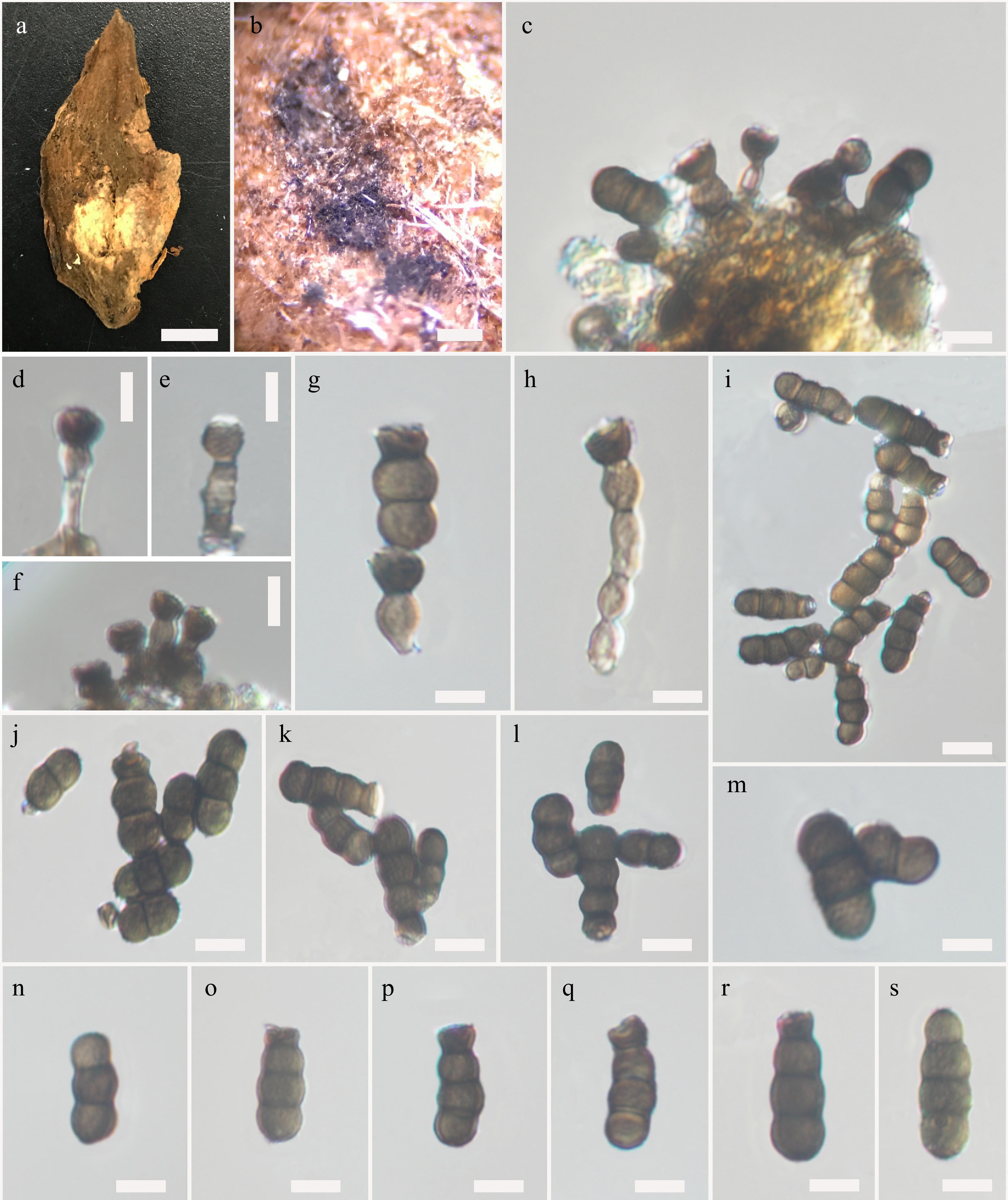
Figure 1.
Torula aquilariae (KUN-HKAS 145332, holotype). (a) A fallen fruit pod of Aquilaria sinensis. (b) Colonies on a fallen fruit pod of A. sinensis. (c) Conidial structure on the substrate. (d)–(h) Conidiophores with conidiogenous cell. (i)–(m) Conidial masses. (n)–(s) Conidia. Scale bars: (a) 0.5 cm, (b) 200 μm, (c)–(h) and (j)–(s) 5 μm, (i) 10 μm.
Etymology: Refers to the host genus, Aquilaria, of which the holotype was collected.
Holotype: KUN-HKAS 145332
Saprobic on fallen fruit pod of Aquilaria sinensis (Thymelaeaceae). Teleomorph: Undetermined. Anamorph: Colonies sporadic on host, black, powdery. Mycelium immersed on the substrate, composed of septate, branched, smooth, light brown hyphae. Conidiophores 6–20 μm long × 3.5–5 μm wide (
$\overline {x} $ $\overline {x} $ $\overline {x} $ Cultural characteristics: Conidia germinating on PDA within 14 h and germ tubes produced from the apical cell. Colonies growing on PDA, reaching 5 cm in 10 d at 28 °C, mycelium partly immersed to superficial, slightly effuse, cottony, with a regular edge, surface greyish-white to brown, reverse pale black, dense, circular, slightly raised, smooth, entire, wrinkled folded, producing brown pigmentation in agar; teleomorph not formed within 60 d.
Material examined: CHINA, Yunnan Province, Yuan Jiang, on a fallen fruit pod of Aquilaria sinensis. (Thymelaeaceae), 12 July 2023, J-F. Li, TA-01 (KUN-HKAS 145332, holotype), ex-type living culture KUNCC 24-18640.
Notes: Torula aquilariae resembles T. masonii in having brown, verruculose, conidia and with constricted at the septa and a dark terminal coronate cell at the apex, but differs in having smaller (T. aquilariae, 14.5 × 4.4 μm vs 21.6 × 9.5 μm, in T. masonii) conidia, shorter (T. aquilariae, 15.6 × 4.6 μm vs 16.8 × 4.5 μm, in T. masonii) conidiophores with smaller conidiogenous cells (T. aquilariae, 4.5 × 4.8 μm vs 6.6 × 5.3 μm, in T. masonii)[2,5]. Phylogenetic analyses showed that T. aquilariae constitutes an independent branch and is sister to T. mackenziei. Morphologically, T. aquilariae differs from T. mackenziei in having conidia with a dark terminal coronate cell at the apex, which is rounded and paler in T. mackenziei; and longer conidiophores (T. aquilariae, 15.6 × 4.6 μm vs 3.8 × 3.5 μm, in T. mackenziei) while other morphological characters are difficult to use to distinguish these two species[5]. Moreover, the comparison of nucleotide pairwise differences indicated that T. aquilariae differs from T. mackenziei (MFLUCC 13-0839, ex-type strain) in 10/488 bp (2%) difference across the ITS gene region, 52/857 bp (6%) difference across the tef1-α gene region and 53/895 bp (5.9% difference, across the rpb2 gene region. Based on morphological characteristics and phylogenetic support coupled with the difference in nucleotide polymorphism, T. aquilariae is introduced as a new species in this study.
Phylogenetic analyses
-
The concatenated ITS, LSU, SSU, tef1-α, and rpb2 dataset comprise 75 taxa with Occultibambusa bambusae (MFLUCC 13–0855) and Neooccultibambusa thailandensis (MFLUCC 16–0274) as the outgroup taxa. Bayesian Inference (BI) and maximum likelihood (ML) analyses of the combined dataset were performed to determine the placement of our new species and clarified relationships at the interspecific level. The phylogenetic trees obtained from BI and ML analyses resulted in trees with largely similar topologies and also similar to those generated from previous studies. The best-scoring RAxML tree is shown in Fig. 2, with the final ML optimization likelihood value of –37630.023997 (ln). The dataset consists of 3,952 total characters including gaps (ITS: 1–625 bp, LSU: 626–1,354 bp, SSU: 1,355–2,294 bp, tef1-α: 2,295–3,067 bp, rpb2: 3,068–3,952 bp). RAxML analysis yielded 960 distinct alignment patterns and 30.36% of undetermined characters or gaps. Estimated base frequencies were as follows: A = 0.244496, C = 0.260259, G = 0.274215, T = 0.221030, with substitution rates AC = 1.131746, AG = 2.587944, AT = 1.227920, CG = 0.930470, CT = 5.073462, GT = 1.000000. The proportion of invariable sites I = 0, the gamma distribution shape parameter alpha = 0.283343, and the tree length = 4.777392. Bayesian posterior probabilities (BYPP) from MCMC were evaluated with the final average standard deviation of split frequencies = 0.008843.
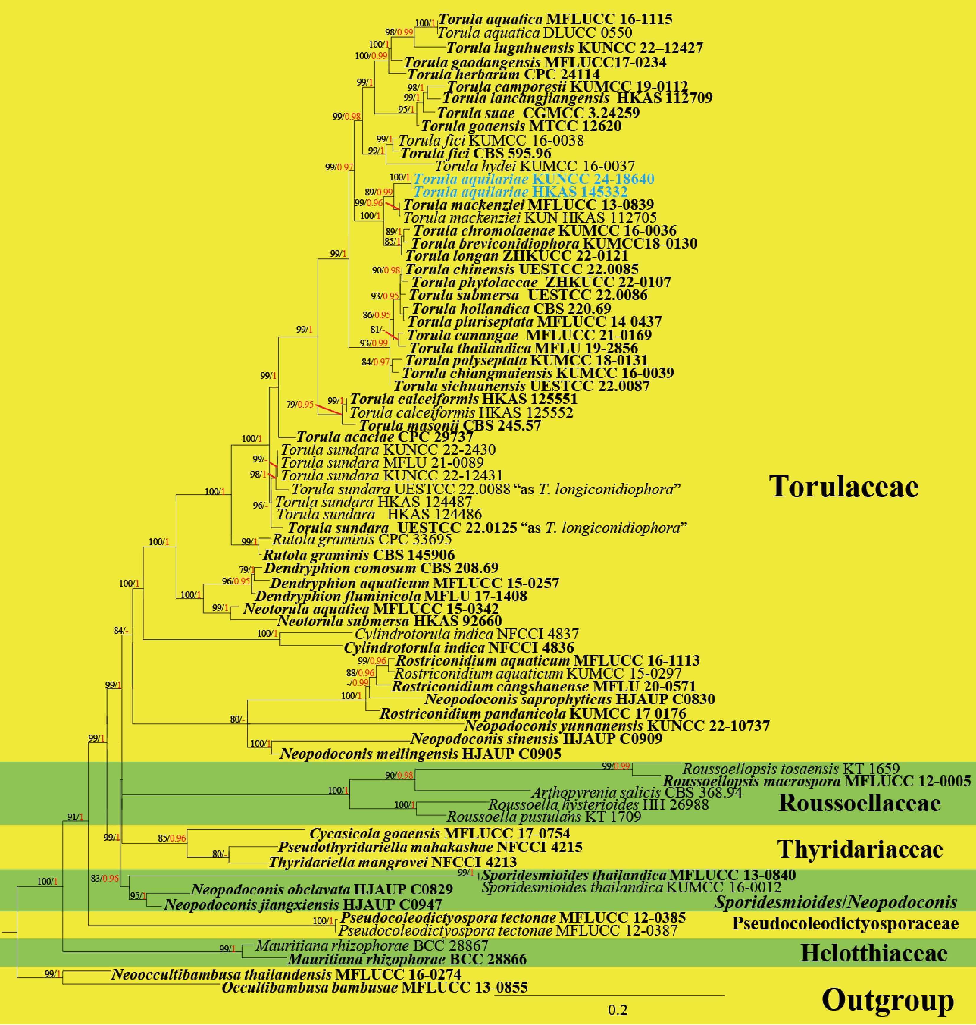
Figure 2.
Phylogenetic construction using RAxML-based analysis of a combined ITS, LSU, SSU, tef1-α, and rpb2 DNA sequence dataset. Bootstrap support values for maximum likelihood (ML) equal to or greater than 70% and Bayesian posterior probabilities (PP) equal to or greater than 0.95 are shown as 'ML/PP' at the nodes. The tree is rooted to Occultibambusa bambusae (MFLUCC 13-0855) and Neooccultibambusa thailandensis (MFLUCC 16–0274). The type strains are in black bold and the newly generated sequences are indicated in blue bold.
Seventy-five available ex-type and representative strains of genera in Torulaceae, and other related families (Halotthiaceae, Pseudocoleodictyosporaceae, Roussoellaceae, and Thyridariaceae) are included in the present phylogenetic analyses (Fig. 2). Multigene phylogenetic analyses demonstrated that these families formed well-resolved monophyletic clades in the present study, except Torulaceae that is well-resolved in ML analysis (84% ML), but has low support in BI analysis. Of these, representative genera in Torulaceae also formed well-resolved subclades in Torulaceae, except Neopodoconis/Rostriconidium that are well-resolved in ML analysis (80% ML), but have low support in BI analysis. Neopodoconis saprophyticus (HJAUP C0830) nested among Rostriconidium species with significant support in BI analyses (0.99 BYPP). Whereas N. meilingensis (HJAUP C0905), N. sinensis (HJAUP C0909), and N. yunnanensis (KUNCC 22-10737) clustered as basal to Rostriconidium species. Besides, Sporidesmioides thailandica (MFLUCC 13-0840 and KUMCC 16-0012) clustered with Neopodoconis jiangxiensis (HJAUP C0947), and N. obclavata (HJAUP C0829) with significant support (83% ML, 0.95 BYPP) and nested between Pseudocoleodictyosporaceae, and Thyridariaceae, distant from Torulaceae.
In the present study, most of the Torula species formed well-supported branches within Torula (≥ 70% ML, and 0.95 BYPP), except for T. canangae, T. hollandica, T. pluriseptata, T. submersa, T. sundara, and T. thailandica. Torula canangae is sister to T. thailandica with 80% ML support but with low support in BI analysis. Torula hollandica is sister to T. pluriseptata. These two species clustered with T. submersa, T. phytolaccae, and T. chinensis with significant support (93% ML, and 0.95 BYPP). Torula sundara formed a well-resolved subclade in ML analysis (96% ML) but had low support in BI analysis. Two new strains (KUNCC 24-18640 and HKAS 145332) formed a robust subclade, sister to T. mackenziei (MFLUCC 13-0839 and KUN-HKAS 112705) with significant support (89% ML and 0.99 BYPP, Fig. 2) and closely related to T. breviconidiophora, T. chromolaenae, and T. longan.
-
Torula have a wide host range in various habitats and are commonly found as saprobes in both aquatic and terrestrial habitats in temperate to tropical climatic zones[2,5,6,56,65,67]. Even though, more than 540 species epithets were listed under the genus Torula, most of which contain ambiguous species that were emphasized by only morphological characteristics. These ambiguous species need to be clarified based on type specimen using a polyphasic taxonomic approach. This study reveals a Torula species isolated from a fallen fruit pod of Aquilaria sinensis in Honghe Dry-hot Valley, Yunnan, China, which morphologically fits well among extant species in having conidia with dark terminal coronate cells at the apex and long, subcylindrical conidiophore with coronate conidiogenous cells. Multigene phylogenetic analyses demonstrated that the new species, Torula aquilariae (KUNCC 24-18640 and KUN-HKAS 145332) clustered with T. mackenziei, but phylogenetic analyses fully support it as the distinct species, constitute an independent lineage with high statistical support (Fig. 2). Torula aquilariae is also the first record on host Aquilaria sinensis from Honghe Dry-hot Valley.
Despite some morphological differences that segregate each species in Torula, the morphology of Torula species are similar and is difficult to use to distinguish them. However, multigene phylogenetic analyses can be utilized in clarifying the interspecific relationships of these species, of which tef1-α and rpb2 genes are reliable phylogenetic markers for delineating Torula species. For instance, Li et al.[5] introduced T. chromolaenae and T. mackenziei that resemble T. herbarum (the type species) in having solitary to catenate, septate, brownish and round-ended conidia, mononematous conidiophores and polyblastic conidiogenous cells. However, these three species formed well-resolved distinct subclades in Li et al.[5,6] and are clearly distinguished when the number of taxon samples were increased[13,14,19,68].
The conspecific status of Torula longiconidiophora and T. sundara is uncertain. Torula sundara was introduced by Jayawardena et al.[31] (published in February) to accommodate the hyphomycete species previously described as Dwayabeeja sundara. The new collection was isolated from bamboo culms in Chiang Mai Province, Thailand, and has shown morphological resemblance with D. sundara. Although, the type specimen of D. sundara was not examined, phylogenetic analyses demonstrated that the new collection belongs to Torula and is sister to T. acaciae. Therefore, the new combination, Torula sundara was proposed by Jayawardena et al.[31]. In the same year, Tian et al.[32] introduced T. longiconidiophora for the new taxon isolated from decaying wood in a damp environment in Sichuan Province, China (published in January). Phylogenetic analyses also demonstrated that T. longiconidiophora formed a sister clade with T. acaciae in Tian et al.[32]. Recent taxonomic work on Torulaceae was carried out by He et al.[14], who investigated phylogenetic relationships of the genera in Torulaceae and reported a new record for T. sundara on decaying wood submerged in freshwater from Yunnan, China. Based on morphological resemblance and phylogenetic evidence, He et al.[14] treated T. longiconidiophora as a synonym of T. sundara, even though T. longiconidiophora was published earlier. Besides, the type strain of T. sundara is unavailable, of which only ITS and LSU genes are available for T. sundara (MFLUCC 21-0067; reference strain for the combination). In the present study, five representative strains of T. sundara (including two strains previously named as T. longiconidiophora) do not form a well-resolved subclade in Torula. Hence, the conspecific status between T. longiconidiophora and T. sundara as well as the intraspecific relationships between representative strains of T. sundara need to be clarified using sufficient phylogenetic markers.
Qiu et al.[12] synonymized Rostriconidium and Sporidesmioides under the genus Neopodoconis based on their morphological resemblance and phylogenetic analyses of combined LSU and SSU gene regions. Qiu et al.[12] introduced five new species in Neopodoconis, namely N. jiangxiensis, N. meilingensis, N. obclavata, N. saprophyticus, and N. sinensis. In their phylogenetic analyses, N. meilingensis and N. saprophyticus clustered with Rostriconidium in Torulaceae. Whereas N. jiangxiensis, N. obclavata, and N. sinensis formed a sister subclade with Sporidesmioides, basal to Rousoellaceae. Unfortunately, the type of Neopodoconis, N. ampullacea, lacks molecular data to clarify its generic placement. Besides, Sporidesmium-like taxa are well-known to be morphologically similar; however, these taxa can be segregated into different genera and families based on molecular data[3,8,9,69]. Even though, the representative Neopodoconis species is phylogenetically separated into two distinctive clades in Qiu et al.[12], Rostriconidium and Sporidesmioides were treated as synonyms of Neopodoconis based solely on morphological characteristics. Subsequently, Wang et al.[13] demonstrated that Neopodoconis formed two distinct clades: clade I comprised N. saprophyticus and other Rostriconidium species in Torulaceae and clade II comprised N. jiangxiensis, N. meilingensis, N. obclavata, N. sinensis, and Sporidesmioides thailandica, as basal to Thyridariaceae. He et al.[14] updated the phylogenetic relationship of the genera in Torulaceae based on multigene phylogenetic analyses of a combined ITS, LSU, SSU, tef1-α, and rpb2 genes. Their phylogenetic results demonstrated that Rostriconidium and Sporidesmioides formed well-resolved distinct subclades in Torulaceae. In the present study, Neopodoconis meilingensis (HJAUP C0905), N. sinensis (HJAUP C0909), N. saprophyticus (HJAUP C0830), and N. yunnanensis (KUNCC 22-10737) clustered with Rostriconidium species in Torulaceae. Whereas, N. jiangxiensis (HJAUP C0947) and N. obclavata (HJAUP C0829) clustered with Sporidesmioides and is basal to Thyridariaceae. Based on phylogenetic evidence and delimitation on molecular data of the type species of Neopodoconis, we therefore, tentatively resurrect the genera Rostriconidium and Sporidesmioides until the generic placement of Neopodoconis is clarified based on type study.
We are grateful to the Yunnan Province Postdoctoral Orientation Training Fund (E33O386261) for supporting this research. The authors acknowledge the Biology Experimental Center, Germplasm Bank of Wild Species, Kunming Institute of Botany, Chinese Academy of Sciences for providing molecular laboratory facilities for the molecular work. Xiao-Hong Li thanks Dr. Shaun Pennycook, Xia Tang, Ming-Yang Kou, and Chao-Yu Luo for their available suggestions and help. Rungtiwa Phookamsak sincerely acknowledges Introducing Talents Start-up Fund of Kunming Institute of Botany, Chinese Academy of Sciences, Yunnan Revitalization Talent Support Program 'Young Talent' Project (Grant No. YNWR-QNBJ-2020-120), Yunnan Revitalization Talent Support Program 'High-end Foreign Expert' Project, and Independent research of Department of Economic Plants and Biotechnology, Yunnan Key Laboratory for Wild Plant Resources, Kunming Institute of Botany, Chinese Academy of Sciences (Grant No. Y537731261), Jianchu Xu thanks the Yunnan Department of Sciences and Technology of China (Grant Nos 202302AE090023, 202303AP140001).
-
The authors confirm contribution to the paper as follows: conceptualization, data curation, and formal analysis: Li XH, Li JF, Phookamsak R; funding acquisition: Xu JC, Li JF; investigation, methodology, and writing—original draft: Li XH, Sun FQ, Jiang HB, Li JF, Phookamsak R; project administration: Li XH, Sun FQ, Li JF; supervision: Xu JC, Li JF, Phookamsak R. writing—review and editing: Li XH, Sun FQ, Li JF, Jiang HB, Phookamsak R. All authors contributed to the article and approved the submitted version.
-
The information of the new species introduced in this study can be found in online repositories. The name is registered for Mycobank repository under MycoBank number: MB 857067. The final tree and sequence matrix are deposited in TreeBASE (2024) under the accession number 31909 and GenBank accession number(s) can be found below: https://www.ncbi.nlm.nih.gov/genbank/ as ITS: PQ788521, PQ788522; LSU: PQ788523, PQ788524; tef1-α: PQ810569, PQ810570; rpb2: PQ81057, PQ810572.
-
The authors declare that they have no conflict of interest.
- Copyright: © 2024 by the author(s). Published by Maximum Academic Press, Fayetteville, GA. This article is an open access article distributed under Creative Commons Attribution License (CC BY 4.0), visit https://creativecommons.org/licenses/by/4.0/.
-
About this article
Cite this article
Li XH, Phookamsak R, Sun F, Jiang HB, Xu JC, et al. 2024. Torula aquilariae sp. nov. (Torulaceae, Pleosporales), a new species associated with Aquilaria sinensis from Yunnan, China. Studies in Fungi 9: e020 doi: 10.48130/sif-0024-0019 

Torula aquilariae sp. nov. (Torulaceae, Pleosporales), a new species associated with Aquilaria sinensis from Yunnan, China
- Received: 18 November 2024
- Revised: 23 December 2024
- Accepted: 23 December 2024
- Published online: 31 December 2024
Abstract: Torula aquilariae sp. nov. was isolated from a fallen fruit pod of Aquilaria sinensis in Yunnan Province, China. Preliminary identification based on morphological characteristics, the new species is typical of Torula forming effuse, dark brown to black colonies, with monoblastic or polyblastic, doliiform to ellipsoid or cupulate conidiogenous cells, and acrogenous, phragmosporous, brown, septate, smooth-walled to verrucose conidia, in short, branched chains. Multigene phylogenetic analyses of a concatenated ITS, LSU, SSU, tef1-α, and rpb2 sequence data demonstrated that Torula aquilariae sp. nov. is a sister taxon of T. mackenziei, and nested between T. breviconidiophora and T. chromolaenae within Torula (Torulaceae, Pleosporales). Morphologically, the new species resembles T. mackenziei, but is different in the size of the conidiophores and conidia. Besides, the nucleotide pairwise comparison of ITS, tef1-α, and rpb2 gene regions also supports their distinction. Based on multigene phylogeny coupled with morphological traits and nucleotide polymorphism analyses, Torula aquilariae sp. nov. is introduced in this study. Detailed descriptions, illustrations, and an updated phylogeny are provided.