REVIEW   Open Access    

Banana somatic embryogenesis and biotechnological application

  • # These authors contributed equally: Jingyi Wang, Shanshan Gan

More Information
  • As one of the most important economic crops for both staple food and fruit widely cultivated in the tropics and subtropics, banana (Musa spp.) is susceptible to a plethora of abiotic and biotic stresses. Breeding cultivars resistant to abiotic and biotic stressors without adverse effects on yield and fruit quality are the objectives of banana improvement programs. However, conventional breeding approaches are time-consuming and severely hampered by inherent banana problems (polyploidy and sterility). Therefore, genetic transformation is becoming increasingly popular and can provide rapid solutions. Numerous efforts have been made to develop superior banana cultivars with better resistance to abiotic and biotic stresses and optimum yields using genetic modification strategies. Somatic embryogenesis (SE) through embryogenic cell suspension (ECS) cultures is an ideal recipient system for genetic transformation in banana. The purpose of this paper is to review the current status of banana somatic embryo research, clarify the process of banana somatic embryo induction and culture, and summarize the main influencing factors in the process of somatic embryogenesis. At the same time, their applications in breeding technologies such as cryopreservation, protoplast culture, genetic transformation and gene editing were also summarized, in order to provide reference for the research and practical application of banana somatic embryogenesis in the future.
  • Cytochrome P450 (P450) is an important superfamily in plants, the members of which play important roles in a wide range of biochemical pathways to produce phytohormones, including auxin, brassinosteroids (BRs) and gibberellins (GAs), as well as secondary metabolites, such as phenylpropanoids and fatty acids[1,2]. Plant P450s were grouped into 11 clans in two categories: multi-family clans (CYP71, CYP72, CYP85, CYP86) and single-family clans (CYP51, CYP74, CYP97, CYP710, CYP711, CYP727, CYP746)[2,3].

    The CYP78A family is one of the families in the CYP71 clan and a plant-specific gene family[3]. The first CYP78A gene, CYP78A5/KLUH, was characterized in Arabidopsis, which is a positive regulator of organ size by promoting cell proliferation. While loss-of-function of Arabidopsis CYP78A5/KLUH results in smaller leaves and floral organs, over-expression of CYP78A5/KLUH produces larger organs, including leaves, seeds and petals[4,5]. Recently, AtKLUH was shown to play positive roles in regulating leaf longevity and drought tolerance by promoting cytokinin signaling and proline metabolism[6]. In addition, cyp78a5 mutants also exhibit reduced plastochron length and early flowering[7,8], indicating the pleiotropic roles of AtKLUH in regulating Arabidopsis growth and development. There are five other Arabidopsis CYP78A members, CYP78A6, CYP78A7, CYP78A8, CYP78A9 and CYP78A10. CYP78A7 and CYP78A5 play redundant roles in regulating plastochron length, leaf size and apical dominance[7,9]. CYP78A6 (EOD3) and CYP78A9 were shown to redundantly regulate seed size and leaf senescence[9,10]. The functions of CYP78A10 are still unknown.

    The orthologs of AtCYP78A5 have been shown to regulate seed or fruit size in other plants. For example, increased expression of tomato KLUH leads to larger fruits and seeds by stimulating cell division[1114]. Over-expression of maize PLASTOCHRON1 (ZmPLA1) stimulates leaf growth by extending cell division duration, leading to increased seed yield[15]. Recent studies in wheat showed that constitutive overexpression of TaCYP78A5 enhances grain weight by accumulating auxin[16,17]. In addition, soybean CYP78A10 and CYP78A72, sweet cherry CYP78A9 and rice GIANT EMBRYO (GE; CYP78A13) have also been demonstrated as key regulators of organ size[1824], showing great potential in yield improvement.

    Eggplant is an important vegetable crop that is cultivated worldwide. Eggplant is also known as an important medical plant due to its high phenolic and alkaloid contents, including chlorogenic acid and acetylcholine (ACh), which can be used to treat human diseases, such as diabetes and high blood pressure[25,26]. With increasing worldwide population, the yield of eggplants is needed to be increased to meet the demands. Considering the potential of CYP78A family members in yield improvement, it would be useful to identify and characterize the CYP78A family in the eggplant genome. However, none of the CYP78A genes have been cloned or characterized in eggplant to date.

    In the present study, the CYP78A gene family was identified in the eggplant genome. Detailed information of the SmCYP78As, including chromosomal locations, phylogenetic relationships, gene structures, conserved motifs, synteny and candidate transcription factors which might directly bind the promoter of SmCYP78As, were investigated. qRT-PCR was performed to analyze the tissue-specific expression patterns of the SmCYP78As. Expression levels of SmCYP78As in young flower buds and developing ovaries were also analyzed using RNA-seq data. Co-expression clustering and GO enrichment analysis were conducted to gain further insights into the functions of SmCYP78As. The results of the present study will be helpful for further functional study of the SmCYP78A genes.

    Amino acid sequences of Arabidopsis CYP78As were downloaded from The Arabidopsis Information Resource (TAIR) database (www.arabidopsis.org). The six AtCYP78A proteins were used as query sequences and searched against the eggplant proteins in the Eggplant Genome Database (http://eggplant-hq.cn/Eggplant/home/index). Then, the Pfam (http://pfam.janelia.org/) and Simple Modular Architecture Research Tool (SMART) (http://smart.embl-heidelberg.de/) programs were used to confirm the existence of the P450 domain. The number of amino acids, isoelectric points (PIs) and molecular weights (MWs) of the 27 CYP78A proteins were determined using ExPASy (https://web.expasy.org/protparam/), and their subcellular localizations were predicted with Cell-PLoc 2.0 (www.csbio.sjtu.edu.cn/bioinf/Cell-PLoc-2/).

    Multiple sequence alignments were performed and the phylogenetic trees were constructed with the full protein sequences of the 27 CYP78As from eggplant, Arabidopsis, rice and tomato using MEGA 7.0. The neighbor-joining (NJ) method was used for the construction of the phylogenetic tree with the following parameters: Poisson correction, pairwise deletion and 1000 bootstrap replicates. Gene structures were analyzed using TBtools[27]. Conserved motifs were identified using MEME (Multiple Expectation Maximization for Motif Elicitation)[28].

    All SmCYP78A genes were mapped to chromosomes based on physical location information from the Eggplant Genome Database using Circos[29]. Multiple Collinearity Scan toolkit (MCScanX) is employed for scanning multiple genomes to align syntenic blocks[30].

    An inbred line '14-345' with round and purple black fruits was used in this study for gene expression analysis and RNA-seq. The plants were grown in the greenhouse at Hebei Agricultural University in Baoding (38° N, 115° E), China.

    Roots, stems, leaves, young flower buds, petals and sepals of 0 DPA (Days post anthesis) flowers, pericarp of 0 DPA ovary, fruit flesh of 10 DPA fruits were collected separately from at least 2−3 individual plants as one biological replicate. Three biological replicates for each tissue sample were carried out for qRT-PCR. Total RNAs were extracted using TRIzol reagent (Invitrogen, USA) and treated with DNase I (Fermentas, Canada) following the manufacturer's protocol. First strand cDNA was synthesized from 1 μg total RNA using PrimeScript 1st Strand cDNA Synthesis Kit (TaKaRa, Japan). qRT-PCR was performed on LightCycler 96 (Roche, Switzerland) using the ChamQ Universal SYBR qPCR Master Mix (Vazyme, China). Clathrin adaptor complexes medium subunit (CAC) gene (Smechr0800014) was used as the housekeeping gene to normalize the gene expression. Primers used for qRT-PCR are listed in Supplemental Table S1. The relative expression level was calculated using the 2− ΔΔCᴛ method.

    Young flower buds and developing ovaries, including 10 DBA (Days before anthesis), 7 DBA and 0 DPA, were collected with four biological replicates for each tissue sample. The cDNA library preparation and sequencing were conducted by the Novogene Bioinformatics Technology Company (Beijing, China) on the Illumina HiSeq TM4000 platform (Illumina Inc., San Diego, CA, USA). The read mapping was performed using the latest version of the Tuxedo protocol with HISAT2 and StringTie[31]. The clean reads from each library were aligned to the eggplant genome of 'HQ-1315' using HISAT2. The mapping results were normalized via Stringtie to compute TPM (Transcripts Per Kilobase Million) values of genes.

    Co-expression genes were clustered using fuzzy C means in the Mfuzz package[32] and gene clusters were visualized by plotting of the normalized expression profiles of each cluster using ggplot2 package in R[33]. For GO enrichment analysis, eggplant geneIDs were converted to Arabidopsis geneIDs using BLASTP and then GO enrichment analysis was performed using clusterProfiler with cnetplot function in R[34]. The significant GO terms were identified with the adjusted value smaller than 0.05.

    The six AtCYP78A proteins were employed as a query to search the SmCYP78As in the eggplant genome via BlastP program, resulting in six putative SmCYP78A genes. The presence of the P450 domains in the six putative SmCYP78A proteins was confirmed via Pfam and SMART, indicating that the six proteins are members of the eggplant CYP78A family (Table 1).

    Table 1.  Summary information of CYP78A family genes in eggplant, Arabidopsis, rice and tomato.
    Gene IDGene nameLocationDeduced polypeptidePSL
    ChrStartEndLength (aa)MW (KDa)pI
    Smechr0101302CYP78A6Chr1126405811264324553860.358.90ER
    Smechr0302971CYP78A5Chr3898868008988918451658.196.79ER
    Smechr0400048CYP78A7Chr442056642292052558.989.23ER
    Smechr0500049CYP78A8Chr578327278534652859.769.30ER
    Smechr0502180CYP78A9Chr5749692627497117753760.557.52ER
    Smechr1100733CYP78A10Chr11105348191053696755161.708.23ER
    AT1G01190CYP78A8Chr1830458494654160.918.22ER
    AT1G13710CYP78A5Chr14702657470469451857.648.57ER
    AT1G74110CYP78A10Chr1278666672786836853860.187.84ER
    AT2G46660CYP78A6Chr2191533281915557953159.578.29ER
    AT3G61880CYP78A9Chr3229058682290795855662.629.04ER
    AT5G09970CYP78A7Chr53111945311423953759.496.69ER
    LOC_Os10g26340CYP78A11/PLA1Chr10136587901366054355659.087.06ER
    LOC_Os11g29720CYP78A5Chr11172342851723817853959.6410.19ER
    LOC_Os03g04190CYP78A9Chr31920043192189651655.808.10ER
    LOC_Os03g30420CYP78A6/GL3.2Chr3173404151734228451656.059.00ER
    LOC_Os03g40600CYP78A7Chr3225676702256868519420.297.99ER
    LOC_Os03g40610CYP78A8Chr3225727062257400830832.8410.12ER
    LOC_Os07g41240CYP78A13/GEChr7247137782471581352655.898.68ER
    LOC_Os08g43390CYP78A15/BSR2Chr8274205012742283655259.799.38ER
    LOC_Os09g35940CYP78A10Chr9206913062069311655460.749.07ER
    Solyc01g096280CYP78A6Chr1796222667962461853961.058.60ER
    Solyc03g114940CYP78A5/KLUHChr3592173895921973051758.386.21ER
    Solyc05g015350CYP78A8Chr5104750281047926733237.856.08ER
    Solyc05g047680CYP78A7Chr5585063905850829253260.499.20ER
    Solyc10g009310CYP78A9Chr103224421322712352661.649.05ER
    Solyc12g056810CYP78A10Chr12625109416251267653760.717.50ER
     | Show Table
    DownLoad: CSV

    The information of the six SmCYP78As, including chromosomal locations, amino acids number (length), PIs, MWs and predicted subcellular localizations (PSL), was listed in Table 1. To gain further insights into the CYP78A family genes in plants, the information of CYP78As from Arabidopsis, rice and tomato was also included in Table 1. The amino acids number of SmCYP78A proteins varies from 516 (SmCYP78A5) to 551 (SmCYP78A10), the PI ranges from 6.79 (SmCYP78A5) to 9.30 (SmCYP78A8) and the MW ranges from 58.19 (SmCYP78A5) to 61.70 KDa (SmCYP78A10). Interestingly, the amino acids lengths of rice CYP78A7 and CYP78A8 as well as tomato CYP78A8 (SlCYP78A8) were much smaller than other CYP78As from Arabidopsis, rice, tomato and eggplant. It would be interesting to know whether the three short CYP78A proteins show similar functions with other CYP78As. Notably, the subcellular localizations of all CYP78As were predicted to be localized in endoplasmic reticulum (ER), which is in agreement with the subcellular localizations of AtCYP78A5 and TaCYP78A5 in vivo[4,16].

    To elucidate the evolutionary relationships of SmCYP78As, an unrooted neighbor-joining (NJ) phylogenetic tree was constructed using the full protein sequences from the six AtCYP78As, nine OsCYP78As, six SlCYP78As and six SmCYP78As. The resulting tree contained five distinct clades (C1-C5) (Fig. 1a). Phylogenetic analysis revealed that C1 and C4 were shared in all the four species. There was equal number of the CYP78A proteins from eggplant, Arabidopsis, tomato and rice in C1 (Fig. 1a). C4 contained two SmCYP78As and one CYP78A from each of tomato, Arabidopsis and rice, indicating the expansion of SmCYP78As in this clade compared with the other three species. C2 and C3 didn't contain SmCYP78As. C2 was shared by rice and tomato, but not in Arabidopsis and eggplant, suggesting the unique roles of the CYP78As in C2 that were likely acquired or expanded in tomato and rice after divergence from the last common ancestor with eggplant and Arabidopsis. It is worth noting that C3 only contains rice CYP78As and OsCYP78A8 didn't fit into any clades, which may have evolved following divergence and have special roles in rice. Moreover, C5 didn't include any rice CYP78As but only members from eggplant, tomato and Arabidopsis, suggesting that the CYP78As in C5 may have been lost in rice during evolution.

    Figure 1.  Phylogenetic relationships, gene structure and conserved protein motifs of CYP78A genes from eggplant, Arabidopsis, rice and tomato. (a) The phylogenetic tree was constructed based on the full-length protein sequences of six AtCYP78As, nine OsCYP78As, six SlCYP78As and six SmCYP78As proteins using MEGA 7.0 software. Eggplant, Arabidopsis, rice and tomato CYP78As were labeled by red, black, pink and green dots. (b) Exon-intron structure of CYP78A genes. Black lines indicate introns. The number indicates the phases of corresponding introns. (c) The motif composition of CYP78A proteins. The motifs, numbers 1–10, are displayed in different colored boxes. The sequence logos and E values for each motif are given in Supplemental Fig. S1.

    Gene structure analysis showed that the number of exons in the 27 CYP78A genes was conserved and most of them contain two exons except OsCYP78A7 and AtCYP78A9, which contains only one and three exons, respectively (Fig. 1b). In addition, the introns of the 27 CYP78As are a phase 0 intron (Fig. 1b), further suggesting the highly conservation of CYP78A genes during the evolution of the four plants.

    Ten conserved motifs that are shared among the 27 CYP78A proteins were identified using the MEME (Fig. 1c; Supplemental Fig. S1). Twenty-four CYP78A proteins contain all 10 motifs with motif 6, 2, 9, 7 and 10 at N terminal and motif 1, 5, 3, 4 and 8 at C terminal (Fig. 1c), suggesting the similar function of these CYP78As. The other three CYP78A proteins with shortest amino acid length did not include some motifs (Fig. 1c). For example, SlCYP78A8 does not have motif 2, 6 and 10. While OsCYP78A7 does not include motif 1, 6, 2, 7, 9 and 10, OsCYP78A8 does not contain motif 1, 5, 3, 4 and 8. Further studies are required to investigate the roles of these motifs regarding the functions of CYP78As.

    The six SmCYP78A genes were mapped on five chromosomes, i.e. E01, E03, E04, E05 and E11 (Fig. 2a). Interestingly, all the six SmCYP78A genes were located at the end of the five chromosomes. Similar locations of CYP78As were also found in Arabidopsis, tomato and rice genomes (Fig. 2). Syntenic analysis of the eggplant genome were performed using MCscanX to identify duplication events among SmCYP78As. Only one gene pair, SmCYP78A6 and SmCYP78A7, were identified in the eggplant genome, indicating that segmental duplication contributes to the expansion of the CYP78A family in eggplant.

    Figure 2.  Gene duplication and synteny analysis of SmCYP78A genes. (a) Schematic representations for the chromosomal distribution and interchromosomal relationships of SmCYP78A genes. Gray lines indicate all synteny blocks in the eggplant genome, and the red lines indicate segmental duplicated SmCYP78A gene pairs. (b) Synteny analysis of CYP78A genes between eggplant and Arabidopsis. (c) Synteny analysis of CYP78A genes between eggplant and tomato. (d) Synteny analysis of CYP78A genes between eggplant and rice. Gray lines in the background indicate the collinear blocks between genomes, while the red lines highlight the syntenic blocks harboring CYP78A gene pairs.

    Comparative syntenic analyses of eggplant genome were performed with genomes of Arabidopsis, tomato and rice. Three (SmCYP78A5, SmCYP78A6 and SmCYP78A7), four (SmCYP78A5, SmCYP78A6, SmCYP78A5, SmCYP78A8 and SmCYP78A9) and one (SmCYP78A5) SmCYP78A gene show syntenic relationships with those in Arabidopsis, tomato and rice, respectively (Fig. 2). Interestingly, SmCYP78A6 and SmCYP78A7 were syntenic with three Arabidopsis CYP78A genes (AtCYP78A6, AtCYP78A8 and AtCYP78A9), respectively. Notably, SmCYP78A5 showed a syntenic relationship with AtCYP78A10, SlCYP78A5/KLUH and OsCYP78A13/GE, indicating that these orthologous pairs likely have existed before the ancestral divergence with conserved functions.

    Real-time quantitative RT-PCR were used to detect the expression patterns for the six SmCYP78A genes in the roots, stems, leaves, young flower buds, petals, sepals, pericarp and fruit flesh. The six SmCYP78A genes showed different patterns of tissue-specific expression and exhibited relatively low expression levels in most tissues (Fig. 3a). SmCYP78A5, SmCYP78A7, SmCYP78A8, SmCYP78A9 and SmCYP78A10 was specifically expressed in young flower buds, roots, petals, roots and stems, respectively (Fig. 3a). SmCYP78A6 showed high levels of transcript abundance in roots and pericarp (Fig. 3a). The different expression patterns of the SmCYP78As indicate their distinct roles in various aspects of physiological and developmental processes.

    Figure 3.  Expression profiles of the six SmCYP78A genes in different tissues. (a) Relative transcript abundances of the SmCYP78A genes examined by qRT-PCR. (b) Expression of the SmCYP78A genes in young flower buds and developing ovaries detected by RNA-seq. Rt, Root; St, Stems; Le, Leaf; Pet, Petal; Se, Sepal; Per, Pericarp; FF, Fruit flesh; YB, Young flower buds; DBA, Days before anthesis; DPA, Days post anthesis.

    Considering the important roles of CYP78A genes in fruit development and the fact that fruit size was largely determined at the early developmental stages, we analyzed the RNA-seq data of young flower buds and developing ovaries in eggplant. The six SmCYP78A genes showed different expression patterns in developing ovaries (Fig. 3b). While SmCYP78A5 and SmCYP78A10 showed high expression, the other four SmCYP78A genes were barely expressed in young flower buds and developing ovaries (Fig. 3b). Moreover, SmCYP78A5 showed highest expression in young flower buds and gradually decreased with the development of eggplant ovary and showed no expression at 0 DPA (Fig. 3b). Interestingly, SmCYP78A5 showed similar expression patterns with tomato KLUH, the closest ortholog of SmCYP78A5, in developing ovaries[11], indicating their conserved roles in regulating fruit size. SmCYP78A10 abundantly expressed in developing ovaries with the expression peak at 10 DBA and very low expression in 0 DPA (Fig. 3b).

    The high expression of SmCYP78A5 and SmCYP78A10 in developing ovaries (Fig. 3b) indicated their important roles in regulating fruit development in eggplant. To gain further insight into the functions of SmCYP78A5 and SmCYP78A10, co-expression analysis was performed using fuzzy C-means clustering. Twelve co-expressed clusters were identified with Cluster 6 and 11 representing SmCYP78A10 and SmCYP78A5, respectively (Fig. 4; Supplemental Table S2).

    Figure 4.  Twelve co-expressed clusters are clustered using fuzzy C-means clustering in Mfuzz with normalized expression values (z-scores). The red lines represent the average of expression values, whereas the gray lines represent the expression values of the co-expressed genes. YB, Young flower buds; DBA, Days before anthesis; DPA, Days post anthesis.

    Cluster 6 represented genes that expressed at higher levels in ovaries at 10 DBA and 7 DBA than young flower buds and ovaries at 0 DPA (Fig. 4). Cluster 6 was significantly enriched with genes involved in cellular processes, such as 'Cell cycle process', 'Organelle fission' and 'Microtubule-based process' (Fig. 5). Genes involved in these processes included SmCYP78A10 and putative orthologs of Arabidopsis SUN1, TON1, TUA6 and NEK1 (Fig. 5). Genes in Cluster 11 showed highest expression in young flower buds and low expression in developing ovaries (Fig. 4). Cluster 11 was enriched with genes involved in photosynthesis related processes, including 'photosynthesis', 'carbon fixation' and 'response to high light intensity'. Genes involved in these processes included NDFs, PRK and PPH1 (Fig. 5). The GO enrichment analysis indicated that SmCYP78A10 and SmCYP78A5 regulate fruit development likely through different mechanisms.

    Figure 5.  Significantly enriched GO terms (biological process) of co-expression genes in (a) Cluster 6 and (b) Cluster 11. Only the top five enriched GO terms are shown. The color of lines represents different GO terms.

    Since transcription factors (TFs) are the main regulators of gene expression, we sought out the TFs in the two clusters. Cluster 6 harbored 77 TFs (7.60%) which were classified into 29 families (Fig. 6a; Supplemental Table S3). The 10 most abundant TF families in cluster 6 were HB (8), GRAS (6), MYB (5), bHLH (5), B3 (5), ERF (4), zf-HD (3), NAC (3), MYB-related (3) and GRF (3) (Fig. 6a; Supplemental Table S3). The Cluster 11 contained 63 TFs (6.52%) mainly from families classified as HB (8), bHLH (5), MIKC (4), MYB (4), NF-YA (4), bZIP (3), C2C2-CO-like (3), C2C2-YABBY (3), C3H (3) and HSF (3) (Fig. 6b; Supplemental Table S4). Interestingly, HB, MYB and bHLH TFs were found in both Cluster 6 and 11, suggesting that HB, MYB and bHLH TFs might play important roles in regulating the expression of CYP78As in eggplant.

    Figure 6.  Overview of distribution of TF families that were co-expressed with (a) SmCYP78A10 in Cluster 6 and (b) SmCYP78A5 in Cluster 11. The Plant Transcription Factor Database v5.0 (http://planttfdb.gao-lab.org) was used to identify TFs in the eggplant genome.

    To gain further insight into the transcriptional regulation of the SmCYP78A5 and SmCYP78A10, we selected a 1.5 kb regulatory region upstream of the ATG of SmCYP78A5 and SmCYP78A10 (Supplemental Table S5) to scan transcription factor binding sites (TFBSs) using PlantRegMap. Interestingly, two HB TFs, Smechr0402062 and Smechr0101299, that are co-expressed with SmCYP78A5 in Cluster 11 were predicted to directly target SmCYP78A5 (Table 2). SmCYP78A10 was identified as candidate target of Smechr0402092 (AP2), Smechr0902218 (Cysteine-rich polycomb-like protein, CPP), Smechr0801604 (MYB) and Smechr0201168 (TCP) that are co-expressed genes of SmCYP78A10 in Cluster 6. Some orthologs of the TFs were known from other studies to be involved in organ size regulation in plants. For example, AINTEGUMENTA (ANT) is an ortholog of Smechr0402092 in Arabidopsis and has been demonstrated as a positive organ size regulator by stimulating cell proliferation and modulating auxin biosynthesis[35,36]. Smechr0902218 encodes a CPP TF and is closely related to Arabidopsis TCX2/SOL2 that has been reported to regulate both cell fate and cell division[37,38]. Smechr0201168 is a putative ortholog of Arabidopsis TCP20 which has been proposed to control cell division and growth by directly binding to the GCCCR element in the promoters of cyclin CYCB1;1[39]. In addition, studies from Arabidopsis have shown the important roles of HB TFs in regulating organ size[40,41]. Therefore, the TFs may function as regulators of eggplant fruit development by directly binding the promoters of CYP78As.

    Table 2.  Candidate transcription factors binding promoters of SmCYP78As identified by PlantRegMap.
    Gene IDTF familyArabidopsis
    ortholog
    Binding sequenceStrandP value
    Smechr0402062HBAT4G08150CACTTCCCTTCTCTCTCTCT+1.71E-05
    Smechr0101299HBAT2G46680TCATTTATTGAAC9.07E-05
    GGAATGATTGTAA9.88E-05
    Smechr0402092AP2AT4G37750CATCACAAATTCCAAAATCCC+2.73E-05
    AAACACTCTCCCCCACGTATA7.73E-05
    Smechr0902218CPPAT4G14770TAAAATTTTAAAA7.34E-05
    TGAAATTTAAAAA8.37E-05
    TCAAATTTAAAAA+8.47E-05
    Smechr0801604MYBCTTGAAGACCGTTGA+9.42E-05
    Smechr0201168TCPAT3G27010TTGCCCCAC+5.27E-05
     | Show Table
    DownLoad: CSV

    The members of P450 superfamily encodes enzymes presenting in the kingdoms of life with functional diversity[2]. CYP78A is a plant-specific P450 family and has been well studied in Arabidopsis, in which CYP78A genes play important roles in growth and development, including plastochron and organ size[9]. However, no related information has been reported in eggplant. In the present study, we identified six CYP78A members in eggplant genome (Table 1), which is same to the number of CYP78A genes in Arabidopsis and tomato. However, compared to rice, the size of CYP78A families was small in Arabidopsis, tomato and eggplant. Considering the number of CYP78A family members was not correlated with genome size, suggesting differential expansion events occurred during the evolution of the CYP78A family between rice and Arabidopsis, tomato and eggplant. Gene duplication events contribute to the expansion and evolution of gene families[42] and one segmental duplication event (SmCYP78A6 and SmCYP78A7) was identified in eggplant genome (Fig. 2a), which may result from the ancient whole genome duplication (WGD) in eggplant before the divergence of asterids and rosids[43].

    Although CYP78A genes have been reported in many plant species, genome-wide identification of CYP78A family were only performed in Arabidopsis[9]. To better understand the phylogenetic relationships of CYP78As in eggplant with those of model plant species, such as Arabidopsis, tomato and rice, we also identified CYP78A genes in the genomes of tomato and rice (Table 1). Phylogenetic trees combining eggplant, Arabidopsis, tomato and rice CYP78As were constructed, which divided the 27 CYP78As into five clades and the six SmCYP78A members into three clades (Fig. 1a). Interestingly, lineage-specific gene loss and expansions were observed in some clades, indicating the divergence of the clades during the evolution of the species. For example, C2 and C3 did not include any eggplant CYP78As, suggesting that the two clades were either lost in eggplant or were acquired in rice and tomato after divergence from the last common ancestor. Similar reasons could explain why none of the rice CYP78As were found in C5 (Fig. 1a).

    To further obtain insight of the evolutionary relationship and diversity/conservativeness of CYP78A genes in eggplant, Arabidopsis, tomato and rice, the gene structure, P450 domain and motif analyses were also conducted (Fig. 1b & c). The results showed that CYP78A members from the four species showed similar gene structure, P450 domain and motif compositions. In addition, all the CYP78A proteins were proposed to be ER-localized (Table 1). These results indicated that the CYP78A family was highly conserved in plants. More importantly, many studies from other plants, such as rice, tomato and maize, also suggested the highly conserved roles of CYP78As in plants[11,15,44].

    Notably, phylogenetic analysis showed that SmCYP78A5 was grouped into the same clade with AtCYP78A5, AtCYP78A10 and SlCYP78A5/KLUH (Fig. 1a). Moreover, syntenic analysis indicated that SmCYP78A5 was an ortholog of AtCYP78A10, SlCYP78A5/KLUH and OsCYP78A13/GE (Fig. 2), which have been identified as key positive organ size regulators in Arabidopsis, tomato and rice, respectively. Therefore, it is reasonable to hypothesize that SmCYP78A5 may positively control organ size in eggplant. SmCYP78A6, SmCYP78A7 and SmCYP78A8 were classified into the same clade with AtCYP78A6, AtCYP78A8 and AtCYP78A9 (Fig. 1a). Furthermore, SmCYP78A6 and SmCYP78A7 were syntenic orthologs of AtCYP78A6, AtCYP78A8 and AtCYP78A9 (Fig. 2b). AtCYP78A6 and AtCYP78A9 have been found to redundantly regulate seed size and leaf senescence, whereas AtCYP78A8 was shown to be involved in seed color regulation but not leaf senescence[9,10,45]. It would be interesting to know whether SmCYP78A6, SmCYP78A7 and SmCYP78A8 play roles in the regulation of organ size, seed color and leaf senescence. Phylogenetic analysis indicated the closest relationships between SmCYP78A9 and SmCYP78A10 with AtCYP78A7 and OsPLA1 (Fig. 1a). Loss of function of AtCYP78A7 and OsPLA1 led to short plastochron in Arabidopsis and rice, respectively[9,46], indicating the important roles of SmCYP78A9 and SmCYP78A10 in plastochron regulation in eggplant, which needs to be confirmed in future studies. In addition, we found that SmCYP78A6, 7, 8 and 9 were barely expressed in developing ovaries but highly expressed in other tissues, such as roots and petals, indicating that they may play important roles in these tissues.

    Tomato KLUH underlies fruit weight QTL fw3.2 and is highly expressed in vegetative meristems and young flower buds[11]. Our previous study indicated that tomato KLUH positively regulates fruit size by promoting cell proliferation in the pericarp at the early stages of fruit development, which has been proposed to be associated with lipid metabolism and photosynthesis[12]. In sweet cherry, PaCYP78A9 showed highest expression in floral organs and was shown to regulate fruit size by affecting cell proliferation and expansion in pericarp. Interestingly, SmCYP78A5 showed similar expression pattern with SlCYP78A5/KLUH and PaCYP78A9 (Fig. 3b)[11,12]. Moreover, gene co-expression analysis in the present study suggested the tight links between SmCYP78A5 and photosynthesis, further supporting the conserved roles of SmCYP78A5 and SlKLUH in regulating fruit development. In the present study, although SmCYP78A5 and SmCYP78A10 had high expression in developing ovaries, they showed different expression patterns. Moreover, GO enrichment of the co-expression genes of SmCYP78A5 and SmCYP78A10 suggested that the two genes regulate fruit development through different pathways. These results indicated the functional divergence of eggplant CYP78A family members. Over-expression and knock-out of SmCYP78A5 and 10 were required to confirm their roles in regulating organ size, especially in fruit size or length in eggplant.

    It has been shown that AtKLUH regulate organ size in non-cell autonomous manner by producing a mobile molecule[4,5,9]. Several lines of evidence suggest that the mobile signal might be fatty acid-derived molecules[5,12,47,48]. A recent study in Arabidopsis showed that AtKLUH promotes organ growth as well as drought tolerance mainly by affecting cytokinin signaling and proline metabolism[6]. However, the studies from maize, wheat and rapeseed indicated the involvement of CYP78As in auxin metabolism[49]. Therefore, more direct evidence is needed to determine the mobile signal generated by CYP78As.

    Identification of TFs that directly target CYP78As would be helpful to gain further insight into the transcriptional regulation of the pathway mediated by CYP78As in eggplant. In Arabidopsis, suppressor of da1-1 (SOD7) encodes B3 domain transcription factor NGATHA-like protein 2 (NGAL2). SOD7/NGAL2 directly binds the promoter of KLUH to repress its expression and thereby negatively regulates seed size[50]. In sweet cherry, AGAMOUS-LIKE transcription factor PavAGL15 regulates fruit size by directly repressing the expression of PavCYP78A9[22]. In this study, several putative TFs binding to the promoters of SmCYP78As were identified through bioinformatic prediction and co-expression analysis (Fig. 4; Fig. 6; Table 2). More interestingly, the orthologs of these TFs in Arabidopsis, including ANT, TCX2/SOL2 and TCP20, have been shown to be associated with organ size regulation[3541]. However, the involvement of these TFs in fruit development needs to be confirmed in eggplant and further studies, such as Yeast One Hybrid and Electrophoretic Mobility Shift Assay (EMSA), are required to dissect the relationships between the TFs and SmCYP78As.

    In this work, we identified six CYP78A family genes in the eggplant genome and provided comprehensive analysis of CYP78A genes from eggplant, Arabidopsis, rice and tomato. The results indicated the close evolutionary relationship and functional conservation of CYP78A genes in plants. The high expression of SmCYP78A5 and SmCYP78A10 in young flower buds and developing ovaries suggested their important roles in controlling fruit development. Co-expression clustering, GO enrichment analysis and TF binding site analysis indicated the different mechanisms underlying fruit development regulation between SmCYP78A5 and SmCYP78A10 and identified six potential upstream TFs that directly bind to the promoters of SmCYP78A5 and SmCYP78A10.

    This work was supported by Natural Science Foundation of Hebei Province (C2021204015), 2021 Project for the Introduction of Oversea Students in Hebei (C20210510), Science and technology research projects of colleges and universities in Hebei Province (ZD2022111), the Introduction of Talents Start-up fund of State Key Laboratory of North China Crop Improvement and Regulation (NCCIR2020RC-13), the Introduction of Talents Start-up fund of Hebei Agricultural University (YJ2020067), Hebei Fruit Vegetables Seed Industry Science and Technology Innovation Team Project (21326309D) and Vegetable Innovation Team Project of Hebei Modern Agricultural Industrial Technology System (HBCT2018030203).

  • Jianjun Zhao is an Editorial Board member of the journal Vegetable Research. He was blinded from reviewing or making decisions on the manuscript. The article was subject to the journal's standard procedures, with peer-review handled independently of the Editorial Board member and his research group.

  • [1]

    Wang Z, Miao H, Liu J, Xu B, Yao X, et al. 2019. Musa balbisiana genome reveals subgenome evolution and functional divergence. Nature Plants 5:810−21

    doi: 10.1038/s41477-019-0452-6

    CrossRef   Google Scholar

    [2]

    Heslop-Harrison JS, Schwarzacher T. 2007. Domestication, genomics and the future for banana. Annals of Botany 100:1073−84

    doi: 10.1093/aob/mcm191

    CrossRef   Google Scholar

    [3]

    Ahmar S, Gill R, Jung K, Faheem A, Qasim M, et al. 2020. Conventional and molecular techniques from simple breeding to speed breeding in crop plants: Recent advances and future outlook. International journal of Molecular Sciences 21:2590

    doi: 10.3390/ijms21072590

    CrossRef   Google Scholar

    [4]

    Muguerza MB, Gondo T, Ishigaki G, Shimamoto Y, Umami N, et al. 2022. Tissue culture and somatic embryogenesis in warm-season grasses—Current status and its applications: A review. Plants 11:1263

    doi: 10.3390/plants11091263

    CrossRef   Google Scholar

    [5]

    Tripathi JN, Oduor RO, Tripathi L. 2015. A high-throughput regeneration and transformation platform for production of genetically modified banana. Frontiers in Plant Science 6:1025

    doi: 10.3389/fpls.2015.01025

    CrossRef   Google Scholar

    [6]

    Tripathi L, Ntui VO, Tripathi JN. 2019. Application of genetic modification and genome editing for developing climate-smart banana. Food and Energy Security 8:e00168

    doi: 10.1002/fes3.168

    CrossRef   Google Scholar

    [7]

    Juarez-Escobar J, Bojórquez-Velázquez E, Elizalde-Contreras JM, Guerrero-Analco JA, Loyola-Vargas VM, et al. 2022. Current proteomic and metabolomic knowledge of zygotic and somatic embryogenesis in plants. International Journal of Molecular Sciences 22:11807

    doi: 10.3390/ijms222111807

    CrossRef   Google Scholar

    [8]

    Ramírez-Villalobos M, de García E. 2009. Secondary somatic embryogenesis in banana cien-bta-03 (Musa sp. AAAA) and regeneration of plants. ISHS Acta Horticulturae 829:45−50

    doi: 10.17660/actahortic.2009.829.4

    CrossRef   Google Scholar

    [9]

    Cronauer-Mitra SS, Krikorian AD. 1988. Plant regeneration via somatic embryogenesis in the seeded diploid banana Musa ornata Roxb. Plant Cell Reports 7:23−25

    doi: 10.1007/BF00272970

    CrossRef   Google Scholar

    [10]

    Escalant JV, Teisson C. 1989. Somatic embryogenesis and plants from immature zygotic embryos of the species Musa acuminata and Musa balbisiana. Plant Cell Reports 7:665−68

    doi: 10.1007/BF00272056

    CrossRef   Google Scholar

    [11]

    Navarro C, Escobedo RM, Mayo A. 1997. In vitro plant regeneration from embryogenic cultures of a diploid and a triploid, Cavendish banana. Plant Cell, Tissue and Organ Culture 51:17−25

    doi: 10.1023/A:1005965030075

    CrossRef   Google Scholar

    [12]

    Uma S, Lakshmi S, Saraswathi MS, Akbar A, Mustaffa MM. 2012. Plant regeneration through somatic embryogenesis from immature and mature zygotic embryos of Musa acuminata ssp. burmannica. In Vitro Cellular & Developmental Biology - Plant 48:539−45

    doi: 10.1007/s11627-012-9462-z

    CrossRef   Google Scholar

    [13]

    Krikorian AD, Scott ME. 1995. Somatic embryogenesis in bananas and plantains (Musa Clones and Species). In Somatic embryogenesis and synthetic seed II. Biotechnology in Agriculture and Forestry, ed. Bajaj YPS. vol 31. Heidelberg: Springer Berlin. pp 183–95. https://doi.org/10.1007/978-3-642-78643-3_16

    [14]

    Escobedo-GraciaMedrano RM, Maldonado-Borges JI, Burgos-Tan MJ, Valadez-González N, Ku-Cauich JR. 2014. Using flow cytometry and cytological analyses to assess the genetic stability of somatic embryo-derived plantlets from embryogenic Musa acuminata Colla (AA) ssp. malaccensis cell suspension cultures. Plant Cell, Tissue and Organ Culture 116:175−85

    doi: 10.1007/s11240-013-0394-z

    CrossRef   Google Scholar

    [15]

    Novak FJ, Afza R, Van Duren M, Perea-Dallos M, Conger BV, et al. 1989. Somatic embryogenesis and plant regeneration in suspension cultures of dessert (AA and AAA) and cooking (ABB) bananas (Musa spp. ). Bio/Technology 7:154−59

    doi: 10.1038/nbt0289-154

    CrossRef   Google Scholar

    [16]

    Ma SS. 1991. Somatic embryogenesis and plant regeneration from cell suspension culture of banana. Proceedings of Symposium on Tissue culture of horticultural crops, Taipei, Taiwan, 8−9 March 1988. Department of Agriculture, National Taiwan university. pp. 181−88

    [17]

    Escalant JV, Teisson C, Cote F. 1994. Amplified somatic embryogenesis from male flowers of triploid banana and plantain cultivars (Musa spp.). In Vitro – Plant 30:181−86

    doi: 10.1007/BF02823029

    CrossRef   Google Scholar

    [18]

    Côte FX, Domergue R, Monmarson S, Schwendiman J, Teisson C, et al. 1996. Embryogenic cell suspensions from the male flower of Musa AAA cv. Grand nain. Physiologia Plantarum 97:285−90

    doi: 10.1034/j.1399-3054.1996.970211.x

    CrossRef   Google Scholar

    [19]

    Grapin A, Ortíz J, Lescot T, Ferriere N, Côté F. 2000. Recovery and regeneration of embryogenic cultures from female flowers of False Horn Plantain. Plant Cell, Tissue and Organ Culture 61:237−44

    doi: 10.1023/A:1006423304033

    CrossRef   Google Scholar

    [20]

    Becker DK, Dugdale B, Smith MK, Harding RM, Dale JL. 2000. Genetic transformation of Cavendish banana (Musa spp. AAA group) cv. Grand Nain via microprojectile bombardment. Plant Cell Reports 19:229−34

    doi: 10.1007/s002990050004

    CrossRef   Google Scholar

    [21]

    Khalil S, Cheah K, Perez E, Gaskill D, Hu J. 2002. Regeneration of banana (Musa spp. AAB cv. Dwarf Brazilian) via secondary somatic embryogenesis. Plant Cell Reports 20:1128−34

    doi: 10.1007/s00299-002-0461-0

    CrossRef   Google Scholar

    [22]

    Dhed'a D, Dumortier F, Panis B, Vuylsteke D. 1991. Plant regeneration in cell suspension cultures of the cooking banana cv. Bluggoe (Musa spp. ABB group). Fruits 46:125−35

    Google Scholar

    [23]

    Schoofs H, Panis B, Swennen R. 1998. Competence of scalps for somatic embryogenesis in Musa. ISHS Acta Horticulturae 490:475−84

    doi: 10.17660/ActaHortic.1998.490.50

    CrossRef   Google Scholar

    [24]

    Pérez-Hernández JB, Rosell-García P. 2008. Inflorescence proliferation for somatic embryogenesis induction and suspension-derived plant regeneration from banana (Musa AAA, cv. 'Dwarf Cavendish') male flowers. Plant Cell Reports 27:965−71

    doi: 10.1007/s00299-008-0509-x

    CrossRef   Google Scholar

    [25]

    Divakaran SP, Nair AS. 2011. Somatic embryogenesis from bract cultures in diploid Musa acuminata cultivars from South India. Scientia Horticulturae 131:99−102

    doi: 10.1016/j.scienta.2011.09.028

    CrossRef   Google Scholar

    [26]

    Remakanthan A, Menon TG, Soniya EV. 2014. Somatic embryogenesis in banana (Musa acuminata AAA cv. Grand Naine): effect of explant and culture conditions. In Vitro Cellular & Developmental Biology − Plant 50:127−36

    doi: 10.1007/s11627-013-9546-4

    CrossRef   Google Scholar

    [27]

    Xu C, Panis B, Strosse H, Li H, Xiao H, et al. 2005. Establishment of embryogenic cell suspensions and plant regeneration of the dessert banana ‘Williams’ (Musa AAA group). The Journal of Horticultural Science and Biotechnology 80:551−56

    doi: 10.1080/14620316.2005.11511972

    CrossRef   Google Scholar

    [28]

    Wei Y, Huang X, Li J, Xiao W, Li X. 2005. The induction of multiple buds and somatic embryogenesis of Musa AAB Silk 'Guoshanxiang'. Acta Horticulturae Sinica 32:414−19

    doi: 10.16420/j.issn.0513-353x.2005.03.007

    CrossRef   Google Scholar

    [29]

    Kulkarni VM, Suprasanna P, Bapat VA. 2006. Plant regeneration through multiple shoot formation and somatic embryogenesis in a commercially important and endangered Indian banana cv. 'Rajeli'. Current Science (India) 90:842−46

    Google Scholar

    [30]

    Strosse H, Schoofs H, Panis B, Andre E, Reyniers K, et al. 2006. Development of embryogenic cell suspensions from shoot meristematic tissue in bananas and plantains (Musa spp.). Plant Science 170:104−12

    doi: 10.1016/j.plantsci.2005.08.007

    CrossRef   Google Scholar

    [31]

    Tripathi JN, Muwonge A, Tripathi L. 2012. Efficient regeneration and transformation of plantain cv. "Gonja manjaya" (Musa spp. AAB) using embryogenic cell suspensions. In Vitro Cellular & Developmental Biology - Plant 48:216−24

    doi: 10.1007/s11627-011-9422-z

    CrossRef   Google Scholar

    [32]

    Kulkarni VM, Bapat VA. 2013. Somatic embryogenesis and plant regeneration from cell suspension cultures of Rajeli (AAB), an endangered banana cultivar. Journal of Plant Biochemistry and Biotechnology 22:132−37

    doi: 10.1007/s13562-012-0119-0

    CrossRef   Google Scholar

    [33]

    Namanya P, Mutumba G, Magambo SM, Tushemereirwe W. 2014. Developing a cell suspension system for Musa-AAA-EA cv. 'Nakyetengu': a critical step for genetic improvement of Matooke East African Highland bananas. In Vitro Cellular & Developmental Biology - Plant 50:442−50

    doi: 10.1007/s11627-014-9598-0

    CrossRef   Google Scholar

    [34]

    Nandhakumar N, Kumar K, Sudhakar D, Soorianathasundaram K. 2018. Plant regeneration, developmental pattern and genetic fidelity of somatic embryogenesis derived Musa spp. Journal of Genetic Engineering and Biotechnology 16:587−98

    doi: 10.1016/j.jgeb.2018.10.001

    CrossRef   Google Scholar

    [35]

    Strosse H, Domergue R, Panis B, Escalant JV, Côte FX. 2003. Banana and plantain embryogenic cell suspensions, INIBAP Technical Guidelines 8. The International Network for the Improvement of Banana and Plantain, Montpellier, France.

    [36]

    Chong B, Gómez R, Reyes M, Bermúdez I, Gallardo J, et al. 2005. New methodology for the establishment of cell suspensions of Grand Naine (AAA). InfoMusa 14:13−17

    Google Scholar

    [37]

    Jalil M, Khalid N, Othman RY. 2003. Plant regeneration from embryogenic suspension cultures of Musa acuminata cv. Mas (AA). Plant Cell, Tissue and Organ Culture 75:209−14

    doi: 10.1023/A:1025814922547

    CrossRef   Google Scholar

    [38]

    Shivani, Tiwaria S. 2019. Enhanced Agrobacterium-mediated transformation efficiency of banana cultivar Grand Naine by reducing oxidative stress. Scientia Horticulturae 246:675−85

    doi: 10.1016/j.scienta.2018.11.024

    CrossRef   Google Scholar

    [39]

    Youssef MA, James A, Mayo-Mosqueda A, Ku-Cauich JR, Grijalva-Arango R, et al. 2010. Influence of genotype and age of explant source on the capacity for somatic embryogenesis of two Cavendish banana cultivars (Musa acuminata Colla, AAA). African Journal of Biotechnology 9:2216

    Google Scholar

    [40]

    Jafari N, Othman RY, Tan BC, Khalid N. 2015. Morphohistological and molecular profiles during the developmental stages of somatic embryogenesis of Musa acuminata cv. 'Berangan' (AAA). Acta Physiologiae Plantarum 37:45

    doi: 10.1007/s11738-015-1796-9

    CrossRef   Google Scholar

    [41]

    Wei Y, Yang H, Huang B, Huang X, Huang X, et al. 2007. Effect of pocloram, ABA and TDZ on somatic embryogenes of banana. Acta Horticulturae Sinica 34:81−86

    doi: 10.16420/j.issn.0513-353x.2007.01.017

    CrossRef   Google Scholar

    [42]

    Murashige T, Skoog F. 1962. A revised medium for rapid growth and bio assays with tobacco tissue cultures. Physiologia Plantarum 15:473−97

    doi: 10.1111/j.1399-3054.1962.tb08052.x

    CrossRef   Google Scholar

    [43]

    Gamborg OL, Miller RA, Ojima K. 1968. Nutrient requirements of suspension cultures of soybean root cells. Experimental Cell Research 50:151−58

    doi: 10.1016/0014-4827(68)90403-5

    CrossRef   Google Scholar

    [44]

    Lloyd G, McCown B. 1981. Commercially-feasible micropropagation of mountain laurel, Kalmia latifolia, by use of shoot tip culture. Proceeding of the International Plant Propagation Society 30:421−27

    Google Scholar

    [45]

    Schenk RU, Hildebrandt AC. 1972. Medium and techniques for induction and growth of monocotyledonous and dicotyledonous plant cell cultures. Canadian Journal of Botany 50:199−204

    doi: 10.1139/b72-026

    CrossRef   Google Scholar

    [46]

    Husin N, Jalil M, Othman RY, Khalid N. 2014. Enhancement of regeneration efficiency in banana (Musa acuminata cv. Berangan) by using proline and glutamine. Scientia Horticulturae 168:33−37

    doi: 10.1016/j.scienta.2014.01.013

    CrossRef   Google Scholar

    [47]

    Zhao H, Peng M, Zeng H, Zhu Y. 2010. Plant regeneration through somatic embryogenesis of Baxi banana sucker. Journal of Fruit Science 27:730−34

    Google Scholar

    [48]

    Maldonado-Borges JI, Ku-Cauich JR, Escobedo-GraciaMedrano RM. 2013. Annotation of differentially expressed genes in the somatic embryogenesis of Musa and their location in the banana genome. The Scientific World Journal 2013:535737

    doi: 10.1155/2013/535737

    CrossRef   Google Scholar

    [49]

    Shivani, Awasthi P, Sharma V, Kaur N, Kaur N, et al. 2017. Genome-wide analysis of transcription factors during somatic embryogenesis in banana (Musa spp. ) cv. Grand Naine. PLoS One 12(8):e0182242

    doi: 10.1371/journal.pone.0182242

    CrossRef   Google Scholar

    [50]

    Shivani, Kaur N, Awasthi P, Tiwari S. 2018. Identification and expression analysis of genes involved in somatic embryogenesis of banana. Acta Physiologiae Plantarum 40:139

    doi: 10.1007/s11738-018-2714-8

    CrossRef   Google Scholar

    [51]

    Kumaravel M, Uma S, Backiyarani S, Saraswathi MS, Vaganan MM, et al. 2017. Differential proteome analysis during early somatic embryogenesis in Musa spp. AAA cv. Grand Naine. Plant Cell Reports 36:163−78

    doi: 10.1007/s00299-016-2067-y

    CrossRef   Google Scholar

    [52]

    Kumaravel M, Uma S, Backiyarani S, Saraswathi MS. 2019. Molecular analysis of somatic embryogenesis through proteomic approach and optimization of protocol in recalcitrant Musa spp. Physiologia Plantarum 167:282−301

    doi: 10.1111/ppl.12966

    CrossRef   Google Scholar

    [53]

    Kumaravel M, Uma S, Backiyarani S, Saraswathi MS. 2020. Proteomic analysis of somatic embryo development in Musa spp. cv. Grand Naine (AAA). Scientific Reports 10:4501

    doi: 10.1038/s41598-020-61005-2

    CrossRef   Google Scholar

    [54]

    Panis B, Withers LA, Langhe ED. 1990. Cryopreservation of Musa suspension cultures and subsequent regeneration of plants. Cryoletters 11:337−50

    Google Scholar

    [55]

    Li Y, Wei Y, Hu G, Chen H, Xu C. 2010. Plant regeneration via somatic embryogenesis after cryoperseration of embryogenic cell suspensions of banana (Musa spp. AAA) by vitrification and the genetic stability of regenerated plant. Acta Horticulturae Sinica 37:899−905

    doi: 10.16420/j.issn.0513-353x.2010.06.006

    CrossRef   Google Scholar

    [56]

    Megia R, Haïcour R, Tizroutine S, Trang VB, Rossignol L, et al. 1993. Plant regeneration from cultured protoplasts of the cooking banana cv. Bluggoe (Musa spp., ABB group). Plant Cell Reports 13:41−44

    doi: 10.1007/BF00232313

    CrossRef   Google Scholar

    [57]

    Panis B, Van Wauwe A, Swennen R. 1993. Plant regeneration through direct somatic embryogenesis from protoplasts of banana (Musa spp.). Plant Cell Reports 12:403−7

    doi: 10.1007/bf00234701

    CrossRef   Google Scholar

    [58]

    Xiao W, Huang X, Wei Y. 2009. Progress in protoplast culture and somatic hybridization in banana (Musa spp. ). Journal of Fruit Science 26:369−74

    Google Scholar

    [59]

    Matsumoto K, Vilarinhos AD, Oka S. 2002. Somatic hybridization by electrofusion of banana protoplasts. Euphytica 125:317−24

    doi: 10.1023/A:1016071624090

    CrossRef   Google Scholar

    [60]

    Assani A, Chabane D, Haïcour R, Bakry F, Wenzel G, et al. 2005. Protoplast fusion in banana (Musa spp.): comparison of chemical (PEG: polyethylene glycol) and electrical procedure. Plant Cell, Tissue and Organ Culture 83:145−51

    doi: 10.1007/s11240-005-4633-9

    CrossRef   Google Scholar

    [61]

    Wu S, Zhu H, Liu J, Yang Q, Shao X, et al. 2020. Establishment of a PEG-mediated protoplast transformation system based on DNA and CRISPR/Cas9 ribonucleoprotein complexes for banana. BMC Plant Biology 20:425

    doi: 10.1186/s12870-020-02609-8

    CrossRef   Google Scholar

    [62]

    Wang X, Yu R, Li J. 2021. Using genetic engineering techniques to develop banana cultivars with Fusarium wilt resistance and ideal plant architecture. Frontiers in Plant Science 11:617528

    doi: 10.3389/fpls.2020.617528

    CrossRef   Google Scholar

    [63]

    Dale J, Paul JY, Dugdale B, Harding R. 2017. Modifying Bananas: From transgenics to organics. Sustainability 9:333

    doi: 10.3390/su9030333

    CrossRef   Google Scholar

    [64]

    Tripathi L, Atkinson H, Roderick H, Kubiriba J, Tripathi JN. 2017. Genetically engineered bananas resistant to Xanthomonas wilt disease and nematodes. Food and Energy Security 6:37−47

    doi: 10.1002/fes3.101

    CrossRef   Google Scholar

    [65]

    Jekayinoluwa T, Tripathi L, Tripathi JN, Ntui VO, Obiero G, et al. 2020. RNAi technology for management of banana bunchy top disease. Food and Energy Security 9:e247

    doi: 10.1002/fes3.247

    CrossRef   Google Scholar

    [66]

    Jekayinoluwa T, Tripathi JN, Dugdale B, Obiero G, Muge E, et al. 2021. Transgenic expression of dsRNA targeting the Pentalonia nigronervosa acetylcholinesterase gene in banana and plantain reduces aphid populations. Plants 10:613

    doi: 10.3390/plants10040613

    CrossRef   Google Scholar

    [67]

    Chakrabarti A, Ganapathi TR, Mukherjee PK, Bapat VA. 2003. MSI-99, a magainin analogue, imparts enhanced disease resistance in transgenic tobacco and banana. Planta 216:587−96

    doi: 10.1007/s00425-002-0918-y

    CrossRef   Google Scholar

    [68]

    Maziah M, Sariah M, Sreeramanan S. 2007. Transgenic banana Rastali (AAB) with β-1,3-glucanase gene for tolerance to Fusarium wilt race 1 disease via Agrobacterium-mediated transformation system. Plant Pathology Journal 6:271−82

    doi: 10.3923/ppj.2007.271.282

    CrossRef   Google Scholar

    [69]

    Paul JY, Becker DK, Dickman MB, Harding RM, Khanna HK, et al. 2011. Apoptosis-related genes confer resistance to Fusarium wilt in transgenic 'Lady Finger' bananas. Plant Biotechnology Journal 9:1141−48

    doi: 10.1111/j.1467-7652.2011.00639.x

    CrossRef   Google Scholar

    [70]

    Ghag SB, Shekhawat UKS, Ganapathi TR. 2012. Petunia floral defensins with unique prodomains as novel candidates for development of fusarium wilt resistance in transgenic banana plants. PLoS One 7:e39557

    doi: 10.1371/journal.pone.0039557

    CrossRef   Google Scholar

    [71]

    Ghag SB, Shekhawat UK, Ganapathi TR. 2014a. Host-induced post-transcriptional hairpin RNA-mediated gene silencing of vital fungal genes confers efficient resistance against Fusarium wilt in banana. Plant Biotechnology Journal 12:541−53

    doi: 10.1111/pbi.12158

    CrossRef   Google Scholar

    [72]

    Ghag SB, Shekhawat UKS, Ganapathi TR. 2014b. Native cell-death genes as candidates for developing wilt resistance in transgenic banana plants. AoB PLANTS 6:plu037

    doi: 10.1093/aobpla/plu037

    CrossRef   Google Scholar

    [73]

    Ghag SB, Shekhawat UKS, Ganapathi TR. 2014c. Transgenic banana plants expressing a Stellaria media defensin gene (Sm-AMP-D1) demonstrate improved resistance to Fusarium oxysporum. Plant Cell, Tissue and Organ Culture 119:247−55

    doi: 10.1007/s11240-014-0529-x

    CrossRef   Google Scholar

    [74]

    Magambo B, Harjeet K, Arinaitwe G, Tendo S, Arinaitwe IK, et al. 2016. Inhibition of cell death as an approach for development of transgenic resistance against Fusarium wilt disease. African Journal of Biotechnology 15:786−97

    doi: 10.5897/AJB2015.15104

    CrossRef   Google Scholar

    [75]

    Mohandasa S, Sowmyaa HD, Saxenaa AK, Meenakshia S, Thilaka Rania R. 2013. Transgenic banana cv. Rasthali (AAB, Silk gp) harboring Ace-AMP1 gene imparts enhanced resistance to Fusarium oxysporum f. sp. cubense race 1. Scientia Horticulturae 164:392−99

    doi: 10.1016/j.scienta.2013.09.018

    CrossRef   Google Scholar

    [76]

    Sunisha C, Sowmya HD, Usharani TR, Umesha M, Gopalkrishna HR, et al. 2020. Deployment of stacked antimicrobial genes in banana for stable tolerance against Fusarium oxysporum f. sp. cubense through genetic transformation. Molecular Biotechnology 62:8−17

    doi: 10.1007/s12033-019-00219-w

    CrossRef   Google Scholar

    [77]

    Pei X, Chen S, Wen R, Ye S, Huang J, et al. 2005. Creation of transgenic bananas expressing human lysozyme gene for Panama wilt resistance. Journal of Integrative Plant Biology 47:971−77

    doi: 10.1111/j.1744-7909.2005.00141.x

    CrossRef   Google Scholar

    [78]

    Yip MK, Lee SW, Su KC, Lin YH, Chen TY, et al. 2011. An easy and efficient protocol in the production of pflp transgenic banana against Fusarium wilt. Plant Biotechnology Reports 5:245−54

    doi: 10.1007/s11816-011-0179-y

    CrossRef   Google Scholar

    [79]

    Mahdavi F, Sariah M, Maziah M. 2012. Expression of Rice Thaumatin-like protein gene in transgenic banana plants enhances resistance to Fusarium wilt. Applied Biochemistry and Biotechnology 166:1008−19

    doi: 10.1007/s12010-011-9489-3

    CrossRef   Google Scholar

    [80]

    Hu C, Wei Y, Huang Y, Yi G. 2013. An efficient protocol for the production of chit42 transgenic Furenzhi banana (Musa spp. AA group) resistant to Fusarium oxysporum. In Vitro Cellular & Developmental Biology - Plant 49:584−92

    doi: 10.1007/s11627-013-9525-9

    CrossRef   Google Scholar

    [81]

    Dale J, James A, Paul JY, Khanna H, Smith M, et al. 2017. Transgenic Cavendish bananas with resistance to Fusarium wilt tropical race 4. Nature Communications 8:1496

    doi: 10.1038/s41467-017-01670-6

    CrossRef   Google Scholar

    [82]

    Baharum NA, Othman RY, Mohd-Yusuf Y, Tan BC, Zaidi K, et al. 2018. The effect of Pathogenesis-related 10 (Pr-10) gene on the progression of Fusarium wilt in Musa acuminata cv. Berangan. Sains Malaysiana 47:2291−300

    doi: 10.17576/jsm-2018-4710-05

    CrossRef   Google Scholar

    [83]

    Zhang L, Yuan L, Staehelin C, Li Y, Ruan J, et al. 2019. The LYSIN MOTIF-CONTAINING RECEPTOR-LIKE KINASE 1 protein of banana is required for perception of pathogenic and symbiotic signals. New Phytologist 223:1530−46

    doi: 10.1111/nph.15888

    CrossRef   Google Scholar

    [84]

    Dou T, Shao X, Hu C, Liu S, Sheng O, et al. 2020. Host-induced gene silencing of Foc TR4 ERG6/11 genes exhibits superior resistance to Fusarium wilt of banana. Plant Biotechnology Journal 18:11−13

    doi: 10.1111/pbi.13204

    CrossRef   Google Scholar

    [85]

    Li H, Hu C, Xie A, Wu S, Bi F, et al. 2022. Overexpression of MpbHLH transcription factor, an encoding ICE1-like protein, enhances Foc TR4-resistance of Cavendish banana. Scientia Horticulturae 291:110590

    doi: 10.1016/j.scienta.2021.110590

    CrossRef   Google Scholar

    [86]

    Vishnevetsky J, White TL, Palmateer AJ, Flaishman M, Cohen Y, et al. 2011. Improved tolerance toward fungal diseases in transgenic Cavendish banana (Musa spp: AAA group) cv. Grand Nain. Transgenic Research 20:61−72

    doi: 10.1007/s11248-010-9392-7

    CrossRef   Google Scholar

    [87]

    Kovács G, Sági L, Jacon G, Arinaitwe G, Busogoro JP, et al. 2013. Expression of a rice chitinase gene in transgenic banana (‘Gros Michel’, AAA genome group) confers resistance to black leaf streak disease. Transgenic Research 22:117−30

    doi: 10.1007/s11248-012-9631-1

    CrossRef   Google Scholar

    [88]

    Tripathi L, Mwaka H, Tripathi JN, Tushemereirwe WK. 2010. Expression of sweet pepper Hrap gene in banana enhances resistance to Xanthomonas campestris pv. musacearum. Molecular Plant Pathology 11:721−31

    doi: 10.1111/j.1364-3703.2010.00639.x

    CrossRef   Google Scholar

    [89]

    Tripathi L, Tripathi JN, Kiggundu A, Korie S, Shotkoski F, et al. 2014a. Field trial of Xanthomonas wilt disease resistant bananas in East Africa. Nature Biotechnology 32:868−70

    doi: 10.1038/nbt.3007

    CrossRef   Google Scholar

    [90]

    Namukwaya B, Tripathi L, Tripathi JN, Arinaitwe G, Mukasa SB, et al. 2012. Transgenic banana expressing Pflp gene confers enhanced resistance to Xanthomonas wilt disease. Transgenic Research 4:855−65

    doi: 10.1007/s11248-011-9574-y

    CrossRef   Google Scholar

    [91]

    Muwonge A, Tripathi JN, Kunert K, Tripathi L. 2016. Expressing stacked Hrap and Pflp genes in transgenic banana has no synergistic effect on resistance to Xanthomonas wilt disease. South African Journal of Botany 104:125−33

    doi: 10.1016/j.sajb.2015.09.017

    CrossRef   Google Scholar

    [92]

    Tripathi JN, Lorenzen J, Bahar O, Ronald P, Tripathi L. 2014b. Transgenic expression of the rice Xa21 pattern-­recognition receptor in banana (Musa sp.) confers resistance to Xanthomonas campestris pv. musacearum. Plant Biotechnology Journal 12:663−73

    doi: 10.1111/pbi.12170

    CrossRef   Google Scholar

    [93]

    Cheah K, Chen Y, Xie WS, Gaskill D, Khalil S, et al. 2009. Transgenic banana plants resistant to banana bunchy top virus infection. V International Symposium on Banana: ISHS-ProMusa Symposium on Global Perspectives on Asian Challenges, Guangzhou, China, 2009. 897:449−57

    [94]

    Ismail RM, El-Domyati FM, Wagih EE, Sadik AS, Abdelsalam AZE. 2011. Construction of banana bunchy top nanovirus-DNA-3 encoding the coat protein gene and its introducing into banana plants cv. Williams. Journal of Genetic Engineering and Biotechnology 9:35−41

    doi: 10.1016/j.jgeb.2011.05.012

    CrossRef   Google Scholar

    [95]

    Shekhawat UKS, Ganapathi TR, Hadapad AB. 2012. Transgenic banana plants expressing small interfering RNAs targeted against viral replication initiation gene display high-level resistance to banana bunchy top virus infection. The Journal of General Virology 93:1804−13

    doi: 10.1099/vir.0.041871-0

    CrossRef   Google Scholar

    [96]

    Shekhawat UKS, Ganapathi TR. 2013. MusaWRKY71 overexpression in banana plants leads to altered abiotic and biotic stress responses. PLoS One 8:e75506

    doi: 10.1371/journal.pone.0075506

    CrossRef   Google Scholar

    [97]

    Shekhawat UKS, Ganapathi TR. 2014. Transgenic banana plants overexpressing MusabZIP53 display severe growth retardation with enhanced sucrose and polyphenol oxidase activity. Plant Cell, Tissue and Organ Culture 116:387−402

    doi: 10.1007/s11240-013-0414-z

    CrossRef   Google Scholar

    [98]

    Dou T, Hu C, Sun X, Shao X, Wu J, et al. 2016. MpMYBS3 as a crucial transcription factor of cold signaling confers the cold tolerance of banana. Plant Cell, Tissue and Organ Culture 125:93−106

    doi: 10.1007/s11240-015-0932-y

    CrossRef   Google Scholar

    [99]

    Tak H, Negi S, Ganapathi TR. 2017. Banana NAC transcription factor MusaNAC042 is positively associated with drought and salinity tolerance. Protoplasma 254:803−16

    doi: 10.1007/s00709-016-0991-x

    CrossRef   Google Scholar

    [100]

    Shekhawat UKS, Srinivas L, Ganapathi TR. 2011. MusaDHN-1, a novel multiple stress-inducible SK3-type dehydrin gene, contributes affirmatively to drought-and salt-stress tolerance in banana. Planta 234:915−32

    doi: 10.1007/s00425-011-1455-3

    CrossRef   Google Scholar

    [101]

    Rustagi A, Jain S, Kumar D, Shekhar S, Jain M, et al. 2015. High efficiency transformation of banana [Musa acuminata L. cv. Matti (AA)] for enhanced tolerance to salt and drought stress through overexpression of a peanut salinity-induced pathogenesis-related class 10 protein. Molecular Biotechnology 57:27−35

    doi: 10.1007/s12033-014-9798-1

    CrossRef   Google Scholar

    [102]

    Sreedharan S, Shekhawat UKS, Ganapathi TR. 2012. MusaSAP1, a A20/AN1 zinc finger gene from banana functions as a positive regulator in different stress responses. Plant Molecular Biology 80:503−17

    doi: 10.1007/s11103-012-9964-4

    CrossRef   Google Scholar

    [103]

    Sreedharan S, Shekhawat UKS, Ganapathi TR. 2013. Transgenic banana plants overexpressing a native plasma membrane aquaporin MusaPIP1;2 display high tolerance levels to different abiotic stresses. Plant Biotechnology Journal 11:942−52

    doi: 10.1111/pbi.12086

    CrossRef   Google Scholar

    [104]

    Sreedharan S, Shekhawat UKS, Ganapathi TR. 2015. Constitutive and stress-inducible overexpression of a native aquaporin gene (MusaPIP2;6) in transgenic banana plants signals its pivotal role in salt tolerance. Plant Molecular Biology 88:41−52

    doi: 10.1007/s11103-015-0305-2

    CrossRef   Google Scholar

    [105]

    Xu Y, Jin Z, Xu B, Li J, Li Y, et al. 2020a. Identification of transcription factors interacting with a 1274 bp promoter of MaPIP1;1 which confers high-level gene expression and drought stress Inducibility in transgenic Arabidopsis thaliana. BMC Plant Biology 20:278

    doi: 10.1186/s12870-020-02472-7

    CrossRef   Google Scholar

    [106]

    Xu Y, Liu J, Jia C, Hu W, Song S, et al. 2021. Overexpression of an banana aquaporin gene MaPIP1;1 enhances tolerance to multiple abiotic stresses in transgenic banana and analysis of its interacting transcription factors. Frontiers in Plant Science 12:699230

    doi: 10.3389/fpls.2021.699230

    CrossRef   Google Scholar

    [107]

    Xu Y, Hu W, Liu J, Song S, Hou X, et al. 2020b. An aquaporin gene MaPIP2-7 is involved in tolerance to drought, cold and salt stresses in transgenic banana (Musa acuminata L.). Plant Physiology and Biochemistry 147:66−76

    doi: 10.1016/j.plaphy.2019.12.011

    CrossRef   Google Scholar

    [108]

    Xu Y, Li J, Song S, Liu J, Hou X, et al. 2020c. A novel aquaporin gene MaSIP2-1 confers tolerance to drought and cold stresses in transgenic banana. Molecular Breeding 40:62

    doi: 10.1007/s11032-020-01143-7

    CrossRef   Google Scholar

    [109]

    Shekhar S, Rustagi A, Kumar D, Yusuf MA, Sarin NB, et al. 2019. Groundnut AhcAPX conferred abiotic stress tolerance in transgenic banana through modulation of the ascorbate-glutathione pathway. Physiology and Molecular Biology of Plants 25:1349−66

    doi: 10.1007/s12298-019-00704-1

    CrossRef   Google Scholar

    [110]

    Kumar GBS, Srinivas L, Ganapathi TR. 2011. Iron fortification of banana by the expression of soybean ferritin. Biological Trace Element Research 142:232−41

    doi: 10.1007/s12011-010-8754-6

    CrossRef   Google Scholar

    [111]

    Paul JY, Khanna H, Kleidon J, Hoang P, Geijskes J, et al. 2017. Golden bananas in the field: elevated fruit pro-vitamin A from the expression of a single banana transgene. Plant Biotechnology Journal 15:520−32

    doi: 10.1111/pbi.12650

    CrossRef   Google Scholar

    [112]

    Elitzur T, Yakir E, Quansah L, Fei Z, Vrebalov J, et al. 2016. Banana MaMADS transcription factors are necessary for fruit ripening and molecular tools to promote shelf-life and food security. Plant Physiology 171:380−91

    doi: 10.1104/pp.15.01866

    CrossRef   Google Scholar

    [113]

    Liu J, Liu M, Wang J, Zhang J, Miao H, et al. 2021. Transcription factor MaMADS36 plays a central role in regulating banana fruit ripening. Journal of Experimental Botany 72:7078−91

    doi: 10.1093/jxb/erab341

    CrossRef   Google Scholar

    [114]

    Tripathi L, Dhugga KS, Ntui VO, Runo S, Syombua ED, et al. 2022. Genome editing for sustainable agriculture in Africa. Frontiers in Genome Editing 4:876697

    doi: 10.3389/fgeed.2022.876697

    CrossRef   Google Scholar

    [115]

    Tripathi L, Ntui VO, Tripathi JN. 2020. CRISPR/Cas9-based genome editing of banana for disease resistance. Current Opinion in Plant Biology 56:118−26

    doi: 10.1016/j.pbi.2020.05.003

    CrossRef   Google Scholar

    [116]

    Tripathi L, Ntui VO, Tripathi JN. 2022. Control of bacterial diseases of banana using CRISPR/Cas-based gene editing. International Journal of Molecular Sciences 23:3619

    doi: 10.3390/ijms23073619

    CrossRef   Google Scholar

    [117]

    Tripathi L, Ntui VO, Tripathi JN, Kumar PL. 2021. Application of CRISPR/Cas for diagnosis and management of viral diseases of banana. Frontiers in Microbiology 11:609784

    doi: 10.3389/fmicb.2020.609784

    CrossRef   Google Scholar

    [118]

    Hu C, Deng G, Sun X, Zuo C, Li C, et al. 2017. Establishment of an efficient CRISPR/Cas9-mediated gene editing system in banana. Scientia Agricultura Sinica 50:1294−301

    doi: 10.3864/j.issn.0578-1752.2017.07.012

    CrossRef   Google Scholar

    [119]

    Kaur N, Alok A, Shivani, Kaur N, Pandey P, et al. 2018. CRISPR/Cas9-mediated efficient editing in phytoene desaturase (PDS) demonstrates precise manipulation in banana cv. Rasthali genome. Functional & Integrative Genomics 18:89−99

    doi: 10.1007/s10142-017-0577-5

    CrossRef   Google Scholar

    [120]

    Naim F, Dugdale B, Kleidon J, Brinin A, Shand K, et al. 2018. Gene editing the phytoene desaturase alleles of Cavendish banana using CRISPR/Cas9. Transgenic Research 27:451−60

    doi: 10.1007/s11248-018-0083-0

    CrossRef   Google Scholar

    [121]

    Ntui VO, Tripathi JN, Tripathi L. 2020. Robust CRISPR/Cas9 mediated genome editing tool for banana and plantain (Musa spp.). Current Plant Biology 21:100128

    doi: 10.1016/j.cpb.2019.100128

    CrossRef   Google Scholar

    [122]

    Zorrilla-Fontanesi Y, Pauwels L, Panis B, Signorelli S, Vanderschuren H, et al. 2020. Strategies to revise agrosystems and breeding to control Fusarium wilt of banana. Nature Food 1:599−604

    doi: 10.1038/s43016-020-00155-y

    CrossRef   Google Scholar

    [123]

    Zhang S, Wu S, Hu C, Yang Q, Dong T, et al. 2022. Increased mutation efficiency of CRISPR/Cas9 genome editing in banana by optimized construct. PeerJ 10:e12664

    doi: 10.7717/peerj.12664

    CrossRef   Google Scholar

    [124]

    Tripathi JN, Ntui VO, Ron M, Muiruri SK, Britt A, et al. 2019. CRISPR/Cas9 editing of endogenous banana streak virus in the B genome of Musa spp. overcomes a major challenge in banana breeding. Communications Biology 2:46

    doi: 10.1038/s42003-019-0288-7

    CrossRef   Google Scholar

    [125]

    Tripathi JN, Ntui VO, Shah T, Tripathi L. 2021. CRISPR/Cas9-mediated editing of DMR6 Orthologue in banana (Musa spp.) confers enhanced resistance to bacterial disease. Plant Biotechnology Journal 19:1291−93

    doi: 10.1111/pbi.13614

    CrossRef   Google Scholar

    [126]

    Maxmen A. 2019. CRISPR might be the banana’s only hope against a deadly fungus. Nature 574:15

    doi: 10.1038/d41586-019-02770-7

    CrossRef   Google Scholar

    [127]

    Kaur N, Alok A, Shivani, Kumar P, Kuar N, et al. 2020. CRISPR/Cas9 directed editing of lycopene epsilon-cyclase modulates metabolic flux for β-carotene biosynthesis in banana fruit. Metabolic Engineering 59:76−86

    doi: 10.1016/j.ymben.2020.01.008

    CrossRef   Google Scholar

    [128]

    Hu C, Sheng O, Deng G, He W, Tong D, et al. 2021. CRISPR/Cas9-mediated genome editing of MaACO1 (aminocyclopropane-1-carboxylate oxidase1) promotes the shelf life of banana fruit. Plant Biotechnology Journal 19:654−56

    doi: 10.1111/pbi.13534

    CrossRef   Google Scholar

    [129]

    Awasthi P, Khan S, Lakhani H, Chaturvedi S, Shivani, et al. 2022. Transgene-free genome editing supports the role of carotenoid cleavage dioxygenase 4 as a negative regulator of β-carotene in banana. Journal of Experimental Botany. Bot 73:3401−16

    doi: 10.1093/jxb/erac042

    CrossRef   Google Scholar

    [130]

    Shao X, Wu S, Dou T, Zhu H, Hu C, et al. 2020. Using CRISPR/Cas9 genome editing system to create MaGA20ox2 gene-modified semi-dwarf banana. Plant Biotechnology Journal 18:17−19

    doi: 10.1111/pbi.13216

    CrossRef   Google Scholar

    [131]

    Cao X, Xie H, Song M, Lu J, Ma P, et al. 2022. Cut-dip-budding delivery system enables genetic modifications in plants without tissue culture. The Innovation 4:100345

    doi: 10.1016/j.xinn.2022.100345

    CrossRef   Google Scholar

  • Cite this article

    Wang J, Gan S, Zheng Y, Jin Z, Cheng Y, et al. 2022. Banana somatic embryogenesis and biotechnological application. Tropical Plants 1:12 doi: 10.48130/TP-2022-0012
    Wang J, Gan S, Zheng Y, Jin Z, Cheng Y, et al. 2022. Banana somatic embryogenesis and biotechnological application. Tropical Plants 1:12 doi: 10.48130/TP-2022-0012

Figures(2)  /  Tables(2)

Article Metrics

Article views(6324) PDF downloads(1719)

REVIEW   Open Access    

Banana somatic embryogenesis and biotechnological application

Tropical Plants  1 Article number: 12  (2022)  |  Cite this article

Abstract: As one of the most important economic crops for both staple food and fruit widely cultivated in the tropics and subtropics, banana (Musa spp.) is susceptible to a plethora of abiotic and biotic stresses. Breeding cultivars resistant to abiotic and biotic stressors without adverse effects on yield and fruit quality are the objectives of banana improvement programs. However, conventional breeding approaches are time-consuming and severely hampered by inherent banana problems (polyploidy and sterility). Therefore, genetic transformation is becoming increasingly popular and can provide rapid solutions. Numerous efforts have been made to develop superior banana cultivars with better resistance to abiotic and biotic stresses and optimum yields using genetic modification strategies. Somatic embryogenesis (SE) through embryogenic cell suspension (ECS) cultures is an ideal recipient system for genetic transformation in banana. The purpose of this paper is to review the current status of banana somatic embryo research, clarify the process of banana somatic embryo induction and culture, and summarize the main influencing factors in the process of somatic embryogenesis. At the same time, their applications in breeding technologies such as cryopreservation, protoplast culture, genetic transformation and gene editing were also summarized, in order to provide reference for the research and practical application of banana somatic embryogenesis in the future.

    • Banana (Musa spp.), the most vital economic crop for both staple food and fruit extensively planted in the tropics and subtropics, are a perennial herbaceous monocotyledonous plant pertaining to the Musaceae family of the order Scitamineae. Based on the data of the United Nations Food and Agriculture Organization, bananas are planted in 138 countries and regions around the world. As a staple food for the largely impoverished continent of Africa, it is the fourth largest staple food crop after rice, wheat and maize. As a fresh fruit, it stands the second largest fruit in the world after citrus, and the consumption and trade volume of fresh fruit rank first in the world[1].

      The most vital cultivated banana cultivars globally are triploids, originating from interspecific or intraspecific hybridization of two wild diploid species, Musa acuminata (A genome) and M. balbisiana (B genome)[1]. Owing to their parthenocarpy and polyploidy, it is very hard to cultivate new varieties through conventional breeding[2]. Banana plants are extremely threatened by diverse biotic and abiotic stresses, such as diseases, salt, drought, and cold. Currently, fusarium wilt (commonly known as panama disease) caused by Fusarium oxysporum f. sp. cubense (Foc) seriously threatens global banana production. At present, there are a lack of banana cultivars with both excellent production and Foc-resistance[2].

      It is a fundamental way out for the sustainable development of global banana production to improve the new varieties with excellent production and Foc-resistance. Biotechnology involving plant tissue culture is a powerful complementary strategy in conventional plant breeding programs[3]. There are two processes of plant regeneration, namely organogenesis and somatic embryogenesis (SE). In general, organogenesis involves the sequential formation of shoots and roots from tissues, relying on the appropriate culture conditions. On the other hand, SE is a totipotent embryonic stem cell formed by dedifferentiation of plant somatic cells. This new embryo can go on to develop into a complete plant[4]. Currently, there are two different ways to induce explants to form SE: direct SE and indirect SE. In the direct SE pathway, explants directly form somatic embryos without callus formation. SE also can be formed indirectly through a callus stage.

      Banana plant regeneration via organogenesis based on meristemic tissue, such as shoot tips and floral apices, are widely used for clonal propagation. Although the regeneration system based on organogenesis has also been applied to genetic transformation, it has the problems of low efficiency of genetic transformation and high proportion of chimeric plants. SE through embryogenic cell suspension (ECS) cultures is an ideal recipient system for genetic transformation in several plants, including banana, due to their oocyte characteristics, strong ability to accept foreign genes and fewer chimeras[5, 6]. Genetic transformation through ECS is a most widely used strategy in different banana varieties[5].

      The purpose of this paper is to review the current status of research on the process of SE for Musa spp. Indirect SE from IMFs and scalps are the focus of this review. At the same time, their applications in breeding technologies were also summarized in order to provide reference for the research and practical application of banana SE in the future.

    • The regenerative system of banana somatic embryogenesis based on ECS provides an ideal raw material for mutation breeding, somatic hybridization and genetic transformation[5, 6]. Based on the type of explant, there are four recognized procedures for the establishment of banana EC and proliferation of embryogenic cell suspension (ECS). In majority of the reports, the immature male flowers (IMFs) and/or shoot-tip derived scalps are a preferred choice for developing ECS cultures. Similar to other plants, banana plant regeneration via somatic embryogenesis based on ECS mainly includes callus induction, embryogenic callus selection, embryogenic callus proliferation and initiation of cell suspension culture, development and maturation of somatic embryo, and plant regeneration[7, 8] (Fig. 1).
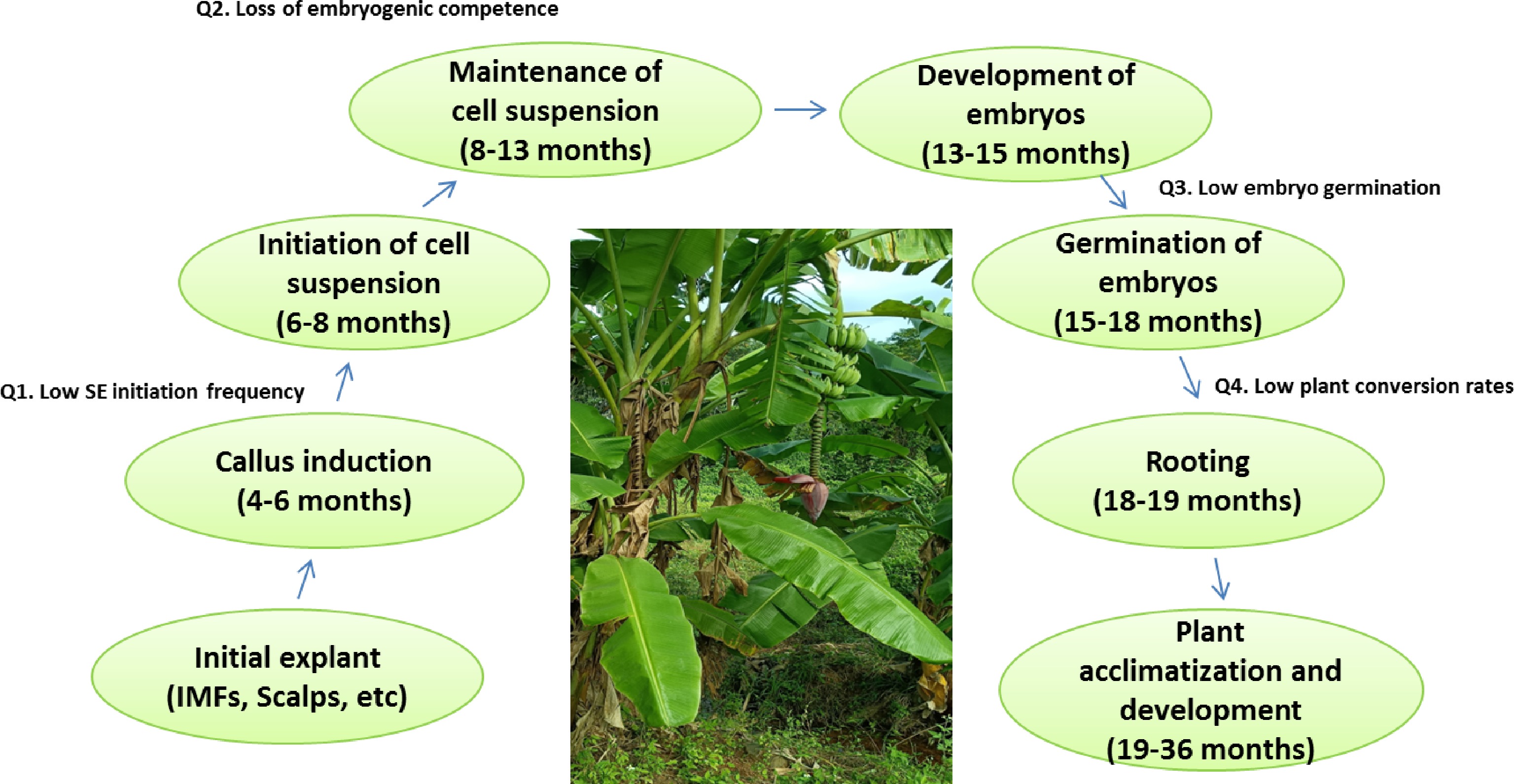
      Figure 1. 

      The main phases and time required for each phase in the somatic embryogenesis of banana and critical questions (Q). (SE) somatic embryo; (IMFs) immature male flowers.

      Although somatic embryogenesis in banana is now a well-established method, the initiation of a 'genotype-independent' embryogenic cell culture is still far from routine. There are still some problems that need to be solved in the reported protocols for banana SE. These problems include either all or some of the following: low SE initiation frequency from the explants, reduction or loss of embryogenic competence concomitant with the increased time of subculture, and low embryo germination and plant conversion rates (Fig. 1).

    • In banana, indirect SE was mostly observed. The main culture stages of indirect SE are induction and proliferation of embryogenic callus (EC), maturation and germination of somatic embryos[7, 8]. Therefore, various factors affect the efficiency and quality of EC formation, including the explant type, the genotype of the donor plant, plant growth regulators, and the media and other additives, etc.

    • The selection of suitable explants is one of the key factors for the success of EC induction. Since early reports in the late 1980s, a series of explants have been successfully used in banana EC. In a word, there are mainly four different types of explants used in banana: immature and mature zygotic embryos[914], Rhizome slices and leaf sheaths[15], IMFs and female flowers[5, 1621], and scalps[22, 23]. Recently, somatic embryos were also successfully induced by secondary explants from male buds and bracts in medium containing TDZ[24, 25]. While, direct somatic embryos developed from split shoot tips under a combination of picloram and 6-benzyladenine (BA)[26]. Despite many options, the most used explants to establish a renewable ECS for seedless banana are still limited to scalps[2731] and IMFs[3234].

      It is reported that factors such as the developmental and physiological state of the explant and the location of the material can affect SE. Strosse et al.[35] suggested that the immature flowers should be taken from position 8 to 16, which were the most responsive ones in terms of embryogenesis. From the reports, sensitive positions are mainly concentrated in 7-13[17]; 8-15[36], 4-11[37], and 6-11[34]. Interestingly, higher efficiency and taking a short time for EC formation were observed by spraying exogenous 2,4-dichlorophenoxyacetic acid (2,4-D) on immature male flower buds[38].

    • SE is highly genome dependent as the efficiency varies with cultivars. Using various explants, SE has been achieved for some genotypes of banana varieties (AA, BB, AB, AAA, ABB, and AAB). Using IMFs as starting materials, three genotypes including six cultivars were tested[17]. The efficiency of EC obtained from different genotypes ranged from 0 to 7%. Even for the different variety of the same genotype, the frequency of EC formation varied differently. Musa AAB cvs. 'French Plantain', 'Mysore' and 'Silk' showed the efficiency of 2%, 3%, and 7%, respectively. As for Musa AAA cv. 'Grande Naine', it had the highest induction rate (37%) of all tested varieties[17]. Among the reported genotypes, two cultivars from Cavendish subgroup (AAA) ranged from 0.7% to 10%, responses[39]. Using the scalps, the mean embryogenic frequency was 6.0%, 3.8%, and 1.8% for cooking bananas (ABB), Cavendish-type bananas (AAA), and plantains (AAB), respectively[30]. Whereas, using inflorescence proliferation for SE induction, the embryogenic frequency was 12.5% and 25% under semisolid and liquid inductive medium, respectively[24].

    • PGRs are crucial in the process of callus formation, proliferation, somatic embryo formation, plant regeneration and rooting. Auxins and cytokinins act a decisive role in somatic embryogenesis in various plant species. At present, the commonly used auxins are 2,4-dichlorophenoxyacetic acid (2,4-D), 1-naphtaleneacetic acid (NAA), indole-3-acetic acid (IAA), Indole-3-butyric acid (IBA), and picloram (4-amino-3,5,6-trichloropicolinic acid). As for CKs, 6-benzylaminopurine (BA), kinetin (KN), and zeatin are the mostly used. 2,4-D is used for EC induction, establishment and proliferation of ECS in most banana cultivars. It is often applied at 1–4 mg L−1, and is combined with low concentrations of cytokinins to control SE. For plant regeneration, BA is often used at concentrations of 0.1–3 mg L−1, and low concentrations of NAA added, or sometimes hormone-free media.

      Different concentrations and various combinations of PGRs were required for different explants. For IMFs method, even though a high level of 2,4-D is needed for the EC induction, prolonged exposure will reduce the embryogenic nature of the callus. At the proliferation of EC and initiation of cell suspension cultures, reduction of the concentration of the sole auxin 2.4-D is improtant for proliferation of somatic embryogenic callus and expression of somatic embryos[18, 37, 40]. However, Nandhakumar et al.[34] reported a MS based ECS medium with 10 mg L−1 resulted in the rapid multiplication of embryogenic cells. In addition, picloram also plays a vital role in SE. It was reported that the induction percentage of EC of M. acuminata cv. 'Mas' (AA) reached 15.6% when 2.4-D in the callus induction medium was substituted by 8.28 μM picloram. The induction efficiency of IMFs on medium with picloram was more than twice that of 2.4-D[41]. On the contrary, the opposite results were observed when the effects of different concentrations of 2,4-D and picloram on callus initiation of M. acuminata cv. 'Berangan' (AAA) were studied[40]. It may be induced by the different genotypes of the explants. As for the other explants, both auxin and cytokinin were used and maintained in the medium. Embryogenic callus (17.5%) was induced from scalps of Musa AAB Silk 'Guoshanxiang' on MS medium with 5 μM 2,4-D and 1 μM Zeatin[28]. Using split shoot tips as explants, maximum embryo induction (100%) for M. acuminata AAA cv. 'Grand Naine' occurred in medium with 4.14 μM picloram and 0.22 μM BA. The plant regeneration (2%–3%) occurred in MS medium with NAA (0.53–2.68 μM) and BA (2.22–44.39 μM), or TDZ (4.54 μM) plus glutamine (200 mg/L)[26].

    • Medium is the basic substance for in vitro plant culture. According to the components, it can be divided into Murashige and Skoog (MS) medium[42], Gamborg's B5[43], Woody Plant Medium (WPM)[44], and Schenk and Hildebrandt (SH) medium[45], etc. The basal medium may be solid, semi-solid, or liquid. The commonly used media for SE in Musa spp. include MS and SH. MS is the preferred medium for callus initiation, establishment of ECS, and plant regeneration. SH medium with MS vitamins or 1/2 MS is often used for the development and maturation of somatic embryos of Musa spp.

      Medium additives, used along with basal media and PGRs, commonly include carbon source, various amino acids, malt extract (ME), and coconut water (CW), etc. Carbon source plays a major role in plant energy metabolism and regulates the osmotic potential of the cell. The most preferred carbon source for banana was sucrose (2%–4.5%; w/v). In addition, maltose, dextrose, fructose, lactose and galactose are also used as carbon sources in some studies. Adding maltose in the medium promoted the formation of banana ECS[32, 34]. The effect of different amino acids (L-Proline, L-Glutamine and L-Asparagine) on somatic embryo production was compared. Among the tested amino acids, L-Glutamine (400 mg L−1) had a significant strengthening effect on primary and secondary somatic embryos in M3 medium[34]. It was in concert with the early report[46]. The presence of 400 mg L−1 L-Glutamine resulted in optimum somatic embryo development and high regeneration efficiency in banana cv. Berangan (AAA). Although L-Glutamine and L-Proline have been shown to promote the embryo development, high concentrations of proline (400 mg L−1) in liquid media caused abnormal embryo differentiation[46]. CW and ME play a promoting role in banana callus induction[32, 47]. To avoid rapid browning of the explants, anti-oxidant like ascorbate (10 mg L−1), melatonin (50 mg L−1), and L-Glutamine (100 mg L−1) were also added to the medium[34].

    • Exploring the molecular regulatory mechanism of plant SE can not only reveal the process of somatic embryo development, but also afford a basis molecular mechanism for somatic embryo development. In most banana genotypes, the potency of explant to develop EC is highly inefficient. Therefore, it is important to find the molecular regulators that can be explored to enhance the SE potential in banana.

      Based on the banana genome database, the differential transcribed fragments between zygotic and somatic embryogenesis were compared by cDNA-AFLP[48]. The role of genes including transcription factors (TFs) was identified in banana SE. The results showed that MaBBM2 and MaWUS2 maybe the prospective candidate TFs and MaPIN1 could be a hopeful gene marker for the embryogenicity in banana[49, 50].

      Recently, differentially expressed proteins during the SE in banana were identified by proteome technology[5153]. Based on comparative proteomics, it is indicated that EC was related to excessive accumulation of ROS scavenging proteins, heat shock proteins (HSP), and growth-regulator related proteins[51]. Furthermore, calcium signaling and PGRs were also involved in the development and germination of somatic embryos. The important role of calcium and PGRs (IAA, BAP, and kinetin) were confirmed by proper induction of five recalcitrant banana cultivars[52]. Based on these results, the medium for optimal SE efficiency in several cultivars could be customized.

    • As the basal materials, ECS is very important for banana germplasm innovation. However, the establishment of banana ECS is very difficult, and after establishment, it needs to be subcultured regularly. Frequent subculture not only consumes a lot of manpower and material resources, but also leads to somatic mutation and the loss of embryogenic characteristics. Furthermore, it is susceptible to bacterial and fungal contamination. Therefore, it is of great significance to study the preservation methods of banana ECS. Cryopreservation is an effective technology that can not only reduce the risk of contamination and gene mutation, but also effectively store plant material for a long time. There are three main methods for cryopreservation of banana germplasm, namely slow-freezing (two-step method), quick-freezing and vitrification. Panis et al.[54] successfully used a two-step method to preserve banana ECS for the first time. In 2010, Li et al. successfully cryopreserved banana ECS by vitrification[55].

    • Protoplast fusion and somatic hybridization offers the potential to produce novel crops and overcome breeding obstacles in polyploid and apomictic banana cultivars. In 1993, the isolation and regeneration of protoplasts from an embryogenic cell suspension culture in banana were successfully received[56, 57]. Science then, a number of banana cultivars including various genotypes (AA, AAA, AAB, ABB) were effectively regenerated through protoplast culture[58].

      Plant regeneration via protoplast culture opens up feasible opportunities for somatic hybridization and protoplast transformation, and eventually leads to genetic modification and breeding of new varieties. Somatic hybridization between triploid (Musa spp. AAB group, cv. 'Maçã') and diploid (Musa spp. AA group, cv. 'Lidi') bananas was attempted using protoplast electrofusion and nurse culture techniques. Somatic hybrids showed different ploidy levels by RAPD and flow cytometric ploidy analyses[59]. Assani et al.[60] successfully obtained banana somatic hybrid plants from Musa spp. triploid cv. 'Gros Michel' (AAA) and diploid cv. 'SF265' (AA). By the comparison of chemical (PEG: polyethylene glycol) and electrical fusion technique, it was found that electric fusion was better for mitotic activities, somatic embryogenesis and plantlet, and chemical procedure was better for the frequency of binary fusion. Xiao et al.[58] developed an asymmetric protoplast fusion with 20% (w/v) PEG and obtained somatic hybrids between Musa Silk cv. 'Guoshanxiang' (AAB) and Musa acuminata cv. 'Mas' (AA). Recently, Wu et al.[61] established a PEG-mediated protoplast transformation, which can serve as an effective and rapid tool for transient expression assays and sgRNA validation in banana.

    • As a perennial fruit crop, banana is susceptible to a plethora of abiotic and biotic stresses[6, 62, 63]. The objectives of banana improvement programs are breeding cultivars resistant to abiotic and biotic stressors without adverse effects on yield and fruit quality. Numerous efforts have been made to breed superior banana cultivars with better resistance to abiotic and biotic stresses and optimum yields at the same time using conventional breeding and genetic modification strategies. However, conventional breeding approaches are time-consuming and severely hampered by inherent banana problems (polyploidy and sterility). Therefore, genetic transformation is becoming increasingly popular and can provide rapid solutions.

      In summary, the recipients used for banana genetic transformation are usually ECS, apical meristem, corm slices, thin cell layers from shoot tips, multiple shoot clumps etc. Among them, genetic transformation through ECS is a most commonly used method in different cultivars of banana owing to its strong ability to accept foreign genes and the lower frequency of chimeras shoot production. The transformation method is mainly mediated by gene gun and Agrobacterium. The flow chart of banana genetic transformation using ECS is shown in Fig. 2. The transformation efficiency was between 1.25% and 50.00%, with a large range of changes. Except for NPTII, GUS, GFP and other screening genes and reporter genes, the transformed functional genes mainly involved in banana fruit quality, disease resistance, drought tolerance, dwarfing and other traits improvement[62]. In this part, the studies for banana genetic transformation with added value from 2000 on were mainly summarized as below.
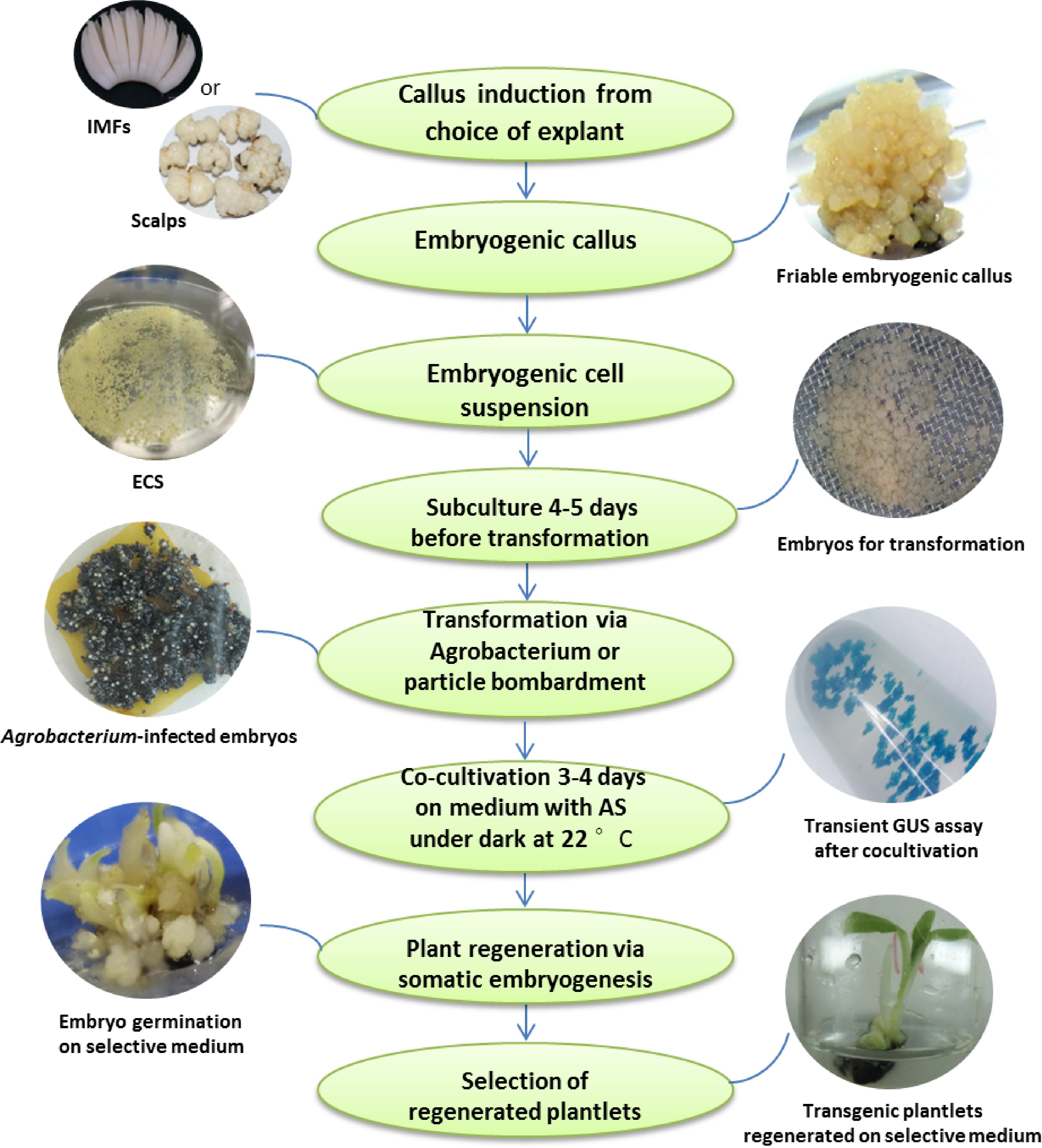
      Figure 2. 

      Schematic representation of genetic transformation steps of banana using embryogenic cell suspension. Scalps and immature male flowers (IMFs) are the most used explants to establish a renewable ECS for seedless banana cultivars. The photos of scalps and friable embryogenic callus are cited from Tripathi et al.[31].

    • In banana, the most serious diseases are fungal (Fusarium wilt, black Sigatoka), bacterial (banana Xanthomonas wilt, BXW), and viral (banana bunchy top disease, and banana streak disease)[63, 64]. Researchers have been working to improve disease and pest resistance in bananas using transgenic technology[6466].

      Various transgenes have been used to develop genetically engineered banana and many conferred significant levels of resistance to fungal pathogens (Table 1). Functional genes used to develop Foc-resistance bananas mainly included the antimicrobial peptides belonging to plant or animal origin[67, 68, 70, 72, 75, 78, 80], apoptosis-inhibition-related animal genes (Bcl-xL, Ced-9 and Bcl-2 3' UTR, Ced9)[69, 74, 81], different cell-death-related genes (MusaDAD1, MusaBAG1 and MusaBI1)[72], and defense-related protein[82]. In addition, Foc-resistance has also been conformed using RNAi silencing of key genes of Foc[71, 84]. Although the above studies demonstrate the transgenic plants resistance to Foc in the greenhouse, field evaluation remains to be seen. Recently, transgenic bananas with resistance gene analog 2 (RGA2), isolated from a seedling of Musa acuminata ssp. malaccensis with resistance to TR4, showed promising resistance against Fusarium wilt after a 3-year field trial in Australia[81]. Similarly, two native genes (MaLYK1 and MabHLH) from banana germplasms with Foc resistance were introduced back to Cavendish banana cv. Brazil, had shown increased resistance to TR4[83, 85]. Several studies from the researchers at the International Institute of Tropical Agriculture (IITA) reported transgenic bananas resistant to BXW disease[8892]. Other studies also dealt with the production of resistance to Black Sigatoka[86, 87] and banana bunchy top disease[9395] (Table 1).

      Table 1.  Genetic transformations of banana (Musa spp.).

      TraitGeneSourcesTransformation methodResultCultivarReference
      Fungal
      Foc Race2 resistanceMSI-99SyntheticAgrobacterium/EHA105/ ECSImproved disease resistance against Foc and black leaf streak diseaseRasthali (AAB)[67]
      Foc Race1 resistanceβ–1,3–endoglucanaseSoybeanAgrobacterium/LBA4404/Single budsIncreased tolerance to Foc Race 1Rasthali (AAB)[68]
      Bcl-xL, Ced-9, Bcl-2 3' UTRAnimalAgrobacterium/LBA4404/ECSApoptosis-inhibition-related genes confer resistance to Foc Race 1Lady Finger (AAB)[69]
      PhDef1 and PhDef2PetuniaAgrobacterium/EHA105/ECSImproved fungal resistance with normal growth and no stunting phenotypeRasthali (AAB)[70]
      ihpRNA-VEL and ihpRNA-FTF1Agrobacterium/EHA105/ECSIncreased resistance to Foc Race 1Rasthali (AAB)[71]
      MusaDAD1, MusaBAG1 and MusaBI1BananaAgrobacterium/EHA105/ECSIncreased resistance to Foc Race 1Rasthali (AAB)[72]
      Sm-AMP-D1Stellaria mediaAgrobacterium/EHA105/ECSImproved resistance against Foc Race 1 and no gross growth abnormalitiesRasthali (AAB)[73]
      Ced9SyntheticIncreased resistance against Fusarium wiltSukali Ndiizi (AAB)[74]
      Ace-AMP1Onion seedsAgrobacterium/LBA4404/ECSEnhanced resistance to Foc race 1Rasthali (AAB)[75]
      Ace-AMP1 + Ca-pflpAllium cepa L; Capsicum annum L.Agrobacterium/AGL1/ ECSStacked Ace-AMP1 and pflp transgenic plants showed resistance to Foc race 1Rasthali (AAB)[76]
      Foc TR4 resistanceHuman lysozyme (HL)Agrobacterium/EHA105/corm slicesImproved resistance to Foc TR4Taijiao (AAA)[77]
      PflpSweet pepperAgrobacterium/EHA105/
      multiple bud clumps
      Enhanced resistance to Foc TR4Pei Chiao (AAA) and Gros Michel (AAA)[78]
      TLP or PR-5RiceBiolistics/Single
      cauliflower-like bodies
      Enhanced resistance to Foc TR4Pisang Nangka (AAB)[79]
      ThChit42Trichoderma harzianumAgrobacterium/EHA105/ECSEnhanced resistance to Foc TR4Furenzhi (AA)[80]
      RGA2 or Ced9BananaAgrobacterium/EHA105/ECSImproved promising resistance against Fusarium wiltGrand Nain (AAA)[81]
      MaPR-10bananaAgrobacterium/−/ECSImproved tolerance against Fusarium infectionBerangan[82]
      MaLYK1BananaAgrobacterium/EHA105/ECSIncreased resistance to Foc TR4Cavendish (AAA)[83]
      Synthesis of ergosterol (ERG6/11)Agrobacterium/EHA105/ECSstrong resistance to Fusarium wiltBrazil (AAA)[84]
      MpbHLHBananaAgrobacterium/EHA105/ECSEnhanced Foc TR4-resistance of Cavendish bananaBrazil (AAA)[85]
      Sigatoka resistanceThEn-42 + StSy + Cu,Zn-SOD co-transformationTrichoderma harzianum
      + grape + tomato
      Biolistics/ECSEnhanced tolerance to SigatokaGrand Nain (AAA)[86]
      rcc2 or rcg3RiceAgrobacterium/EHA105/ ECSEnhanced host plant resistance to black SigatokaGros Michel (AAA)[87]
      Bacterial
      BXW resistanceHrapSweet pepperAgrobacterium/AGL1/ ECSAbout 20% of the Hrap lines showed
      100% resistance for both mother and
      ratoon crops under field conditions
      Sukali Ndiizi (AAB) and Mpologoma (AAA)[88, 89]
      PflpSweet pepperAgrobacterium/EHA105/ ECSAbout 16% of the Pflp lines showed
      100% resistance for both mother and
      ratoon crops under field conditions
      Sukali Ndiizi (AAB) and Nakinyika (AAA)[89, 90]
      Stacked Harp and PflpSweet pepperAgrobacterium/AGL1/ ECSStacked Harp and Pflp transgenic plants
      had higher resistance to X cm
      Gonja manjaya (AAB)[91]
      Xa21RiceAgrobacterium/EHA105/ ECS50% of the transgenic lines showed complete resistance to X cmGonja manjaya (AAB)[92]
      ViralRepBBTVCompletely resistant to BBTV infection was found under glasshouse conditionsBrazilian (AAA)[93]
      BBTV-G- cpBBTVBiolistics/apical meristemWilliams (AAA)[94]
      Rep, ProRepBBTVRasthali (AAB)[95]
      Abiotic stress
      Salt, oxidative stressMusaWRKY71bananaAgrobacterium/EHA105/ ECSEnhanced tolerance towards oxidative and salt stressRasthali (AAB)[96]
      Cold, drought, saltMusabZIP53bananaAgrobacterium/EHA105/ ECSTransgenic plants displayed severe growth retardationRasthali (AAB)[97]
      ColdMpMYBS3bananaAgrobacterium/EHA105/ ECSThe transgenic lines had higher cold toleranceBrazil (AAA)[98]
      Salt, droughtMusaNAC042bananaAgrobacterium/EHA105/ ECSMusaNAC042 is positively associated with drought and salinity toleranceRasthali (AAB)[99]
      Drought, saltMusa-DHN-1bananaAgrobacterium/-/ ECSImproved tolerance to drought and salt-stressRasthali (AAB)[100]
      Salt, droughtAhSIPR10Arachis hypogaeaAgrobacterium/EHA105/
      multiple shoot clump
      Transgenic plants showed better tolerance of salt and drought conditionsMatti (AA)[101]
      Drought, salt, oxidative stressMusaSAP1bananaAgrobacterium/EHA105/ ECSTransgenic plants displayed better stress endurance characteristicsRasthali (AAB)[102]
      Cold, salt, droughtMusaPIP1;2bananaAgrobacterium/EHA105/ ECSTransgenic plants showed better abiotic stress survival characteristicsRasthali (AAB)[103]
      SaltMusaPIP2;6bananaAgrobacterium/EHA105/ ECSTransgenic plants showed better tolerance under salt stressRasthali (AAB)[104]
      Salt, droughtMaPIP1;1bananaAgrobacterium/EHA105/
      thin cell layers from shoot tips
      Improved tolerance to salt and drought stressesBrazilian (AAA)[105, 106]
      Drought, cold ,saltMaPIP2-7bananaAgrobacterium/EHA105/
      thin cell layers from shoot tips
      Improved tolerance to salt, drought, and cold stressesBrazilian (AAA)[107]
      MaSIP2-1bananaAgrobacterium/EHA105/
      thin cell layers from shoot tips
      Transgenic plants had a stronger drought and cold tolerance than the controlBrazilian (AAA)[108]
      Salt, droughtAhcAPXArachis hypogeaAgrobacterium/EHA105/
      multiple shoot clump
      Enhanced the tolerance to drought and salt stressGrand naine (AAA)[109]
      Fruit quality and others
      Biofortified Iron ferritinsoybeanAgrobacterium/EHA105/ ECSA 6.32-fold increase in iron accumulation and a 4.58-fold increase in the zinc levels were noted in the leaves of transgenic plantsRasthali (AAB)[110]
      Biofortified pro-vitamin AMtPsy2abanaanAgrobacterium/AGL1/ ECSA high content of β-CE (75.1 µg/g dw) was found in the fourth generation with no variation in critical agronomical features such as yield and cycle timeDwarf Cavendish (AAA)[111]
      Fruit ripeningMaMADS1 and MaMADS2bananaAgrobacterium/-/ ECSRepression of either MaMADS1 or
      MaMADS2, resulted in delayed ethylene
      synthesis and maturation
      Grand Nain (AAA)[112]
      Sense and anti-sense MaMADS36bananaAgrobacterium/GV3101/
      thin cell layers from shoot tips
      MaMADS36 represents a central molecular switch in regulating banana fruit ripeningRed banana (AAA)[113]
      Foc, Fusarium oxysporum f. sp. cubense; ECS, embryogenic cell suspension; BBTV, Banana bunchy top virus; NM, not mention.
    • Many transcription factors (TFs) and downstream genes which respond to abiotic stress have been identified in banana. They mainly include WRKY[96], bZIP[97], MYB[98], NAC[99], dehydrins (DHN)[100], SAP1[102], and aquaporins (AQP)[103108], and so on (Table 1). In transgenic plants, overexpression of these TFs let them withstand and survive under stress conditions. Because of their important role in plant growth and development, it can cause abnormal growth in transgenic plants by the constitutive overexpression of these TFs, such as bZIP53[97]. Identifying and overexpressing a key gene participated in stress tolerance is a good option. Ectopic expression of stress-related genes has been introduced into banana to enhance the tolerance to some abiotic stresses[100109]. However, the majority of these studies have been reported from a glasshouse evaluation. Trials in the field are necessary to further prove their worthiness.

    • Recently, genetic engineering was also employed to improve fruit nutrient content and control fruit ripening (Table 1). Transgenic bananas with biofortified iron content and pro-vitamin A were tested in the green house and field, respectively. The transgenic banana plants overexpressing soybean ferritin accumulated the higher levels of iron and zinc under in vitro conditions as well as in the green house[110]. PVA-biofortified transgenic Cavendish bananas were also developed[111].

      Banana MaMADS transcription factors are necessary for fruit ripening and molecular tools to promote shelf-life. Repression of either MaMADS1 or MaMADS2, resulted in delayed ethylene synthesis and maturation[112]. Similarly, transgenic red bananas were obtained with sense and anti-sense constructs of MaMADS36. Further study demonstrated that MaMADS36 directly binds to the CA/T(r)G box of the MaBAM9b promoter to regulate enzyme activity and starch degradation during ripening[113].

    • Genome-editing technologies using various site-directed nucleases (SDNs) have become powerful tools for modifying plant genomes. SDNs include meganucleases, ZFNs (zinc finger nucleases), TALENs (transcription activator-like effector nucleases), and CRISPR/Cas (clustered regularly interspaced short palindromic repeats/CRISPR-associated protein)[114]. The CRISPR/Cas system has been widely adopted for plants genetic improvement due to its simplicity and high-efficiency[115].

      Although the application of the CRISPR/Cas9 system in banana is still at the preliminary stage, CRISPR/Cas9 mediated genome editing has been applied to improve banana nutrient contents, storage time, disease resistance, and alter the plant architecture[6, 114, 116, 117]. In summary, the CRISPR/Cas9-based genome editing utilized in banana are outline in Table 2.

      Table 2.  Application of CRISPR/Cas9 gene editing technology in Musa.

      CultivarExplantStrategy of transformationCas9 promotersgRNA promoterTarget geneTarget traitResultEditing efficiencyReference
      Baxi; AAAECSAgrobacterium-mediated transformation
      EHA105
      UbiOsU6aMaPDSAlbino and variegated phenotypeMurtation in target genes; Albino phenotype in transgenic plants55%[118]
      Rasthali; AABECSAgrobacterium-mediated transformation
      Agl1
      2 × CaMV35SOsU3MaPDSAlbino and variegated phenotypeMurtation in target genes; Albino phenotype in transgenic plants59%[119]
      Williams; AAAECSAgrobacterium-mediated transformation
      EHA105
      Ubi;CaMV35sOsU3MaPDSAlbino and dwarf phenotypeMurtation in target genes; Albino and dwarfing phenotype in transgenic plants63%[120]
      Sukali Ndiizi; AAB;Gonja Manjaya; AABECSAgrobacterium-mediated transformation
      EHA105
      2 × CaMV35SOsU6MaPDSalbino and variegated phenotypesGeneration of mutants with albino and variegated phenotypes100%[121]
      Williams; AAAECSAgrobacterium-mediated transformationNMNMMaCHAOSPale-green phenotypesMurtation in target genes; Pale-green phenotypes and normal growthNM[122]
      Baxi; AAAprotoplastPEG-mediated transformationUbiOsU3MaPDS-The efficiency of CRISPR/Cas9-mediated mutagenesis was higher than that of CRISPR/Cas12a, and RNP-CRISPR-Cas91.04% (Cas9), 0.92% (RNP), 0.39% (Cas12a)[61]
      Brazilian; AAAprotoplastPEG-mediated transformationUbiMaU6MaPDS-Increased mutation efficiency of CRISPR/Cas9 genome editing in banana by optimized construct4-fold[123]
      Gonja Manjaya; AABECSAgrobacterium-mediated transformation
      EHA105
      UbiOsU6Viral genesBanana streak virus (BSV)Inactivation of endogenous banana streak virus (eBSV) intergated in host genome and generated resistant banana plants against eBSV95%[124]
      Sukali Ndiizi; AABECSAgrobacterium-mediated transformation
      EHA105
      2 × CaMV35SOsU6MusaDMR6Banana Xanthomonas wilt (BXW)Improved resistance to BXW disease in mutants with normal growth100%[125]
      Grand Naine; AAAECSAgrobacterium-mediated transformation
      Agl1
      CaMV35SOsU3MaLCYεRegulation synthesis of β-caroteneImproved nutritional trait in transgenic plants with normal growthNM[127]
      Rasthali; AABprotoplasts and ECSelectroporation-mediated transformation;
      particle bombardment method
      CaMV35SOsU3MaCCD4Regulatory mechanism of β-carotene homeostasisCCD4 negatively regulates β-carotene biosynthesisNM[128]
      Brazilian; AAAECSAgrobacterium-mediated transformationUbiOsU6aMaACO1Shelf lifeMore vitamin C and improved shelf life in transgenic plants98%[129]
      Gros Michel; AAAECSAgrobacterium-mediated transformationUbiOsU6a/
      OsU3
      MaGA20ox2Semi-dwarf phenotypeA lower active GA content and a semi-dwarf phenotype in transgenic plantsNM[130]
      NM, not mentioned.
    • Plant albino phenotype is a classic and indicative phenotype for testing and judging whether the CRISPR/Cas9 system is effective. As an indicator gene, phytoene desaturase (PDS) can easily obtain the target albino trait and has been knocked out in most fruit trees. Recently, CRISPR/Cas9-based genome editing in banana has been established using the PDS as a marker gene[118121]. However, knockout of PDS has adverse effects on plant growth. Optionally, RP43/CHAOS39-edited banana plants were obtained with pale-green phenotype and no negative effects on plant growth[122]. Recently, the transient delivery system by a PEG-mediated protoplast was established[61]. The editing efficiency of the CRISPR/Cas9, CRISPR/Cas12a, and ribonucleoprotein-CRISPR-Cas9 (RNP-CRISPR-Cas9) for targeting the PDS gene in banana protoplasts was compared. The results showed that the efficiency of CRISPR/Cas9-mediated mutagenesis was higher than that of the other two systems. In addition, it was the first report by a RNP-CRISPR-Cas9 system for genome editing in banana[61]. In comparison to the previous report in banana, using endogenous U6promoter and banana codon-optimized Cas9 in CRISPR/Cas9 cassette, the mutagenesis efficiency has a fourfold increase[123].

    • Banana streak virus (BSV), a double stranded DNA Badnavirus which integrated in the B genome derived from M. balbisiana, is called endogenous BSV (eBSV). It severely affects production of plantain (AAB) in Africa. To inactivate the virus, a multiplexed gRNA strategy targeting all three ORFs of eBSV was constructed and transformed into Gonja Manjaya (AAB). Compared with the controls, the eBSV-edited plants exhibited resistance against eBSV and normal growth. A very high mutation effciency of 95% using three gRNAs were observed[124]. Recently, CRISPR/Cas9 mediated gene editing for banana resistance against bacteria was also reported. To obtain the banana cultivar against Xan­thomonas Wilt (BXW), a Musadmr6 gene was edited[125]. The edited plants had a higher resistance to BXW without adverse affecting on plant growth. Researchers are also trying to breed TR4 resistance cultivars by CRISPR[126].

    • Fruit quality is an important indicator to measure the value of fruit commodities. Carotenoids are essential for human nutrition. Most Cavendish group cultivars have low β-carotene content in the fruit pulp. Using CRISPR/Cas9 technology, β-carotene-enriched banana plants were created by editing the fifth exon of LCYε gene from A genome, which determines a high α-/β-carotene ratio[127]. Compared with the unedited fruits, the β-carotene count of the fruit pulp of the edited lines increased by 6-fold. More recently, CRISPR/Cas9-mediated editing of CCD4 was conducted in Rasthali. In comparison to the controls, the accumulation of β-carotene in roots was increased in the CCD4-edited plants[128].

      The shelf life of post-harvest fruits is an important factor affecting fruit quality. The production of ethylene is closely related to the storage time of banana fruits. Thus, it is the first consideration for developing postharvest preservation technology. MaACO1 encodes for an O2-activating ascorbate-dependent non-heme iron enzyme that catalyzes the last step in ethylene biosynthesis. The MaACO1-editted banana fruit extended shelf life and had more Vitamin C compared with the wild-type fruit[129].

    • Developing semi-dwarf and dwarf banana varieties is also one of the objectives of banana improvement programs. Gibberellin (GA) is a key gene which determines plant height and the mutations in its biosynthesis genes often leads to dwarf plants. CRISPR/Cas9 technology was applied to generate a semi-dwarf banana cultivar 'Gros Michel' by manipulating the M. acuminata gibberellin 20ox2 (MaGA20ox2) gene, disrupting the gibberellin (GA) pathway[130].

    • At present, extensive advances has been made on banana SE. Reports of banana genetic improvement using ECS in the past five years has increased dramatically. Nevertheless, there are still many problems to be solved in the research on banana SE and genetic modification. Little information is available on the molecular mechanisms of banana SE. The embryogenic capacity and efficiently propagated plantlets are very low. The repeatability of the protocols early reported for SE in banana is poor. So far, SE in bananas is far from being considered a conventional technique and has not even been successfully used in some varieties.

      Hence, an important consideration for future work is to explore the basic molecular mechanism of banana embryogenic potency. The gradual application of multi-omics technique in plant SE provides the feasibility to uncover the regulatory mechanism of SE development at the molecular level. Further continuous work is needed for optimizing a highly effcient and versatile transformation and regeneration system which independent on genotype. And the genetic improvement at present only aims at single gene or single trait. More genes associated with disease-resistance, as well as with other important agronomic traits, should be characterized and utilized in target breeding programs. Molecular designing breeding with multi-gene superposition should be carried out to breed new banana varieties with good comprehensive characters.

      Despite the rapid progress of banana transgenic, there are no commercial transgenic varieties applied to the production. With the continuous optimization and improvement of CRISPR/Cas and other gene editing technologies, it is possible to obtain an ideal mutant by accurately targeting target sites. In addition, the key technology of modern biotechnology breeding is the delivery system of plant genetic modification. The application of nanocarriers in plant genetic engineering shows a broad application prospect. The introduction of nanotechnology into banana tissue culture showed significant positive effects on callus induction, somatic embryogenesis and other regeneration aspects. More recently, a cut-dip-budding delivery (CBD) system enables genetic modifications in plants without tissue culture[131]. It overcomes the difficulties posed by the traditional technology due to the plant tissue culture process. Therefore, it would be very interesting to explore a simple, fast and efficient method for banana genetic transformation or genome editing without the need for tissue culture.

      • This research was supported by the specific research fund of The Innovation Platform for Academicians of Hainan Province (YSPTZX202101), the Hainan Provincial Natural Science Foundation (321RC638), the National Natural Science Foundation of China (32172269, 31501043) and the Earmarked Fund for Modern Agro-industry Technology Research System (CARS-31).

      • The authors declare that they have no conflict of interest.

      • Received 30 September 2022; Accepted 29 November 2022; Published online 23 December 2022

      • # These authors contributed equally: Jingyi Wang, Shanshan Gan

      • Copyright: © 2022 by the author(s). Published by Maximum Academic Press on behalf of Hainan University. This article is an open access article distributed under Creative Commons Attribution License (CC BY 4.0), visit https://creativecommons.org/licenses/by/4.0/.
    Figure (2)  Table (2) References (131)
  • About this article
    Cite this article
    Wang J, Gan S, Zheng Y, Jin Z, Cheng Y, et al. 2022. Banana somatic embryogenesis and biotechnological application. Tropical Plants 1:12 doi: 10.48130/TP-2022-0012
    Wang J, Gan S, Zheng Y, Jin Z, Cheng Y, et al. 2022. Banana somatic embryogenesis and biotechnological application. Tropical Plants 1:12 doi: 10.48130/TP-2022-0012

Catalog

  • About this article

/

DownLoad:  Full-Size Img  PowerPoint
Return
Return