RESEARCH ARTICLE   Open Access    

Lignocellulolytic microbiomes orchestrating degradation cascades in the rumen of dairy cattle and their diet-influenced key degradation phases

More Information
  • Received: 20 August 2024
    Revised: 14 September 2024
    Accepted: 18 September 2024
    Published online: 21 October 2024
    Animal Advances  1 Article number: e002 (2024)  |  Cite this article
  • Dairy cattle (Bos taurus) can convert lignocellulosic biomass into milk efficiently via their rumen symbiotic microbiota. However, the mechanisms by which the rumen microbiota of cows mediate the degradation cascades of lignocellulose and the specific stages primarily affected by dietary interventions remain unclear. Herein, 244 rumen metagenome samples from Holstein cows were used, identifying 1353 high-quality microbial metagenome-assembled genomes (MAGs) responsible for the degradation cascades of lignocellulose. It was revealed that Fibrobacter spp. and Ruminococcus spp. exhibited numerous endo-/exo-glucanases with accessory non-catalytic multi-carbohydrate binding modules for highly efficient cellulolytic abilities. Prevotella spp. and Cryptobacteroides spp. developed diverse polysaccharide utilization loci (PULs) to tackle the main and side chains of hemicellulose, particularly acetylxylan esterase-contained PULs. Notably, novel and potential lignocellulolytic microbiomes were identified in the rumen of dairy cattle, such as Hallerella spp., Sodaliphilus spp., and Mageeibacillus spp. Through in vivo diet intervention and in sacco rumen incubation, it was discovered that high-grain diets primarily affected Prevotella spp., leading to a reduction in the initial degradation of amorphous regions in lignocellulose. Therefore, the present findings systematically illustrate the orchestrated enzymatic strategies of the cow rumen microbiota for the degradation cascades of lignocellulose, contributing to the dietary regulation of dairy cattle.
  • Wheat stands as one of the world’s most crucial staple food crops, furnishing 20% of the global population’s calorie intake and holding a pivotal role in ensuring food security worldwide. Wheat yield is mainly determined by three factors: thousand grain weight (TGW), spike number per unit area, and grain number per spike[1]. The optimization of these three components is of great significance for improving yield. Among these, increasing the grain weight emerges as a particularly significant avenue for boosting wheat productivity. Traits shaping grain morphology, including grain length, grain width, and grain thickness, directly affect grain size, which in turn affects grain weight.

    The development of seeds significantly influences grain weight. The mature seeds of angiosperms are composed of the embryo, endosperm, and seed coat. The maternal and zygotic tissues jointly participate in the regulation of the growth and development of seeds as well as control of the synergistic growth of embryo, endosperm, and seed coat. As grain development advances, seed coat cells perpetually undergo division and expansion, accompanied by continuous carbohydrate accumulation in the endosperm[2]. Genes pertinent to transport, carbohydrate metabolism, and starch synthesis also become active during development. Starch is the main storage component of the wheat endosperm, and its content is a key regulator of grain weight. In addition, plant hormone contents exhibit significant changes during seed development, and genes related to metabolism participate extensively in seed development.

    In this review, we summarize recent research on key regulators of wheat grain weight including transcriptional regulatory factors, post-translation modification factors, the G-protein signaling pathway, and phytohormone signalings to understand the regulatory mechanisms of wheat grain weight (Fig. 1, Table 1).

    Figure 1.  Regulatory networks involved in grain weight in wheat. Several genes involved in transcriptional regulatory factors, post-translation modification factors, the G-protein signaling pathway, and phytohormone signalings participate in wheat grain weight regulation. Broken lines indicate inhibitory regulation. Arrowheads represent positive regulation. Elliptical overlaps represent interactions.
    Table 1.  Genes involved in wheat grain weight regulation.
    Protein nameGene IDProtein categoryPositive(+)/negative(−)
    regulator
    Elite haplotype for
    high grain weight
    Reference
    Starch synthesis-related genes reported to be involved in wheat grain weight
    TaCwi-A1TraesCS2A03G0736600Cell wall invertase+TaCwi-A1a[3]
    TaCWI-5DTraesCS5D03G1216700Cell wall invertase+Hap-5D-C[4]
    TaSUT1-ATraesCS4A03G0027400Sucrose transporters+TaSUT1 in Kauz[5]
    TaSUT1-BTraesCS4B03G0758500Sucrose transporters+TaSUT1 in Kauz[5]
    TaSUT1-DTraesCS4D03G0679400Sucrose transporters+TaSUT1 in Kauz[5]
    TaSus1-ATraesCS7A03G0375000Sucrose synthase+TaSus1-7A-Hap-1[6, 7]
    TaSus1-BTraesCS7B03G0171900Sucrose synthase+TaSus1-7B-Hap-T[6, 7]
    TaSus1-DTraesCS7D03G0358200Sucrose synthase+[6, 7]
    TaSus2-ATraesCS2A03G0349200Sucrose synthase+TaSus2-2A-Hap-A[6, 8]
    TaSus2-BTraesCS2B03G0468900Sucrose synthase+TaSus2-2B-Hap-H[6, 8]
    TaSus2-DTraesCS2D03G0366700Sucrose synthase+[6, 8]
    TaBT1-ATraesCS6A03G0433200Sucrose transporter+[9]
    TaBT1-BTraesCS6B03G0559700Sucrose transporter+Hap1 and Hap2[9]
    TaBT1-DTraesCS6D03G0376900Sucrose transporter+[9]
    TaAGPL1-BTraesCS1B03G1206000Large subunit gene of the AGPase+TaAGP-L-1B-Hap-I[10, 11]
    TaAGPS-1TraesCS7A03G0682600Small subunit gene of the AGPase+TaAGP-S1-7A-Hap-I[10]
    TaSBEIII-ATraesCS7A03G0826800Starch-branching enzyme+Allele-T[12]
    TaSSIV-ATraesCS1A03G0866200Starch synthases+Hap-2-1A[13, 14]
    TaSSIV-BTraesCS1B03G1004700Starch synthases+Hap-3-1B[13, 14]
    TaSSIV-DTraesCS1D03G0838700Starch synthases+[13, 14]
    GWD-ATraesCS6A03G0662800Glucan, water dikinase[15]
    GWD-BTraesCS6B03G0813900Glucan, water dikinase[15]
    GWD-DTraesCS6D03G0552200Glucan, water dikinase[15]
    Transcriptional regulatory factors
    TaNAC019-ATraesCS3A03G0172000NAC transcription factor+[16]
    TaNAC019-BTraesCS3B03G0216600NAC transcription factor+TaNAC019-BI[16]
    TaNAC019-DTraesCS3D03G0154500NAC transcription factor+[16]
    TaNAC100-ATraesCS2A03G0808100NAC transcription factor+TaNAC100-2A-H1[17]
    TaNAC100-BTraesCS2B03G0891700NAC transcription factor+[17]
    TaNAC100-DTraesCS2D03G0746900NAC transcription factor+[17]
    TaPGS1TraesCS1D03G0219000bHLH transcription factor+[18]
    TaPGS1TraesCS1D03G0219700bHLH transcription factor+[18]
    TaFI3TraesCS3A03G1169900PLATZ transcription factor+[18]
    TaGSNETraesCS5B03G0668000WRKY transcription factor+TaGSNE-Hap-2[19]
    TaHDZ-A34TraesCS7A03G0760400HD-Zip transcription factor+Hap-ABD[20]
    TaHDZ-B34TraesCS7B03G0590000HD-Zip transcription factor+Hap-ABD[20]
    TaHDZ-D34TraesCS7D03G0729900HD-Zip transcription factor+Hap-ABD[20]
    TaGW8-B1TraesCS7B03G0430500SPL transcription factor+ TaGW8-B1a[21]
    TaSPL14-ATraesCS5A03G0658100SPL transcription factor+[22]
    TaSPL14-BTraesCS5B03G0692900SPL transcription factor+[22]
    TaSPL14-DTraesCS5D03G0627900SPL transcription factor+[22]
    TaSPL14-7ATraesCS7A03G0567100SPL transcription factor+TaSPL14-7A-Hap1/2[23]
    TaSPL14-7BTraesCS7B03G0393600SPL transcription factor+[23]
    TaSPL14-7DTraesCS7D03G0548900SPL transcription factor+[23]
    Post-Translational Modifications (PTMs)
    Ubiquitin–proteasome pathway
    TaGW2-6ATraesCS6A03G0480200RING-type E3 ubiquitin ligaseHap-A[24, 25]
    TaGW2-6BTraesCS6B03G0578500RING-type E3 ubiquitin ligaseTaGW2-B-HapI/II[24, 25]
    TaGW2-6DTraesCS6D03G0404800RING-type E3 ubiquitin ligase[24, 25]
    TaDA1-ATraesCSU03G0004100LCUbiquitin receptorTaDA1-A-HapI[26]
    TaDA1-BTraesCS2B03G0048000Ubiquitin receptor[26]
    TaDA1-DTraesCS2D03G0031900Ubiquitin receptor[26]
    TaSDIR1-4ATraesCS4A03G0197400RING-type E3 ubiquitin ligaseTaSDIR1-4A-2[27]
    TaPUB1-ATraesCS5A03G1197700U-box E3 ligase+[28]
    TaPUB1-BTraesCS4B03G0885300U-box E3 ligase+[28]
    TaPUB1-DTraesCS4D03G0783100U-box E3 ligase+[28]
    ZnF-ATraesCS4A03G0701600RING-type E3 ubiquitin ligase+[29]
    ZnF-BTraesCS4B03G0092600RING-type E3 ubiquitin ligase+[29]
    ZnF-DTraesCS4D03G0066800RING-type E3 ubiquitin ligase+[29]
    SnRK and phosphatase pathways
    TaSnRK2.3-ATraesCS1A03G0569000Sucrose non-fermenting 1 (SNF1)-related protein kinaseHap-1A-1[30]
    TaSnRK2.3-BTraesCS1B03G0660500Sucrose non-fermenting 1 (SNF1)-related protein kinaseHap-1B-1[30]
    TaSnRK2.9-ATraesCS5A03G0177100Sucrose non-fermenting 1 (SNF1)-related protein kinase Hap-5A-1/2[31]
    TaSnRK2.9-BTraesCS5B03G0188000Sucrose non-fermenting 1 (SNF1)-related protein kinase[31]
    TaSnRK2.9-DTraesCS5D03G0195600Sucrose non-fermenting 1 (SNF1)-related protein kinase[31]
    TaSnRK2.10-ATraesCS4A03G0621500Sucrose non-fermenting 1 (SNF1)-related protein kinaseHap-4A-H[32]
    TaSnRK2.10-BTraesCS4B03G0179500Sucrose non-fermenting 1 (SNF1)-related protein kinase[32]
    TaSnRK2.10-DTraesCS4D03G0149100Sucrose non-fermenting 1 (SNF1)-related protein kinase[32]
    TaPSTOLTraesCS5A03G0115500LCPhosphate Starvation Tolerance 1+[33]
    TaGL3-5ATraesCS5A03G0897900PPKL family—Ser/Thr phosphatase+TaGL3-5A-G[1]
    TaGL3-5BTraesCS5B03G0943200PPKL family—Ser/Thr phosphatase+[1]
    TaGL3-5DTraesCS5D03G0859400PPKL family—Ser/Thr phosphatase+[1]
    TaGL3.3-ATraesCS5A03G0073900PPKL family—Ser/Thr phosphatase+[34]
    TaGL3.3-BTraesCS5B03G0068000PPKL family—Ser/Thr phosphatase+TaGL3.3-5B-C[34]
    TaGL3.3-DTraesCS5D03G0098300PPKL family—Ser/Thr phosphatase+[34]
    TaTPP-7ATraesCS7A03G0422300Trehalose-6-phosphate phosphatase+TaTPP-7A-HapI[35]
    TaTPP-7BTraesCS7B03G0228800Trehalose-6-phosphate phosphatase+[35]
    TaTPP-7DTraesCS7D03G0410500Trehalose-6-phosphate phosphatase+[35]
    Asparagine N-glycosylation pathway
    TaSTT3b-2ATraesCS2A03G1282700Catalytic subunit of oligosaccharyltransferase+[36]
    TaSTT3b-2BTraesCS2B03G1473200Catalytic subunit of oligosaccharyltransferase+[36]
    TaSTT3b-2DTraesCS2D03G1245300Catalytic subunit of oligosaccharyltransferase+[36]
    G-protein signaling pathway
    TaGS3-4ATraesCS4A03G1194500Gγ subunit[37, 38]
    TaGS3-7ATraesCS7A03G0037700Gγ subunit[37, 38]
    TaGS3-7DTraesCS7D03G0033100Gγ subunit[37, 38]
    TaDEP1-ATraesCS5A03G0545300Gγ subunit+TaDEP1-Hap1[39]
    TaDEP1-BTraesCS5B03G0555000Gγ subunit+[39]
    TaDEP1-DTraesCS5D03G0509000Gγ subunit+[39]
    Phytohormone signalings
    CK
    TaCKX2TraesCS3A03G0298200Cytokinin oxidase/dehydrogenase (CKX) enzymes+TaCKX2A-2[40]
    TaCKX4TraesCS3A03G1128900Cytokinin oxidase/dehydrogenase (CKX) enzymes+TaCKX4A-2[40]
    TaCKX5TraesCS3A03G0763900Cytokinin oxidase/dehydrogenase (CKX) enzymes+TaCKX5A-3[40]
    TaCKX9TraesCS1A03G0609600Cytokinin oxidase/dehydrogenase (CKX) enzymes+TaCKX9A-2[40]
    TaCKX6a02TraesCS3D03G0306000Cytokinin oxidase/dehydrogenase (CKX) enzymes+TaCKX6a02-D1a[41]
    TaCKX6-D1TraesCS3D03G0305400Cytokinin oxidase/dehydrogenase (CKX) enzymesTaCKX6-D1-a[42]
    GA
    TaGASR7-ATraesCS7A03G0485700Gibberellin-regulated proteinH1c[43, 44]
    TaGASR7-BTraesCS7B02G115300Gibberellin-regulated protein[43, 44]
    TaGASR7-DTraesCS7D02G210500Gibberellin-regulated protein[43, 44]
    Auxin
    TaTGW-7ATraesCS7A03G0542800Involved in the tryptophan biosynthetic pathway+TaTGW-7Aa[45]
    TaTGW-7BTraesCS7B03G0358400Involved in the tryptophan biosynthetic pathway+[45]
    TaTGW-7DTraesCS7D03G0520200Involved in the tryptophan biosynthetic pathway+[45]
    TaTGW6TraesCS7D03G0173900IAA–glucose (IAA-Glc) hydrolase activity+[46]
    TaIAA21-ATraesCS7A03G0816300Auxin/indole acetic acid repressorHap2, Hap3, Hap5
    TaIAA21-BTraesCS7B03G0674700Auxin/indole acetic acid repressor
    TaIAA21-DTraesCS7D03G0801000Auxin/indole acetic acid repressor
    TaARF25-ATraesCS5A03G0098100Auxin response factor (ARF) protein+
    TaARF25-BTraesCS5B03G0104300Auxin response factor (ARF) protein+
    TaARF25-DTraesCS5D03G0114800Auxin response factor (ARF) protein+
    BR
    TaD11-2ATraesCS2A03G0818100BR biosynthesis enzymes+TaD11-2A-HapI[47]
    TaD11-2BTraesCS2B03G0904700BR biosynthesis enzymes+[47]
    TaD11-2DTraesCS2D03G0759600BR biosynthesis enzymes+[47]
    Tasg-D1TraesCS3D03G0288900STKc_GSK3 Kinase[48]
    ABA
    TaPYL1-1ATraesCS1A03G0514200Abscisic acid (ABA) receptor PYL+[49]
    TaPYL1-1BTraesCS1B03G0603200Abscisic acid (ABA) receptor PYL+TaPYL1-1BIn-442[49]
    TaPYL1-1DTraesCS1D03G0499200Abscisic acid (ABA) receptor PYL+[49]
    TaMYB70-ATraesCS5A03G0432900MYB transcription factor+[49]
    TaMYB70-BTraesCS5B03G0428700MYB transcription factor+[49]
    TaMYB70-DTraesCS5D03G0401500MYB transcription factor+[49]
    TaABI5-ATraesCS3A03G0880400Basic/region leucine zipper transcription factor[28]
    TaABI5-BTraesCS3B03G1006600Basic/region leucine zipper transcription factor[28]
    TaABI5-DTraesCS3D03G0808000Basic/region leucine zipper transcription factor[28]
    JA
    KAT-2BTraesCS6B03G1211100Keto-acyl thiolase 2B+[50]
    TaPAP6-ATraesCS2A03G0298800Fibrillin family member+[51]
    TaPAP6-BTraesCS2B03G0419200Fibrillin family member+[51]
    TaPAP6-DTraesCS2D03G0317100Fibrillin family member+[51]
    TaGL1-B1TraesCS1B03G0239600Carotenoid isomerase gene+TaGL1-B1b[51]
    Other regulators
    TaCYP78A3-ATraesCS7A03G0630800Cytochrome P450(CYP) 78A3 protein+[52]
    TaCYP78A3-BTraesCS7B03G0455800Cytochrome P450(CYP) 78A3 protein+[52]
    TaCYP78A3-DTraesCS7D03G0611800Cytochrome P450(CYP) 78A3 protein+[52]
    TaGW7-ATraesCS2A03G0367000TONNEAU1-recruiting motif (TRM1) proteinH1a[53]
    TaGW7-BTraesCS2B03G0488200TONNEAU1-recruiting motif (TRM1) proteinH1b[53]
    TaGW7-DTraesCS2D03G0384600TONNEAU1-recruiting motif (TRM1) proteinH1d[53]
    TaFlo2-A1TraesCS2A03G1201700FLOURY ENDOSPERM2 (Flo2) gene+TaFlo2-A1b[54, 55]
    TaGS5-3ATraesCS3A03G0396700LCSerine carboxypeptidases+TaGS5-3A-T[56]
    TaMGD-ATraesCS6A03G0937800Monogalactosyl diacylglycerol+[57]
    TaMGD-BTraesCS6B03G1143600Monogalactosyl diacylglycerol+[57]
    TaMGD-DTraesCS6D03G0814200Monogalactosyl diacylglycerol+[57]
     | Show Table
    DownLoad: CSV

    Grain weight hinges on both grain size and endosperm constituents. Within monocot plants, the endosperm has a pivotal role in determining seed size and weight. This prominence arises because the endosperm occupies most of the volume of a mature grain. Consequently, endosperm components exert defining influences on grain weight. In general, among seeds of comparable size, those with higher oil contents have lower seed weights, and those with higher starch contents have higher seed weights. Notably, starch is an important component of wheat grains, accounting for approximately 70% of the dry weight[58]. Starch synthesis and accumulation are related to the development of wheat endosperm and contribute directly to grain weight[59]. Furthermore, the starch content within grains of the same variety exhibits a significantly positive correlation with grain size. The filling process and endosperm development also affect the accumulation, conversion, and starch synthesis of photosynthetic products. Several starch synthesis-related genes have important roles in controlling size and weight in wheat grains. These include the cell wall invertase genes TaCwi-A1 and TaCWI-5D, the sucrose transporter gene TaSUT1, sucrose synthase genes TaSus1 and TaSus2, ADP-Glc pyrophosphorylase genes TaAGPL1/ TaLSU1, BRITTLE1 (BT1), and TaBT16B, starch synthase TaSSIV, starch branching enzyme TaSBEIII-A, and Glucan, Water-Dikinase gene GWD; these genes play vital roles in starch accumulation and are all associated with TGW ( Table 1)[312,15].

    The division and elongation of seed coat cells affect the volume of the cavity wherein both the embryo and the endosperm develop, and they determine traits related to the final grain size, including grain length, width, and thickness. Several signaling pathways have been shown to control seed size by regulating the growth of maternal tissues in wheat. We summarize the grain weight regulatory pathways in wheat from the following aspects: transcriptional regulatory factors, post-translation modification factors, the G-protein signaling pathway, and phytohormone signalings.

    Transcription factors (TFs) are general regulators of functional genes that bind to specific motifs of target gene promoters, thereby activating or suppressing transcription. Numerous TFs have been identified as participants in the intricate orchestration of wheat grain weight.

    Notably, several SQUAMOSA PROMOTER-BINDING PROTEIN-LIKE (SPL) family TFs are associated with grain weight. OsSPL16 positively regulates grain weight by enhancing cell proliferation and grain filling in rice[60]. Its ortholog, TaSPL16, also known as TaGW8, is reported to have a similar function to OsSPL16 in wheat grain weight regulation and is regulated by miR156[21,53]. Correlation analysis between TaGW8-B1a, TaGW8-B1b alleles and agronomic traits showed that wheat cultivars with the allele TaGW8-B1a exhibit a significantly larger grain size and higher TGW compared to those with TaGW8-B1b, because TaGW8-B1b possesses a 276-bp indel in its first intron[21]. Knockdown lines of TaGW7, the ortholog of GRAIN WIDTH7 (OsGW7), showed increases in grain width and weight but reductions in grain length by regulating cell division and organ growth[53]. OsSPL16 directly interacts with the promoter of OsGW7, and represses OsGW7’s expression[61]. Therefore, it is possible that TaSPL16 could bind directly to the promoter of TaGW7 to regulate wheat grain weight. MiR156 cleaves TaSPL14 mRNA, with knockout lines exhibiting a reduced TGW[22]. Another SPL TF, TaSPL14-7A, has a similar function, and its elite alleles, TaSPL14-7A-Hap1/2, are significantly correlated with a higher TGW; expression levels are higher for TaSPL14-7A-Hap1/2 than for TaSPL14-7A-Hap3 and the locus underwent positive selection during global wheat breeding over the last century[23]. Given the conservation of SPL family TF binding motifs and miR156-regulated SPLs, the miR156-SPLs-TaGW7 pathway emerges as a potential regulator of wheat grain weight.

    NAC TFs belong to a plant-specific TF family. As one of the largest TF families, its members are widely involved in the regulation of many biological processes in plants, including stress responses, seed development, and nutrient accumulation. Recently, NAC TFs have been reported to participate in grain weight regulation. For example alterations in TaNAC019 and TaNAC100 could affect TaSus expression, thereby affecting grain starch content and grain size[16,17]. Remarkably, OsNAC20 and OsNAC26 in rice and ZmNAC128 and ZmNAC130 in maize have been recently reported to regulate starch synthesis-related genes to impact grain size and weight[62,63]. Notably, these NAC genes are specifically expressed in endosperm tissue, except for TaNAC100.

    Ectopic overexpression of the basic helix-loop-helix (bHLH) TF TaPGS1 (T. aestivum Positive Regulator of Grain Size 1) within the wheat endosperm yields increases carbohydrate and total protein levels, thereby increasing grain weight[18]. The plant AT-rich zinc-binding proteins (PLATZ), OsFI3 and ZmFI3, which are orthologs of TaFI3 in wheat, are associated with a high TGW, grain width, and grain length in rice and maize[18,64]. TaPGS1 regulates TaFI3 expression in wheat and the PGS1-Fl3 regulatory system is conserved in different cereals[18].

    Grain Size and Number Enhancer (TaGSNE) encodes a WRKY TF and has the highest expression in young roots at the flowering stage[19]. TaGSNE not only governs root length but also adeptly balances the trade-off between grain size and number in wheat[19]. Further, TaGSNE displays responsive behavior to abscisic acid (ABA) and environmental cues. As evaluated using a generalized linear model, the TaGSNE-Hap-2 allele exhibits a significant positive correlation with TGW in three environments[19]. TaGSNE is a candidate gene for breeding high-yield, abiotic-stress-resistant wheat varieties.

    The homeodomain-leucine zipper (HD-Zip) TF, TaHDZ34, plays an important role in modulating wheat TGW. TaHDZ34 can be classified into eight haplotype combinations: Hap-ABD, Hap-Abd, Hap-aBd, Hap-AbD, Hap-aBD, Hap-abD, Hap-ABd, and Hap-abd. A correlation analysis based on two populations (172 lines and162 lines) and eight haplotype combinations of TaHDZ34 showed that the Hap-ABD allele is associated with a higher TGW than those of the other seven haplotype combinations, revealing that it is a superior haplotype for wheat breeding[20]. The regulatory mechanism of TaHDZ34 warrants comprehensive exploration in future studies.

    Post-translation modifications (PTMs) constitute a cornerstone in plant development’s regulatory landscape, and are flexibly responsive to plant signals through protein ubiquitination, phosphorylation, glycosylation, and methylation. These modifications exert influence over gene expression and protein stabilization. Within this intricate framework, the OST pathway, and sucrose non-fermentation-1-related protein kinases (SnRKs) pathway, PPKL family Ser/Thr phosphatase protein phosphatases pathway, collectively contribute to grain weight regulation.

    The ubiquitin–proteasome pathway plays a critical role in seed development by ubiquitinating and degrading proteins. This ubiquitination reaction requires a series of special enzymes: ubiquitin-activating enzymes (E1s), ubiquitin-conjugating enzymes (E2s), and ubiquitin ligases (E3s)[65]. Notably, the ubiquitin–proteasome pathway assumes a conserved role in crop grain weight regulation. We summarize some new recently reported genes involved in this pathway in wheat and regulatory networks that differ from those of other crops.

    TaGW2, a well-known negative regulator of grain weight, encodes an E3 RING ubiquitin ligase and has a similar function to that of its ortholog OsGW2 in rice. In Arabidopsis, rice, and wheat, the ubiquitin receptor DA1, a conserved component of the ubiquitin–proteasome pathway, restricts the proliferation of maternal pericarp cells and in wheat, TaDA1 has an additive effect on TaGW2 by physically interacting with TaGW2, which shares significant sequence similarity with DA2 in Arabidopsis[26,66]. TaDA1 and TaGW2 function in partially overlapping but relatively independent regulatory networks because the abundance of downstream proteins in lines with TaGW2 silencing and lines with TaDA1 silencing differ[26]. In wheat, TaGW2 ubiquitinates TaAGPS via the 26S proteasome pathway and is a negative regulator of TGW[24,25]. Meanwhile, TaGW2-6A has a negative correlation with cytokinin (CK) and gibberellin (GA) synthesis genes, thereby leading to negative control of endosperm cell elongation and division during grain filling[59,67].

    The RING-type E3 ubiquitin ligase TaSDIR1-4A also negatively regulates grain size in common wheat, and Hap-4A-2, a elite allele of TaSDIR1-4A, is associated with a higher TGW because its expression is repressed by the ethylene response factor TaERF3[27]. Overexpression of the E3 ligase TaPUB1 results in a larger seed size and higher TGW than those of WT lines[28]. A recent report published in Nature showed that ZnF-B, a zinc-finger RING-type E3 ligase, ubiquitinates the brassinosteroid (BR) signaling repressor BRI1 kinase inhibitor 1 (TaBKI1), and degrade it to affect wheat plant height and yield. The loss of ZnF stabilizes TaBKI1 to block BR signal transduction to reduce plant height and improve grain size and weight[29].

    Protein phosphorylation, mediated by protein kinases, is one of the most important post-translational modifications and is critically involved in almost every biological process, including defense responses, sugar synthesis, seed dormancy, and germination. Reported functions of protein kinases are mainly focus on their responses to biotic and abiotic stresses, while few studies have focused on seed traits in wheat. A notable exception lies in the SnRKs, which have been reported to be associated with wheat grain traits. Whereas mitogen-activated protein kinases (MAPKs) have been reported to play important roles in regulating the grain size in other plants.

    The SnRK family is a class of Ser/Thr protein kinases; according to sequence homology and protein structural characteristics, it can be divided into three families: SnRK1, SnRK2, and SnRK3[68]. SnRK1 exhibits an important role in carbon metabolism regulation, and SnRK2 and SnRK3 are related to ABA-mediated signaling pathways[69].

    Trehalose-6-phosphate (T6P), a signal hub for sucrose abundance and carbon availability, is important in the regulation of plant growth, development, and yield in major cereal crops[70]. During early grain development stages, T6P directly inhibits SnRK1 activity in response to sucrose availability and promots carbon biosynthesis in wheat grains[71,72]. With a dramatic decrease in T6P levels, SnRK1 activity is activated, and many genes dependent on SnRK1 and related to starch synthesis are triggered to initiate grain filling and maturation[35]. Additionally, ABA is involved in SnRK-related sugar signaling and promotes starch accumulation during grain development. TaTPP-7A encodes the functional T6P dephosphorylation enzyme[35]. In TaTPP-7A overexpression lines, SnRK1-dependent gene (PYL3-7D, PP2C-7D, and SnRK2-1B) expression levels, as well as the expression levels of NCED, a key rate-limiting enzyme coding gene in the ABA biosynthetic pathway, were higher than those in WT[35]. Thus, TaTPP-7A enhances starch synthesis and grain filling mainly through the T6P–SnRK1 pathway and sugar–ABA interaction[35]. A haplotype association analysis show that varieties with HapI of TaTPP-7A have a high TGW and long grain length, whereas those with HapII show a low TKW and short grains. Therefore, HapI is the elite allele for TGW[35].

    SnRK2 is a plant-specific protein kinase family, and is instrumental in the regulation of carbon metabolism[73]. TaSnRK2.3-1A and TaSnRK2.3-1B affect TGW in different environments[30]. Hap-1A-1 and Hap-1B-1, which are associated with a higher TGW, are considered elite haplotypes. Hap-5A-1/2 of TaSnRK2.9-5A and Hap-4A-H of TaSnRK2.10-4A are significantly associated with a higher TGW[31,32]. Regulatory relationships between these SnRK2 haplotypes and TGW were found by association analyses. However, studies on the TGW regulatory mechanisms of SnRK2s are lacking. These regulatory mechanisms are worthy of further exploration.

    The SnRK pathway is a new pathway that regulates grain weight in wheat. Other protein kinases are also involved in regulating grain weight. TaPSTOL (Phosphate Starvation Tolerance 1) is a putative kinase gene that promotes flowering time and seed size, and these traits are correlated with the expression of TaPSTOL under different P concentrations in wheat[33]. However, the regulatory mechanism linking TaPSTOL to grain weight is still unclear. Owing to the functional versatility of protein kinases, the regulation of these genes on grain weight may have indirect or secondary effects. Precise regulatory mechanisms need to be determined.

    Protein phosphatases and kinases have opposing functions and regulate the reversible phosphorylation of proteins[74]. The qGL3 gene encodes the phosphatase kelch (PPKL) family Ser/Thr phosphatase and is associated with a higher grain size and yield in rice[75,76]. GL3.1 directly dephosphorylates Cyclin-T1;3 in rice and results in a shorter grain[75]. TaGL3-5A, an ortholog of GL3.1 in wheat, and the other PPKL-related gene TaGL3.3 are significantly associated with a higher TGW in common wheat[1,34]. These regulatory mechanisms may be conserved, but still need to be verified.

    Asparagine N-glycosylation is one of the most abundant post-translational protein modifications in eukaryotic cells. This biochemical process is catalyzed by the oligosaccharyltransferase (OST) complex and plays a pivotal role in various biological processes in plant development[77,78]. The STAUROSPORINE AND TEMPERATURE SENSITIVE3 (STT3) subunit is a subunit of the OST complex and is important for the catalytic activity of OST[79]. Overexpression of TaSTT3b-2B significantly increases wheat grain weight by affecting the expression of a series of starch synthase, sucrose synthase, and jasmonate (JA) biosynthesis related genes[36]. These recent findings support the role of the OST pathway in the regulation of grain weight in wheat.

    The G-protein signaling pathway is one of the most crucial pathways for grain weight regulation in rice[80]. And this regulatory mechanism is also conserved in wheat. Heterotrimeric G-proteins, comprising Gα, Gβ, and Gγ subunits, could transmit signals from transmembrane receptors to target proteins[37]. A plant-specific organ size regulation (OSR) domain exists at the N-terminus of the G-protein γ-subunit[81]. OsGS3, a Gγ subunit in rice, is identified as a negative regulator of grain weight and length[82]. Correspondingly, in wheat, TaGS3, an ortholog of OsGS3, negatively regulates grain weight and size[38]. However, TaGS3 has five splicing variants, among which GS3.1 is a negative regulator and GS3.5 is a positive regulator because of their different OSR domains[37]. The TaGS3.1 variant can bind to WGB1 to form a functional Gβγ heterodimer and regulate grain weight and size, while TaGS3.5 with an incomplete OSR domain does not interact with WGB1[37].

    DENSE AND ERECT PANICLE 1 (DEP1) was identified a genomic loci associated with grain thickness by genome-wide association study (GWAS)[39]. TaDEP1, which encodes the G-protein γ-subunit, is essential for wheat grain development, and its knockout lines exhibit decreased grain size and TGW[39]. HapI is the elite allele of TaDEP1 and manifests as a major factor with a grain-weight-improving effect of 32%[39]. The SKP1 gene encodes a critical component of the DELLA protein degradation complex within the GA pathway, and it is downregulated in TaDEP1 mutants[39]. This observation hints at an interaction between the G-protein pathway and the GA pathway. This finding provides a novel insight intowheat grain weight regulation, even though the G-protein signaling pathway is conserved in wheat and rice. Nonetheless, the mechanisms by which TaDEP1 regulates grain weight and the interaction between the G-protein pathway and GA pathway are still unclear and should be elucidated in further studies.

    Plant hormones play significant roles in seed development[83,84]. The concentrations of many hormones show large transient changes during grain filling and development. CK, GA, auxin, BR, ABA, and JA are involved in wheat grain weight regulation.

    CK is a classic plant hormone with crucial roles in plant growth and development. Recent studies in model plants have unveiled its pivotal role in regulating the number of endosperm cells and grain-filling patterns by modulating CK metabolic genes' expression; this affects the size and weight of wheat grains and significantly affects the wheat grain yield[85,86]. Cytokinin oxidase/dehydrogenase (CKX) enzymes impact plant growth and development by catalyzing the irreversible degradation of CKs[87]. The TaCKX gene family is linked to TGW and plant height in common wheat. Haplotype variants such as TaCKX2A_2, TaCKX4A_2, TaCKX5A_3, and TaCKX9A_2 show significantly associated with a higher TGW and shorter plant height in both Chinese wheat micro-core collection and GWAS open population[40]. Haplotype variants TaCKX6a02-D1a of TaCKX6a02 (TaCKX2.1)and TaCKX6-D1-a of TaCKX6-D1 (TaCKX2.2) are associated with higher filling rates and grain sizes[41,42]. While numerous CKXs regulators have been reported in rice, relatively little is known about their roles in wheat, necessitating further exploration.

    GA plays a crucial role in plant growth and is associated with seed development. TaGW2-6A negatively regulates GA synthesis and GA response genes. TaGW2-6A allelic variant TaGW2-6ANIL31 regulates GA synthesis via regulating GA 3-oxidases, thereby leading to higher expression of GASA4 and promoting endosperm cell elongation and division during grain filling[67]. TaGASR7, a gibberellin-regulated gene, is identified as a negative regulator of wheat grain weight[44]. However, the regulatory mechanism has not been studied yet in both rice and wheat.

    Auxin, the first plant hormone discovered, contributes substantially to plant growth and development. Auxins exhibit polar transport characteristics, and their concentrations have important effects on plant morphogenesis. Auxin/INDOLE-3-ACETIC ACID (Aux/IAA) repressors and the AUXIN RESPONSE FACTOR (ARF) TFs are two core components of the auxin signaling pathway[88]. Aux/IAA repressors negatively regulate auxin signal transduction and often form dimers with ARF TFs to prevent their transcriptional activation functions of ARFs to their targets[89]. TaIAA21 encodes an Aux/IAA repressor, and mutation in this gene increases grain length, grain width, and grain weight significantly by restricting maternal cell elongation in wheat grains[90]. TaIAA21 interacts with TaARF25, which can directly regulate TaERF3, thereby regulating grain size and weight[90]. The Aux/IA-ARF-ERF regulatory module is relatively conserved in rice and wheat, but target genes of ARFs are different between rice and wheat[90].

    Grain carbohydrates primarily arise from pre-heading and post-heading photosynthesis-derived carbohydrates[91]. In rice, THOUSAND-GRAIN WEIGHT 6 (TGW6) encodes a protein with IAA-glucose hydrolase activity[91]. Loss of function of TGW6 can increase the grain length and grain weight by controlling the IAA supply and increasing the accumulation of carbohydrates before heading[91]. In contrast, Kabir & Nonhebel[46] gave a different viewpoint, declaring that TaTGW6 and OsTGW6 do not regulate grain size via the hydrolysis of IAA-glucose because developing wheat grains do not express an IAA-glucose synthase and have undetectable levels of TaTGW6 and OsTGW6[46]. This is a controversial result and requires further study.

    TaTGW-7A has an N-terminal domain of sigma 54-dependent transcriptional activators[45]. TaTGW-7A is positively correlated with TGW because it encodes a key enzyme in auxin biosynthesis[45,46].TaTGW-7Aa is associated with a high TGW and is the predominant allele[45].

    BRs are a class of plant steroid hormones. Despite their low content, they have high activity and play key roles in the growth and development of plants[92]. BR content is positively correlated with grain weight. Genetic networks of BR level or BR sensitivity to improve rice yield has established in rice, but BR’s impact on wheat remains less understood.

    TaGS5-3A encodes a putative serine carboxypeptidase and is a positive regulator of grain size[56]. TaGS5-3A-T is an elite haplotype and is significantly correlated with a larger grain size and higher TGW[56]. In rice, GS5 regulates grain width by interacting with OsBAK1-7 to affect endocytosis and enhance BR signaling, thereby promoting cell proliferation and palea/lemma expansion[93]. GS5’s role in grain weight regulation of crop might be conserved, because ZmGS5 in maize has similar function with GS5 in rice.

    TaD11, the ortholog of D11 in rice, encodes a enzyme involved in BR biosynthesis, and the expression of TaD11 is significantly suppressed by exogenous BR (24-epiBL)[47]. Overexpressing TaD11-2A in rice could increase endogenous BR levels and improve grain weight. The tad11-2a-1 mutant exhibited a lower grain size than that of the WT. TaD11-2A-HapI is the elite allele and positively selected with wheat breeding development[47]. Tasg-D1, an ortholog of OsGSK2, encodes a Ser/Thr protein kinase glycogen synthase kinase3 and negatively regulates BR signaling, resulting in a reduced TGW[48]. As mentioned above, ZnF-B is a BR signaling activator that regulates the BR signaling pathway to affect wheat grain size[29].

    ABA plays a pivotal role in plant growth, development, and other processes, like grain development, seed dormancy, germination, and seedling establishment. The ABA signal transduction pathway is regulated by a variety of factors. In the presence of ABA, soluble pyrabactin resistance 1 (PYR1)/PYR1-like (PYL)/regulatory components of ABA receptors bind ABA and undergo conformational changes[94]. They can then interact with clade A type 2C protein phosphatases (PP2Cs) and release SnRK2s, which are inhibited by PP2Cs[95,96]. SnRK2s could phosphorylate the downstream ABA-responsive proteins AREB/ABFs[97,98]. TaPYL1-1B encodes an ABA receptor[49]. TaPYL1-1B overexpression lines show higher ABA sensitivity, larger grain sizes, and higher grain yields, water-use efficiency, and drought tolerance than those of WT lines[49]. The TaPYL1-1BIn-442 allele is targeted by TaMYB70 and associated with larger kernel size and higher TGW[49]. The wheat E3 ligase TaPUB1 acts as a negative regulator of the ABA signaling pathway by mediating TaABI5 degradation and positively controlling seed TGW in wheat[28].

    JA has a significant impact on crop growth and defense. Overexpression of the ketoacyl thiolase 2B gene (KAT-2B), which is involved in oxidation during JA synthesis, increases grain weight, thereby enhancing yield[50]. TaPAP6 could promote the accumulation of JA contents by suppressing the jasmonic acid-amino synthetase (JAR) gene[51]. TaGL1-B1 encodes a carotenoid isomerase[51]. The interaction relationship between TaGL1-B1 and TaPAP6 could increase JA accumulation, carotenoid contents, and photosynthesis, thereby increasing wheat grain weight[51]. TaSTT3b-2B impacts grain weight through regulating the expression of JA biosynthesis genes[36].

    Cytochrome P450 (CYP) 78A protein (CYP78A) belongs to a plant-specific gene family. Several cytochrome P450s have been reported to be involved in seed weight regulation in in rice and Arapidopsis. In wheat, the activity of TaCYP78A3 is positively correlated with the final seed size by affecting the cell number in the seed coat[52].

    The function of FLOURY ENDOSPERM2 (Flo2) is conserved across plants. OsFlo2 is positively correlated with the amylose content and grain weight by influencing the expression of starch synthesis-related genes in rice[55,99]. In wheat, TaFlo2-A1, an ortholog of rice OsFlo2, exhibits the same function; furthermore the haplotype TaFlo2-A1b, which is highly expressed levels, is an elite haplotype associated with a high TGW[54].

    Monogalactosyl diacylglycerol (MGDG) is the major glycolipid of the amyloplast membrane and is essential for chloroplast photosynthesis[57]. Overexpressing MGDG synthase gene TaMGD could increase the expression of most starch synthesis-related genes, therefore increasing starch accumulation and grain weight[57].

    Wheat grain weight is regulated by multiple signaling pathways. These signaling pathways are relatively conserved across crops and involve the transcriptional regulation, post-translational modifications, G-protein signaling pathway, and phytohormone signalings. Due to the large and complex genome of wheat, the moleculer basis of wheat grain weight cannot be directly compared with that of rice grain weight. For example, while a number of rice genes has been studied to regulate grain weight through the BR signaling pathway, wheat research has only revealed two such genes. The regulation pathways of grain weight are conserved among different crops. Many grain weight regulatory genes in wheat are orthologous to genes identified in rice. For example, TaGS3, a Gγ subunit identified as a negative regulator of grain weight and length in wheat, is the ortholog of OsGS3. The huge genome and redundant gene functions in wheat make it difficult to explore functions of such orthologous genes. With the development of biotechnology, it is becoming easier to knock out multiple genes simultaneously and explore function of genes in wheat. Moreover, wheat-specific genes, like those in the OST pathway, important candidates for functional studies. The wheat genome is hexaploid with high heterozygosity, presenting substantial opportunities for discovering new grain weight-regulated genes, and for overcoming yield bottlenecks.

    Despite numerous studies on wheat grain weight, the regulatory mechanisms of wheat gain weight genes have not been systematically analyzed. Starch synthesis-related genes are regulated by lots of factors in many pathways related to grain weight. Plant hormones vary substantially across time and post-translational modifications are often involved in hormone signal transduction. SKP1 is downregulated in the TaDEP1 mutant, and this observation suggests that there is an interaction between the G-protein pathway and GA pathway. Pathways contributing to the regulation grain weight are related. However, the interrelationships between regulatory pathways still need to be systematically studied. Most genes affect not only grain weight but also other functional traits. Future challenges in wheat grain weight research involve unraveling the molecular mechanisms of identified regulators, identifying novel regulators, and enhancing grain weight without compromising other traits by establishing appropriate genetic frameworks. The work described in this review provide an important basis for enhancing grain weight through multi-gene-based breeding strategies.

    The authors confirm contribution to the paper as follows: Gao Y, Li Y wrote the article; Dai D and Xia W collected the data; Dai Y, Wang Y, Ma Haigang and Ma Hongxiang modified the manuscript. All authors have reviewed and approved the final version of the manuscript.

    Data sharing not applicable to this article as no datasets were generated or analyzed during current study.

    This work was funded by Natural Science Foundation of Jiangsu Province (BK20220568), Jiangsu Key Project for the Research and Development (BE2022346), Natural Science Fund for Colleges and Universities in Jiangsu Province (22KJB210018), National Natural Science Foundation of China (32201772).

  • The authors declare that they have no conflict of interest.

  • Supplementary Table S1 226 published ruminal metagenome samples from dairy cattle.
    Supplementary Table S2 Sample source of 5034 MAGs from dairy cattle rumen.
    Supplementary Table S3 Genomic statistics for 1374 non-redundant high-quality MAGs (Completeness > 80% and Contamination < 10%).
    Supplementary Table S4 1353 high-quality MAGs involved in the degradation process of lignocellulose, including cellulose, hemicellulose, and lignin.
    Supplementary Table S5 Genome-wide prediction of CBM-containing CAZymes in lignocellulolytic MAGs in the rumen of dairy cattle.
    Supplementary Table S6 Genome-wide prediction of total polysaccharide utilization loci (PULs) in lignocellulolytic MAGs in the rumen of dairy cattle.
    Supplementary Table S7 Ingredients and nutritional compositions of the high-forage (CON) and high-grain (HG) diets.
    Supplementary Table S8 The significant changes of microbial genomes and key enzymes involved in the degradation cascade of various lignocellulosic components caused by high-grain diets.
    Supplementary Table S9 Significantly different abundance of 786 microbial ASVs colonized leymus chinensis between the high-forage (CON) and high-grain (HG) diets during the rumen incubation.
    Supplementary Table S10 The abundance of 740 LM-MAGs colonized leymus chinensis during rumen incubation
    Supplementary Fig. S1 A: Pipeline for data processing and integration. B: Distribution of quality metrics across the high-quality MAGs (n = 1374), showing the minimum value, first quartile, median, third quartile and maximum value.
    Supplementary Fig. S2 Heatmap of the abundance and distribution of Prevotella-affiliated LM-MAGs at 0.5, 8, 36 h during the rumen incubation between the CON and HG groups, and a Z-score was used for correction. CON, high-forage diet; HG, high-grain diet.
  • [1]

    Johansen KS. 2016. Lytic polysaccharide monooxygenases: the microbial power tool for lignocellulose degradation. Trends in Plant Science 21:926−36

    doi: 10.1016/j.tplants.2016.07.012

    CrossRef   Google Scholar

    [2]

    Shahab RL, Brethauer S, Davey MP, Smith AG, Vignolini S, et al. 2020. A heterogeneous microbial consortium producing short-chain fatty acids from lignocellulose. Science 369:eabb1214

    doi: 10.1126/science.abb1214

    CrossRef   Google Scholar

    [3]

    King AJ, Cragg SM, Li Y, Dymond J, Guille MJ, et al. 2010. Molecular insight into lignocellulose digestion by a marine isopod in the absence of gut microbes. Proceedings of the National Academy of Sciences of the United States of America 107:5345−50

    doi: 10.1073/pnas.0914228107

    CrossRef   Google Scholar

    [4]

    Lin L, Lai Z, Zhang J, Zhu W, Mao S. 2023. The gastrointestinal microbiome in dairy cattle is constrained by the deterministic driver of the region and the modified effect of diet. Microbiome 11:10

    doi: 10.1186/s40168-022-01453-2

    CrossRef   Google Scholar

    [5]

    Hess M, Sczyrba A, Egan R, Kim TW, Chokhawala H, et al. 2011. Metagenomic discovery of biomass-degrading genes and genomes from cow rumen. Science 331:463−67

    doi: 10.1126/science.1200387

    CrossRef   Google Scholar

    [6]

    Xie F, Jin W, Si H, Yuan Y, Tao Y, et al. 2021. An integrated gene catalog and over 10, 000 metagenome-assembled genomes from the gastrointestinal microbiome of ruminants. Microbiome 9:137

    doi: 10.1186/s40168-021-01078-x

    CrossRef   Google Scholar

    [7]

    Artzi L, Bayer EA, Moraïs S. 2017. Cellulosomes: bacterial nanomachines for dismantling plant polysaccharides. Nature Reviews Microbiology 15:83−95

    doi: 10.1038/nrmicro.2016.164

    CrossRef   Google Scholar

    [8]

    Gharechahi J, Vahidi MF, Sharifi G, Ariaeenejad S, Ding XZ, et al. 2023. Lignocellulose degradation by rumen bacterial communities: New insights from metagenome analyses. Environmental Research 229:115925

    doi: 10.1016/j.envres.2023.115925

    CrossRef   Google Scholar

    [9]

    Seshadri R, Leahy SC, Attwood GT, Teh KH, Lambie SC, et al. 2018. Cultivation and sequencing of rumen microbiome members from the Hungate1000 Collection. Nature Biotechnology 36:359−67

    doi: 10.1038/nbt.4110

    CrossRef   Google Scholar

    [10]

    Michalak L, Gaby JC, Lagos L, La Rosa SL, Hvidsten TR, et al. 2020. Microbiota-directed fibre activates both targeted and secondary metabolic shifts in the distal gut. Nature Communications 11:5773

    doi: 10.1038/s41467-020-19585-0

    CrossRef   Google Scholar

    [11]

    Gálvez EJC, Iljazovic A, Amend L, Lesker TR, Renault T, et al. 2020. Distinct polysaccharide utilization determines interspecies competition between intestinal Prevotella spp. Cell Host & Microbe 28:838−852.E6

    doi: 10.1016/j.chom.2020.09.012

    CrossRef   Google Scholar

    [12]

    Novy V, Aïssa K, Nielsen F, Straus SK, Ciesielski P, et al. 2019. Quantifying cellulose accessibility during enzyme-mediated deconstruction using 2 fluorescence-tagged carbohydrate-binding modules. Proceedings of the National Academy of Sciences of the United States of America 116:22545−51

    doi: 10.1073/pnas.1912354116

    CrossRef   Google Scholar

    [13]

    Shi Q, Abdel-Hamid AM, Sun Z, Cheng Y, Tu T, et al. 2023. Carbohydrate-binding modules facilitate the enzymatic hydrolysis of lignocellulosic biomass: Releasing reducing sugars and dissociative lignin available for producing biofuels and chemicals. Biotechnology Advances 65:108126

    doi: 10.1016/j.biotechadv.2023.108126

    CrossRef   Google Scholar

    [14]

    Moraïs S, Mizrahi I. 2019. Islands in the stream: from individual to communal fiber degradation in the rumen ecosystem. FEMS microbiology reviews 43:362−79

    doi: 10.1093/femsre/fuz007

    CrossRef   Google Scholar

    [15]

    Gharechahi J, Vahidi MF, Ding XZ, Han JL, Salekdeh GH. 2020. Temporal changes in microbial communities attached to forages with different lignocellulosic compositions in cattle rumen. FEMS Microbiology Ecology 96:fiaa069

    doi: 10.1093/femsec/fiaa069

    CrossRef   Google Scholar

    [16]

    Gharechahi J, Vahidi MF, Bahram M, Han JL, Ding XZ, et al. 2021. Metagenomic analysis reveals a dynamic microbiome with diversified adaptive functions to utilize high lignocellulosic forages in the cattle rumen. The ISME Journal 15:1108−20

    doi: 10.1038/s41396-020-00837-2

    CrossRef   Google Scholar

    [17]

    Van Soest PJ, Robertson JB, Lewis BA. 1991. Methods for dietary fiber, neutral detergent fiber, and nonstarch polysaccharides in relation to animal nutrition. Journal of Dairy Science 74:3583−97

    doi: 10.3168/jds.S0022-0302(91)78551-2

    CrossRef   Google Scholar

    [18]

    Van Keulen J, Young BA. 1977. Evaluation of acid-insoluble ash as a natural marker in ruminant digestibility studies. Journal of Animal Science 44:282−87

    doi: 10.2527/jas1977.442282x

    CrossRef   Google Scholar

    [19]

    Yu Z, Morrison M. 2004. Improved extraction of PCR-quality community DNA from digesta and fecal samples. Biotechniques 36:808−12

    doi: 10.2144/04365ST04

    CrossRef   Google Scholar

    [20]

    Magoč T, Salzberg SL. 2011. FLASH: fast length adjustment of short reads to improve genome assemblies. Bioinformatics 27:2957−63

    doi: 10.1093/bioinformatics/btr507

    CrossRef   Google Scholar

    [21]

    Callahan BJ, McMurdie PJ, Rosen MJ, Han AW, Johnson AJA, et al. 2016. DADA2: High-resolution sample inference from Illumina amplicon data. Nature Methods 13:581−83

    doi: 10.1038/nmeth.3869

    CrossRef   Google Scholar

    [22]

    Bolyen E, Rideout JR, Dillon MR, Bokulich NA, Abnet CC, et al. 2019. Reproducible, interactive, scalable and extensible microbiome data science using QIIME 2. Nature Biotechnology 37:852−57

    doi: 10.1038/s41587-019-0209-9

    CrossRef   Google Scholar

    [23]

    Quast C, Pruesse E, Yilmaz P, Gerken J, Schweer T, et al. 2013. The SILVA ribosomal RNA gene database project: improved data processing and web-based tools. Nucleic Acids Research 41:D590−D596

    doi: 10.1093/nar/gks1219

    CrossRef   Google Scholar

    [24]

    Bokulich NA, Kaehler BD, Rideout JR, Dillon M, Bolyen E, et al. 2018. Optimizing taxonomic classification of marker-gene amplicon sequences with QIIME 2's q2-feature-classifier plugin. Microbiome 6:90

    doi: 10.1186/s40168-018-0470-z

    CrossRef   Google Scholar

    [25]

    Li J, Zhong H, Ramayo-Caldas Y, Terrapon N, Lombard V, et al. 2020. A catalog of microbial genes from the bovine rumen unveils a specialized and diverse biomass-degrading environment. Gigascience 9:giaa057

    doi: 10.1093/gigascience/giaa057

    CrossRef   Google Scholar

    [26]

    Glendinning L, Genç B, Wallace RJ, Watson M. 2021. Metagenomic analysis of the cow, sheep, reindeer and red deer rumen. Scientific reports 11:1990

    doi: 10.1038/s41598-021-81668-9

    CrossRef   Google Scholar

    [27]

    Wolff SM, Ellison MJ, Hao Y, Cockrum RR, Austin KJ, et al. 2017. Diet shifts provoke complex and variable changes in the metabolic networks of the ruminal microbiome. Microbiome 5:60

    doi: 10.1186/s40168-017-0274-6

    CrossRef   Google Scholar

    [28]

    Li W, Han Y, Yuan X, Wang G, Wang Z, et al. 2017. Metagenomic analysis reveals the influences of milk containing antibiotics on the rumen microbes of calves. Archives of Microbiology 199:433−43

    doi: 10.1007/s00203-016-1311-8

    CrossRef   Google Scholar

    [29]

    Wang L, Zhang G, Xu H, Xin H, Zhang Y. 2019. Metagenomic analyses of microbial and carbohydrate-active enzymes in the rumen of holstein cows fed different forage-to-concentrate ratios. Frontiers in Microbiology 10:649

    doi: 10.3389/fmicb.2019.00649

    CrossRef   Google Scholar

    [30]

    Xue MY, Sun HZ, Wu XH, Liu JX, Guan LL. 2020. Multi-omics reveals that the rumen microbiome and its metabolome together with the host metabolome contribute to individualized dairy cow performance. Microbiome 8:64

    doi: 10.1186/s40168-020-00819-8

    CrossRef   Google Scholar

    [31]

    Xue MY, Xie YY, Zhong Y, Ma XJ, Sun HZ, et al. 2022. Integrated meta-omics reveals new ruminal microbial features associated with feed efficiency in dairy cattle. Microbiome 10:32

    doi: 10.1186/s40168-022-01228-9

    CrossRef   Google Scholar

    [32]

    Mu YY, Qi WP, Zhang T, Zhang JY, Mao SY. 2021. Gene function adjustment for carbohydrate metabolism and enrichment of rumen microbiota with antibiotic resistance genes during subacute rumen acidosis induced by a high-grain diet in lactating dairy cows. Journal of Dairy Science 104:2087−105

    doi: 10.3168/jds.2020-19118

    CrossRef   Google Scholar

    [33]

    Wu X, Huang S, Huang J, Peng P, Liu Y, et al. 2021. Identification of the potential role of the rumen microbiome in milk protein and fat synthesis in dairy cows using metagenomic sequencing. Animals 11:1247

    doi: 10.3390/ani11051247

    CrossRef   Google Scholar

    [34]

    Chen S, Zhou Y, Chen Y, Gu J. 2018. fastp: an ultra-fast all-in-one FASTQ preprocessor. Bioinformatics 34:i884−i890

    doi: 10.1093/bioinformatics/bty560

    CrossRef   Google Scholar

    [35]

    Kang DD, Froula J, Egan R, Wang Z. 2015. MetaBAT, an efficient tool for accurately reconstructing single genomes from complex microbial communities. PeerJ 3:e1165

    doi: 10.7717/peerj.1165

    CrossRef   Google Scholar

    [36]

    Li D, Liu CM, Luo R, Sadakane K, Lam TW. 2015. MEGAHIT: an ultra-fast single-node solution for large and complex metagenomics assembly via succinct de Bruijn graph. Bioinformatics 31:1674−76

    doi: 10.1093/bioinformatics/btv033

    CrossRef   Google Scholar

    [37]

    Wu YW, Simmons BA, Singer SW. 2016. MaxBin 2.0: an automated binning algorithm to recover genomes from multiple metagenomic datasets. Bioinformatics 32:605−7

    doi: 10.1093/bioinformatics/btv638

    CrossRef   Google Scholar

    [38]

    Alneberg J, Bjarnason BS, De Bruijn I, Schirmer M, Quick J, et al. 2014. Binning metagenomic contigs by coverage and composition. Nature Methods 11:1144−46

    doi: 10.1038/nmeth.3103

    CrossRef   Google Scholar

    [39]

    Uritskiy GV, DiRuggiero J, Taylor J. 2018. MetaWRAP − a flexible pipeline for genome-resolved metagenomic data analysis. Microbiome 6:158

    doi: 10.1186/s40168-018-0541-1

    CrossRef   Google Scholar

    [40]

    Parks DH, Imelfort M, Skennerton CT, Hugenholtz P, Tyson GW. 2015. CheckM: assessing the quality of microbial genomes recovered from isolates, single cells, and metagenomes. Genome Research 25:1043−55

    doi: 10.1101/gr.186072.114

    CrossRef   Google Scholar

    [41]

    Olm MR, Brown CT, Brooks B, Banfield JF. 2017. dRep: a tool for fast and accurate genomic comparisons that enables improved genome recovery from metagenomes through de-replication. The ISME Journal 11:2864−68

    doi: 10.1038/ismej.2017.126

    CrossRef   Google Scholar

    [42]

    Seemann T. 2014. Prokka: rapid prokaryotic genome annotation. Bioinformatics 30:2068−69

    doi: 10.1093/bioinformatics/btu153

    CrossRef   Google Scholar

    [43]

    Nayfach S, Shi ZJ, Seshadri R, Pollard KS, Kyrpides NC. 2019. New insights from uncultivated genomes of the global human gut microbiome. Nature 568:505−10

    doi: 10.1038/s41586-019-1058-x

    CrossRef   Google Scholar

    [44]

    Parks DH, Chuvochina M, Waite DW, Rinke C, Skarshewski A, et al. 2018. A standardized bacterial taxonomy based on genome phylogeny substantially revises the tree of life. Nature Biotechnology 36:996

    doi: 10.1038/nbt.4229

    CrossRef   Google Scholar

    [45]

    Segata N, Börnigen D, Morgan XC, Huttenhower C. 2013. PhyloPhlAn is a new method for improved phylogenetic and taxonomic placement of microbes. Nature Communications 4:2304

    doi: 10.1038/ncomms3304

    CrossRef   Google Scholar

    [46]

    Letunic I, Bork P. 2016. Interactive tree of life (iTOL) v3: an online tool for the display and annotation of phylogenetic and other trees. Nucleic Acids Research 44:W242−W245

    doi: 10.1093/nar/gkw290

    CrossRef   Google Scholar

    [47]

    Zhang H, Yohe T, Huang L, Entwistle S, Wu P, et al. 2018. dbCAN2: a meta server for automated carbohydrate-active enzyme annotation. Nucleic Acids Research 46:W95−W101

    doi: 10.1093/nar/gky418

    CrossRef   Google Scholar

    [48]

    Potter SC, Luciani A, Eddy SR, Park Y, Lopez R, et al. 2018. HMMER web server: 2018 update. Nucleic Acids Research 46:W200−W204

    doi: 10.1093/nar/gky448

    CrossRef   Google Scholar

    [49]

    Lombard V, Golaconda Ramulu H, Drula E, Coutinho PM, Henrissat B. 2014. The carbohydrate-active enzymes database (CAZy) in 2013. Nucleic Acids Research 42:D490−D495

    doi: 10.1093/nar/gkt1178

    CrossRef   Google Scholar

    [50]

    Stewart RD, Auffret MD, Roehe R, Watson M. 2018. Open prediction of polysaccharide utilisation loci (PUL) in 5414 public Bacteroidetes genomes using PULpy. bioRxiv Preprint:421024

    doi: 10.1101/421024

    CrossRef   Google Scholar

    [51]

    Yeoman CJ, Fields CJ, Lepercq P, Ruiz P, Forano E, et al. 2021. In Vivo competitions between Fibrobacter succinogenes, Ruminococcus flavefaciens, and Ruminoccus albus in a gnotobiotic sheep model revealed by multi-omic analyses. mBio 12:e03533-20

    doi: 10.1128/mBio.03533-20

    CrossRef   Google Scholar

    [52]

    Wylensek D, Hitch TCA, Riedel T, Afrizal A, Kumar N, et al. 2020. A collection of bacterial isolates from the pig intestine reveals functional and taxonomic diversity. Nature Communications 11:6389

    doi: 10.1038/s41467-020-19929-w

    CrossRef   Google Scholar

    [53]

    Terry SA, Badhan A, Wang Y, Chaves AV, McAllister TA. 2019. Fibre digestion by rumen microbiota—a review of recent metagenomic and metatranscriptomic studies. Canadian Journal of Animal Science 99:678−92

    doi: 10.1139/cjas-2019-0024

    CrossRef   Google Scholar

    [54]

    Wang K, Gao P, Geng L, Liu C, Zhang J, et al. 2022. Lignocellulose degradation in Protaetia brevitarsis larvae digestive tract: refining on a tightly designed microbial fermentation production line. Microbiome 10:90

    doi: 10.1186/s40168-022-01291-2

    CrossRef   Google Scholar

    [55]

    Hagen LH, Brooke CG, Shaw CA, Norbeck AD, Piao H, et al. 2021. Proteome specialization of anaerobic fungi during ruminal degradation of recalcitrant plant fiber. The ISME Journal 15:421−34

    doi: 10.1038/s41396-020-00769-x

    CrossRef   Google Scholar

    [56]

    Cabral L, Persinoti GF, Paixão DAA, Martins MP, Morais MAB, et al. 2022. Gut microbiome of the largest living rodent harbors unprecedented enzymatic systems to degrade plant polysaccharides. Nature Communications 13:629

    doi: 10.1038/s41467-022-28310-y

    CrossRef   Google Scholar

    [57]

    Froidurot A, Julliand V. 2022. Cellulolytic bacteria in the large intestine of mammals. Gut Microbes 14:2031694

    doi: 10.1080/19490976.2022.2031694

    CrossRef   Google Scholar

    [58]

    Beckham GT, Matthews JF, Peters B, Bomble YJ, Himmel ME, et al. 2011. Molecular-level origins of biomass recalcitrance: decrystallization free energies for four common cellulose polymorphs. The Journal of Physical Chemistry B 115:4118−27

    doi: 10.1021/jp1106394

    CrossRef   Google Scholar

    [59]

    Zhang KD, Li W, Wang YF, Zheng YL, Tan FC, et al. 2018. Processive degradation of crystalline cellulose by a multimodular endoglucanase via a wirewalking mode. Biomacromolecules 19:1686−96

    doi: 10.1021/acs.biomac.8b00340

    CrossRef   Google Scholar

    [60]

    Ding S, Cao J, Zhou R, Zheng F. 2007. Molecular cloning, and characterization of a modular acetyl xylan esterase from the edible straw mushroom Volvariella volvacea. FEMS Microbiology Letters 274:304−10

    doi: 10.1111/j.1574-6968.2007.00844.x

    CrossRef   Google Scholar

    [61]

    Rivera-Chacon R, Pacífico C, Ricci S, Petri RM, Reisinger N, et al. 2024. Prolonged feeding of high-concentrate diet remodels the hindgut microbiome and modulates nutrient degradation in the rumen and the total gastrointestinal tract of cows. Journal of Dairy Science In Press

    doi: 10.3168/jds.2024-24919

    CrossRef   Google Scholar

    [62]

    Xie F, Xu L, Wang Y, Mao S. 2021. Metagenomic sequencing reveals that high-grain feeding alters the composition and metabolism of cecal microbiota and induces cecal mucosal injury in sheep. mSystems 6:e00915-21

    doi: 10.1128/mSystems.00915-21

    CrossRef   Google Scholar

    [63]

    Li Z, Wang X, Zhang Y, Yu Z, Zhang T, et al. 2022. Genomic insights into the phylogeny and biomass-degrading enzymes of rumen ciliates. The ISME Journal 16:2775−87

    doi: 10.1038/s41396-022-01306-8

    CrossRef   Google Scholar

    [64]

    Peng X, Wilken SE, Lankiewicz TS, Gilmore SP, Brown JL, et al. 2021. Genomic and functional analyses of fungal and bacterial consortia that enable lignocellulose breakdown in goat gut microbiomes. Nature Microbiology 6:499−511

    doi: 10.1038/s41564-020-00861-0

    CrossRef   Google Scholar

  • Cite this article

    Lin L, Ma H, Zhang J, Yang H, Zhang J, et al. 2024. Lignocellulolytic microbiomes orchestrating degradation cascades in the rumen of dairy cattle and their diet-influenced key degradation phases. Animal Advances 1: e002 doi: 10.48130/animadv-0024-0002
    Lin L, Ma H, Zhang J, Yang H, Zhang J, et al. 2024. Lignocellulolytic microbiomes orchestrating degradation cascades in the rumen of dairy cattle and their diet-influenced key degradation phases. Animal Advances 1: e002 doi: 10.48130/animadv-0024-0002

Figures(5)

Article Metrics

Article views(4448) PDF downloads(1426)

RESEARCH ARTICLE   Open Access    

Lignocellulolytic microbiomes orchestrating degradation cascades in the rumen of dairy cattle and their diet-influenced key degradation phases

Animal Advances  1 Article number: e002  (2024)  |  Cite this article

Abstract: Dairy cattle (Bos taurus) can convert lignocellulosic biomass into milk efficiently via their rumen symbiotic microbiota. However, the mechanisms by which the rumen microbiota of cows mediate the degradation cascades of lignocellulose and the specific stages primarily affected by dietary interventions remain unclear. Herein, 244 rumen metagenome samples from Holstein cows were used, identifying 1353 high-quality microbial metagenome-assembled genomes (MAGs) responsible for the degradation cascades of lignocellulose. It was revealed that Fibrobacter spp. and Ruminococcus spp. exhibited numerous endo-/exo-glucanases with accessory non-catalytic multi-carbohydrate binding modules for highly efficient cellulolytic abilities. Prevotella spp. and Cryptobacteroides spp. developed diverse polysaccharide utilization loci (PULs) to tackle the main and side chains of hemicellulose, particularly acetylxylan esterase-contained PULs. Notably, novel and potential lignocellulolytic microbiomes were identified in the rumen of dairy cattle, such as Hallerella spp., Sodaliphilus spp., and Mageeibacillus spp. Through in vivo diet intervention and in sacco rumen incubation, it was discovered that high-grain diets primarily affected Prevotella spp., leading to a reduction in the initial degradation of amorphous regions in lignocellulose. Therefore, the present findings systematically illustrate the orchestrated enzymatic strategies of the cow rumen microbiota for the degradation cascades of lignocellulose, contributing to the dietary regulation of dairy cattle.

    • Lignocellulose is a complex biopolymer composed primarily of cellulose, hemicellulose, and lignin, displaying significant resistance to hydrolysis[1,2]. Deriving nutritional value from lignocellulose is challenging due to the highly resistant crystalline cellulose regions and the lignin coating that encapsulates the polysaccharide network[3]. Mammals typically lack the enzymes to efficiently break down complex lignocellulosic biomass, relying heavily on microbes residing in the digestive tract to perform this function[4]. Especially in ruminants, the symbiotic microbiota process the efficient conversion of lignocellulose into high-nutritious foods. For decades, studies have shown that lignocellulolytic degradation is primarily conducted by the rumen microbial community sequentially and synergistically, and the rumen microbiota has thus been employed as a model system to discover the enzyme repertoires for lignocellulose depolymerization[5,6]. Despite the extensive research on lignocellulose degradation in the rumen, the specific diversity of the rumen microbiota responsible for each stage of the degradation cascades of various lignocellulosic components remains to be elucidated. Understanding the microbial diversity and substrate specificity is crucial for developing targeted strategies to enhance the efficiency of lignocellulose degradation and harness its full potential as a sustainable resource.

      Microorganisms employ diverse enzymatic strategies for lignocellulose degradation. One strategy involves cellulosomes, multienzyme assemblies efficiently degrading lignocellulose[7,8]. These assemblies consist of cell surface-anchored scaffolding proteins with cohesion and dockerin domains, binding multiple carbohydrate-active enzymes (CAZymes, such as glycoside hydrolases)[9]. Another strategy, encoded by specific polysaccharide utilization loci (PULs), exhibits somewhat 'selfish' behavior by transporting depolymerized products of complex polysaccharides into microbial cells, limiting sugar release into the environment and access for other scavenging populations[10,11]. Additionally, non-catalytic module carbohydrate-binding modules (CBMs) with various substrate-binding capabilities play an important role in lignocellulose degradation[12,13]. Despite the identification of numerous lignocellulose-degrading enzymes at the molecular level, the enzymatic strategies employed by ruminal microbiota for the degradation cascades of lignocellulosic components remain limited.

      The lignocellulose degradation process involves fiber colonization, amorphous region degradation, specific bacterial population increase, and crystalline region degradation[14]. Different microorganisms sequentially dominate these phases, driven by niche partitioning and microbial interactions[14]. Previous studies have reported that the physicochemical properties of feed can be the primary factors that determine microbial colonization and digestion in the rumen[8,15,16]. In particular, the diversity and composition of microbiota in the rumen differ between forage-fed and grain-fed animals[4], implying that their microbial colonization and digestion in lignocellulosic biomass is different. Despite the recognized influence of high-grain diets on altering microbial communities and reducing lignocellulose degradation, it remains unclear which phase of degradation cascades is mainly affected and how this reduction occurs.

      Here, 244 rumen metagenome samples were analyzed from Holstein cows, constructing 5034 microbial metagenome-assembled genomes (MAGs). These MAGs underwent functional comparison, serving as a base for subsequent experiments, including in vivo high-grain diet interventions and in sacco rumen incubation. This comprehensive approach provided insights into lignocellulose degradation from both spatial and temporal perspectives. The integrated datasets allowed (1) identification of the diversity of lignocellulolytic microbiomes (LMs) and their diverse enzymatic strategies for specific lignocellulosic components during the degradation cascades, (2) elucidate the primary lignocellulosic components affected by high-grain diets and identify the key microbial players involved, and (3) clarify which stage of the lignocellulolytic cascades is predominantly influenced by high-grain diets and identify the primary microbial contributors to these effects. Based on a vast array of uncultured microbial genomes, the findings provide a more in-depth understanding of the lignocellulose degradation cascades of rumen microbiota, particularly in relation to diet, offering insights for promoting the efficient conversion of low-quality lignocellulosic biomass into highly nutritious milk in dairy cattle in the future.

    • Twelve lactating Holstein cows with rumen fistulas, weighing 651 ± 54 kg, were housed in tie stalls for a one-month experiment[4]. Before the animal trial, all cows were fed a diet with a forage-to-grain ratio of 6:4 for one week. After this preparation period, six cows were fed on a high-forage diet with a forage-to-grain ratio of 6:4 on a dry matter (DM) basis (CON group), whereas the six other cows were fed a high-grain diet with a forage-to-grain ratio of 4:6 on a DM basis (HG group; Supplementary Table S7). The feeding trial lasted for 21 d, and the animals were fed ad libitum twice a day (07:00 and 19:00). The feed and fecal samples were collected at 07:00 and 19:00 for three consecutive days before the cows were slaughtered. The feed and fecal samples were processed for chemical composition analysis, including dry matter (DM), neutral detergent fiber (NDF), and acid detergent fiber (ADF)[17]. To assess the apparent digestibility of DM, NDF, and ADF, acid insoluble ash (AIA) was used as an internal marker[18]. After the experiment, all cows were slaughtered to collect rumen contents and stored at −80 °C. The rumen contents were further used for DNA extraction.

    • Six lactating rumen-fistulated Holstein cows (CON group: n = 3; HG group: n = 3) were selected for in sacco rumen incubation. The Leymus chinensis materials were dried and cut into 2.5 mm pieces. The pieces were weighed and 4 g of them were placed in each heat-sealed nylon bag (bag size: 8 cm × 12 cm; pore size: 300 μm). A total of 96 heat-sealed bags, 16 per cows, were simultaneously placed into each rumen before morning feeding. After 0.5, 2, 4, 6, 8, 12, 24, and 36 h of incubation, two nylon bags were retrieved at each time point from each cow's rumen, washed three times with distilled water to eliminate liquid-borne and loosely attached microbes, and then squeezed to remove excess water. The incubated Leymus chinensis samples in nylon bags were transferred to the laboratory in liquid nitrogen. One replicate was used for subsequent DNA extraction, while the other was subjected to chemical composition analysis, including DM, NDF, and ADF[17]. The Leymus chinensis samples were also stored for chemical composition analysis.

    • The DNA from the incubated Leymus chinensis samples (including 0.5, 2, 4, 6, 8, 12, 24, and 36 h of rumen incubation) was extracted using a microbead stirrer (BioSpec Products, Inc., Bartlesville, OK, USA)[19] and the E.Z.N.A.® Stoo1 DNA Kit (Omega Bio-tek, Norcross, GA, USA) according to the manufacturer's protocols. The quality and quantity of the extracted DNA were determined using the Nanodrop ND-1000 (Thermo Scientific, Wilmington, USA), and the integrity of the DNA was evaluated through electrophoresis on 0.8% agarose gels. The high-quality DNA samples were then stored at −80 °C until further processing.

    • The extracted high-quality DNA was further used for 16S rRNA gene sequencing. The V3 and V4 regions of the gene were amplified using universal primers (341F: 5′-CCTAYGGGRBGCASCAG-3′, 806R: 5′-GGACTACNNGGGTATCTAAT-3′), with a 6 bp barcode unique to each sequence. The Illumina MiSeq platform was used for sequencing, and the barcodes and sequencing primers were removed for data processing. After removing low-quality reads, the remained paired-end reads were merged using FLASH[20] (v.1.2.7). The sequences were further screened to remove chimeras and dereplication by the procedure of 'removeBimeraDenovo', and ASV feature tables were constructed using the DADA2[21] (v.1.18) plug-in in QIIME 2[22] (v.2021.08). The ASVs were taxonomically assigned against the SILVA v.138 database[23] using the naive Bayes classifier[24].

    • The microbial DNA extracted from 18 ruminal incubated Leymus chinensis samples (0.5, 8, and 36 h) was used for metagenomic sequencing. For each sample, the high-quality DNA samples were utilized to generate a metagenomic library with an insert size of 350 bp following the manufacturer's instructions for the TruSeq DNA PCR-Free Library Preparation Kit (Illumina, San Diego, CA, USA). The resulting library was sequenced on the Illumina NovaSeq platform to obtain the sequence data.

    • The Illumina data from 18 fiber-adherent rumen metagenome samples and 226 published ruminal metagenome samples from dairy cattle was processed[46,2533]. First, quality control was performed on the data using Fastp[34] (v.0.20.1) to trim adapters, then the host (Bos taurus, GCA_002263795.2), food was removed, and human sequences (Homo sapiens, GCA_000001405.28) by using BWA-MEM[35] (v.0.7.17) according to a previous study[4]. The reference genome sets of plants in feed included wheat (Triticum aestivum, GCA_002220415.3), medicago (Medicago truncatula, GCA_000219495.2), rice (Oryza sativa, GCF_000005425.2), maize (Zea mays, GCA_003185045.1 and GCA_000005005.6), and soybean (Glycine max, GCA_000004515.4). MEGAHIT[36] (v.1.2.9) was applied to assemble the high-quality reads from each sample (parameter: --min-contig-len 500 --presets meta-large). The remaining high-quality contigs were binned into genomes by three different approaches, including MaxBin[37] (v.2.2.4), MetaBAT2[35] (v.2.11.1), and CONCOCT[38] (v.0.4.0) with default parameters. The obtained genomes were integrated using the bin refinement module of metaWRAP[39] (v.1.3). Prokaryotic metagenome-assembled genomes (MAGs) were evaluated for completeness and contamination using CheckM[40] (v.1.0.7). Among them, 5,034 rumen microbial MAGs exhibited completeness over 50% and contamination below 10%. The non-redundant 3,808 MAGs were remained, with a dereplication threshold of 99% average nucleotide identity by using dRep[41] (v3.4.0). After filtering for completeness > 80% and contamination < 10%, 1374 MAGs were obtained to predict ORFs by Prokka[42] (v.1.14.6). The estimated genome size of 5,034 MAGs was corrected based on completeness and contamination using the algorithm from Nayfach et al.[43].

    • All 1374 high-quality genomes were subjected to annotation using GTDB-Tk[44] (v.0.1.6) based on the Genome Taxonomy Database. Subsequently, a maximum-likelihood phylogenomic tree was constructed using PhyloPhlAn[45] (v.1.0) and visualized using iTol[46] (v.4.3.1). The carbohydrate-active enzyme (CAZyme) profiles of each MAG were annotated using dbCAN2[47]. The assignment of dockerin domains of each MAG was predicted based on the hidden Markov model (HMM) using HMMER[48] (v.3.2.1), according to the CAZyme database[49]. Putative lignocellulolytic microbiomes (LMs) as genomes containing any of the CAZyme families capable of lignocellulose degradation were identified, including GH5, GH51, GH48, GH9, GH44, GH74, GH124, GH148, GH45, GH8, GH10, GH2, GH3, GH1, GH116, GH43, GH30, GH98, GH11, GH141, GH39, GH54, GH120, CE1, GH67, CE3, CE5, CE7, GH159, GH4, GH110, GH26, GH113, GH164, CE2, CE4, GH27, GH31, GH36, GH57, CE6, CE12, GH97, CE15, AA1, AA3, AA4, AA6, and AA7. The polysaccharide utilization loci (PUL) of all 1374 MAGs were predicted following PULpy[50] (v.1.0) pipeline. Finally, all 1353 LM-MAGs were employed as a genomic database to assign metagenomic samples from the 12 dairy cattle rumen and 18 fiber-adherent rumen by using CoverM (v.0.6.1; https://github.com/wwood/CoverM) (parameter: --min-read-percent-identity 0.95 --min-read-aligned-percent 0.75 --trim-min 0.10 --trim-max 0.90 -m tpm --proper-pairs-only). Subsequently, the transcripts per million (TPM) calculation process was employed to quantify the abundance levels of each genome in these samples.

    • To compare the feed apparent digestibility of DM, NDF, and ADF between the CON and HG groups, a t-test model was used. For the digestibility of DM, NDF, and ADF in the incubated Leymus chinensis material, a t-test analysis was performed at each time point to compare between the CON and HG groups. To identify the differences between the two groups at the ASV level, Principal Coordinates Analysis (PCoA) based on the Bray-Curtis distance was performed and an ANOSIM test conducted with 9999 permutations using the R vegan package (v.2.6-4). The changes of ASVs between the CON and HG groups among the eight time points during rumen incubation were analyzed using the R packages indicspecies (v.1.7.12) and edgeR (v.3.36.0). Weighted Correlation Network Analysis (WGCNA, v.1.71) was employed to construct co-occurrence modules based on the ASVs with significantly changed abundance, with MEDissThres set to 0.2. Additionally, a Wilcoxon rank-sum test was performed to compare the abundance of MAGs between the CON and HG groups. Relationships between the changes and the number of genes encoding lignocellulolytic CAZymes of Prevotella-affiliated MAGs were based on Spearman.

    • To establish a potent lignocellulolytic consortium in the rumen of dairy cattle, 244 metagenome samples from Holstein cows were used to construct 5034 rumen microbial MAGs, which had completeness of over 50% and contamination below 10% (Supplementary Fig. S1a; Supplementary Tables S1 & S2). The non-redundant 3808 MAGs with a dereplication threshold of 99% average nucleotide identity were observed. Within this subset, 1374 high-quality MAGs were identified with > 80% completeness and < 10% contamination, which had a mean completeness of 89.51% (± 0.15%) and a mean contamination of 3.04% (± 0.06%) (Supplementary Fig. S1b; Supplementary Table S3). For taxonomic profiling, 100%, 99.56%, and 81.88% of MAGs were classified into microbes at the phylum, genus, and species levels, respectively (Supplementary Table S3). The genomic repertoire of the rumen microbiome encompassed 23 phyla, 86 families, and 268 genera in dairy cattle (Supplementary Table S3). The integrated microbial MAGs from dairy cattle rumen are more representative than those previously reported[4].

      In the CAZyome analysis, 1353 high-quality MAGs (98.47%) were identified to be involved in the degradation process of lignocellulose, including cellulose, hemicellulose, and lignin (Fig. 1a, Supplementary Table S4). This ability enables cows to efficiently convert complex and recalcitrant plant biomass into valuable nutrients. All 1353 MAGs represented LMs consisting of 23 phyla, with the distribution including Bacteroidota (519), Firmicutes_A (510), Firmicutes (90), Spirochaetota (58), Firmicutes_C (48), Proteobacteria (42), Methanobacteriota (19), Actinobacteriota (18), Fibrobacterota (10), Elusimicrobiota (9), Cyanobacteria (8), and Verrucomicrobiota (8). These LMs contained 35,043 genes encoding lignocellulolytic CAZymes, covering 49 distinct families (Supplementary Table S4). The number of genes encoding lignocellulolytic CAZymes in LMs was strongly positively correlated with their genome size (Mantel test, R = 0.661, p < 0.001; Fig. 1b), suggesting that the lignocellulose degrading ability is closely related to other microbial functions. Certain LM-MAGs, particularly those belonging to the Verrucomicrobiota, Bacteroidota, and Fibrobacterota phyla exhibited the most extensive and diverse repertoire of CAZymes for the lignocellulose degradation (Fig. 1a, c). Members of the Fibrobacterota phylum demonstrated a more varied repertoire of CAZyme families for lignocellulose degradation compared to those in the Actinobacteriota phylum. Additionally, Firmicutes_A exhibited a less diverse repertoire of CAZymes for lignocellulose degradation compared to the Bacteroidota phylum. Furthermore, the Proteobacteria phylum had the lowest number of genes and families encoding lignocellulolytic CAZymes. These observations imply that the diversity and redundancy of lignocellulolytic CAZymes within microbial consortia may contribute to variations in the breakdown and utilization of plant fibers.
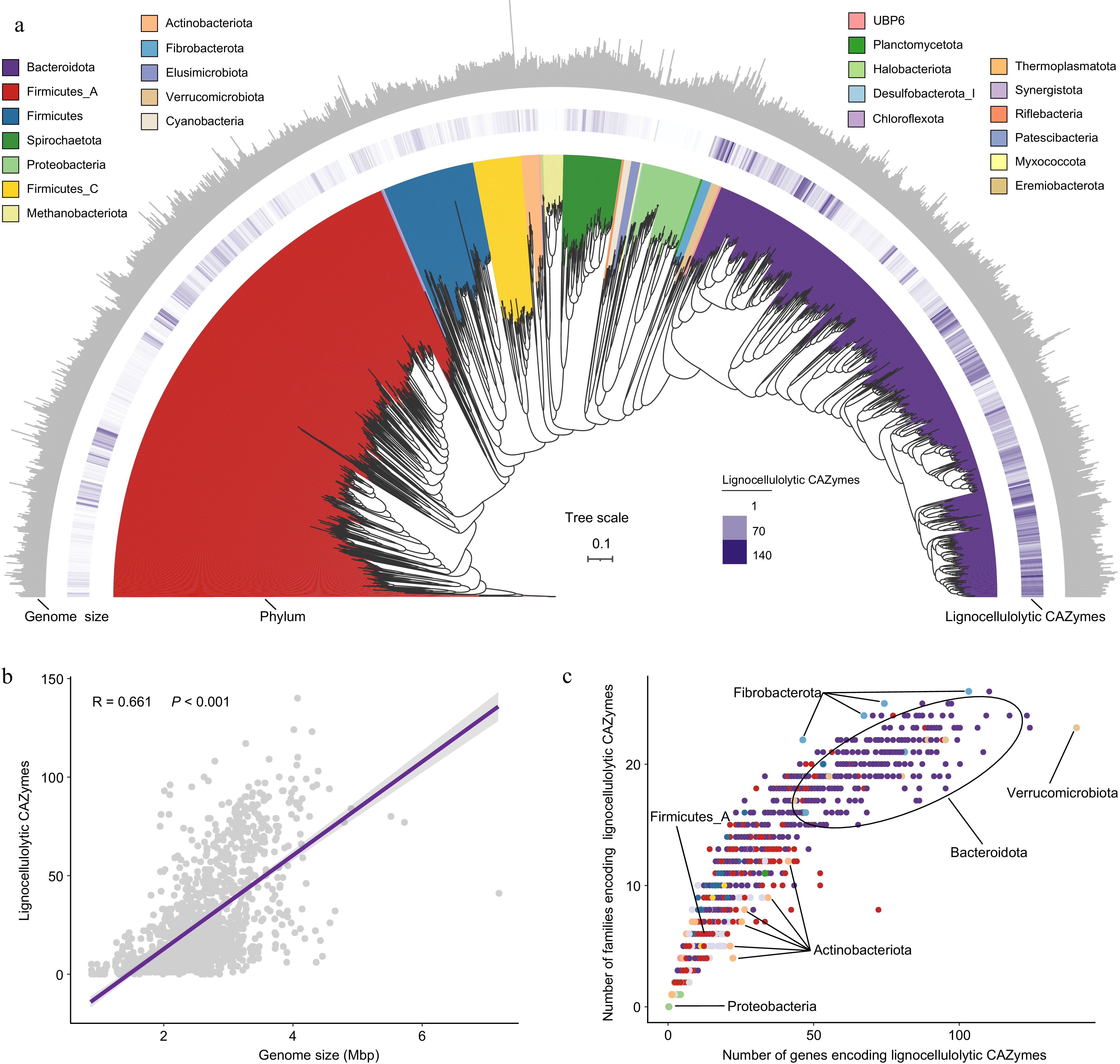
      Figure 1. 

      The CAZyme profiles in lignocellulolytic microbiomes (LMs) from dairy cattle rumen. (a) Phylogenetic tree of 1353 microbial metagenome-assembled genomes (MAGs) coding lignocellulolytic CAZymes. The maximum-likelihood tree is constructed using PhyloPhlAn. Branches are shaded with color to highlight phylum-level affiliations. The inside layer of the heat map represents number of genes encoding lignocellulolytic CAZymes of each LM-MAG. The outside layer of the bar graph represents the genome size of each LM-MAG. (b) The correlation between number of genes encoding lignocellulolytic CAZymes and genome size of LM-MAGs. (c) Number of degradative CAZymes in distinct families in each LM-MAG. Genomes are colored by phylum.

    • Lignocellulose is a complex and recalcitrant structure composed primarily of cellulose, hemicellulose, and lignin, which provide plants with rigidity and durability. For cellulose, GH5 was identified as the most abundant cellulolytic enzyme in rumen LMs, particularly within the Bacteroidota phylum (Fig. 2a). Members of Fibrobacter and Ruminococcus (e.g., Ruminococcus flavefaciens) had the largest number of CAZyme genes coding endo- and exoglucanases (primarily GH5 and GH9; Fig. 2b). Therefore, these microbial populations play a significant role in cellulose degradation by primarily attacking cellulose fibrils at the amorphous regions, followed by cutting at the crystalline regions from both the reducing and non-reducing ends. Ruminococcus MAGs were found to encode GH48, enabling them to target both amorphous and crystalline cellulose (Fig. 2b). Compared with Ruminococcus spp., Fibrobacter spp. exhibited a more diverse array of CAZyme families to attack amorphous regions of cellulose by encoding endoglucanases GH45 and GH8 (Fig. 2b). In addition, the enzymes involved in lignocellulose degradation often feature a CBM attached to the catalytic domain[12]. Notably, Ruminococcus spp. (25) and Fibrobacter spp. (23) displayed the highest counts of CBM-containing enzymes, many of which had N-terminal signal peptides (Supplementary Table S5). This highlights the abundance of secreted or periplasmic multi-domain enzymes in these populations, which are highly effective in lignocellulose degradation. Ruminococcus spp., in particular, had adapted to produce complex proteins with multiple catalytic domains, often accompanied by one or two CBMs to target amorphous cellulose and hemicellulose. This was particularly evident in numerous PULs (CBM4 + GH9; Fig. 3). In contrast, certain proteins identified in Fibrobacter spp. contained three tandemly arranged CBMs and one GH domain (CBM11 + CBM11 + CBM11 + GH51) to adhere cellulosic biomass (Fig. 3). The most significant disparity between Ruminococcus and Fibrobacter was that Ruminococcus had a multitude of dockerin and cohesion domains, whereas Fibrobacter lacked such domains, implying that Ruminococcus possesses the ability to produce cellulosomes for fiber digestion[51] (Supplementary Table S3). Additionally, no genes encoding lytic polysaccharide monooxygenases (LPMOs) targeting cellulosic crystalline substrates were found in the LMs of dairy cattle rumen. For β-glucosidases, members of the Bacteroidales order were the major agents of hydrolyzing glucose dimers into glucose (e.g., GH2 and GH3; Supplementary Table S4).
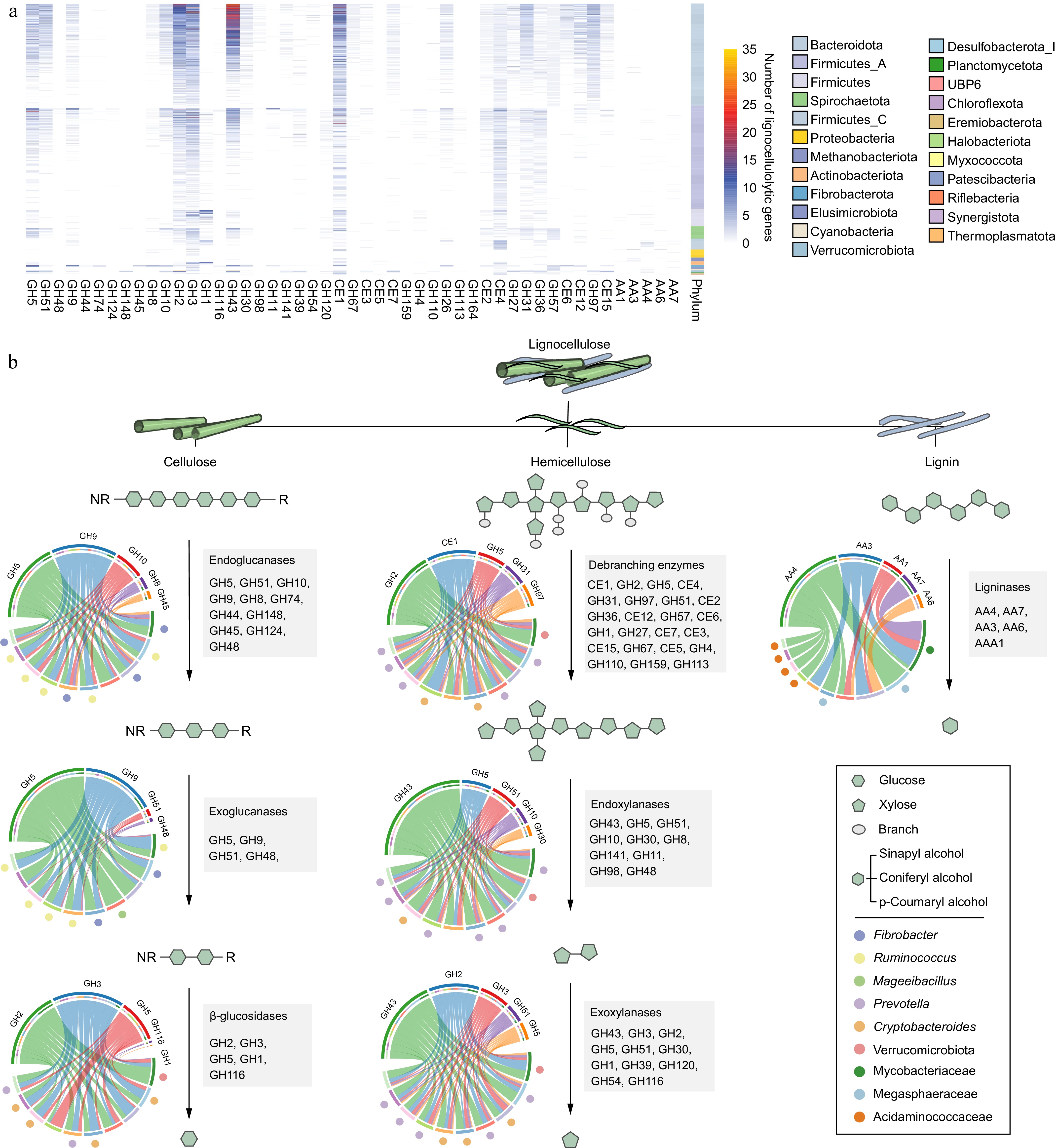
      Figure 2. 

      The distribution and diversity of lignocellulolytic microbiomes (LMs) from dairy cattle rumen. (a) Distribution of lignocellulolytic CAZymes (GH, glycoside hydrolases; CE, carbohydrate esterases; AA, Auxiliary Activities) in LM-MAGs. Genomes are colored by phylum. (b) Cooperative model of cellulases, hemicellulases, and ligninases in lignocellulose degradation in the LM-MAGs. Chord Diagram represent the top families of lignocellulolytic CAZymes contributed by ruminal LM-MAGs. The detailed information regarding LMs involved in the degradation process of lignocellulose, including cellulose, hemicellulose, and lignin, can be found in Table S4.
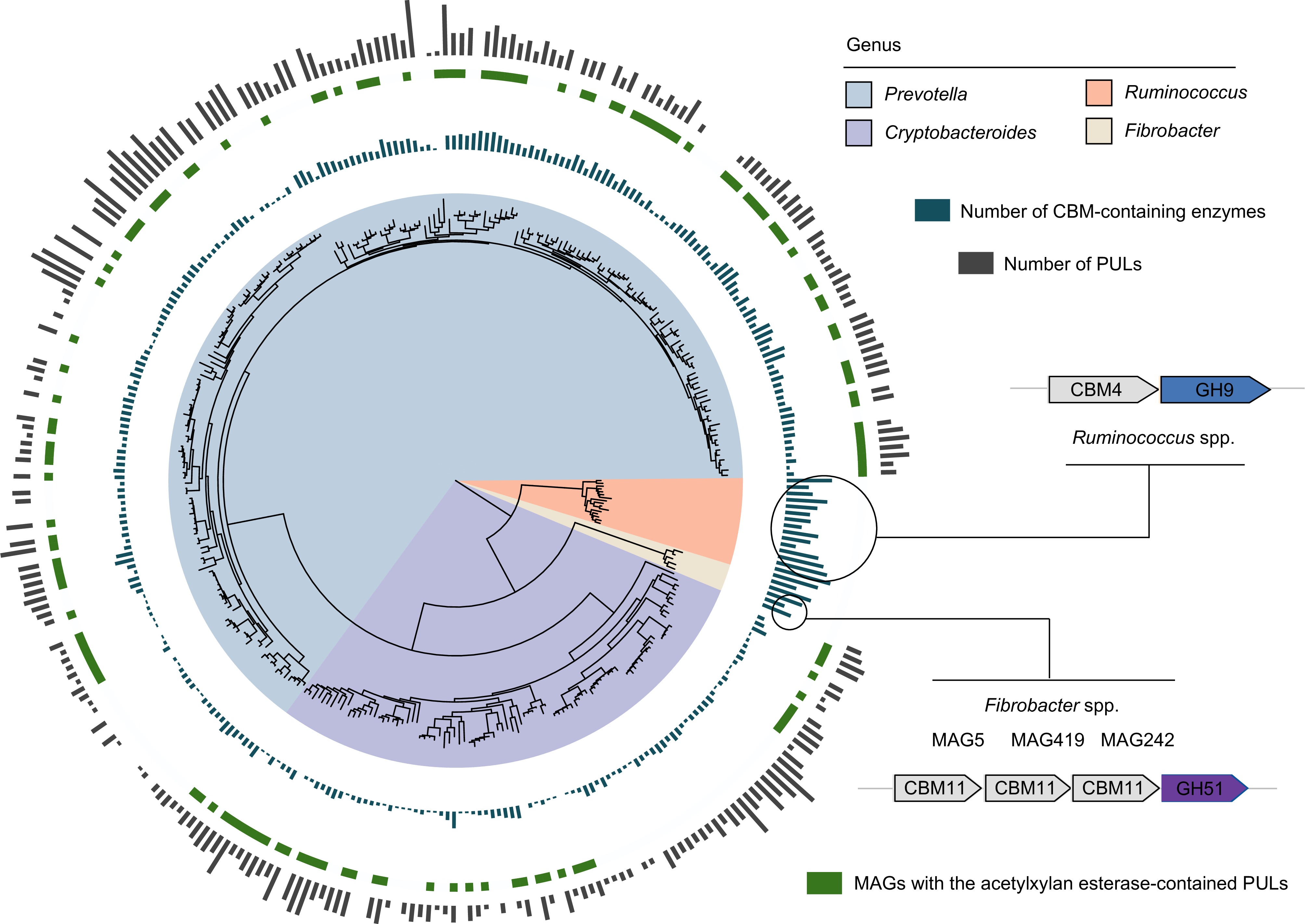
      Figure 3. 

      Different lignocellulose degradation strategies used by taxa present in the dairy cattle rumen. Phylogenetic tree of lignocellulolytic microbiomes (LMs) belonged to genera Prevotella, Cryptobacteroides, Ruminococcus, and Fibrobacter. The maximum-likelihood tree is constructed using PhyloPhlAn. The background of branches is shaded with color to highlight these four taxa. The inside layer of the bar graph represents number of CBM-containing enzymes. The green color represents the specific MAGs with the acetylxylan esterase-contained PULs. The outside layer of the bar graph represents the number of polysaccharide utilization loci (PULs) encoded by the targeted MAGs.

      For hemicellulose degradation, the predominant CAZyme family identified within LMs was GH43, with a notable presence in the Bacteroidota phylum (Fig. 2a). Within the Bacteroidota population, the distribution of lignocellulolytic CAZymes exhibited a similar pattern (Fig. 2a), suggesting a considerable degree of conservation in their enzymatic systems. Members of the Bacteroidota phylum, primarily Prevotella spp. and Cryptobacteroides spp., were found to encode enzymes such as endoxylanase, β-xylosidase, and de-branching activities (primarily GH43, GH2, GH3, and CE1; Fig. 2b). Furthermore, it was observed that Prevotella spp. and Cryptobacteroides spp. encoded a substantial number of PULs to handle the chemical and structural complexity of hemicelluloses such as xylose, arabinose, galactose, mannose, and ferulic acid (Supplementary Table S6). It is noteworthy that most of the Prevotella and Cryptobacteroides MAGs contained acetylxylan esterase-containing PULs (Fig. 3), facilitating the breakdown of the xylan backbone. These results indicate that Prevotella spp. and Cryptobacteroides spp. have developed PULs to address the challenges posed by complex and diverse hemicelluloses, which are subsequently transported into the cells for their utilization.

      Despite the presence of diverse CAZyme repertoires, only a few enzymes involved in lignin breakdown were identified in the rumen microbiome of dairy cattle (Fig. 2a). Specifically, only 135 MAGs (9.82%) encoded AA1, AA3, AA4, AA6, or AA7 enzymes involved in lignin modification and degradation, primarily belonging to the classes Clostridia (47 MAGs), Negativicutes (26 MAGs), and Methanobacteria (17 MAGs; Supplementary Table S4). The AA family with the highest number of genes was vanillyl-alcohol oxidase AA4 (consisting of 89 genes) used for lignin degradation (Fig. 2a). In contrast, only eight genes coding for laccase AA1 were identified for lignin modification (Fig. 2a). These findings suggest that the rumen microbiota in dairy cattle has a limited capacity to degrade lignin from plant biomass.

    • Among the 1353 LM-MAGs identified, some members of the Fibrobacteraceae family, such as Hallerella spp., were isolated from pig intestines in 2020[52] and had not been previously reported in ruminants. Genome annotation against the CAZy database revealed that four Hallerella-affiliated MAGs possessed a large number of cellulases belonging to the families GH5 and GH9, but only a few of these cellulases were found to be fused with CBM domains (Supplementary Tables S4 & S5). This suggests that Hallerella spp. may have the potential to utilize cellulose as a carbohydrate substrate[52]. Additionally, Mageeibacillus spp., previously isolated from the human vagina, contained the highest number of endo- and exoglucanases belonging to the GH5 family, as well as dockerin (Fig. 2b; Supplementary Table S3). Sodaliphilus is a recently described genus, with its type species S. pleomorphus, initially isolated from pig feces[52]. It is noteworthy that it was identified that 32 MAGs could be annotated to Sodaliphilus (including S. pleomorphus) in the dairy cattle rumen, which had the largest number of dockerins and was enriched in hemicellulose and cellulose-degrading enzymes (Supplementary Table S3). However, Sodaliphilus did not contain any cohesion domains, suggesting that the numerous non-cellulosomal dockerins may have other functions[7]. Analysis of PULs revealed that each of the Sodaliphilus-affiliated MAGs contained seven PULs, with S. pleomorphus having more than ten PULs (Supplementary Table S6). Therefore, we hypothesize that Sodaliphilus is a potential lignocellulose degrader in the rumen. Overall, the identification of potential lignocellulolytic consortia may open new opportunities for enhancing the degradation of plant fibers in dairy cattle production.

    • The 1353 LM-MAGs were further employed as a genomic database to assign metagenomic samples from the CON and HG groups in dairy cattle rumen to explore the substantial alterations in microbial communities and enzyme abundances during the degradation processes of cellulose, hemicellulose, and lignin affected by high-grain diets (Fig. 4a; Supplementary Table S7). In the depolymerization phase of celluloses, genomes containing endoglucanases, particularly from the genera Ruminococcus and Hallerella exhibited a significant increase in abundance under high-grain diet conditions (Wilcoxon rank-sum test, p < 0.05; Fig. 4b & Supplementary Table S8). Meanwhile, the grain-based diet increased the abundance of endo-β-1,4-glucanase GH124 (Fig. 4b). Therefore, Ruminococcus spp. and Hallerella spp. emerged as the primary agents targeting cellulose fibrils at the amorphous regions during high-grain diet feeding conditions. In the subsequent cellulose degradation process, a noteworthy shift transpired in the majority of Prevotella-affiliated genomes encoding exoglucanases and β-glucosidases under high-grain diet feeding conditions, notably marked by the decreased abundance in Prevotella ruminicola (Supplementary Table S8).
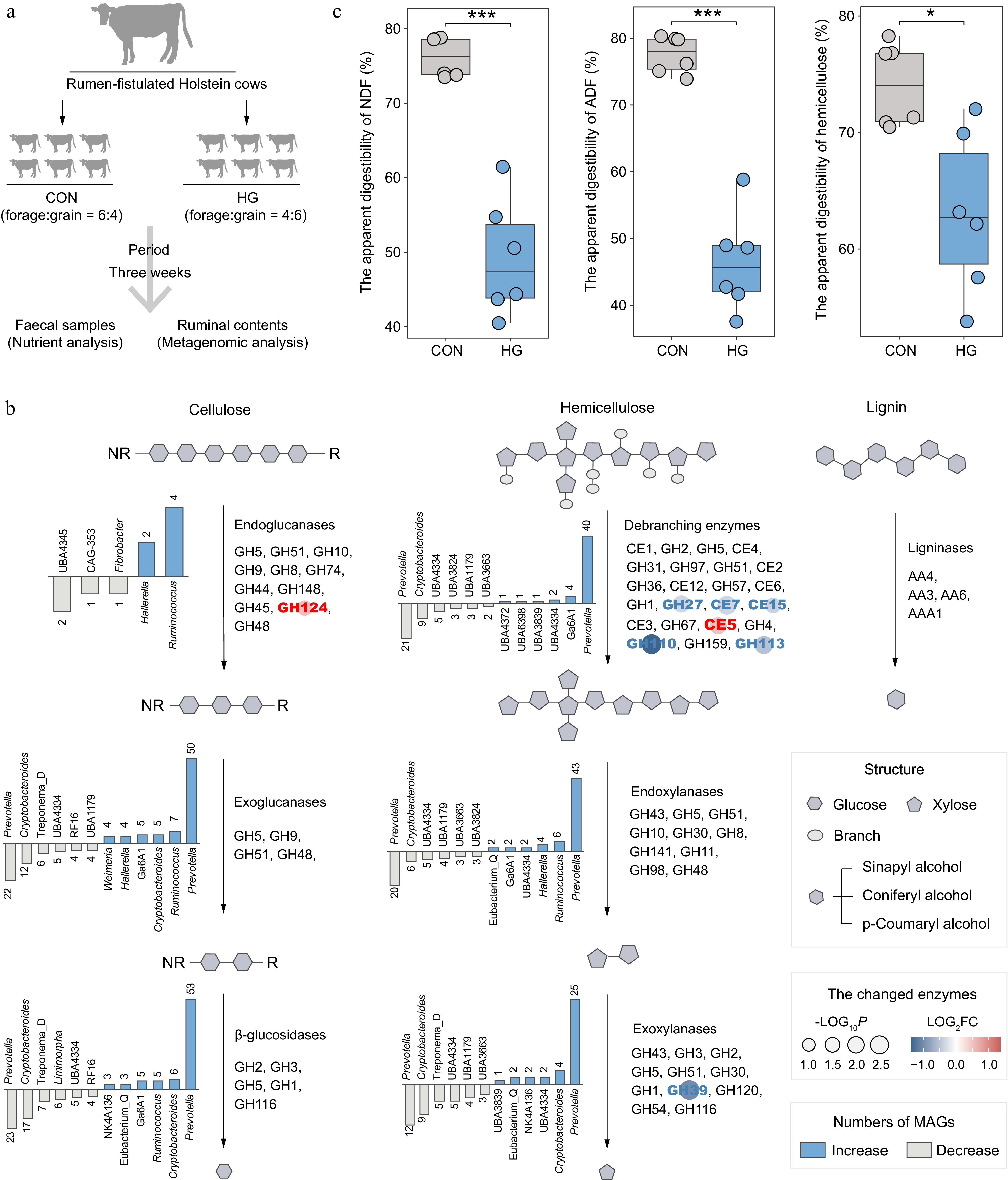
      Figure 4. 

      Alterations in the lignocellulose degradation potential of rumen microbiota between the CON and HG groups in dairy cattle. (a) Experimental scheme of high-grain diet intervention. (b) The number and top taxonomic populations of the significantly increased and decreased abundances of lignocellulolytic microbiome (LM-MAGs) during the degradation of various lignocellulosic components in the rumen in the HG group, compared with the CON group (Wilcoxon rank-sum test, p < 0.05). The bar chart illustrates the number of genomes classified into specific genera with significantly different abundances, containing at least half of the CAZyme families encoding the same type of enzyme. (c) Comparison of the apparent digestibility in neutral detergent fiber (NDF), acid detergent fiber (ADF), and hemicellulose between the CON and HG groups, respectively. Significance is based on the relative index of each cohort according to the t-test. * p < 0.05, ** p < 0.01, *** p < 0.001.

      In the depolymerization process of hemicelluloses, it is notable that the high-grain diet significantly decreased the abundance of various debranching enzymes, including acetylxylan esterases (CE7 and CE15), α-galactosidases (GH27 and GH110), and mannosidase (GH113) (Fig. 4b). Furthermore, a subsequent decline in the abundance of exoxylanases GH39 was observed after feeding the high-grain diets (Fig. 4b), leading to the inhibition of debranching activity in hemicellulose and subsequent degradation of xylan chains. This reduction was primarily attributed to a substantial decrease in the abundance of Prevotella-affiliated genomes (Fig. 4b). However, enzymes involved in lignin modification and degradation processes exhibited no significant changes after high-grain diet feeding.

      As expected, nutrient content analysis of fecal samples revealed a significant reduction in the apparent digestibility of neutral detergent fiber (NDF) and acid detergent fiber (ADF) in response to the high-grain diet (t-test, p < 0.001; Fig. 4c). In addition, the high-grain diet significantly decreased the apparent digestibility of hemicellulose (p = 0.01). Therefore, the high-grain diet reduces the degradation of lignocellulose, primarily manifested in its impact on the degradation of hemicellulose.

    • To gain a deeper understanding of the specific stage of lignocellulose degradation affected by high-grain diets, resulting in an overall decrease in digestibility, the study was extended to encompass a more extensive temporal dimension. Leymus chinensis was subjected to incubation within the rumens of the six fistulated cows, comprising three forage-fed and three grain-fed cows, over 36 h (Fig. 5a). The samples were taken at nine different time points (0, 0.5, 2, 4, 6, 8, 12, 24, and 36 h) for nutrient analysis, 16S rRNA gene sequencing, and metagenomics (Fig. 5a). Nutrient analysis revealed a decrease in the digestibility of NDF, cellulose, and hemicellulose of leymus chinensis during in situ rumen later-stage incubation, especially at 36 h (t-test, p < 0.05; Fig. 5b). It is noteworthy that high-grain diets significantly impacted hemicellulose degradation during the initial phase of rumen incubation, resulting in reduced digestibility at 0.5 h and 2 h (t-test, p < 0.05; Fig. 5b). However, the digestibility of cellulose during the early stage of rumen incubation was unaffected by high-grain diets (t-test, p < 0.05; Fig. 5b). Therefore, the effect of high-grain diets on the degradation of hemicellulose during the initial stage of rumen incubation emerges as a crucial factor contributing to the overall reduction in lignocellulose degradation.
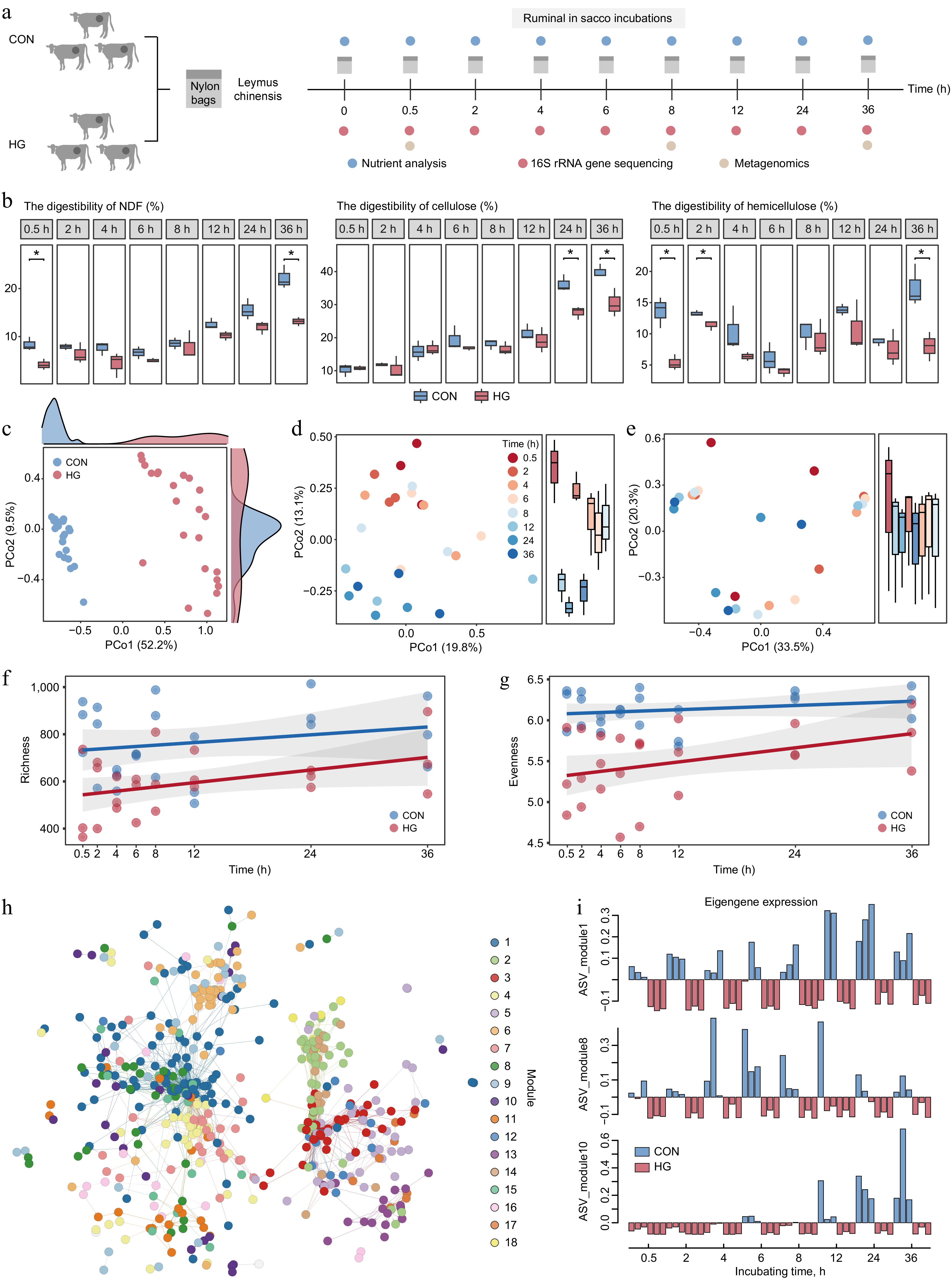
      Figure 5. 

      Key stages of reduction in lignocellulose degradation with high-grain diet intervention. (a) Experimental scheme of rumen in situ incubations. (b) Comparison of digestibility in neutral detergent fiber (NDF), cellulose, and hemicellulose of incubated Leymus chinensis materials at each time point between the CON and HG groups according to the t-test, respectively. * p < 0.05, ** p < 0.01, *** p < 0.001. (c) Principal Coordinate Analysis (PCoA) plot generated from Bray–Curtis dissimilarity matrices using Leymus chinensis-adherent ASV abundances during rumen incubation in both the CON and HG groups. (d) Principal Coordinate Analysis (PCoA) plot generated from Bray–Curtis dissimilarity matrices using Leymus chinensis-adherent ASV abundances at 0.5, 2, 4, 6, 8, 12, 24, and 36 h during rumen incubation in the CON group. (e) Principal Coordinate Analysis (PCoA) plot generated from Bray–Curtis dissimilarity matrices using Leymus chinensis-adherent ASV abundances at 0.5, 2, 4, 6, 8, 12, 24, and 36 h during rumen incubation in the HG group. Temporal shift of richness ((f), Observed number of ASVs) and evenness ((g), Shannon diversity) indexes of ASVs between the CON and HG groups, respectively. (h) The co-occurrence network visualizing significant correlations of diet-sensitive 786 ASVs among eight time points during the rumen incubation (R > 0.7). (i) Eigengene expression of module 1, module 8, and module 10 in the CON and HG groups. CON, high-forage diet; HG, high-grain diet; ASV, amplicon sequence variants.

      To understand the effect of high-grain diets on microbial colonization trajectories, fiber-adherent 16S rRNA gene sequencing data was analyzed at the amplicon sequence variant (ASV) level. The ordination analysis revealed a distinct distribution between the CON and HG groups (p = 0.0001, ANOSIM 9999 permutations; Fig. 5c). Considering the variable 'timepoint' showed that microbial taxa in the CON group exhibited a clear separation over time, indicating a regular colonization pattern following the degradation process (Fig. 5d). In contrast, the high-grain diet disrupted the colonization pattern of microorganisms, resulting in a mixing of microbiota at different time points (Fig. 5e). I was speculated that the high-grain diet affects the initial stage of the degradation period, resulting in no separation of microbial colonization in different degradation periods. Moreover, the diversity curve showed that alpha diversity (richness and evenness) in the HG group was lower than that in the CON group from 0.5 h during the rumen incubation (Fig. 5f, g).

      Indicator species and edge analysis were conducted to identify 786 ASVs with significantly changed abundance during rumen incubation under grain introduction, mainly belonging to Prevotella (120 ASVs), Rikenellaceae RC9 gut group (72 ASVs), Unclassified F082 (59 ASVs), Christensenellaceae R-7 group (57 ASVs), and Ruminococcus (39 ASVs) (indicator species: p < 0.05 and edgeR: FDR < 0.05; Supplementary Table S9). These diet-sensitive 786 ASVs were further clustered into 18 dominant co-occurrence modules using weighted correlation network analysis (WGCNA) among eight time points during the rumen incubation (Fig. 5h). Specifically, the ASVs from modules 1, 8, and 10 significantly decreased in abundance at different time points under the high-grain diet conditions (Fig. 5i). The ASVs from module 1 belonged tothe Rikenellaceae RC9 gut group and showed a notable decline in abundance during the later stages (12−36 h) of rumen incubation under high-grain diet conditions (Supplementary Table S9). The ASVs from module 10 belonged to Treponema and Fibrobacter also demonstrated a substantial reduction in abundance during the later stages (12−36 h) of rumen incubation under high-grain diet conditions (Supplementary Table S9). Furthermore, ASVs from module 8, predominantly attributed to Prevotella (70.97%), exhibited a significant decrease in abundance during the early stages (0.5−8 h) of rumen incubation under high-grain diet conditions (Supplementary Table S9). This implies that Prevotella spp. may play a pivotal role as a key executor in the reduction of early-stage hemicellulose degradation.

      The 1353 LM-MAGs were further utilized as a genomic database to analyze the metagenomic data from fiber-adherent samples and found that 740 LM-MAGs colonized leymus chinensis during rumen incubation (TPM > 0), with the majority (about 19.6%) belonging into Prevotella (Supplementary Table S10). Notably, a decrease in the abundance of Prevotella-affiliated MAGs were observed in the HG-fed cow rumen, which was most significant from 0.5 to 8 h of rumen incubation compared to the CON group (Supplementary Fig. S2). This phenomenon reiterated the initial 16S rRNA gene observations. In detail, MAG168, MAG160, MAG421, MAG1102, MAG646, MAG1207, and MAG763 mainly colonized from 0.5 to 8 h of rumen incubation in the CON group, while their colonization abundance was much lower in the HG group. Additionally, Prevotella ruminicola (MAG71, MAG1076, and MAG906) and Prevotella sp900100635 (MAG170 and MAG413) mainly colonized at 8 h of rumen incubation in the CON group. These Prevotella spp. showed a strong potential for hemicellulose degradation, as inferred from the enrichment family GH43 (Supplementary Table S4). The present genome-centric analysis further supports the hypothesis that the high-grain diet affects the degradation of amorphous regions in lignocellulose during the initial stage of rumen incubation. This effect was largely dependent on the Prevotella-dominated reduction in hemicellulose degradation.

    • Despite the impressive ability of rumen microbiota in dairy cattle to convert low-quality lignocellulose into nutrient-rich milk, approximately 50% of plant biomass is still resistant to degradation[53]. Previous studies have explored carbohydrate-degrading enzyme libraries within the rumen microbiomes of dairy cattle[4,6]. However, the limited number of dairy cattle has hindered our understanding of the microbial mechanisms involved in the degradation cascades of lignocellulose. In this study, all publicly available rumen metagenomes of dairy cattle were compiled and supplemented them with our own data, creating a comprehensive dataset to elucidate 1353 high-quality microbial genomes involved in lignocellulose degradation in the rumen. The present study provided a systematic description of these 1353 high-quality LM-MAGs spanning 23 phyla, representing a crucial foundation for a comprehensive understanding of rumen lignocellulose degradation efficiency in dairy cattle and creating opportunities for further improvement.

      Given the diversity and redundancy of lignocellulolytic CAZymes, it was observed that different microbial consortia employed distinct enzymatic strategies in the degradation cascades of specific lignocellulosic components, including cellulose, hemicellulose, and lignin. Cellulose, the main component of lignocellulose, is degraded through the synergistic action of three classes of enzymes, including endoglucanases, exoglucanases, and β-glucosidases[5456]. Members of Fibrobacter and Ruminococcus (e.g., Ruminococcus flavefaciens) were predicted as powerful degraders to process endo- and exoglucanases to target amorphous and crystalline cellulose[57]. An interesting phenomenon was the striking enrichment of non-catalytic domain CBMs in genera Fibrobacter and Ruminococcus. It was found that Fibrobacter spp. contained tandemly arranged CBM families anchored to cellulosic biomass. The absence of CBMs leads to a significant reduction in the binding affinity and enzymatic activity of enzyme proteins towards crystalline cellulose[58]. In contrast, the substrate adhesion effects of multiple CBMs can greatly promote the enzymatic activities of the catalytic domains of CAZymes[13,59]. Therefore, the presence of multiple CBMs-harboring GH domains in Fibrobacter spp. in cow rumen is more advantageous for cellulases to attach to the hydrophobic surface of the crystalline substrate, thereby enhancing the degradation processivity. Compared to cellulose-degrading taxa, the main players in hemicellulose degradation, Prevotella spp. and Cryptobacteroides spp., had relatively fewer CBM domains but encoded numerous PULs to degrade the main and side chains of hemicellulose[14]. The enrichment of acetylxylan esterase-contained PULs contributes largely to hemicellulose degradation. A significant proportion of the xylose residues in hemicellulose are estimated to be substituted with acetyl groups at the O-2 or O-3 position, ranging from approximately 22% to 50%[60]. The acetylxylan esterase encoded by Prevotella spp. and Cryptobacteroides spp. can remove these acetyl groups of hemicellulose to improve the degradation efficiency of other enzymes. Therefore, the different bacterial consortia employed diverse enzymatic strategies for degradation cascades of various lignocellulosic components.

      Previous studies have shown that feeding high-grain diets impacts the ability of ruminal microbes to degrade fiber[4,61]. The present findings further suggest that a high-grain diet specifically disrupts the degradation cascade of hemicellulose. This was evident in the significant decrease of acetylxylan esterases (CE7 and CE15), α-galactosidases (GH27 and GH110), mannosidases (GH113), and exoxylanases (GH39) under high-grain diet conditions, which leads to a decrease in debranching activity in hemicellulose and subsequent degradation of xylan chains. Notably, this process was primarily driven by the reduction of Prevotella spp. The decreased apparent digestibility of hemicellulose under high-grain diet conditions supported this phenomenon. Through further extending the present study to multiple time points, it was discovered that a high-grain diet decreased the early-stage degradation of hemicellulose by Prevotella spp., leading to the reduced degradation of the amorphous regions of lignocellulose. This reduction in turn reduces the accessibility of microbial degrading enzymes to the ordered crystalline cellulose and results in ineffective degradation of lignocellulose[12]. The present results showed that feeding high-grain diets disrupted the ordered colonization of microorganisms on Leymus chinensis, resulting in the non-separation of microbiota at different colonization time points. Therefore, the Prevotella-dominated reduction in hemicellulose degradation during the initial stage of rumen incubation may be the primary reason for the reduced digestibility of lignocellulose under high-grain diet feeding.

      In addition, it was found that some lignocellulose-degrading bacteria were significantly enriched under high-grain conditions, and importantly, they belong to microbial taxa that have not been described before in ruminants. For example, Hallerella spp., the nearest phylogenetic neighbor of Fibrobacter spp.[52], were found to possess a large amount of endoglucanases and exoglucanases. Hallerella-affiliated MAGs were found to only attach to fiber in cows fed high-grain diets, indicating their important role in lignocellulose degradation in such environments. Despite both belonging to the Fibrobacteraceae family, Fibrobacter spp. and Hallerella spp. had different adaptabilities in various environments. In detail, Fibrobacter was more suitable for high-fiber environments, while Hallerella exhibited fiber degradation ability in low-fiber environments. Additionally, members of Sodaliphilus were predicted to possess the capability of lignocellulose degradation through extracellular enzymes or polysaccharide utilization loci (PULs), and their abundance exhibited a significant increase under high-grain diet conditions. Previous studies have shown that high-grain diets can lead to a decrease in the abundance of lignocellulose-degrading microorganisms[4,62]. Therefore, these newly discovered potential lignocellulose-degrading bacteria in the rumen of dairy cattle represent important resources for development. These bacteria could help mitigate the reduced lignocellulose degradation capacity associated with high-grain feeding. Although further research is needed to confirm the actual lignocellulose degradation capabilities of these microorganisms, our findings provide valuable support for future efforts aimed at enhancing the degradation efficiency of lignocellulose in the rumen of dairy cattle fed high-grain diets.

      In addition to bacteria, several studies have explored the genomes of eukaryotic organisms, such as fungi and ciliates, in the rumen[63,64]. These studies have revealed that these organisms encode a variety of carbohydrate-related genes and enzymes, showing their significant capabilities for degrading plant fiber. The present study primarily focuses on prokaryotic genomic research related to the microbial degradation of rumen carbohydrates. Future research should clarify how high-grain diets influence different lignocellulosic components and the various stages of lignocellulolytic cascades through specific protozoa and fungi. This will contribute to a more comprehensive understanding of the role of rumen microorganisms in carbohydrate degradation.

    • The present study utilized a genome-centric approach to identify 1353 high-quality MAGs involved in lignocellulose degradation in the cow rumen. By analyzing their enzymatic strategies for different substrate types, insights into the complexity and specialization of ruminal microbial populations in degrading various lignocellulosic components were gained, with a particular emphasis on cellulose and hemicellulose degradation, while highlighting their limited role in lignin degradation. Through spatial and temporal studies involving diet interventions and rumen in situ incubation, it was discovered that a high-grain diet primarily interfered with the degradation of amorphous regions of lignocellulose and significantly reduced hemicellulose degradation by Prevotella-dominated communities. These findings underscore the intricate interplay among diet, microbial consortia, and enzymatic strategies in the rumen, highlighting the potential for manipulating the rumen microbiota to improve the efficiency of lignocellulose degradation in dairy cattle.

    • All procedures were reviewed and preapproved by the the Nanjing Agricultural University Institutional Animal Care and Use Committee, identification number: SYXK-2017–0027, approval date: 2019-3-12. The research followed the 'Replacement, Reduction, and Refinement' principles to minimize harm to animals. This article provides details on the housing conditions, care, and pain management for the animals, ensuring that the impact on the animals is minimized during the experiment.

    • The authors confirm contribution to the paper as follows: study conception and design: Mao S, Zhu W; samples collection and experiments conduction: Lin L, Yang H, Zhang Jiyou, Lai Z, Qi W, Ma H, Zhang Jiawei; published rumen metagenome collection: Xie F, Lin L; bioinformatic analyses, data visualization and interpretation, draft manuscript preparation: Lin L; manuscript revision: Mao S. All authors read, edited, and approved the final manuscript.

    • Raw sequence reads for all 18 incubated leymus chinensis samples are available under European Nucleotide Archive (ENA) project PRJNA955930. All MAGs produced and utilized in this study have been deposited in Figshare (https://figshare.com/s/dcaad3555e23f2551029). The data sources for an additional 226 published ruminal metagenome samples from dairy cattle are provided in Supplementary Table S1. Please note that although there were 48 samples available under PRJNA214227 as reported by Wolff et al.[27], we exclusively utilized 16 rumen samples from dairy cattle. For other projects, we also exclusively utilized rumen samples from dairy cattle.

      • The current project was supported by the high-performance computing platform of Bioinformatics Center, Nanjing Agricultural University. This research was funded by the National Key R&D Program of China (2022YFD1301001).

      • The authors declare that they have no conflict of interest. Shengyong Mao is the Editorial Board member of Animal Advances who was blinded from reviewing or making decisions on the manuscript. The article was subject to the journal's standard procedures, with peer-review handled independently of this Editorial Board member and the research groups.

      • Supplementary Table S1 226 published ruminal metagenome samples from dairy cattle.
      • Supplementary Table S2 Sample source of 5034 MAGs from dairy cattle rumen.
      • Supplementary Table S3 Genomic statistics for 1374 non-redundant high-quality MAGs (Completeness > 80% and Contamination < 10%).
      • Supplementary Table S4 1353 high-quality MAGs involved in the degradation process of lignocellulose, including cellulose, hemicellulose, and lignin.
      • Supplementary Table S5 Genome-wide prediction of CBM-containing CAZymes in lignocellulolytic MAGs in the rumen of dairy cattle.
      • Supplementary Table S6 Genome-wide prediction of total polysaccharide utilization loci (PULs) in lignocellulolytic MAGs in the rumen of dairy cattle.
      • Supplementary Table S7 Ingredients and nutritional compositions of the high-forage (CON) and high-grain (HG) diets.
      • Supplementary Table S8 The significant changes of microbial genomes and key enzymes involved in the degradation cascade of various lignocellulosic components caused by high-grain diets.
      • Supplementary Table S9 Significantly different abundance of 786 microbial ASVs colonized leymus chinensis between the high-forage (CON) and high-grain (HG) diets during the rumen incubation.
      • Supplementary Table S10 The abundance of 740 LM-MAGs colonized leymus chinensis during rumen incubation
      • Supplementary Fig. S1 A: Pipeline for data processing and integration. B: Distribution of quality metrics across the high-quality MAGs (n = 1374), showing the minimum value, first quartile, median, third quartile and maximum value.
      • Supplementary Fig. S2 Heatmap of the abundance and distribution of Prevotella-affiliated LM-MAGs at 0.5, 8, 36 h during the rumen incubation between the CON and HG groups, and a Z-score was used for correction. CON, high-forage diet; HG, high-grain diet.
      • Copyright: © 2024 by the author(s). Published by Maximum Academic Press on behalf of Nanjing Agricultural University. This article is an open access article distributed under Creative Commons Attribution License (CC BY 4.0), visit https://creativecommons.org/licenses/by/4.0/.
    Figure (5)  References (64)
  • About this article
    Cite this article
    Lin L, Ma H, Zhang J, Yang H, Zhang J, et al. 2024. Lignocellulolytic microbiomes orchestrating degradation cascades in the rumen of dairy cattle and their diet-influenced key degradation phases. Animal Advances 1: e002 doi: 10.48130/animadv-0024-0002
    Lin L, Ma H, Zhang J, Yang H, Zhang J, et al. 2024. Lignocellulolytic microbiomes orchestrating degradation cascades in the rumen of dairy cattle and their diet-influenced key degradation phases. Animal Advances 1: e002 doi: 10.48130/animadv-0024-0002

Catalog

  • About this article

/

DownLoad:  Full-Size Img  PowerPoint
Return
Return