ARTICLE   Open Access    

Quality differences and profiling of volatile components between fermented and unfermented cocoa seeds (Theobroma cacao L.) of Criollo, Forastero and Trinitario in China

  • # These authors contributed equally: Dewei Yang, Baoduo Wu

More Information
  • Fermentation plays a crucial role and contributes to the quality of cocoa beans, the basis of chocolate products. The aim of this study was to evaluate the levels of chemical and aromatic compounds in the cocoa beans of Criollo, Forastero and Trinitario in China. Besides fermentation, unfermented beans of the three genetic groups were also analyzed. The results indicated that genotype involvement played an important role, contributing to cocoa volatile profiling and quality traits. The three cocoa groups' mass indicators all demonstrated some degree of decline after fermentation. After HS-SPME-GC-MS testing, 48 compounds were found in raw beans and 79 compounds in fermented beans. Trinitario contained the most volatile compounds with the highest levels of alcohol, while Criollo contained the highest ester content in unfermented beans. Alcohols and esters were characteristic components of Criollo fermented beans with 37.33% and 14.81%, respectively. The content of acetic acid was as high as 50% in the fermented beans of Forastero and Trinitario and only 35.42% in Criollo. The feature compounds of Criollo fermented beans showed an aromatic profile related to flower flavor, fruit flavor, green flavor, baking flavor and nut flavor. While the characteristic volatile compounds were mainly associated with descriptors of sour, buttery, fruity, roasted and nutty notes for Forastero and Trinitario fermented beans. The results of this study revealed the characteristics of three genetic groups of cocoa beans that will ultimately contribute to the breeding of excellent aroma varieties and cocoa production sustainability in Hainan Province, China.
  • Salvia rosmarinus L. (old name Rosmarinus officinalis), common name Rosemary thrives well in dry regions, hills and low mountains, calcareous, shale, clay, and rocky substrates[1]. Salvia rosmarinus used since ancient times in traditional medicine is justified by its antiseptic, antimicrobial, anti-inflammatory, antioxidant, and antitumorigenic activity[1,2]. The main objective of the study is to evaluate the antimicrobial activity of different extracts of Salvia rosmarinus in vitro, and its compounds related to in silico targeting of enzymes involved in cervical cancer. Since the start of the 20th century, some studies have shown that microbial infections can cause cervical cancers worldwide, infections are linked to about 15% to 20% of cancers[3]. More recently, infections with certain viruses like Human papillomaviruses (HPV) and Human immunodeficiency virus (HIV), bacteria like Chlamydia trachomatis, and parasites like schistosomiasis have been recognized as risk factors for cancer in humans[3]. Then again, cancer cells are a group of diseases characterized by uncontrolled growth and spread of abnormal cells. Many things are known to increase the risk of cancer, including dietary factors, certain infections, lack of physical activity, obesity, and environmental pollutants[4]. Some studies have found that unbalanced common flora Lactobacillus bacteria around the reproductive organ of females increases the growth of yeast species (like Candida albicans) and some studies have found that women whose blood tests showed past or current Chlamydia trachomatis infection may be at greater risk of cervical cancer. It could therefore be that human papillomavirus (HPV) promotes cervical cancer growth[3]. Salvia rosmarinus is traditionally a healer chosen as a muscle relaxant and treatment for cutaneous allergy, tumors, increases digestion, and the ability to treat depressive behavior; mothers wash their bodies to remove bacterial and fungal infections, promote hair growth, and fight bad smells[5] .

    The study of plant-based chemicals, known as phytochemicals, in medicinal plants is gaining popularity due to their numerous pharmacological effects[6] against drug resistance pathogens and cancers. The causes of drug resistance to bacteria, fungi, and cancer are diverse, complex, and only partially understood. The factors may act together to initiate or promote infections and carcinogenesis in the human body is the leading cause of death[7]. Antimicrobial medicines are the cornerstone of modern medicine. The emergence and spread of drug-resistant pathogens like bacteria and fungi threaten our ability to treat common infections and to perform life-saving procedures including cancer chemotherapy and cesarean sections, hip replacements, organ transplantation, and other surgeries[7]. On the other hand, information about the current magnitude of the burden of bacterial and fungal drug resistance, trends in different parts of the world, and the leading pathogen–drug combinations contributing to the microbial burden is crucial. If left unchecked, the spread of drug resistance could make many microbial pathogens much more lethal in the future than they are today. In addition to these, cancers can affect almost any part of the body and have many anatomies and molecular subtypes that each require specific management strategies to avoid or inhibit them. There are more than 200 different types of cancer that have been detected. The world's most common cancers affecting men are lung, prostate, colorectal, stomach, and liver cancers[8]. While breast, cervix, colorectal, lung, and stomach cancers are the most commonly diagnosed among women[8]. Although some cancers said to be preventable they seem to still be one of the causes of death to humans, for example cervical cancer. The need to fill the gap to overcome the problem of searching for antimicrobials and anticancers from one source of Salvia rosmarinus is of importance.

    Cervical cancer is a common cancer in women and a prominent cause of death[9]. In Ethiopia, cervical cancer is a big deal for women aged 15 to 44, coming in as the second most common cancer[9]. Globally, it's the fourth most common prevalent disease for women[10]. Aberrant methylation of tumor-suppressor genes' promoters can shut down their important functions and play a big role in causing cervical tumors[10]. There are various cervical cancer repressor genes (proteins turn off or reduce gene expression from the affected gene), such as CCNA1, CHF, HIT, PAX1, PTEN, SFRP4, and TSC1. The genes play a crucial role in causing cervical cancer by regulating transcription and expression through promoter hypermethylation, leading to precursor lesions during cervical development and malignant transformation[11]. The process of DNA methylation is primarily carried out by a group of enzymes known as DNA methyltransferases (DNMT1). It has been reported that DNMT1 (PDB ID: 4WXX), a protein responsible for DNA methylation can contribute to the development of cervical cancer. DNMT1 inhibits the transcription of tumor suppressor genes, facilitating tumorigenesis, which finally develops into cervical cancer. Tumor suppressor gene transcription is inhibited by DNMT1, which helps cancer grow and eventually leads to cervical cancer. Repressive genes' hypermethylation may be decreased, their expression can be increased, and the phenotype of malignant tumors can be reversed by inhibiting the DNMT1 enzyme.

    On the other hand, infection by the human papilloma virus (HPV) phenotype 16, enzyme 6 (PDB ID: 4XR8) has been correlated with a greatly increased risk of cervical cancer worldwide[12]. Based on variations in the nucleotide sequences of the virus genome, over 100 distinct varieties of the human papilloma virus (HPV) have been identified (e.g. type 1, 2 etc.). Genital warts can result from certain types 6 and 11 of sexually transmitted HPVs. Other HPV strains, still, that can infect the genitalia, do not show any symptoms of infection[8]. Persistent infection with a subset of approximately 13 so-called 'high-risk' sexually transmitted HPVs, including such as types 16, 18, 31, 33, 35, 39, 45, 51, 52, 56, 58, 59, and 68 different from the ones that cause warts may lead to the development of cervical intraepithelial neoplasia (CIN), vulvar intraepithelial neoplasia (VIN), penile intraepithelial neoplasia (PIN), and/or anal intraepithelial neoplasia (AIN). These are precancerous lesions and can progress to invasive cancer. Almost all occurrences of cervical cancer have HPV infection as a required component[13]. Superfluous infection by HPV type 16 E6 (PDB ID: 4XR8) has been correlated with a greatly increased genital risk of precursor cervical cancer worldwide[11]. Scholars more defined in major biochemical and biological activities of HPV type 16 E6 (PDB ID: 4XR8) in high-risk HPV oncogenes and how they may work together in the development of cervical disease and cancer[13].

    One potential approach to treat cervical cancer is to inhibit the activity of the DNMT1 and HPV type 16 E6 enzymes specifically[1316]. Over 50% of clinical drug forms worldwide originate from plant compounds[17]. In the past, developing new drugs was a lengthy and costly process. However, with the emergence of bioinformatics, the use of computer-based tools and methods have become increasingly important in drug discovery. One such method is molecular docking and ADMET profiling which involves using the structure of a drug to screen for potential candidates. This approach is known as structure-based drug design and can save both time and resources during the research process[15]. Structural-based drug designing addresses ligand binding sites with a known protein structure[15]. Using free binding energies, a computational method known as docking examines a large number of molecules and suggests structural theories for impeding the target molecule[17]. Nowadays, due to increasing antibiotic resistance like bacteria, fungi, and cancer cells, natural products remain an important source for discovering antimicrobial compounds and novel drugs for anti-cancers like cervical cancers. Therefore, the purpose of this research is to assess the antimicrobial activity of extracts, molecular docking, ADMET profiling in anticancer properties of compounds isolated from Salvia rosmarinus, on a targeting DNMT1 and HPV type 16 E6 in human cervical cancer. In the present study, various solvent crude extracts obtained from Salvia rosmarinus were used for antimicrobial activity and the isolated compounds 1 and 2 were submitted for in silico study to target the DNMT1 and HPV type 16 E6 enzymes to inhibit the growth of human cervical cancer cells.

    Healthy Salvia rosmarinus leaves were collected in Bacho district, Southwest Showa, Oromia, Ethiopia, during the dry season of November 2022. The plant materials were authenticated by Melaku Wondafrish, Natural Science Department, Addis Ababa University and deposited with a voucher number 3/2-2/MD003-80/8060/15 in Addis Ababa University's National Herbarium.

    The most common organic solvent used in extractions of medicinal plants is 2.5 L of petroleum ether, chloroform/methanol (1:1), and methanol. The test culture medium for microbes was used and performed in sterile Petri dishes (100 mm diameter) containing sterile Muller–Hinton Agar medium (25 mL, pH 7) and Sabouraud Dextrose Agar (SDA) for bacteria and fungi, respectively. A sterile Whatman filter paper (No. 1) disc of 6 mm diameter was used to determine which antibiotics an infective organism is sensitive to prescribed by a minimum zone of inhibition (MZI). Ciprofloxacin antibiotic reference (manufactured by Wellona Pharma Ciprofloxacin tablet made in India) and Ketoconazole 2% (made in Bangladesh) were used as a positive controls for antibacterial and antifungal, respectively and Dimethyl sulfoxide (DMSO) 98.9% was used as a negative control for antimicrobial tests. In the present study, the height of the column was 650 mm and the width was 80 mm. Several studies by previous researchers showed the acceptable efficiency of column chromatography (up to 43.0% w/w recovery) in the fractionation and separation of phenolic compounds from plant samples[18]. In column chromatography, the ideal stationary phase used silica gel 60 (0.200 mm) particles. The 1H-NMR spectrums of the compounds were analyzed using a 600 MHz NMR machine and 150 MHz for 13C NMR. The compounds were dissolved in MeOD for compound 1 and in DMSO for compound 2 for NMR analysis. On the other hand, UV spectroscopy (made in China) used 570 nm ultraviolet light to determine the absorbency of flavonoids (mg·g−1) phytochemicals.

    The samples (extracts) were analyzed to detect the presence of certain chemical compounds such as alkaloids (tested using Wagner's reagents), saponins (tested using the froth test), steroids (tested with Liebermann Burchard's tests), terpenoids (tested with Lidaebermann Burchard's tests), quinones, and flavonoids (tested using Shinoda tests)[19].

    The leaves of Salvia rosmarinus (500 g) were successively extracted using maceration using petroleum ether, chloroform/methanol (1:1), and methanol, every one 2.5 L for 72 h to afford 3.6, 6, and 53 g crude extracts, respectively. The methanol/chloroform (1:1) extract (6 g) was loaded to silica gel (150 g) column chromatography using the increasing polarity of petroleum ether, methanol/chloroform (1:1) solvent system to afford 80 fractions (100 mL each). The fraction obtained from chloroform/methanol 1:1 (3:2) after repeated column chromatography yielded compound 1 (18 mg). Fractions 56-65, eluted with chloroform/methanol (1:1) were combined and purified with column chromatography to give compound 2 (10 mg).

    The microorganisms were obtained from the Ethiopia Biodiversity Institution (EBI). Two gram-positive bacteria namely Staphylococcus aureus serotype (ATCC 25923) and Streptococcus epidermidis (ATCC14990); and three gram-negative bacteria, namely Escherichia coli (ATCC 25922), Pseudomonas aeruginosa (ATCC 5702), and Klebsiella pneumonia (ATCC e13883) were inoculated overnight at 37 °C in Muller–Hinton Agar/MHA culture medium and two fungus strains of Candida albicans (ATCC 16404) and Aspergillus niger (ATCC 11414) were inoculated overnight at 27−30 °C in Sabouraud Dextrose Agar/SDA culture medium[20].

    The antibacterial and antifungal activities of different crude extracts obtained from Salvia rosmarinus plant leaves were evaluated by the disk diffusion method (in accordance with the 13th edition of the CLSI M02 document on hardydiagnostics.com/disk-diffusion). Briefly, the test was performed in sterile Petri dishes (100 mm diameter) containing solid and sterile Muller–Hinton Agar medium (25 mL, pH 7) and Sabouraud Dextrose Agar (SDA) for bacteria and fungi, respectively. The extracts were placed on the surface of the media that had previously been injected with a sterile microbial suspension (one microbe per petri dish) after being adsorbed on sterile paper discs (5 μL per Whatman disc of 6 mm diameter). To prevent test samples from eventually evaporating, all Petri dishes were sealed with sterile laboratory films. They were then incubated at 37 °C for 24 h, and the zone diameter of the inhibition was measured and represented in millimeters. Ciprofloxacin antibiotic reference (manufactured by Wellona Pharma Ciprofloxacin tablet, India) was used as a positive control and DMSO was used as a negative control for antibacterial activity test while Ketoconazole 2% (Bangladesh) was used as a positive control and 10 μL of 0.2% agar as a negative control for antifungal activity tests[20]. The term 'inhibitory concentration' refers to the minimum sample concentration required to kill 99.9% of the microorganisms present[21]. Three repetitions of the crude extract sample were used to precisely measure the inhibitory halo diameter (in mm), which was then expressed as mean ± standard deviation to assess the anti-microbial activity.

    Cervical cancer-causing protein was identified through relevant literature. The protein molecule structure of DNA (cytosine-5)-methyltransferase 1 (DNMT1) (PDB ID: 4WXX)[21] and HPV type 16 E6 (PDB ID: 4XR8)[21] - a protein known to cause cervical cancer - were downloaded from the Protein Data Bank[22]. The stability of the protein molecule was assessed using Rampage[23].

    Phytochemical constituents of Salvia rosmarinus plant leaves were used to select a source of secondary metabolites (ligands). Ligand molecules were obtained through plant extraction, and isolation, and realized with PubChem (https://pubchem.ncbi.nlm.nih.gov/). The ligands were downloaded in Silver diamine fluoride format (SDF) and then converted to PDB format using an online SMILES translator (https://cactus.nci.nih.gov/translate/). The downloaded files were in PDB format, which was utilized for running various tools and software[24].

    The Biovia Discovery Studio Visualizer software was used to analyze the protein molecule. The protein molecule was converted into PDB format and its hierarchy was analyzed by selecting ligands and water molecules. Both the protein molecule and the water molecules lost their attached ligands during the analysis. Finally, the protein's crystal structure was saved in a PDB file[25].

    PyRx software was utilized to screen secondary metabolites and identify those ligands with the lowest binding energy to the protein target. The ligands with the lowest binding energy were further screened for their drug-likeliness property through analysis. It is worth noting that PyRx runs on PDBQT format. To begin using PyRx, it needs to load a protein molecule. This molecule should be converted from PDB to the protein data bank, partial charge (Q), and Atom Type (PDBQT) format. Once the protein molecule is loaded, it can import ligands from a specific folder in Silver diamine fluoride format. The ligand energy was minimized and changed to PDBQT format. The protein was docked with the ligand and screened based on minimum binding energy (https://cactus.nci.nih.gov/translate/).

    The optimal ligand was selected for final docking using AutoDock Vina and Biovia by modifying the reference of Discovery Studio Client 2021 (https://cactus.nci.nih.gov/translate/).

    The protein target from the Protein Data Bank (PDB) was loaded onto the graphical interface of AutoDock Vina. To prepare the protein for docking, water molecules were removed, hydrogen polar atoms were added, and Kollman charges were assigned to the protein molecule. Ultimately, PDBQT format was used to store the protein. After being imported in PDB format, the Ligand molecule was transformed to PDBQT format. Next, a grid box was chosen to represent the docked region. The command prompt was used to run AutoDock Vina and the outcomes were examined (https://cactus.nci.nih.gov/translate/).

    Docking the ligand with the protein target DNMT1(PDB ID: 4WXX)[22] and HPV type 16 E6 (PDB ID: 4XR8)[21] enzymes were performed using Biovia Discovery Studio Client 2021 by loading the protein target first followed by the ligand in PDB format. The charges were attached to the protein molecule, and the energy was minimized for the ligands. Both the protein and ligand molecules were prepared for docking. Once the docking process was complete, the results were analyzed based on several parameters, including absolute energy, clean energy, conf number, mol number, relative energy, and pose number. The interaction between the protein and ligand was analyzed using structure visualization tools, such as Biovia Discovery Studio Visualizer and PyMol (https://cactus.nci.nih.gov/translate/).

    The process of visualizing the structure was carried out using the PyMol tool. PyMol is a freely available software. Firstly, the protein molecule in PDBQT form was loaded on the PyMol graphical screen. Then, the output PDBQT file was added. The docked structure was visualized and the 'molecule' option was changed to 'molecular surface' under the 'shown as' menu (https://cactus.nci.nih.gov/translate/).

    Drug likeliness properties of the screened ligands were evaluated using the SwissADME online server. SMILE notations were obtained from PubChem and submitted to the SwissADME web server for analysis. The drugs were subjected to Lipinski's rule of five[20] for analysis. Lipinski's rules of five were selected for final docking through AutoDock Vina and Biovia Discovery Studio Client 2021. Ligands 1 and 2 were analyzed using Lipinski's rule of five for docking with AutoDock Vina and Biovia Discovery Studio Client 2021.

    The antimicrobial analysis data generated by triplicate measurements reported as mean ± standard deviation, and a bar graph also generated by GraphPad Prism version 8.0.1 (244) for Windows were used to perform the analysis. GraphPad Prism was used and combined with scientific graphing, comprehensive bar graph fitting (nonlinear regression), understandable statistics, and data organization. Prism allows the performance and modification of basic statistical tests commonly used and determined through the statistical applications in microbiology labs (https://graphpad-prism.software.informer.com/8.0/).

    Phytochemical screening of the different extracts for the presence (+) and absence (−) of alkaloids, steroids, glycosides, coumarins, terpenoids, flavonoids, carbohydrates, tannins, and saponins were done. The present study showed that alkaloids, terpenoids, flavonoids, and tannins tests in S. rosmarinus leaves of petroleum ether, chloroform/methanol (1:1), and methanol extracts were high whereas glycoside, coumarins, and carbohydrates had a moderate presence. The extract of S. rosmarinus leaves contain commonly bioactive constituents such as alkaloids, steroids, terpenoids, flavonoids, tannins, and saponins. These bioactive chemicals have active medicinal properties. Phytochemical compounds found in S. rosmarinus leaves have the potential to treat cancer cells and pathogens. The study also found that these flavonoids are related to natural phenolic compounds with anticancer and antimicrobial properties in the human diet (Table 1).

    Table 1.  Phytochemical screening tests result of petroleum ether, chloroform/methanol (1:1) and methanol extracts of Salvia rosmarinus leaves.
    Botanical name Phytochemicals Phytochemical screening tests Different extracts
    Petroleum ether Chloroform/methanol (1:1) Mehanol
    Salvia rosmarinus Alkaloids Wagner's test ++ ++ ++
    Steroids Libermann Burchard test ++ + ++
    Glycoside Keller-Killiani test +
    Coumarins Appirade test + +
    Terpenoids Libermann Burchard test ++ ++ ++
    Flavonoids Shinoda test ++ ++ ++
    Carbohydrate Fehling's test ++ ++
    Tannins Lead acetate test ++ ++ ++
    Saponins Foam test + + +
    + indicates moderate presence, ++ indicates highly present, − indicates absence.
     | Show Table
    DownLoad: CSV

    Two compounds were isolated and characterized using NMR spectroscopic methods (Fig. 1 & Supplementary Fig. S1ac). Compound 1 (10 mg) was isolated as yellow crystals from the methanol/chloroform (1:1) leaf extract of Salvia rosmarinus. The TLC profile showed a spot at Rf 0.42 with methanol/chloroform (3:2) as a mobile phase. The 1H-NMR spectrum (600 MHz, MeOD, Table 2, Supplementary Fig. S1a) of compound 1 showed the presence of one olefinic proton signal at δ 5.3 (t, J = 3.7 Hz, 1H), two deshielded protons at δ 4.7 (m, 1H), and 4.1 (m, 1H) associated with the C-30 exocyclic methylene group, and one O-bearing methine proton at δH 3.2 (m, 1H), and six methyl protons at δ 1.14 (s, 3H), 1.03 (d, J = 6.3 Hz, 3H), 1.00 (s, 3H), 0.98 (s, 3H), 0.87 (s, 3H), and 0.80 (s, 3H). A proton signal at δ 2.22 (d, J = 13.5 Hz, 1H) was attributed to methine proton for H-18. Other proton signals integrate for 20 protons were observed in the range δ 2.2 to 1.2. The proton decoupled 13C-NMR and DEPT-135 spectra (151 MHz, MeOD, Supplementary Fig. S1b & c) of compound 1 revealed the presence of 30 well-resolved carbon signals, suggesting a triterpene skeleton. The analysis of the 13C NMR spectrum displayed signals corresponding to six methyl, nine methylene, seven methine, and eight quaternary carbons. Among them, the signal observed at δ 125.5 (C-12) belongs to olefinic carbons. The methylene carbon showed signals at δC 39.9, 28.5, 18.1, 36.7, 23.9, 30.4, 26.5, 32.9, and 38.6. The quaternary carbons showed a signal at δC 39.4, 41.9, 38.4, 138.2, 41.8, and 47.8. The signals of exocyclic methylene carbon signals appeared at δ 153.1 and 103.9. The spectrum also showed sp3 oxygenated methine carbon at δ 78.3 and carboxyl carbon at δ 180.2. The spectrum revealed signals due to methyl groups at δC 27.4, 16.3, 15.0, 20.2, 22.7, and 16.4. The remaining carbon signals for aliphatic methines were shown at δC 55.3, 55.2, 53.0, and 37.1. The NMR spectral data of compound 1 is in good agreement with data reported for micromeric acid, previously reported from the same species by Abdel-Monem et al.[26]. (Fig. 1, Table 2).

    Figure 1.  Structure of isolated compounds from the leaves of Salvia rosmarinus.
    Table 2.  Comparison of the 13C-NMR spectral data of compound 1 and micromeric acid (MeOD, δ in ppm).
    Position NMR data of compound 1 Abdel-Monem
    et al.[26]
    1H-NMR 13C-NMR 13C-NMR
    1 38.60 39.9
    2 27.8 28.5
    3 3.2 (m, 1H) 78.3 80.3
    4 39.4 39.9
    5 55.3 56.7
    6 18.1 18.3
    7 36.7 34.2
    8 41.9 40.7
    9 53 48.8
    10 38.4 38.2
    11 23.9 24.6
    12 5.3 (t, J = 3.7 Hz, 1H) 125.5 127.7
    13 138.2 138
    14 41.8 43.3
    15 30.4 29.1
    16 26.5 25.6
    17 47.8 48
    18 δ 2.22 (d, J = 13.5 Hz, 1H) 55.2 56.1
    19 37.1 38.7
    20 153.1 152.8
    21 32.9 33.5
    22 39.0 40.1
    23 27.4 29.4
    24 16.3 16.9
    25 15.0 16.6
    26 20.2 18.3
    27 22.7 24.6
    28 180.2 177.8
    29 16.4 17.3
    30 4.7 (m, 1H), and 4.1 (m, 1H) 103.9 106.5
     | Show Table
    DownLoad: CSV

    Compound 2 (18 mg) was obtained as a white amorphous isolated from 40% methanol/chloroform (1:1) in petroleum ether fraction with an Rf value of 0.49. The 1H NMR (600 MHz, DMSO, Supplementary Fig. S2a) spectral-data showed two doublets at 7.79 (d, J = 8.7 Hz, 2H), and 6.90 (d, J = 8.7 Hz, 2H) which are evident for the presence of 1,4-disubstituted aromatic group. The oxygenated methylene and terminal methyl protons were shown at δ 4.25 (q, J = 7.1 Hz, 2H) and 1.29 (t, J = 7.1 Hz, 3H), respectively. The13C-NMR spectrum, with the aid of DEPT-135 (151 MHz, DMSO, Table 3, Supplementary Fig. S2b & c) spectra of compound 2 confirmed the presence of well-resolved seven carbon peaks corresponding to nine carbons including threee quaternary carbons, one oxygenated methylene carbon, one terminal methyl carbon, and two symmetrical aromatic methine carbons. The presence of quaternary carbon signals was shown at δ 120.9 (C-1), 148.2 (C-4), and ester carbonyl at δ 166.0 (C-7). The symmetry aromatic carbons signal was observed at δ 131.4 (C-2, 6), and 116.8 (C-3, 5). The oxygenated methylene and terminal methyl carbons appeared at δC 60.4 (C-8) and 14.7 (C-9), respectively. The spectral results provided above were in good agreement with those for benzocaine in the study by Alotaibi et al.[27]. Accordingly, compound 2 was elucidated to be benzocaine (4-Aminobenzoic acid-ethyl ester) (Table 3, Fig. 1, Supplementary Fig. S2ac), this compound has never been reported before from the leaves of Salvia rosmarinus.

    Table 3.  Comparison of the 1H-NMR, and 13C-NMR spectral data of compound 2 and benzocaine (DMSO, δ in ppm).
    Position NMR data of compound 2 Alotaibi et al.[27]
    1H-NMR 13C-NMR 1H-NMR 13C-NMR
    1 120.9 119
    2 7.79 (d, J = 8.7 Hz, 2H) 131.4 7.86 (d, J = 7.6 Hz) 132
    3 6.90 (d, J = 8.7 Hz, 2H) 116.8 6.83 (d, J = 7.6 Hz) 114
    4 148.2 151
    5 6.90 (d, J = 8.7 Hz, 2H) 116.8 6.83 (d, J = 7.6 Hz) 114
    6 7.79 (d, J = 8.7 Hz, 2H) 131.4 7.86 (d, J = 7.6 Hz) 132
    7 166.0 169
    8 4.3 (q, J = 7.1 Hz, 2H) 60.4 4.3 (q, J = 7.0 Hz) 61
    9 1.3 (t, J = 7.1 Hz, 3H) 14.7 1.36 (t, J = 7.0 Hz) 15
     | Show Table
    DownLoad: CSV

    The extracts and isolated compounds from Salvia rosmarinus were evaluated in vitro against microbes from gram-positive bacteria (S. aureus and S. epidermidis), gram-negative bacteria (E. coli, P. aeruginosa, and K. pneumoniae) and fungi (C. albicans and A. Niger) (Table 4). The petroleum ether extracts exhibited significant activity against all the present study-tested microbes at 100 μg·mL−1, resulting in an inhibition zone ranging from 7 to 21 mm. Chloroform/methanol (1:1) and methanol extracts demonstrated significant activity against all the present study-tested microbes at 100 μg·mL−1 exhibiting inhibition zones from 6 to 14 mm and 6 to 13 mm, respectively (Table 4). The chloroform/methanol (1:1) extracts were significantly active against bacteria of E. coli and K. pneumonia, and A. Niger fungi at 100 μg·mL−1. On the other hand, chloroform/methanol (1:1) extracts were significantly inactive against the S. rosmarinus and P. aeruginosa of bacteria and C. albicans of fungi, and again chloroform/methanol (1:1) extracts overall significantly active produced an inhibition zone of 12 to 14 mm (Table 4). Methanol extracts exhibited significant activity against S. aureus, E. coli bacteria, and A. Niger fungi at 100 μg·mL−1. The inhibition zone was recorded to be 11 to 13 mm. However, methanol extracts exhibited significant inactivity against K. pneumoniae (Table 4). The overall result of our studies shows that Salvia rosmarinus was extracted and evaluated in vitro, exhibiting significant antibacterial and antifungal activity, with inhibition zones recorded between 6 to 21 mm for bacteria and 5 to 21 mm for fungi. In our study, the positive control for ciprofloxacin exhibited antibacterial activity measured at 21.33 ± 1.15 mm, 15.00 ± 0.00 mm, and 14.20 ± 0.50 mm for petroleum ether, chloroform/methanol (1:1), and methanol extracts, respectively. Similarly, the positive control for ketoconazole demonstrated antifungal activity of 22.00 ± 1.00 mm, 13.67 ± 0.58 mm, and 15.00 ± 0.58 mm for petroleum ether, chloroform/methanol (1:1), and methanol extracts, respectively. Additionally, our findings indicated that the mean values of flavonoids (mg/g) tested were 92.2%, 90.4%, and 94.0% for petroleum ether, chloroform/methanol (1:1), and methanol extracts, respectively. This suggests that the groups of phenolic compounds evaluated play a significant role in antimicrobial activities, particularly against antibiotic-resistant strains.

    Table 4.  Comparison of mean zone of inhibition (MZI) leaf extracts of Salvia rosmarinus.
    Type of specimen, and standard antibiotics for
    each sample
    Concentration (μg·mL−1) of extract
    in 99.8% DMSO
    Average values of the zone of inhibition (mm)
    Gram-positive (+) bacteria Gram-negative (−) bacteria Fungai
    S. aurous S. epidermidis E. coli P. aeruginosa K. pneumoniae C. albicans A. niger
    Petroleum ether extracts
    S. rosmarinus 50 18.50 ± 0.50 15.33 ± 0.58 0.00 ± 0.00 0.00 ± 0.00 10.00 ± 0.00 15.93 ± 0.12 4.47 ± 0.50
    75 19.87 ± 0.06 17.00 ± 0.00 9.33 ± 0.29 10.53 ± 0.50 10.93 ± 0.12 18.87 ± 0.23 5.47 ± 0.50
    100 21.37 ± 0.78 17.50 ± 0.50 11.47 ± 0.50 13.17 ± 0.29 12.43 ± 0.51 20.83 ± 0.76 6.70 ± 0.10
    Standard antibiotics Cipro. 21.33 ± 1.15 18.33 ± 0.58 9.33 ± 0.58 12.30 ± 0.52 15.00 ± 0.00
    Ketocon. 22.00 ± 1.00 10.67 ± 0.58
    Chloroform/methanol (1:1) extracts
    50 5.47 ± 0.42 0.00 ± 0.00 10.33 ± 0.00 0.00 ± 0.00 9.70 ± 0.00 0.00 ± 0.12 8.47 ± 0.50
    S. rosmarinus
    75 5.93 ± 0.06 0.00 ± 0.00 11.33 ± 0.29 0.00 ± 0.50 12.50 ± 0.12 0.00 ± 0.23 10.67 ± 0.50
    100 6.47 ± 0.06 0.00 ± 0.00 14.17 ± 0.50 7.33 ± 0.29 14.17 ± 0.51 0.00 ± 0.76 12.67 ± 0.10
    Standard antibiotics Cipro. 15.00 ± 0.00 11.00 ± 1.00 11.33 ± 0.58 10.00 ± 0.52 12.67 ± 0.00
    Ketocon. 7.00 ± 1.00 13.67 ± 0.58
    Methanol extracts
    50 9.17 ± 0.29 5.50 ± 0.50 0.00 ± 0.00 7.50 ± 0.00 0.00 ± 0.00 6.57 ± 0.12 0.00 ± 0.50
    S. rosmarinus
    75 9.90 ± 0.10 6.93 ± 0.12 9.33 ± 0.29 8.50 ± 0.50 0.00 ± 0.00 8.70 ± 0.23 0.00 ± 0.50
    100 11.63 ± 0.55 7.97 ± 0.06 11.47 ± 0.50 9.90 ± 0.10 0.00 ± 0.00 10.83 ± 0.76 13.13 ± 0.10
    Standard antibiotics Cipro. 13.00 ± 0.00 11.50 ± 0.50 14.20 ± 0.58 13.33 ± 0.29 10.00 ± 0.00
    Ketocon. 12.00 ± 1.00 15.00 ± 0.58
    Mean values of flavonoids (mg·g−1) by 570 nm
    S. rosmarinus
    Petroleum ether extracts Chloroform/methanol (1:1) extracts Methanol extracts
    50 0.736 0.797 0.862
    75 0.902 0.881 0.890
    100 0.922 0.904 0.940
    Samples: Antibiotics: Cipro., Ciprofloxacin; Ketocon., ketoconazole (Nizoral); DMSO 99.8%, Dimethyl sulfoxide.
     | Show Table
    DownLoad: CSV

    Determining the three solvent extracts in S. rosmarinus plants resulted in relatively high comparable with positive (+) control. Especially, the S. rosmarinus petroleum ether leaf extracts against drug resistance human pathogenic bacteria S. aureus, S. epidermidis, E. coli, P. aeruginosa, and K. pneumoniae were minimum zone of inhibition (MZI) recorded that 21.37 ± 0.78, 17.50 ± 0.50, 11.47 ± 0.50, 13.17 ± 0.29, and 12.43 ± 0.51 mm, respectively and against human pathogenic fungi C. albicans and A. niger were minimum zone of inhibition (MZI) recorded that 20.83 ± 0.76 and 6.70 ± 0.10 mm, respectively which was used from bacteria against S. aureus MZI recorded that 21.37 ± 0.78 mm higher than the positive control (21.33 ± 1.15 mm). The S. rosmarinus of chloroform/methanol (1:1) extracts were found to be against E. coli (14.17 ± 0.50 mm) and K. pneumoniae (14.17 ± 0.51 mm) higher than the positive control 11.33 ± 0.58 and 12.67 ± 0.00 mm, respectively. The methanol extracts of leaves in the present study plants were found to have overall MZI recorded less than the positive control. The Salvia rosmarinus crude extracts showed better antifungal activities than the gram-negative (−) bacteria (Table 4, Fig 2, Supplementary Fig. S3). Therefore, the three extracts, using various solvents of different polarity indexes, have been attributed to specific biological activities. For example, the antimicrobial activities of Salvia rosmarinus extracts may be due to the presence of alkaloids, terpenoids, flavonoids, tannins, and saponins in natural products (Table 1).

    Figure 2.  Microbes' resistance with drugs relative to standard antibiotics in extracts of Salvia rosmarinus. The figures represent understudy of three extracts derived from Salvia rosmarinus. (a) Petroleum ether, (b) chloroform/methanol (1:1), and (c) methanol extracts tested in Salvia rosmarinus.

    Compounds 1 and 2 were isolated from chloroform/methanol (1:1) extract of Salvia rosmarinus (Fig. 1, Tables 2 & 3). The plant extract exhibited highest antibacterial results recorded a mean inhibition with diameters of 21 and 14 mm at a concentration of 100 mg·mL−1 against S. aureus and E. coli/K. pneumoniae, respectively. After testing, overall it was found that the highly active petroleum ether extract of Salvia rosmarinus was able to inhibit the growth of S. aureus and C. albicans, with inhibition zones of 21 and 20 mm, respectively. The petroleum ether extracts showed good efficacy against all tested microbes, particularly gram-positive bacteria and fungi (Table 4). This is noteworthy because gram-negative bacteria generally exhibit greater resistance to antimicrobial agents. Petroleum ether and chloroform/methanol (1:1) extracts of the leaves were used at a concentration of 100 mg·mL−1, resulting in impressive inhibition zone diameters of 11 and 14 mm for E. coli, 13 and 7 mm for P. aeruginosa, and 12 and 14 mm for K. pneumoniae, respectively.

    The present study found that at a concentration of 50 μg·mL−1, petroleum ether, chloroform/methanol (1:1), and MeOH extracts did not display any significant inhibition zone effects against the tested microbes. This implies that the samples have a dose-dependent inhibitory effect on the pathogens. The leaves of Salvia rosmarinus have been found to possess remarkable antimicrobial properties against gram-negative bacteria in different extracts such as E. coli, P. aeruginosa, and K. pneumoniae with 14.17 ± 0.50 in chloroform/methanol (1:1), 13.17 ± 0.29 in petroleum ether and 14.17 ± 0.51 in chloroform/methanol (1:1), respectively. However, in the present study, Salvia rosmarinus was found to possess remarkable high zones of inhibition with diameters of 21.37 ± 0.78 and 17.50 ± 0.50 mm antimicrobial properties against S. aureus, and S. epidermidis of gram-positive bacteria, respectively (Supplementary Fig. S3). The results are summarized in Fig. 2ac.

    The crystal structure of human DNMT1 (351-1600), classification transferase, resolution: 2.62 Å, PDB ID: 4WXX. Active site dimensions were set as grid size of center X = −12.800500 Å, center Y = 34.654981 Å, center Z = −24.870231 Å (XYZ axis) and radius 59.081291. A study was conducted to investigate the binding interaction of the isolated compounds 1 and 2 of the leaves of Salvia rosmarinus with the binding sites of the DNMT1 enzyme in human cervical cancer (PDB ID: 4WXX), using molecular docking analysis.

    The study also compared the results with those of standard anti-cancer agents Jaceosidin (Table 5 & Fig. 3). The compounds isolated had a final fixing energy extending from −5.3 to −8.4 kcal·mol−1, as shown in Table 4. It was compared to jaceosidin (–7.8 kcal·mol−1). The results of the molecular docking analysis showed that, compound 1 (−8.4 kcal·mol−1) showed the highest binding energy values compared with the standard drugs jaceosidin (–7.8 kcal·mol−1). Compound 2 has shown lower docking affinity (–5.3 kcal·mol−1) but good matching amino acid residue interactions compared to jaceosidin. After analyzing the results, it was found that the isolated compounds had similar residual interactions and docking scores with jaceosidin.

    Table 5.  Molecular docking results of ligand compounds 1 and 2 against DNMT1 enzyme (PDB ID: 4WXX).
    Ligands Binding affinity

    ( kcal·mol−1)
    H-bond Residual interactions
    Hydrophobic/electrostatic Van der Waals
    1 −8.4 ARG778 (2.85249), ARG778 (2.97417), VAL894 (2.42832) Lys-889, Pro-879, Tyr-865, His-795, Cys-893, Gly-760, Val-759, Phe-892, Phe-890, Pro-884, Lys-749
    2 −5.3 ARG596 (2.73996), ALA597 (1.84126), ILE422 (2.99493), THR424 (2.1965), ILE422 (2.93653) Electrostatic Pi-Cation-ARG595 (3.56619), Hydrophobic Alkyl-ARG595 (4.15839), Hydrophobic Pi-Alkyl-ARG595 (5.14967) Asp-423, Glu-428, Gly-425, Ile-427, Trp-464, Phe-556, Gln-560, Gln-594, Glu-559, Gln-598, Ser-563
    Jaceosidin −7.8 ASP571 (2.93566), GLN573 (2.02126), GLU562 (2.42376), GLN573 (3.49555), GLU562 (3.46629) Hydrophobic Alkyl-PRO574 (4.59409), Hydrophobic Alkyl-ARG690 (5.09748), Hydrophobic Pi-Alkyl-PHE576 (5.1314), Hydrophobic Pi-Alkyl-PRO574 (4.97072), Hydrophobic Pi-Alkyl-ARG690 (5.07356) Glu-698, Cys-691, Ala-695, Pro-692, Val-658, Glu-566, Asp-565
     | Show Table
    DownLoad: CSV
    Figure 3.  The 2D and 3D binding interactions of compounds against DNMT1 enzyme (PDB ID: 4WXX). The 2D and 3D binding interactions of compound 1 and 2 represent against DNMT1 enzyme, and jaceosidin (standard) against DNMT1 enzyme.

    Hence, compound 1 might have potential anti-cancer agents. However, anti-cancer in vitro analysis has not yet been performed. Promising in silico results indicate that further research could be beneficial. The 2D and 3D binding interactions of compounds 1 and 2 against human cervical cancer of DNMT1 enzyme (PDB ID: 4WXX) are presented in Fig. 3. The binding interactions between the DNMT1 enzyme (PDB ID: 4WXX), and compound 1 (Fig. 3) and compound 2 (Fig. 3) were displayed in 3D. Compounds and amino acids are connected by hydrogen bonds (green dash lines) and hydrophobic interactions (non-green lines).

    Crystal structure of the HPV16 E6/E6AP/p53 ternary complex at 2.25 Å resolution, classification viral protein, PDB ID: 4XR8. Active site dimensions were set as grid size of center X = −43.202782 Å, center Y = −39.085513 Å, center Z = −29.194115 Å (XYZ axis), R-value observed 0.196, and Radius 65.584122. A study was conducted to investigate the binding interaction of the isolated compounds 1 and 2 of the leaves of Salvia rosmarinus with the binding sites of the enzyme of human papilloma virus (HPV) type 16 E6 (PDB ID: 4XR8), using molecular docking analysis software. The study also compared the results with those of standard anti-cancer agents jaceosidin (Table 6 & Fig. 4). The compounds isolated had a bottom most fixing energy extending from −6.3 to −10.1 kcal·mol−1, as shown in Table 6. It was compared to jaceosidin (–8.8 kcal·mol−1). The results of the molecular docking analysis showed that, compound 1 (−10.1 kcal·mol−1) showed the highest binding energy values compared with the standard drugs jaceosidin (–8.8 kcal·mol−1). Compound 2 has shown lower docking affinity (–6.3 kcal·mol−1) but good matching amino acid residue interactions compared to jaceosidin. After analyzing the results, it was found that the isolated compounds had similar residual interactions and docking scores with jaceosidin.

    Table 6.  Molecular docking results of ligand compounds 1 and 2 against HPV type 16 E6 (PDB ID: 4XR8).
    Ligands Binding affinity
    (kcal·mol−1)
    H-bond Residual interactions
    Hydrophobic/electrostatic Van der Waals
    1 −10.1 ASN101 (2.25622), ASP228 (2.88341) Asp-148, Lys-176, Lys-180, Asp-178, Ile-179, Tyr-177, Ile-334, Glu-382, Gln-336, Pro-335, Gln-73, Arg-383, Tyr-100
    2 −6.5 TRP63 (1.90011), ARG67 (2.16075), ARG67 (2.8181) Hydrophobic Pi-Sigma-TRP341 (3.76182), Hydrophobic Pi-Pi Stacked-TYR156 (4.36581), Hydrophobic Pi-Pi T-shaped-TRP63 (5.16561), Hydrophobic Pi-Pi T-shaped-TRP63 (5.44632), Hydrophobic Alkyl-PRO155 (4.34691), Hydrophobic Pi-Alkyl-TRP341 (4.11391), Hydrophobic Pi-Alkyl-ALA64 (4.61525) Glu-154, Arg-345, Asp-66, Met-331, Glu-112, Lys-16, Trp-231
    Jaceosidin −8.8 ARG146 (2.06941), GLY70 (3.49991), GLN73 (3.38801) Electrostatic Pi-Cation-ARG67 (3.93442), Hydrophobic Pi-Alkyl-PRO49 (5.40012) Tyr-342, Tyr-79, Ser-338, Arg-129, Pro-335, Leu-76, Tyr-81, Ser-74, Tyr-71, Ser-80, Glu-46
     | Show Table
    DownLoad: CSV
    Figure 4.  The 2D and 3D binding interactions of compounds against HPV type 16 E6 (PDB ID: 4XR8). The 2D and 3D binding interactions of compound 1 and 2 represent against HPV type 16 E6 enzyme, and jaceosidin (standard) against HPV type 16 E6 enzyme.

    Hence, compounds 1 and 2 might have potential anti-cancer agents of HPV as good inhibitors. However, anti-cancer in vitro analysis has not been performed yet on HPV that causes cervical cancer agents. Promising in silico results indicate that further research could be beneficial. The 2D and 3D binding interactions of compounds 1 and 2 against human papilloma virus (HPV) type 16 E6 enzyme (PDB ID: 4XR8) are presented in Fig. 4. The binding interactions between the HPV type 16 E6 enzyme (PDB ID: 4XR8) and compound 1 (Fig. 4) and compound 2 (Fig. 4) were displayed in 3D. Compounds and amino acids are connected by hydrogen bonds (magenta lines) and hydrophobic interactions (non-green lines).

    In silico bioactivities of a drug, including drug-likeness and toxicity, predict its oral activity based on the document of Lipinski's Rule[25] was stated and the results of the current study showed that the compounds displayed conform to Lipinski's rule of five (Table 7). Therefore, both compounds 1 and 2 should undergo further investigation as potential anti-cancer agents. Table 8 shows the acute toxicity predictions, such as LD50 values and toxicity class classification (ranging from 1 for toxic, to 6 for non-toxic), for each ligand, revealing that none of them were acutely toxic. Furthermore, they were found to be similar to standard drugs. Isolated compound 1 has shown toxicity class classification 4 (harmful if swallowed), while 2 showed even better toxicity prediction giving results of endpoints such as hepatotoxicity, mutagenicity, cytotoxicity, and irritant (Table 8). All the isolated compounds were predicted to be non-hepatotoxic, non-irritant, and non-cytotoxic. However, compound 1 has shown carcinogenicity and immunotoxicity (Table 9). Hence, based on ADMET prediction analysis, none of the compounds have shown acute toxicity, so they might be proven as good drug candidates.

    Table 7.  Drug-likeness predictions of compounds computed by Swiss ADME.
    Ligands Formula Mol. Wt. (g·mol−1) NRB NHA NHD TPSA (A°2) Log P (iLOGP) Log S (ESOL) Lipinski's rule of five
    1 C30H46O3 454.68 1 3 2 57.53 3.56 −6.21 1
    2 C 9H11NO2 165.19 3 2 1 52.32 1.89 −2.21 0
    Jaceosidin C17H14O7 330.3 3 7 3 105 1.7 1 0
    NHD, number of hydrogen donors; NHA, number of hydrogen acceptors; NRB, number of rotatable bonds; TPSA, total polar surface area; and log P, octanol-water partition coefficients; Log S, turbid metric of solubility.
     | Show Table
    DownLoad: CSV
    Table 8.  Pre ADMET predictions of compounds, computed by Swiss ADME.
    Ligands Formula Skin permeation value
    (logKp - cm·s−1)
    GI
    absorption
    Inhibitor interaction
    BBB permeability Pgp substrate CYP1A2 inhibitor CYP2C19 inhibitor CYP2C9 inhibitor CYP2D6 inhibitor
    1 C30H46O3 −4.44 Low No No No No No No
    2 C 9H11NO2 −5.99 High Yes No No No No No
    Jaceosidin C17H14O7 −6.13 High No No Yes No Yes Yes
    GI, gastrointestinal; BBB, blood brain barrier; Pgp, P-glycoprotein; and CYP, cytochrome-P.
     | Show Table
    DownLoad: CSV
    Table 9.  Toxicity prediction of compounds, computed by ProTox-II and OSIRIS property explorer.
    Ligands Formula LD50
    (mg·kg−1)
    Toxicity
    class
    Organ toxicity
    Hepatotoxicity Carcinogenicity Immunotoxicity Mutagenicity Cytotoxicity Irritant
    1 C30H46O3 2,000 4 Inactive Active Active Inactive Inactive Inactive
    2 C 9H11NO2 NA NA Inactive Inactive Inactive Inactive Inactive Inactive
    Jaceosidin C17H14O7 69 3 Inactive Inactive Inactive Inactive Inactive Inactive
    NA, not available.
     | Show Table
    DownLoad: CSV

    Rosemary is an evergreen perennial plant that belongs to the family Lamiaceae, previously known as Rosmarinus officinalis. Recently, the genus Rosmarinus was combined with the genus Salvia in a phylogenetic study and became known as Salvia rosmarinus[28,29] and it has been used since ancient times for various medicinal, culinary, and ornamental purposes. In the field of food science, rosemary is well known as its essential oil is used as a food preservative, thanks to its antimicrobial and antioxidant properties, rosemary has many other food applications such as cooking, medicinal, and pharmacology uses[30]. According to the study, certain phytochemical compounds found in Salvia rosmarinus leaves have the potential to halt the growth of cancer cells, and pathogens or even kill them[31]. In literature, alkaloids are found mostly in fungi and are known for their strong antimicrobial properties, which make them valuable in traditional medicine[32,33]. However, in the present study, S. rosmarinus species have been shown to possess alkaloids. Most alkaloids have a bitter taste and are used to protect against antimalarial, antiasthma, anticancer, antiarrhythmic, analgesic, and antibacterial[33] also some alkaloids containing nitrogen such as vincristine, are used to treat cancer.

    Steroids occur naturally in the human body. They are hormones that help regulate our body's reaction to infection or injury, the speed of metabolism, and more. On the other hand, steroids are reported to have various biological activities such as chronic obstructive pulmonary disease (COPD), multiple sclerosis, and imitate male sex hormones[34]. It is a natural steroid compound occurring both in plants and animals[35]. Thus, were found in the present study. Terpenoids are derived from mevalonic acid (MVA) which is composed of a plurality of isoprene (C5) structural units. Terpenoids, like mono-terpenes and sesquiterpenes, are widely found in nature and more than 50,000 have been found in plants that reduce tumors and cancers. Many volatile terpenoids, such as menthol and perillyl alcohol, are used as raw materials for spices, flavorings, and cosmetics[36]. In the present study, high levels of these compounds were found in Salvia rosmarinus leaves.

    Flavonoids are a class of phenolic compounds commonly found in fruits and vegetables and are considered excellent antioxidants[37]. Similarly, the results of this study revealed that S. rosmarinus contain flavonoids. According to the literature, these flavonoids, terpenoids, and steroid activities include anti-diabetic, anti-inflammatory, anti-cancer, anti-bacterial, hepatic-protective, and antioxidant effects[36]. Tannins are commonly found in most terrestrial plants[38] and have the potential to treat cancer, and HIV/AIDS as well as to treat inflamed or ulcerated tissues. Similarly, in the present study, tannins were highly found in the presented plant. On the other hand, due to a sudden rise in the number of contagious diseases and the development of antimicrobial resistance against current drugs, drug development studies are vital to discovering novel medicinal compounds[30] and add to these cancer is a complex multi-gene disease[39] as in various cervical cancer repressor genes[11] that by proteins turn off or reduce gene expression from the affected gene to cause cervical cancer by regulating transcription and expression through promoter hypermethylation (DNMT1), leading to precursor lesions during cervical development and malignant transformation.

    In a previous study[40], a good antibacterial result was recorded at a median concentration (65 μg·mL−1). Methanol extract showed a maximum and minimum zone antibacterial result against negative bacteria E. coli 14 + 0.71 and most of the petroleum ether tests show null zone of inhibition. However, in the present study at a concentration of 100 μg·mL−1, the methanol extract demonstrated both maximum and minimum antibacterial zones against E. coli 11.47 ± 0.50. Conversely, the test conducted with petroleum ether exhibited a good zone of inhibition by increasing concentration. Further research may be necessary to determine the optimal concentration for this extract to maximize its efficacy. The results obtained in gram-negative bacteria such as E. coli, P. aeruginosa, and K. pneumoniae are consistent with previous research findings[41]. However, in the present study, Salvia rosmarinus has been found to possess high zones of inhibition with diameters of 21.37 ± 0.78 and 17.50 ± 0.50 mm antimicrobial properties against S. aureus, and S.epidermidis of gram-positive bacteria, respectively (Table 4 & Fig. 2, Supplementary Fig. S3). According to a previous study[42], the ethanolic leaf extract of Salvia rosmarinus did exhibit activity against C. albicans strains. In the present study, the antifungal activity of petroleum ether extracts from Salvia rosmarinus were evaluated against two human pathogenic fungi, namely C. albicans and A. niger. The findings showed that at a concentration of 100 μg·mL−1, the extracts were able to inhibit the growth of C. albicans 20.83 ± 0.76 resulting in a minimum zone of inhibition.

    Antimicrobial agents can be divided into groups based on the mechanism of antimicrobial activity. The main groups are: agents that inhibit cell wall synthesis, depolarize the cell membrane, inhibit protein synthesis, inhibit nucleic acid synthesis, and inhibit metabolic pathways in bacteria. On the other hand, antimicrobial resistance mechanisms fall into four main categories: limiting the uptake of a drug; modifying a drug target; inactivating a drug; and active drug efflux. Because of differences in structure, etc., there is a variation in the types of mechanisms used by gram-negative bacteria vs gram-positive bacteria. Gram-negative bacteria make use of all four main mechanisms, whereas gram-positive bacteria less commonly use limiting the uptake of a drug[43]. The present findings showed similar activity in chloroform/methanol (1:1) and methanol extracts of leaves of Salvia rosmarinus than gram-negative bacteria like P. aeruginosa and Klebsiella pneumoniae. However, Staphylococcus epidermidis of gram-positive bacteria under chloroform/methanol (1:1) extracts have similarly shown antimicrobial résistance. This occurred due to intrinsic resistance that may make use of limiting uptake, drug inactivation, and drug efflux that need further study. The structure of the cell wall thickness and thinners of gram-negative and gram-positive bacteria cells, respectively when exposed to an antimicrobial agent, there happen two main scenarios may occur regarding resistance and persistence. In the first scenario, resistant cells survive after non-resistant ones are killed. When these resistant cells regrow, the culture consists entirely of resistant bacteria. In the second scenario, dormant persistent cells survive. While the non-persistent cells are killed, the persistent cells remain. When regrown, any active cells from this group will still be susceptible to the antimicrobial agent.

    Ferreira et al.[44] explained that with molecular docking, the interaction energy of small molecular weight compounds with macromolecules such as target protein (enzymes), and hydrophobic interactions and hydrogen bonds at the atomic level can be calculated as energy. Several studies have been conducted showing natural products such as epigallocatechin-3-gallate-3-gallate (EGCG), curcumin, and genistein can be used as an inhibitor of DNMT1[4547] . In the literature micromenic (1) is used for antimicrobial activities and for antibiotic-resistance like methicillin-resistant Staphylococcus aureus (MRSA)[48], and benzocaine (2) is used to relieve pain and itching caused by conditions such as sunburn or other minor burns, insect bites or stings, poison ivy, poison oak, poison sumac, minor cuts, or scratches[49]. However, in the present study, Salvia rosmarinus was used as a source of secondary metabolites (ligands) by using chloroform/methanol (1:1) extract of the plant leaves yielded to isolate micromeric (1) and benzocaine (2) in design structure as a candidate for drugs as inhibitors of the DNMT1 enzyme by inhibiting the activity of DNMT1 that prevent the formation of cervical cancer cells.

    Cervical cancer is one of the most dangerous and deadly cancers in women caused by Human papillomaviruses (HPV). Some sexually transmitted HPVs (type 6 owner of E6) may cause genital warts. There are several options for the treatment of early-stage cervical cancer such as surgery, nonspecific chemotherapy, radiation therapy, laser therapy, hormonal therapy, targeted therapy, and immunotherapy, but there is no effective cure for an ongoing HPV infection. In the present study, Salvia rosmarinus leaves extracted and isolated compounds 1 and 2 are one of the therapeutic drugs design structure as a candidate drug for inhibiting HPV type 16 E6 enzyme. Similarly, numerous researchers have conducted studies on the impact of plant metabolites on the treatment of cervical cancer. Their research has demonstrated that several compounds such as jaceosidin, resveratrol, berberin, gingerol, and silymarin may be active in treating the growth of cells[47].

    Small-molecule drugs are still most commonly used in the treatment of cancer[50]. Molecular docking in in silico looks for novel small-molecule (ligands) interacting with genes or DNA or protein structure agents which are still in demand, newly designed compounds are required to have a specific even multi-targeted mechanism of action to anticancer and good selectivity over normal cells. In addition to these, in the literature, anti-cancer drugs are not easily classified into different groups[51]. Thus, drugs have been grouped according to their chemical structure, presumed mechanism of action, and cytotoxic activity related to cell cycle arrest, transcription regulation, modulating autophagy, inhibition of signaling pathways, suppression of metabolic enzymes, and membrane disruption[52]. Another problem for grouping anticancers often encountered is the resistance that may emerge after a brief period of a positive reaction to the therapy or may even occur in drug-naïve patients[50]. In recent years, many studies have investigated the molecular mechanism of compounds affecting cancer cells and results suggest that compounds exert their anticancer effects by providing free electron charge inhibiting some of the signaling pathways that are effective in the progression of cancer cells[53] and numerous studies have shown that plant-based compounds such as phenolic acids and sesquiterpene act as anticancer agents by affecting a wide range of molecular mechanisms related to cancer[53]. The present investigations may similarly support molecular mechanisms provided for the suppression of metabolic enzymes of cervical cancer.

    The main aim of the study was to evaluate the antimicrobial activity of different extracts of Salvia rosmarinus in vitro, and its compounds related to in silico targeting of enzymes involved in cervical cancer. The phytochemical screening tests indicated the presence of phytochemicals such as alkaloids, terpenoids, flavonoids, and tannins in its extracts. The plant also exhibited high antimicrobial activity, with varying efficacy in inhibiting pathogens in a dose-dependent manner (50−100 μg·mL−1). However, this extract exhibited a comparatively high inhibition zone in gram-positive and gram-negative bacteria had lower inhibition zones against E. coli, P. aeruginosa, and K. pneumoniae, respectively, and stronger antifungal activity 20.83 ± 0.76 mm inhibition zone against C. albicans fungi. Molecular docking is a promising approach to developing effective drugs through a structure-based drug design process. Based on the docking results, the in silico study predicts the best interaction between the ligand molecule and the protein target DNMT1 and HPV type 16 E6. Compound 1 (–8.3 kcal·mol−1) and 2 (–5.3 kcal·mol−1) interacted with DNMT1 (PDB ID: 4WXX) and the same compound 1 (–10.1 kcal·mol−1) and 2 (–6.5 kcal·mol−1) interacted with HPV type 16 E6 (PDB ID: 4XR8). Compounds 1 and 2 may have potential as a medicine for treating agents of cancer by inhibiting enzymes DNMT1 and HPV type 16 E6 sites, as well as for antimicrobial activities. None of the compounds exhibited acute toxicity in ADMET prediction analysis, indicating their potential as drug candidates. Further studies are required using the in silico approach to generate a potential drug through a structure-based drug-designing approach.

  • The authors confirm contribution to the paper as follows: all authors designed and comprehended the research work; plant materials collection, experiments performing, data evaluation and manuscript draft: Dejene M; research supervision and manuscript revision: Dekebo A, Jemal K; NMR results generation: Tufa LT; NMR data analysis: Dekebo A, Tegegn G; molecular docking analysis: Aliye M. All authors reviewed the results and approved the final version of the manuscript.

  • All data generated or analyzed during this study are included in this published article.

  • This work was partially supported by Adama Science and Technology University under Grant (ASTU/SP-R/171/2022). We are grateful for the fellowship support from Adama Science and Technology University (ASTU), the identification of plants by Mr. Melaku Wendafrash, and pathogenic strain support from the Ethiopian Biodiversity Institute (EBI). We also thank the technical assistants of the Applied Biology and Chemistry departments of Haramaya University (HU) for their help.

  • The authors declare that they have no conflict of interest.

  • Supplemental Table S1 Agronomic traits and pictures of each material.
    Supplemental Table S2 Quality characteristics of fermented and unfermented beans for all cocoa materials.
    Supplemental Fig. S1 Heat map of quality traits and volatile compound correlations. (a) unfermented beans. (b) fermented beans. The black diagonal separates them. The pie charts in the boxes indicate the strength of the correlations, red is positive, blue is negative, and "×" represents significance (< 0.05).
  • [1]

    Predan GMI, Lazăr DA, Lungu II. 2019. Cocoa industry—from plant cultivation to cocoa drinks production. In Caffeinated and Cocoa Based Beverages, ed. Grumezescu AM, Holban AM. UK: Academic Press, Elsevier. pp. 489−507. https://doi.org/10.1016/b978-0-12-815864-7.00015-5

    [2]

    Kongor JE, Hinneh M, Van de Walle D, Afoakwa EO, Boeckx P, et al. 2016. Factors influencing quality variation in cocoa ( Theobroma cacao) bean flavour profile—A review. Food Research International 82:44−52

    doi: 10.1016/j.foodres.2016.01.012

    CrossRef   Google Scholar

    [3]

    Barbosa-Pereira L, Rojo-Poveda O, Ferrocino I, Giordano M, Zeppa G. 2019. Assessment of volatile fingerprint by HS-SPME/GC-qMS and E-nose for the classification of cocoa bean shells using chemometrics. Food Research International 123:684−96

    doi: 10.1016/j.foodres.2019.05.041

    CrossRef   Google Scholar

    [4]

    Nair KP. 2021. Cocoa (Theobroma cacao L.). In Tree Crops. Cham: Springer. pp. 153−213. https://doi.org/10.1007/978-3-030-62140-7_5

    [5]

    Food and Agriculture Organizationof the United Nations. 2023. FAO STAT: Crops and livestock products. www.fao.org/faostat/zh/#data/QCL.

    [6]

    Khan N, Khymenets O, Urpí-Sardà M, Tulipani S, Garcia-Aloy M, et al. 2014. Cocoa polyphenols and inflammatory markers of cardiovascular disease. Nutrients 6(2):844−80

    doi: 10.3390/nu6020844

    CrossRef   Google Scholar

    [7]

    Sioriki E, Tuenter E, Van de Walle D, Lemarcq V, Cazin CSJ, et al. 2022. The effect of cocoa alkalization on the non-volatile and volatile mood-enhancing compounds. Food Chemistry 318:132082

    doi: 10.1016/j.foodchem.2022.132082

    CrossRef   Google Scholar

    [8]

    Montagna MT, Diella G, Triggiano F, Caponio GR, Giglio OD, et al. 2019. Chocolate, “food of the gods”: History, science, and human health. International Journal of Environmental Research and Public Health 16(24):4960

    doi: 10.3390/ijerph16244960

    CrossRef   Google Scholar

    [9]

    Tafuri A, Ferracane R, Ritieni A. 2004. Ochratoxin A in Italian marketed cocoa products. Food Chemistry 88(4):487−94

    doi: 10.1016/j.foodchem.2004.01.061

    CrossRef   Google Scholar

    [10]

    Li F, Wu B, Yan L, Qin X, Lai J. 2021. Metabolome and transcriptome profiling of Theobroma cacao provides insights into the molecular basis of pod color variation. Journal of Plant Research 134(6):1323−34

    doi: 10.1007/s10265-021-01338-9

    CrossRef   Google Scholar

    [11]

    Adewale DB, Adeigbem OO, Adenuga OO, Adepoju AF, Muyiwa AA, et al. 2013. Descriptive and discriminatory significance of pod phenotypic traits for diversity analysis of cocoa genotypes. Journal of Plant Breeding and Genetics 1(3):131−37

    Google Scholar

    [12]

    De Vuyst L, Weckx S. 2016. The cocoa bean fermentation process: from ecosystem analysis to starter culture development. Journal of Applied Microbiology 121(1):5−17

    doi: 10.1111/jam.13045

    CrossRef   Google Scholar

    [13]

    Motamayor JC, Lachenaud P, Da Silva E Mota JW, Loor R, Kuhn DN, et al. 2008. Geographic and genetic population differentiation of the Amazonian chocolate tree ( Theobroma cacao L). PLoS ONE 3(10):e3311

    doi: 10.1371/journal.pone.0003311

    CrossRef   Google Scholar

    [14]

    Li Y, Zhang D, Motilal LA, Lachenaud P, Mischke S, et al. 2021. Traditional varieties of cacao ( Theobroma cacao) in Madagascar: their origin and dispersal revealed by SNP markers. Beverage Plant Research 1:4

    doi: 10.48130/bpr-2021-0004

    CrossRef   Google Scholar

    [15]

    Graziani De Fariñas L, Ortiz De Bertorelli L, Angulo J, Parra P. 2002. Características físicas del fruto de cacaos tipos criollo, forastero y trinitario de la localidad de Cumboto, Venezuela. Agronomía tropical 52(3):343−62

    Google Scholar

    [16]

    Asep EK, Jinap S, Tan TJ, Russly AR, Harcharan S, et al. 2008. The effects of particle size, fermentation and roasting of cocoa nibs on supercritical fluid extraction of cocoa butter. Journal of Food Engineering 85(3):450−58

    doi: 10.1016/j.jfoodeng.2007.08.008

    CrossRef   Google Scholar

    [17]

    Gu F, Tan L, Wu H, Fang Y, Xu F, et al. 2013. Comparison of cocoa beans from china, indonesia and papua new guinea. Foods 2(2):183−97

    doi: 10.3390/foods2020183

    CrossRef   Google Scholar

    [18]

    Oracz J, Żyżelewicz D. 2020. Antioxidants in Cocoa. Antioxidants 9(12):1230

    doi: 10.3390/antiox9121230

    CrossRef   Google Scholar

    [19]

    Scalone GLL, Textoris-Taube K, De Meulenaer B, De Kimpe N, Wöstemeyer J, et al. 2019. Cocoa-specific flavor components and their peptide precursors. Food Research International 123:503−15

    doi: 10.1016/j.foodres.2019.05.019

    CrossRef   Google Scholar

    [20]

    Joel N, Pius B, Deborah A, Chris U. 2013. Production and quality evaluation of cocoa products (plain cocoa powder and chocolate). American Journal of Food and Nutrition 3(1):31−38

    Google Scholar

    [21]

    Albertini B, Schoubben A, Guarnaccia D, Pinelli F, Della Vecchia M, et al. 2015. Effect of fermentation and drying on cocoa polyphenols. Journal of Agricultural and Food Chemistry 63(45):9948−53

    doi: 10.1021/acs.jafc.5b01062

    CrossRef   Google Scholar

    [22]

    Peláez PP, Bardón I, Camasca P. 2016. Methylxanthine and catechin content of fresh and fermented cocoa beans, dried cocoa beans, and cocoa liquor. Scientia Agropecuaria 7(4):355−65

    doi: 10.17268/sci.agropecu.2016.04.01

    CrossRef   Google Scholar

    [23]

    Castro-Alayo EM, Idrogo-Vásquez G, Siche R, Cardenas-Toro FP. 2019. Formation of aromatic compounds precursors during fermentation of Criollo and Forastero cocoa. Heliyon 5(1):E01157

    doi: 10.1016/j.heliyon.2019.e01157

    CrossRef   Google Scholar

    [24]

    De Vuyst L, Leroy F. 2020. Functional role of yeasts, lactic acid bacteria and acetic acid bacteria in cocoa fermentation processes. FEMS Microbiology Reviews 44(4):432−53

    doi: 10.1093/femsre/fuaa014

    CrossRef   Google Scholar

    [25]

    International Cocoa Organization. 2023. Fine or Flavor Cocoa. www.icco.org/fine-or-flavor-cocoa/

    [26]

    Sukha DA, Butler DR, Umaharan P, Boult E. 2008. use of an optimised organoleptic assessment protocol to describe and quantify different flavour attributes of cocoa liquors made from Ghana and Trinitario beans. European Food Research & Technology 226(3):405−13

    doi: 10.1007/s00217-006-0551-2

    CrossRef   Google Scholar

    [27]

    Tran PD, Van de Walle D, De Clercq N, De Winne A, Kadow D, et al. 2015. Assessing cocoa aroma quality by multiple analytical approaches. Food Research International 77:657−69

    doi: 10.1016/j.foodres.2015.09.019

    CrossRef   Google Scholar

    [28]

    Qin X, Lai J, Tan L, Hao C, Li F, et al. 2017. Characterization of volatile compounds in Criollo, Forastero, and Trinitario cocoa seeds ( Theobroma cacao L.) in China. International Journal of Food Properties 20(10):2261−75

    doi: 10.1080/10942912.2016.1236270

    CrossRef   Google Scholar

    [29]

    Cevallos-Cevallos JM, Gysel L, Maridueña-Zavala MG, Molina-Miranda MJ. 2018. Time-related changes in volatile compounds during fermentation of bulk and fine-flavor cocoa ( Theobroma cacao) beans. Journal of Food Quality 2018:1758381

    doi: 10.1155/2018/1758381

    CrossRef   Google Scholar

    [30]

    Xie W, Lai J, Qin X, Zhu Z, Li F. 2021. Evaluation and correlation analysis of main quality traits of 54 cacao germplasm resources. Journal of Southern Agriculture 52(8):2174−82

    doi: 10.3969/j.issn.2095-1191.2021.08.016

    CrossRef   Google Scholar

    [31]

    Kan Y, Zhang L, Dong Y, Chen J, Gong M. 2000. Determination of theobromine and caffeine in cocoa powder and cacao shell tincture by high performance liquid chromatography. Tobacco Science & Technology 142:26−28

    Google Scholar

    [32]

    Standardization Administration of the People's Republic of China. 2018. Determination of total polyphenols and catechins content in tea. GB/T 8313-2018. Beijing: Standards press of China.

    [33]

    Duan M, Qin X, He S, Li F, Xie W, et al. 2021. Study on Variation of Volatile Components in Bottled Cocoa Beans during Fermentation Process. China Tropical Agriculture 2021(4):57−65

    doi: 10.3969/j.issn.1673-0658.2021.04.012

    CrossRef   Google Scholar

    [34]

    Fang Y, Li R, Chu Z, Zhu K, Gu F, et al. 2020. Chemical and flavor profile changes of cocoa beans ( Theobroma cacao L.) during primary fermentation. Food Science & Nutrition 8(8):4121−33

    doi: 10.1002/fsn3.1701

    CrossRef   Google Scholar

    [35]

    Barišić V, Kopjar M, Jozinović A, Flanjak I, Ačkar , et al. 2019. The chemistry behind chocolate production. Molecules 24(17):3163

    doi: 10.3390/molecules24173163

    CrossRef   Google Scholar

    [36]

    Agudelo C, Acevedo S, Carrillo-Hormaza L, Galeano E, Osorio E. 2022. Chemometric Classification of Colombian Cacao Crops: Effects of Different Genotypes and Origins in Different Years of Harvest on Levels of Flavonoid and Methylxanthine Metabolites in Raw Cacao Beans. Molecules 27(7):2068

    doi: 10.3390/molecules27072068

    CrossRef   Google Scholar

    [37]

    Carrillo LC, Londoño-Londoño J, Gil A. 2014. Comparison of polyphenol, methylxanthines and antioxidant activity in Theobroma cacao beans from different cocoa-growing areas in Colombia. Food Research International 60:273−80

    doi: 10.1016/j.foodres.2013.06.019

    CrossRef   Google Scholar

    [38]

    Trognitz B, Cros E, Assemat S, Davrieux F, Forestier-Chiron N, et al. 2013. Diversity of cacao trees in Waslala, Nicaragua: associations between genotype spectra, product quality and yield potential. PLoS ONE 8(1):e54079

    doi: 10.1371/journal.pone.0054079

    CrossRef   Google Scholar

    [39]

    Santander Muñoz M, Rodríguez Cortina J, Vaillant FE, Escobar Parra S. 2020. An overview of the physical and biochemical transformation of cocoa seeds to beans and to chocolate: Flavor formation. Critical Reviews in Food Science and Nutrition 60(10):1593−613

    doi: 10.1080/10408398.2019.1581726

    CrossRef   Google Scholar

    [40]

    Marseglia A, Musci M, Rinaldi M, Palla G, Caligiani A. 2020. Volatile fingerprint of unroasted and roasted cocoa beans ( Theobroma cacao L.) from different geographical origins. Food Research International 132:109101

    doi: 10.1016/j.foodres.2020.109101

    CrossRef   Google Scholar

    [41]

    Schlüter A, Hühn T, Kneubühl M, Chatelain K, Rohn S, et al. 2020. Novel time-and location-independent postharvest treatment of cocoa beans: investigations on the aroma formation during “moist incubation” of unfermented and dried cocoa nibs and comparison to traditional fermentation. Journal of Agricultural and Food Chemistry 68(38):10336−44

    doi: 10.1021/acs.jafc.9b06119

    CrossRef   Google Scholar

    [42]

    Servent A, Boulanger R, Davrieux F, Pinot MN, Tardan E, et al. 2018. Assessment of cocoa ( Theobroma cacao L.) butter content and composition throughout fermentations. Food Research International 107:675−82

    doi: 10.1016/j.foodres.2018.02.070

    CrossRef   Google Scholar

    [43]

    Rodriguez-Campos J, Escalona-Buendía HB, Contreras-Ramos SM, Orozco-Avila I, Jaramillo-Flores E, et al. 2012. Effect of fermentation time and drying temperature on volatile compounds in cocoa. Food Chemistry 132(1):277−88

    doi: 10.1016/j.foodchem.2011.10.078

    CrossRef   Google Scholar

    [44]

    Tuenter E, Delbaere C, De Winne A, Bijttebier S, Custers D, et al. 2020. Non-volatile and volatile composition of West African bulk and Ecuadorian fine-flavor cocoa liquor and chocolate. Food Research International 130:108943

    doi: 10.1016/j.foodres.2019.108943

    CrossRef   Google Scholar

    [45]

    Erazo Solorzano CY, Disca V, Muñoz-Redondo JM, Tuárez García DA, Sánchez-Parra M, et al. 2023. Effect of drying technique on the volatile content of ecuadorian bulk and fine-flavor cocoa. Foods 12:1065

    doi: 10.3390/foods12051065

    CrossRef   Google Scholar

    [46]

    Utrilla-Vázquez M, Rodríguez-Campos J, Avendaño-Arazate CH, Gschaedler A, Lugo-Cervantes E. 2020. Analysis of volatile compounds of five varieties of Maya cocoa during fermentation and drying processes by Venn diagram and PCA. Food Research International 129:108834

    doi: 10.1016/j.foodres.2019.108834

    CrossRef   Google Scholar

    [47]

    Dulce VR, Anne G, Manuel K, Carlos AA, Jacobo RC, et al. 2021. Cocoa bean turning as a method for redirecting the aroma compound profile in artisanal cocoa fermentation. Heliyon 7(8):e7694

    doi: 10.1016/j.heliyon.2021.e07694

    CrossRef   Google Scholar

    [48]

    Pino JA, Ceballos L, Quijano CE. 2010. Headspace volatiles of Theobroma cacao L. pulp from Colombia. Journal of Essential Oil Research 22(2):113−15

    doi: 10.1080/10412905.2010.9700276

    CrossRef   Google Scholar

    [49]

    Adler P, Frey LJ, Berger A, Bolten CJ, Hansen CE, et al. 2014. The key to acetate: metabolic fluxes of acetic acid bacteria under cocoa pulp fermentation-simulating conditions. Applied and Environmental 80(15):4702−16

    doi: 10.1128/AEM.01048-14

    CrossRef   Google Scholar

    [50]

    Magagna F, Guglielmetti A, Liberto E, Reichenbach SE, Allegrucci E, et al. 2017. Comprehensive chemical fingerprinting of high-quality cocoa at early stages of processing: Effectiveness of combined untargeted and targeted approaches for classification and discrimination. Journal of Agricultural and Food Chemistry 65(30):6329−41

    doi: 10.1021/acs.jafc.7b02167

    CrossRef   Google Scholar

    [51]

    Chetschik I, Kneubühl M, Chatelain K, Schlüter A, Bernath K, et al. 2018. Investigations on the aroma of cocoa pulp ( Theobroma cacao L.) and its influence on the odor of fermented cocoa beans. Journal of Agricultural and Food Chemistry 66(10):2467−72

    doi: 10.1021/acs.jafc.6b05008

    CrossRef   Google Scholar

    [52]

    Frauendorfer F, Schieberle P. 2008. Changes in key aroma compounds of Criollo cocoa beans during roasting. Journal of Agricultural and Food Chemistry 56(21):10244−51

    doi: 10.1021/jf802098f

    CrossRef   Google Scholar

    [53]

    Lin LY, Chen KF, Changchien LL, Chen KC, Peng RY. 2022. Volatile variation of Theobroma cacao Malvaceae L. beans cultivated in Taiwan affected by processing via fermentation and roasting. Molecules 27(10):3058

    doi: 10.3390/molecules27103058

    CrossRef   Google Scholar

    [54]

    Schlüter A, Hühn T, Kneubühl M, Chatelain K, Rohn S, et al. 2022. Comparison of the Aroma Composition and Sensory Properties of Dark Chocolates Made with Moist Incubated and Fermented Cocoa Beans. Journal of Agricultural and Food Chemistry 70(13):4057−65

    doi: 10.1021/acs.jafc.1c08238

    CrossRef   Google Scholar

    [55]

    Acierno V, Fasciani G, Kiani S, Caligiani A, van Ruth S. 2019. PTR-QiToF-MS and HSI for the characterization of fermented cocoa beans from different origins. Food Chemistry 289:591−602

    doi: 10.1016/j.foodchem.2019.03.095

    CrossRef   Google Scholar

  • Cite this article

    Yang D, Wu B, Qin X, Zhao X, Zhu Z, et al. 2024. Quality differences and profiling of volatile components between fermented and unfermented cocoa seeds (Theobroma cacao L.) of Criollo, Forastero and Trinitario in China. Beverage Plant Research 4: e010 doi: 10.48130/bpr-0024-0002
    Yang D, Wu B, Qin X, Zhao X, Zhu Z, et al. 2024. Quality differences and profiling of volatile components between fermented and unfermented cocoa seeds (Theobroma cacao L.) of Criollo, Forastero and Trinitario in China. Beverage Plant Research 4: e010 doi: 10.48130/bpr-0024-0002

Figures(6)  /  Tables(2)

Article Metrics

Article views(4521) PDF downloads(715)

ARTICLE   Open Access    

Quality differences and profiling of volatile components between fermented and unfermented cocoa seeds (Theobroma cacao L.) of Criollo, Forastero and Trinitario in China

Beverage Plant Research  4 Article number: e010  (2024)  |  Cite this article

Abstract: Fermentation plays a crucial role and contributes to the quality of cocoa beans, the basis of chocolate products. The aim of this study was to evaluate the levels of chemical and aromatic compounds in the cocoa beans of Criollo, Forastero and Trinitario in China. Besides fermentation, unfermented beans of the three genetic groups were also analyzed. The results indicated that genotype involvement played an important role, contributing to cocoa volatile profiling and quality traits. The three cocoa groups' mass indicators all demonstrated some degree of decline after fermentation. After HS-SPME-GC-MS testing, 48 compounds were found in raw beans and 79 compounds in fermented beans. Trinitario contained the most volatile compounds with the highest levels of alcohol, while Criollo contained the highest ester content in unfermented beans. Alcohols and esters were characteristic components of Criollo fermented beans with 37.33% and 14.81%, respectively. The content of acetic acid was as high as 50% in the fermented beans of Forastero and Trinitario and only 35.42% in Criollo. The feature compounds of Criollo fermented beans showed an aromatic profile related to flower flavor, fruit flavor, green flavor, baking flavor and nut flavor. While the characteristic volatile compounds were mainly associated with descriptors of sour, buttery, fruity, roasted and nutty notes for Forastero and Trinitario fermented beans. The results of this study revealed the characteristics of three genetic groups of cocoa beans that will ultimately contribute to the breeding of excellent aroma varieties and cocoa production sustainability in Hainan Province, China.

    • Cacao (Theobroma cacao L.) is a tropical species native to the Amazon basin of South America[1]. Owing to the main ingredient of chocolate, cocoa has a high economic value and is widely cultivated throughout the world's tropical regions[24]. The livelihoods of 40-50 million people worldwide depend on the production of cocoa beans, which amounted to 5.7 million tons of production from cultivated areas of 12.3 million hectares in 2020[5]. The antioxidant, cardiovascular protective and anti-inflammatory properties of cocoa beans are due to their abundance in theobromine, flavonoids, and other vital components[68]. Cocoa is also used in beverages, desserts, cosmetics and other industries[1,9,10].

      Given the late-acting or ovarian form of self-incompatibility, cross-pollination operates at higher fruit set rates and generates plenty of genetic diversity for the natural cacao germplasm[4,11]. Generally, cocoa has been subdivided into three traditional groups: Criollo, Forastero, and Trinitario, based on morphological traits and geographical origins[4,12]. The Trinitario group has been recognized as the hybrids of Criollo and Forastero[13,14]. It should be noted that Motomayor et al.[13] proposed a new classification system for cacao germplasm using microsatellite marker technology, which included nine new groups and the Criollo group. There are large differences in trait phenotypes between cocoa groups[13,15].

      Unfermented cocoa beans show a high bitterness and astringency taste, hard for consumers to accept[16,17]. Products from roasted and fermented cocoa beans can obtain the distinctive cocoa flavor[18,19]. Some investigations have proven that the content of theobromine, caffeine, and polyphenols are decreased to different degrees after the fermentation process[2022]. The most crucial aspect for the consumer market is the aroma composition of cocoa bean, which also affects the bean quality[23]. Numerous volatile organic compounds, including volatile (acids, alcohol, ketone, etc.) and non-volatile organic compounds (amino acids, sugar, etc.), which are generated during the fermentation, drying, and roasting of the beans, contribute to the aroma of cocoa[19,24].

      As a generalization, the cocoa groups mentioned above are classified commercially into fine flavor cocoa (Criollo and Trinitario) and bulk cocoa (Forastero) based on their aroma texture. A significant amount of the world's production of cocoa is in bulk, compared to 12% of the fine flavor cocoa, it lacks floral, fruity, and other premium aromas[25]. Of course, high-quality aroma is only an important basis in classification, and it is also related to many factors, such as planting and processing methods[25]. The demand for fine flavor cocoa on the commercial market is constantly increasing, which encourages the urgent expansion of new sources of fine flavor cocoa[14,25,26].

      The compositions of the volatile compounds released by cocoa beans of various genetic groups have already been the subject of previous studies[2729]. For the cocoa beans from Hainan Province, China, high levels of furans, hydrocarbons, alcohols and esters were found in unfermented Trinitario beans. Very high levels of hydrocarbons, alcohol and acids were characterized in Criollo unfermented beans. However, Forastero has no distinctive performance in terms of volatile compounds[28]. As for the fermented bean samples from Ecuador, Criollo generated volatiles of floral, fruity and woody aromas, while Forastero generated a large number of floral and sweet volatiles[29]. Criollo could be well identified in terms of aromatic composition, which distinguishes it from the other two groups[27].

      In contrast to cultivation, fermentation, or roasting processes, the quality and aroma of fermented and unfermented beans in the Criollo, Forastero and Trinitario groups from the same region have not yet been investigated. Therefore, the present study aims to assess the quality phenotype and volatile characteristics variations of fermented and unfermented Criollo, Forastero and Trinitario cocoa beans. The investigation results will be used to evaluate volatile polymorphisms in different groups of cocoa beans in China, thus providing aromatic information as the basis for future breeding or biotechnological applications.

    • In the present study, nine cacao lines representing the three genetic groups, Criollo, Forastero, and Trinitario, were cultivated in the Cacao Germplasm Repository of the Spice and Beverage Research Institute, CATAS, Hainan Province, China. All the germplasm was introduced from southeast Asia and west Africa, and studies were conducted to select germplasm with prominent phenotypes from natural hybrid offspring. It should be noted that the selected Forastero lines were introduced from Africa and they have the characteristics of the Amelonado variety. Additionally, the three Criollo lines were introduced from Indonesia and showed typical characteristic traits. During the major ripening period (February to April) of 2022, 15 to 20 healthy cacao pods were harvested from each line respectively, then transported to the laboratory, where they were dealt with individually as per standard methods in a timely manner. The pods were opened and the beans were taken out. Beans of three pods were washed with clean water and dried at 45 °C in a drying oven (Blast high-temperature oven, Shanghai Heheng Instrument Equipment Co., Ltd., China) to a moisture content of 5%−7% for the unfermented treatment beans. The remaining beans were placed in a jar and fermented, temperature-controlled in an incubator. The fermentation process was as follows: 48 h at 35 °C, 48 h at 40 °C, 48 h at 45 °C, and finally 24 h at 50 °C. During this whole process, the beans were mixed well after every 24 h, and excess fermentation broth was removed. Finally, the fermented beans were also dried in a 45 °C oven to a moisture content of 5%−7%, turned every 2 h for the first 12 h, and then every 8 h to ensure even heating and easier drying.

      During sampling, the beans were ground in a rotating blade mill (JC-FW200, Qingdao Juchuang Jiaheng Analytical Instrument Co., Ltd., China) and sieved through a pore size of 0.425 mm. The powder obtained was finally stored at −20 °C until sample preparation. The unfermented samples of three groups were designated as C01, C02, C03 (Criollo), F01, F02, F03 (Forastero), and T01, T02, T03 (Trinitario), and the fermented samples of three groups were correspondingly designated as C11, C12, C13 (Criollo), F11, F12, F13 (Forastero), and T11, T12, T13 (Trinitario). The details of each line are listed in Supplemental Table S1.

    • Cocoa butter content was analyzed through ultrasound-assisted extraction using a previously validated method of petroleum ether extraction[30]. Approximately 1.50 g of each sample was extracted with 15.00 mL of petroleum ether using an ultrasonic bath at a fixed power of 250 W (FRQ-1006HT, Hangzhou Frante Ultrasonic Technology Co., Ltd., China) for 10 min and centrifuged for 10 min at 8,000 rpm. The extraction process was repeated three times for each sample. Then, the obtained precipitates were vacuum-dried, and weighed after drying. The content of cocoa butter is calculated by dividing the quantity difference by the sample quality.

    • In the quantification of theobromine and caffeine levels, each sample was analyzed using a previously used liquid chromatography method[31]. Firstly, 0.50 g of cocoa powder was weighed, then operated as follows: adding 90.00 mL of distilled water, extracting by ultrasonic (room temperature, 30 min, 250 W), diluting to 100.00 mL, and finally filtering with the pore size of 0.22 μm filter (13 mm × 0.22 μm, nylon, ANPEL Laboratory Technologies (Shanghai) Inc. China). The samples were analyzed by a semi-preparative HPLC Agilent 1260 Series LC system (Agilent Technologies, Palo Alto, CA, USA) equipped with an auto sampler, a diode array detector (DAD) set to 275 nm. The compounds were separated on a Zorbax column (XDB-C18, 250 × 4.6 mm × 1.8 μm) at 40 °C. The flow system consisted of acetic acid to methanol and water in a ratio of 1:20:79, with an injection volume of 10.00 μL and a flow rate of 1.00 mL/min.

    • The Folin-Ciocalteu method, with some changes, was employed to determine the total content of polyphenols[32]. Approximately 0.20 g of each sample was extracted with 10.00 mL of a 70% methanol solution using a water bath for 10 min at 70 °C and centrifuged for 10 min at 8,000 rpm. The supernatant was filtered through a filter membrane with a 0.22 μm filter, and then the obtained supernatant was diluted 100 times with distilled water. Subsequently, 1.00 mL of the diluted extract was transferred to a 15.00 mL centrifuge tube and reacted with 5.00 mL of 10% Folin-Ciocalteu solution for 5 min, followed by the addition of 4.00 mL of 7.5% Na2CO3 solution for 60 min. Finally, the absorbance of the solution at 765 nm was measured in comparison with the standard curve of prepared gallic acid solutions using a UV-Vis spectrophotometer (UV2310 II, Shanghai, China). All tests were carried out in triplicate. Polyphenol content was expressed as gallic acid equivalents (GAE).

    • Volatile compounds were performed using a previously reported Head-Space Solid Phase Micro-Extraction (HS-SPME) and Gas Chromatography-Mass Spectrometry (GC-MS) method[33]. Approximately 2.00 g of each sample was placed in a 10.00 mL head-space vial. The sample was incubated at 60 °C for 10 min, then the fiber (50/30 μm DVB/CAR/PMDS, Agilent, USA) was exposed for 30 min at the same temperature. Subsequently, the volatile compounds were analyzed by GC-MS (7890B-5977B, Agilent, USA), equipped with a DB-WAX column (30 m × 0.25 mm × 0.25 μm). The operating conditions were programmed as follows: 40 °C of initial oven temperature for 4 min, from 40 to 96 °C at 3 °C/min, from 96 to 150 °C at 1.5 °C/min, from 150 to 210 °C at 10 °C/min. High purity helium gas was used as a carrier gas at a flow rate of 1.00 mL/min. The injector was operated in split less mode at 230 °C with an inlet and quadruple temperature of 250 °C and a scan range of 30 to 450 m/z in full scan mode. The identification of volatile compounds was conducted by comparing the mass spectra of the compounds in the samples with the NIST 17 Library. An alkane series (C7-C30) was a run under the same GC-MS conditions to obtain linear retention indices (LRI) for non-isothermal Kovats. Aroma descriptors for the compounds were identified by the online databases VCF 16.9 (www.vcf-online.nl), and the Good Scents Company (www.thegoodscentscompany.com/indeX.html).

    • A one-way ANOVA was employed to test the variation of quality traits among the different cacao groups (p < 0.05) by the Duncan method. Venn diagrams were performed on BioLadder platform (www.bioladder.cn). Origin 2021 is used for PCA analysis and drawing bar charts. Box plot, clustering heat maps, and correlation heat maps were conducted using R package version 4.0.4 for Windows. The clustering method was hierarchical clustering.

    • The cocoa butter content of unfermented beans were higher than those of fermented beans in the three cacao groups[20], with a range of 37.98%−44.00% (Table 1). Statistical analysis indicated that Trinitario showed a significant change(p < 0.05) in cocoa butter content after fermentation, with a reduction of 5.70%. It is well known that cocoa butter plays a crucial role in the production of chocolate, and variations in its content may have a direct impact on the taste buds and physical characteristics of cocoa products[16,34].

      Table 1.  Quality traits of unfermented and fermented beans for the three cacao groups.

      Germplasm Cocoa butter
      (%)
      Theobromine
      (mg/g)
      Caffeine
      (mg/g)
      Polyphenol
      (mg GAE/g)
      F0 42.37 ± 1.94ab 13.83 ± 0.50a 1.92 ± 0.83a 63.54 ± 4.69a
      C0 42.12 ± 1.46ab 14.49 ± 0.84a 1.24 ± 0.11a 64.40 ± 2.96a
      T0 44.00 ± 2.24a 13.56 ± 1.67a 2.69 ± 1.10a 66.21 ± 1.92a
      F1 38.76 ± 2.58b 12.01 ± 1.24a 1.73 ± 0.75a 53.64 ± 6.53ab
      C1 37.98 ± 1.93b 11.68 ± 1.53a 1.18 ± 0.09a 45.35 ± 12.44b
      T1 38.30 ± 1.36b 12.49 ± 1.18a 2.04 ± 0.58a 46.41 ± 5.49b
      The value for each group is the mean ± SD of the triplicate material in the group. Mean values assigned with a common letter within the same column are not significantly different according to Duncan's multiple range tests at the 5% level.
    • The maximum contents of theobromine and caffeine were found in Forastero (13.83 mg/g) and Trinitario (2.69 mg/g) in unfermented beans. Measurements of theobromine and caffeine were consistent with previous research, resulting in lower levels after fermentation due to their diversion and diffusion during the fermentation process[22,35,36]. As the fermented beans with the lowest theobromine and caffeine concentrations (11.68 and 1.18 mg/g, respectively), Criollo is less bitter than Forastero and Trinitario[37].

    • In the three groups of cocoa beans, the total polyphenol content ranged from 45.35 to 66.21 mg GAE/g (GAE, gallic acid equivalent)[6]. The astringent taste of cocoa beans is mainly caused by polyphenols, but fermentation reduces their content to mitigate this taste[21,38]. The total polyphenol content of Criollo and Trinitario was statistically significantly decreased after fermentation (p < 0.05). The fermented beans of Criollo showed the smallest total polyphenol content and are therefore less astringent[18,39].

      We discuss this based on the average values of three germplasm within the same group. The average data for all quality traits showed a decrease in the level of quality after fermentation. But it has to be said that after fermentation, the caffeine content of F01 and the polyphenol content of T01 have both increased (Supplemental Table S2). The fermentation conditions of cocoa beans is difficult to control in the natural environment, and susceptible to environmental influences[40], and the quality of fermented beans fluctuates widely[39,41]. The fermentation treatment in this study was performed under strict control conditions in the room, avoiding the interference of environmental factors. Therefore, the changes or differences in different cacao groups are mainly caused by genetic background[38]. However, it is undeniable that these quality traits are also influenced to a large extent by environmental, fermentation, and drying factors[18,22,36,38,42].

    • Similarly, the mean of the three materials in the group were represented as the corresponding cacao group. Forty-eight volatile compounds were detected in unfermented beans and 79 volatile compounds were detected in fermented cocoa beans from the three groups (Table 2 & Fig. 1c). A total of 81 volatile compounds were detected, and the same classes of compounds were found in other experiments, including 19 alcohols, 21 esters, eight acids, eight aldehydes, seven ketones, four hydrocarbons, three pyrazines, two furans, four lactones and other types of volatile components (two phenols, one pyridine, one sulfide, and one pyrrole) collectively[26,37].

      Table 2.  Volatile compounds before and after fermentation of three groups cocoa beans.

      Code Compound LRI Relative content (%) Aroma description
      F0 C0 T0 F1 C1 T1
      Alcohols
      A1 2-Methyl-3-buten-2-ol 1002 1.98 ± 0.58 0.65 ± 0.65 1.01 ± 0.53 0.03 ± 0.03 0.08 ± 0.08 0.01 ± 0.01 Herbal, earth, oily
      A2 Isobutanol 1068 0.46 ± 0.27 0.28 ± 0.26 0.47 ± 0.31 0.07 ± 0.03 0.22 ± 0.14 0.10 ± 0.07 Apple, cocoa, wine
      A3 2-Pentanol 1108 46.33 ± 13.39 29.22 ± 7.88 38.31 ± 24.80 NA NA NA Green, fusel oil
      A4 3-Methyl-1-butanol 1202 3.37 ± 1.45 3.85 ± 3.29 5.05 ± 0.69 0.32 ± 0.22 1.44 ± 1.17 0.72 ± 0.08 Banana, fruity, fusel oil
      A5 1-Pentanol 1247 0.17 ± 0.17 0.12 ± 0.12 0.06 ± 0.04 NA 0.03 ± 0.03 NA Fruity, green
      A6 2-Hexanol 1311 0.50 ± 0.50 0.26 ± 0.01 0.43 ± 0.14 NA NA NA Wine, fruity, fatty
      A7 2-Heptanol 1322 1.04 ± 1.72 6.31 ± 3.44 6.80 ± 1.27 0.06 ± 0.05 0.51 ± 0.57 0.13 ± 0.07 Fruity, citrus
      A8 1-Hexanol 1342 0.18 ± 0.18 0.40 ± 0.05 0.23 ± 0.04 NA 0.04 ± 0.04 NA Green, herbal, fruity
      A9 3-Ethoxy-1-propanol 1374 NA NA NA 0.06 ± 0.06 0.13 ± 0.08 0.06 ± 0.02 Fruity
      A10 2-Octanol 1421 NA NA NA 0.08 ± 0.05 0.06 ± 0.02 NA Spicy, green, wood
      A11 2-Nonanol 1519 NA 0.27 ± 0.10 0.83 ± 0.83 0.19 ± 0.08 1.01 ± 0.21 0.27 ± 0.20 Fruity, green
      A12 2,3-Butanediol 1536 1.03 ± 0.91 1.55 ± 0.62 1.19 ± 0.55 13.24 ± 7.34 10.22 ± 4.77 12.89 ± 2.84 Creamy, fruity
      A13 Linalool 1544 0.49 ± 0.49 0.30 ± 0.30 0.35 ± 0.16 0.27 ± 0.16 0.16 ± 0.14 0.08 ± 0.02 Rose, floral, green
      A14 1-Octanol 1555 NA 0.14 ± 0.14 0.07 ± 0.07 0.03 ± 0.03 0.08 ± 0.08 0.05 ± 0.01 Waxy, green
      A15 1,3-Butanediol 1574 3.15 ± 1.97 3.12 ± 1.55 4.23 ± 3.31 4.74 ± 1.67 4.04 ± 0.93 4.74 ± 0.87
      A16 Propylene glycol 1587 NA 0.13 ± 0.13 0.08 ± 0.06 0.07 ± 0.07 0.11 ± 0.04 0.07 ± 0.03
      A17 1-Phenylethanol 1798 0.38 ± 0.38 NA 0.17 ± 0.17 0.09 ± 0.05 0.20 ± 0.15 0.04 ± 0.00 Floral, honey, rose
      A18 Benzyl alcohol 1857 NA NA NA 0.02 ± 0.00 0.12 ± 0.08 0.03 ± 0.02 Floral, rose
      A19 2-Phenylethanol 1890 0.63 ± 0.21 1.19 ± 0.19 1.42 ± 0.75 6.17 ± 5.01 18.88 ± 13.74 8.10 ± 6.24 Floral, honey, rose
      Esters
      E1 Ethyl acetate 903 10.09 ± 4.66 13.03 ± 4.23 9.70 ± 6.52 2.34 ± 2.08 8.94 ± 6.12 3.90 ± 2.51 Fruity, sweet, grape
      E2 Isobutyl acetate 973 NA NA NA 0.12 ± 0.01 0.10 ± 0.03 0.12 ± 0.01 Apple, floral, herbal
      E3 Ethyl butanoate 994 0.57 ± 0.57 0.74 ± 0.18 0.15 ± 0.15 NA 0.12 ± 0.12 NA Fruity, green, apple
      E4 2-Pentyl acetate 1039 1.53 ± 0.32 7.32 ± 3.01 1.92 ± 0.84 0.03 ± 0.01 0.05 ± 0.05 0.05 ± 0.01 Herbal, fruity, green
      E5 Isopentyl acetate 1104 NA NA NA 0.72 ± 0.44 1.01 ± 0.02 1.34 ± 0.50 Fruity, sweet, banana
      E6 Ethyl hexanoate 1225 0.27 ± 0.27 0.26 ± 0.16 0.22 ± 0.13 0.13 ± 0.10 0.21 ± 0.13 0.12 ± 0.14 Fruity, green, banana
      E7 2-Heptyl acetate 1257 0.19 ± 0.19 1.95 ± 0.58 0.27 ± 0.27 0.04 ± 0.04 0.17 ± 0.17 0.13 ± 0.13 Fenugreek, fruity
      E8 Ethyl heptanoate 1329 NA NA 0.07 ± 0.07 0.03 ± 0.01 0.04 ± 0.02 0.02 ± 0.02 Fruit, wine
      E9 Acetoin acetate 1377 NA NA NA 0.32 ± 0.15 0.19 ± 0.11 0.31 ± 0.14 Sweet, creamy
      E10 Ethyl octanoate 1429 NA NA NA 0.09 ± 0.03 0.22 ± 0.12 0.09 ± 0.08 Fruity, apricot, brandy
      E11 2-Hydroxyethyl acetate 1627 NA NA NA 0.19 ± 0.04 NA 0.19 ± 0.04
      E12 Ethyl decanoate 1630 NA NA NA 0.01 ± 0.01 0.02 ± 0.01 NA Grape, brandy, grape
      E13 Diethyl succinate 1667 NA NA 0.22 ± 0.22 NA 0.24 ± 0.05 NA Fruity, floral
      E14 1-Phenylethyl acetate 1684 NA 0.11 ± 0.11 0.08 ± 0.08 0.02 ± 0.00 0.02 ± 0.00 0.02 ± 0.02 Green, fruity
      E15 Ethyl phenylacetate 1766 NA 0.11 ± 0.11 NA 0.08 ± 0.02 0.15 ± 0.06 0.12 ± 0.05 Floral, rose, honey
      E16 2-Phenylethyl acetate 1793 NA NA NA 2.58 ± 2.01 2.94 ± 2.08 3.14 ± 0.50 Floral, rose, honey
      E17 Isobutyl benzoate 1816 NA NA 0.44 ± 0.17 0.04 ± 0.01 0.04 ± 0.02 0.07 ± 0.01 Sweet, fruity
      E18 Ethyl laurate 1834 NA NA NA 0.02 ± 0.02 0.02 ± 0.00 0.01 ± 0.01 Sweety, floral
      E19 Ethyl cinnamate 2097 NA NA NA 0.02 ± 0.02 0.03 ± 0.02 0.02 ± 0.02 Balsamic, fruity
      E20 Eugenyl acetate 2239 NA NA NA 0.02 ± 0.02 0.17 ± 0.17 0.25 ± 0.25 Woody, clove, floral
      E21 Ethyl palmitate 2246 NA NA NA 0.12 ± 0.01 0.13 ± 0.07 0.12 ± 0.02 Waxy
      Acids
      Ac1 Acetic acid 1443 6.30 ± 3.52 9.15 ± 0.82 6.22 ± 4.56 52.56 ± 9.01 35.42 ± 20.58 52.57 ± 8.27 Sour, vinegar, pungent
      Ac2 Propanoic acid 1531 NA NA NA 0.06 ± 0.03 0.04 ± 0.04 0.04 ± 0.01 Acidic, pungent, cheesy
      Ac3 Isobutyric acid 1561 NA NA NA 0.28 ± 0.17 0.30 ± 0.22 0.20 ± 0.04 Acidic, buttery, cheese
      Ac4 Butanoic acid 1622 NA NA NA 0.03 ± 0.01 0.03 ± 0.01 0.03 ± 0.03 Acetic, cheese, butter
      Ac5 2-Methylbutanoic acid 1662 NA NA NA 2.94 ± 2.94 0.28 ± 0.28 NA Pungent, cheese
      Ac6 Isovaleric acid 1664 0.30 ± 0.30 0.36 ± 0.07 0.17 ± 0.08 0.68 ± 0.42 0.68 ± 0.49 0.58 ± 0.14 Sour, stinky
      Ac7 Hexanoic acid 1839 NA NA NA 0.03 ± 0.01 0.05 ± 0.03 0.03 ± 0.00 Sour, fatty
      Ac8 Octanoic acid 2056 NA NA NA 0.05 ± 0.01 0.05 ± 0.02 0.04 ± 0.01 Acid, cheese
      Aldehydes
      Ald1 Hexanal 1046 0.24 ± 0.24 0.54 ± 0.54 0.37 ± 0.37 NA 0.01 ± 0.01 NA Grass, fresh, fruity
      Ald2 Octanal 1279 NA 0.12 ± 0.12 0.10 ± 0.05 0.02 ± 0.01 0.02 ± 0.01 NA Citrus, fatty
      Ald3 Nonanal 1387 1.19 ± 0.78 0.99 ± 0.67 0.77 ± 0.47 0.15 ± 0.02 0.14 ± 0.07 0.13 ± 0.07 Citrus, fatty
      Ald4 2-Furaldehyde 1448 0.58 ± 0.58 0.54 ± 0.54 NA NA 0.15 ± 0.15 0.14 ± 0.14 Baked bread, almond
      Ald5 Decanal 1480 NA NA NA 0.07 ± 0.02 0.04 ± 0.02 0.27 ± 0.01 Orange peel, floral
      Ald6 Benzaldehyde 1503 0.13 ± 0.13 NA NA 0.10 ± 0.06 0.35 ± 0.27 0.17 ± 0.09 Almond, cherry
      Ald7 Phenylacetaldehyde 1619 NA NA NA 0.05 ± 0.02 0.08 ± 0.03 0.04 ± 0.03 Honey, nutty
      Ald8 2-Phenyl-2-butenal 1899 NA NA NA NA 0.09 ± 0.06 0.03 ± 0.03 Cocoa, honey, roast
      Ketones
      K1 2-Pentanone 946 11.10 ± 1.17 9.06 ± 0.52 10.71 ± 5.75 0.26 ± 0.25 0.13 ± 0.09 0.12 ± 0.02 Fruity
      K2 2-Heptanone 1166 0.57 ± 0.71 2.64 ± 1.20 4.01 ± 3.35 0.10 ± 0.10 0.24 ± 0.22 0.08 ± 0.07 Fruity, green
      K3 Acetoin 1271 1.02 ± 0.26 0.84 ± 0.21 0.69 ± 0.45 3.67 ± 2.59 1.63 ± 0.74 2.09 ± 0.39 Buttery, creamy
      K4 Hydroxyacetone 1285 NA 0.13 ± 0.13 NA 0.02 ± 0.00 0.03 ± 0.02 0.02 ± 0.01 Caramellic, buttery
      K5 2-Hydroxy-3-pentanone 1348 NA NA NA 0.19 ± 0.19 0.04 ± 0.02 0.02 ± 0.02 Truffle, earthy, nutty
      K6 2-Nonanone 1383 NA 0.70 ± 0.06 0.26 ± 0.26 0.05 ± 0.05 1.73 ± 1.46 0.03 ± 0.03 Green, herbal
      K7 Acetophenone 1625 3.20 ± 3.20 0.17 ± 0.17 0.72 ± 0.43 0.35 ± 0.10 0.65 ± 0.37 NA Floral, almond
      Hydrocarbons
      H1 β-Myrcene 1148 0.98 ± 0.98 NA 0.66 ± 0.41 0.22 ± 0.16 0.13 ± 0.10 0.04 ± 0.01 Balsam, spicy
      H2 Styrene 1242 NA NA NA 0.03 ± 0.01 0.02 ± 0.00 0.02 ± 0.02 Balsam, plastic
      H3 Alloocimene 1366 NA NA NA 0.08 ± 0.08 0.02 ± 0.02 NA Floral, herbal
      H4 α-Humulene 1643 0.38 ± 0.10 0.29 ± 0.29 0.48 ± 0.26 0.08 ± 0.03 0.08 ± 0.02 0.05 ± 0.01 Woody
      Pyrazines
      P1 2,3-Dimethylpyrazine 1338 0.16 ± 0.12 0.16 ± 0.11 0.11 ± 0.03 0.25 ± 0.20 3.31 ± 3.86 0.27 ± 0.03 Caramel, roasted, nutty
      P2 Trimethylpyrazine 1397 NA NA NA 0.18 ± 0.16 0.06 ± 0.03 0.18 ± 0.02 Nutty, cocoa, baked potato
      P3 Tetramethylpyrazine 1464 NA NA NA 4.43 ± 6.06 0.51 ± 0.36 4.27 ± 1.19 Chocolate, coffee, cocoa
      Furans
      F1 trans-Furan linalool oxide 1461 0.66 ± 0.66 NA 0.21 ± 0.05 0.05 ± 0.03 0.07 ± 0.04 0.05 ± 0.01 Floral, citrus
      F2 Furaneol 2017 NA NA NA 0.02 ± 0.02 0.05 ± 0.05 0.05 ± 0.05 Caramel, sweet, strawberry
      Lactone
      L1 γ-Valerolactone 1583 0.15 ± 0.15 NA NA 0.04 ± 0.03 NA 0.09 ± 0.09 Herbal, sweet
      L2 γ-Butyrolactone 1599 0.57 ± 0.32 0.56 ± 0.47 0.60 ± 0.76 0.30 ± 0.16 0.50 ± 0.18 0.42 ± 0.36 Creamy, caramel
      L3 Pantolactone 2001 NA NA NA 0.08 ± 0.03 0.12 ± 0.06 0.10 ± 0.05 Cotton candy, burnt
      L4 5-Acetyldihydro-2(3H)-furanone 2024 NA NA NA 0.02 ± 0.00 0.04 ± 0.02 0.06 ± 0.01 Wine
      Others
      O1 Methionol 1704 NA 0.11 ± 0.11 NA 0.06 ± 0.06 0.43 ± 0.39 0.12 ± 0.17 Onion, soup
      O2 trans-Pyran linalool oxide 1756 NA NA NA 0.03 ± 0.01 0.04 ± 0.01 0.03 ± 0.01 Floral, sweet
      O3 2-Acetylpyrrole 1946 NA NA NA 0.04 ± 0.01 0.03 ± 0.02 0.07 ± 0.02 Bready, cocoa, hazelnut
      O4 Eugenol 2152 0.12 ± 0.12 0.39 ± 0.39 0.17 ± 0.17 0.03 ± 0.03 0.18 ± 0.18 0.04 ± 0.02 Clove, spicy
      O5 Elemicin 2207 NA 1.93 ± 1.93 NA 0.04 ± 0.03 0.13 ± 0.16 0.20 ± 0.20 Spicy, floral
      The value for each group is the mean ± SD of the triplicate material in the group. 'NA' indicates that the substance has not been detected.
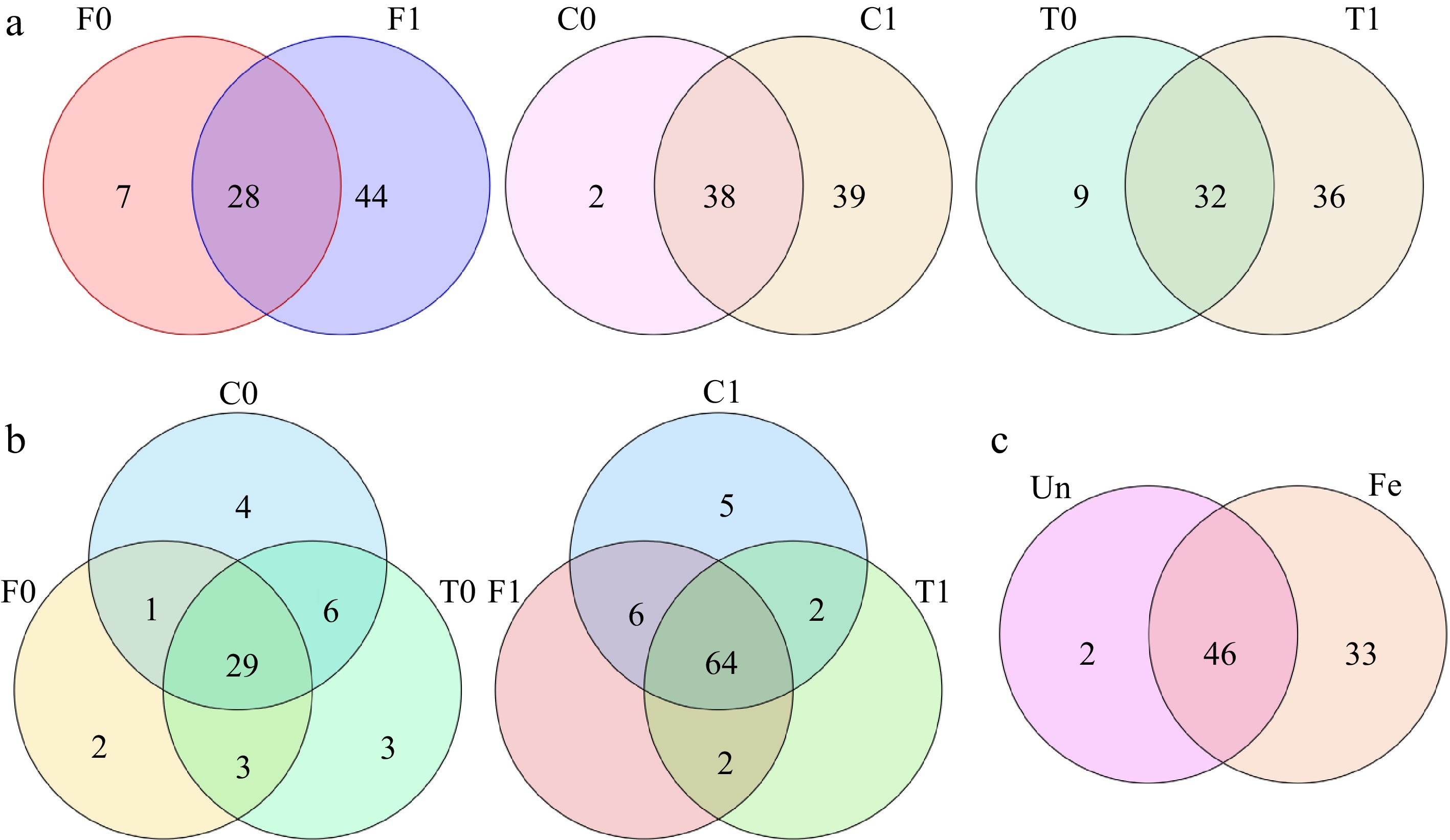
      Figure 1. 

      Venn diagram of volatile compounds. (a) Comparison of the number of volatile compounds in unfermented and fermented beans within the same group. (b) Comparison of the number of volatile compounds between unfermented and fermented beans in the three groups. (c) Number of volatile compounds shared by unfermented and fermented beans in the three groups (Un: unfermented beans, Fe: fermented beans).

      Among the unfermented beans, Trinitario had the most volatile compounds (41 compounds), followed by Criollo (40 compounds) and Forastero (35 compounds) contained the lowest compounds (Fig. 2b). On average of each group, the Trinitario group exhibited the highest volatile compounds, followed by the Criollo group. The Forastero group showed the lowest volatile compounds (Fig. 3). The Criollo, Forastero and Trinitario groups exhibited four, two and three specialized volatile compounds respectively (Fig. 1b). Diversity of the volatile component number and average number within groups were converged with different cacao genotypes, in agreement with the results of Qin et al.[28]. Trinitario contained the highest total volatility, approximately twice that of Forastero (Fig. 2a).
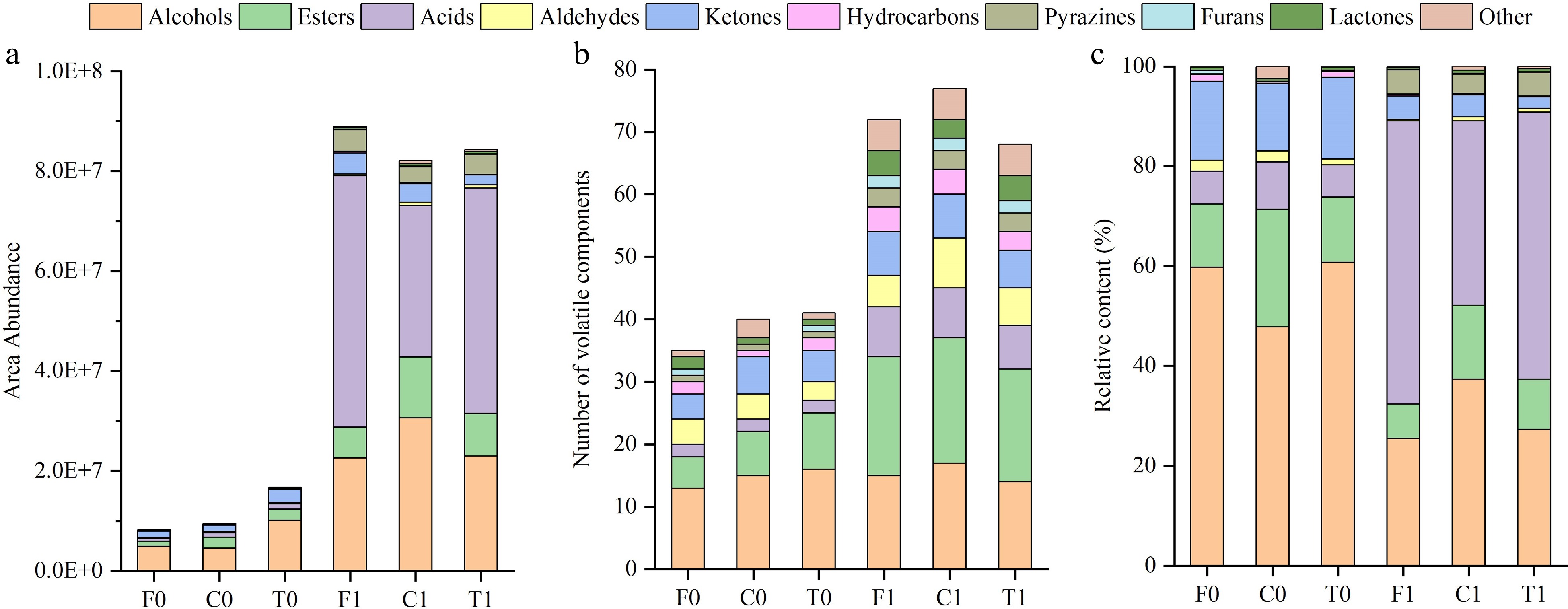
      Figure 2. 

      Volatile compound buildup map. (a) Total area abundance of volatile components. (b) Number of volatile components. (c) Relative content of volatile components.
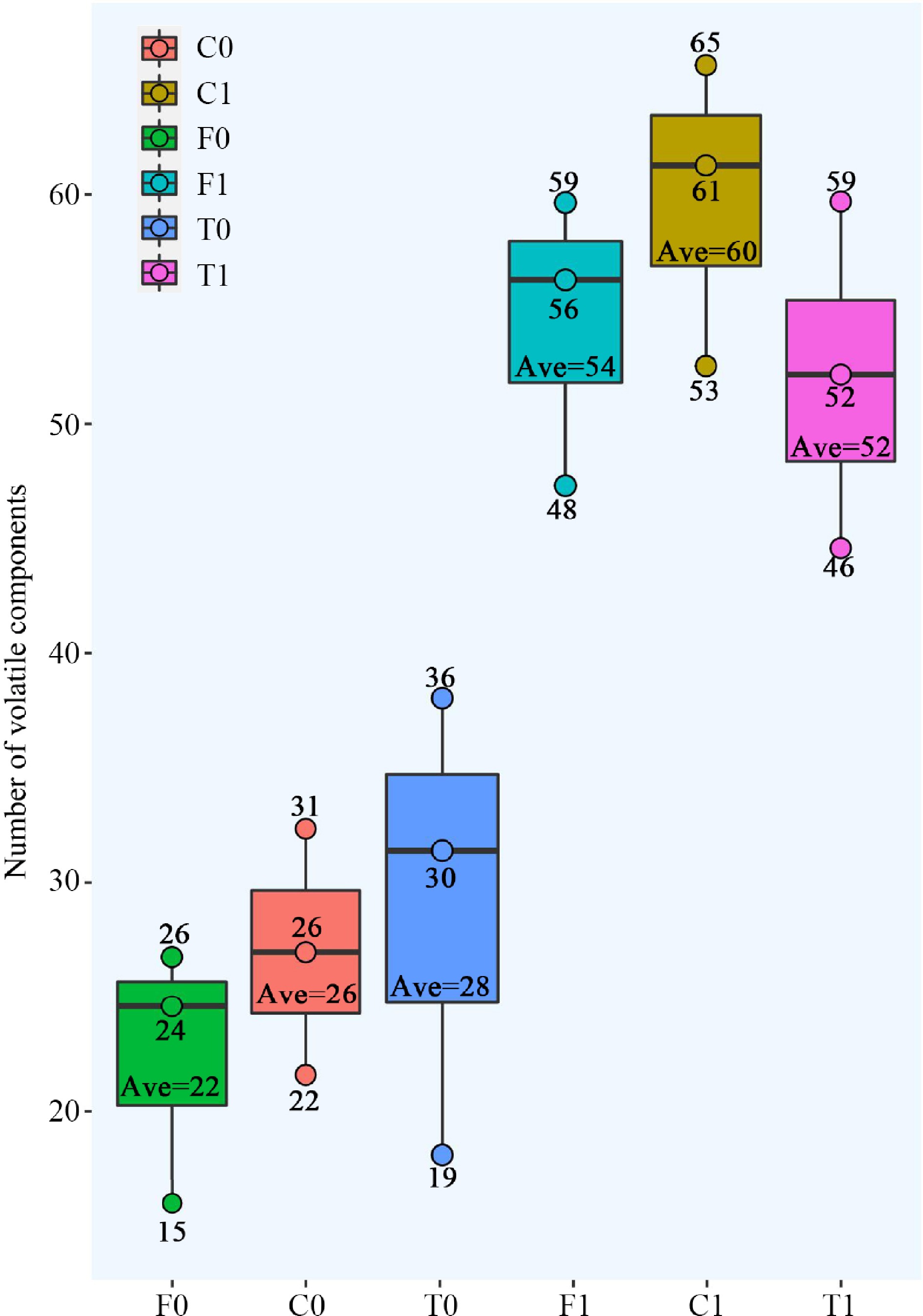
      Figure 3. 

      Number of volatile compounds within the three cacao groups. The three dots on the box represent the number of volatile compounds for the three replicates; 'Ave' represents the average (rounded).

      As for the fermented beans, Criollo had the highest number of volatile compounds (77 compounds), followed by Forastero (72 compounds) and Trinitario (68 compounds). On average of each group, the Criollo group exhibited the highest volatile compounds, followed by the Forastero group. The Trinitario group showed the lowest volatile compounds (Fig. 3). Only the Criollo group exhibited five specialized volatile compounds (1-pentanol, 1-hexanol, ethyl butanoate, diethyl succinate, and hexanal) compared to the other two groups (Fig. 1b). These compounds have good aromas (fruity, green, etc.) and may be responsible for the good quality of Criollo.

      In both unfermented and fermented beans, there were significant differences in the number of volatile components among the three cacao groups (Fig. 1c). After the fermentation process, the total amount of volatile compounds in cocoa beans increased by 5 to 10-fold (Fig. 2a) and the number of volatile compounds (Fig. 2b) significantly increased. The results revealed that the fermentation process is a crucial step in enhancing the flavor of cocoa beans[24].

    • The relative contents of alcohols was about 50% in unfermented beans and 20%−40% in fermented beans (Fig. 2), where large amounts were essential due to the production of floral, fruity and green aromas[28,40,43]. 2-pentanol was the most abundant substance in the volatile compounds of unfermented beans (Table 2 & Fig. 4). Interestingly, 2-pentanol was not detected in the fermented beans, and the same was true for 2-hexanol (Fig. 1c). While the final profiles of fermented cocoa beans were characterized by a higher content of 2,3-butanediol, 1,3-butanediol and 2-phenylethanol.
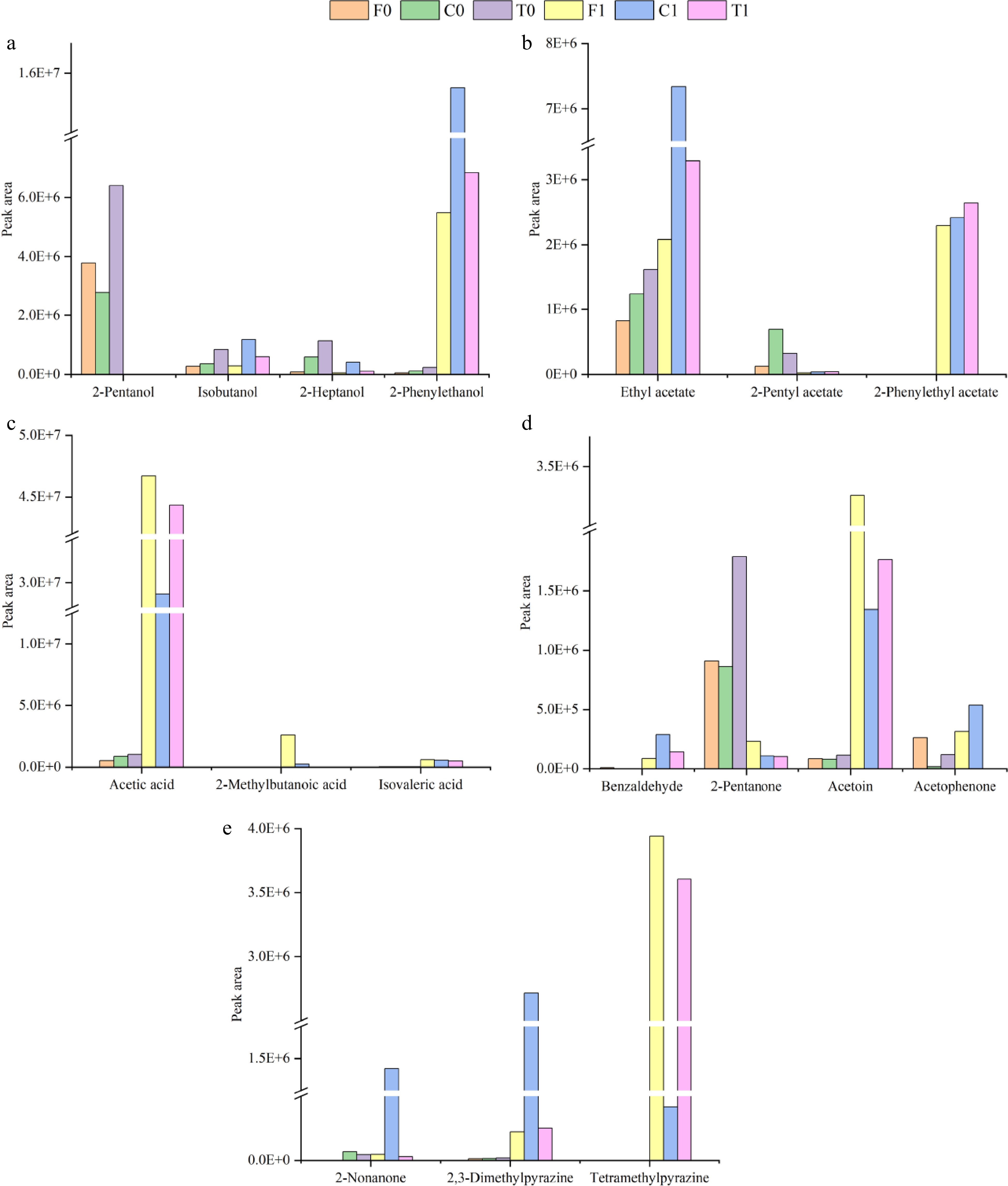
      Figure 4. 

      Peak areas of major volatile compounds.

      Isoamyl alcohol, 2-heptanol and 2-phenylethanol are considered important volatile compounds because they produce desirable flavors (fruity, floral)[43,44]. In particular, 2-phenylethanol has been identified as an important compound in fine flavor cocoa[45]. However, the 2-heptanol content was decreased after fermentation (Fig. 4a). The final profile of the unfermented cocoa beans was characterized by the highest content of isoamyl alcohol, 2-heptanol, and 2-phenylethanol in Trinitario, followed by Criollo and Forastero. The characteristics of the fermented cocoa beans were clearly different, with Criollo containing the highest content of the three alcohol compounds mentioned above, in particular producing about twice as much 2-phenylethanol as the other two groups, which gives it a more fruity and floral aroma, followed by Trinitario (Fig. 4a).

    • The esters are mainly derived from the reaction between organic acids and alcohols during anaerobic fermentation, providing the fruity flavor of cocoa beans[46]. A total of 10 esters were detected before fermentation, which was increased to 21 kinds after fermentation (Fig. 2b). The highest levels of esters were found in the unfermented cocoa beans of Criollo and, interestingly, in the fermented beans as well (Fig. 2c), confirming that esters were the characteristic volatile compounds of Criollo[47]. Ethyl acetate was the most abundant ester in both unfermented and fermented beans, with the amount in fermented beans increasing by a factor of two to four in comparison.

      The fruity and green aromas are stronger in Criollo fermented beans because of the highest content of ethyl acetate, which is also one of the main volatile components of cocoa[43]. Directly related to the fine flavor of cocoa is 2-phenylethyl acetate[40], which appeared only after fermentation and released notes of rose and honey, with Trinitario fermented beans showing the highest level and Forastero fermented beans showing the lowest level, although the differences were not significant (Fig. 4b). 2-pentyl acetate, the main volatile compound in cocoa pulp[48], was also present at high levels in the volatile composition of unfermented beans but its level was reduced in fermented beans, likely due to its conversion to other substances during fermentation (Fig. 4b). The excellent flavor of fermented beans is largely due to acetate compounds produced by acetic acid bacteria (AAB)[49], and both the quantity and content of these compounds increased after fermentation.

    • Through the fermentation process, acids were advanced from two components (acetic acid and isovaleric acid) to eight components (Table 2). The content of acetic acid and isovaleric acid was observed 30 to 90 times higher and 20 times boost in fermented beans compared to unfermented beans. Acetic acid was the highest component of volatile compounds in fermented beans and was also highly odour-active[43,50], with relative levels as high as 35%−52% (Table 2). Acetic acid permeates into the inside of the cocoa beans and forms aromatic compounds (esters and higher alcohols, etc.), influencing the flavor profile of fermented beans[51]. Furthermore, the roasting process adversely affects the concentration of acetic acid, resulting from its high volatility[50,52]. In the current study, acetic acid content was significantly lower in Criollo group fermented beans than those in Trinitario and Forastero groups, supporting the viewpoint of Santander et al.[39]. Generally, the pulp of Criollo group fresh cocoa beans is thinner than that of Trinitario and Forastero groups, in which carbohydrates are consumed and transformed into acetic acid, carbon dioxide, and flavor compounds under the action of yeasts and AAB. 2-methylbutanoic acid also showed a higher content in Forastero fermented beans (Fig. 4c). It is best to avoid producing isovaleric acid in large quantities because it has an unpleasant flavor (souring) and is produced during the fermentation and drying processes[46].

    • Aldehydes further contribute to the development of desirable flavors and influence the formation of pyrazines[35]. The components of aldehyde in the final volatile profile were different between fermented and unfermented treatments. Eight aldehydes were detected in fermented cocoa beans, which is twice the number of aldehydes in unfermented cocoa beans, and all aldehydes can be found in Criollo fermented beans (Table 2). The aldehyde content of fermented cocoa beans increased about threefold, with phenylethylaldehyde being detected after fermentation and being the main flavour marker of chocolate, imparting floral and other flavours[47]. Benzaldehyde endows roasted almond flavor, which showed the highest content in Criollo and the lowest content in Forastero (Fig. 4d), and it is one of the volatiles that strongly enriched the characteristic aroma of fermented cocoa beans[44].

    • Ketones such as 2-pentanone and 2-heptanone, which were natural compounds present in the unfermented beans, decreased throughout fermentation and resulted in a lower content level in the fermented beans. Following fermentation, beans displayed an increased concentration of several key ketones, particularly acetoin, which is known as a technological indicator of cocoa processing and a key component of the buttery and creamy flavors in chocolate[46,50]. Additionally, 2-nonanone was detected in much higher concentrations in the Criollo group fermented beans than the Trinitario and Forastero groups (Fig. 4e), imparting a more fruity flavor[44]. Acetophenone produces floral aromas that contribute to the flavor and quality of the cocoa[35,45], and it is interesting to note that this compound was not detected in Trinitario fermented beans (Fig. 4d).

    • Pyrazines are mainly generated during the Maillard reaction and Strecker degradation of the roasting process and also form in a small amount during the drying process, imparting roasted incense and nutty notes[35,40,46]. The final profile of fermented beans was characterized by three pyrazine components of 2,3-dimethylpyrazine, trimethylpyrazine, and tetramethylpyrazine. The three group's cocoa beans presented an approximate percentage of pyrazines. Trimethylpyrazine and tetramethylpyrazine are considered important and were mainly produced after fermentation[53], with tetramethylpyrazine being present in large quantities in fermented beans of the Trinitario and Forastero groups, while 2,3-dimethylpyrazine was predominant in Criollo fermented beans (Fig. 4e). However, pyrazine is not thought to be responsible for determining the superior aroma of Criollo[23,54].

    • Two furans, recognized as trans-furan linalool oxide and furaneol, were found in the present study, of which furaneol was specifically presented in fermented beans. Four types of lactones have been discovered, and interestingly, 5-acetyldihydro-2(3H)-furanone has a wine aroma. Some other volatile compounds, including one pyran, one sulfide, one pyrrole and two phenolic compounds, were detected in the present study and showed differences between fermented and unfermented beans. We detected two phenolic compounds (eugenol and elemicin) that released floral flavors instead of smoky flavors, despite the fact that phenolic compounds are frequently believed to be connected with smoky flavors in cocoa (Table 2)[40,43]. 2-acetylpyrrole is unique to fermented beans and provides the flavor of roasted cocoa[43].

    • Principal component analysis (PCA) is a routinely employed statistical analysis method and has been successfully applied to analytical results, both for individual compounds and component combinations[37]. In order to evaluate the possibility of differentiating the samples taking into account their genotype, this statistical tool was applied. In both unfermented and fermented beans, the two first principal components (PCs) were sufficient to explain more than 99.5% of the variation in the original data (Fig. 5).
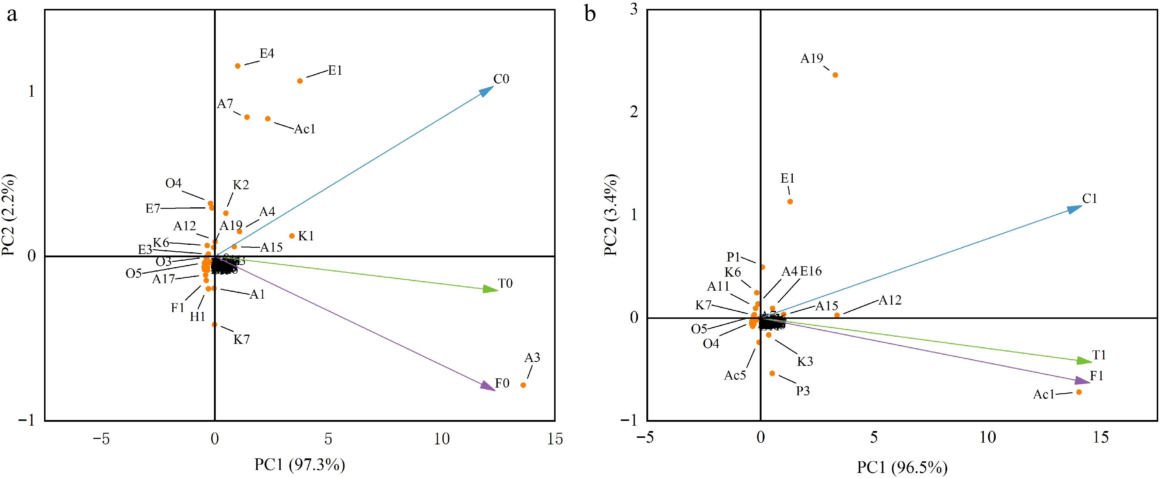
      Figure 5. 

      PCA analysis of unfermented and fermented beans in three cacao groups. (a) unfermented beans. (b) Fermented beans. The code in the figure corresponds to the corresponding compound.

      As illustrated in Fig. 5a, the unfermented beans of Criollo, which situated in the positive region of PC2, were clearly differentiated from those of the Trinitario and Forastero groups. Criollo beans combine high contents of 2-heptanol, ethyl acetate, 2-Pentyl acetate and acetic acid, giving it high scores on PC2[27]. Conversely, the high content of 2-pentanol caused the Forastero and Trinitario groups to be located in the negative regions of PC2. Additionally, Trinitario, derived from the hybrid of Criollo and Forastero, was also located in the position of PC2 between the two groups. The positional relationship of the three groups of fermented beans in the principal component analysis was similar to that of unfermented beans (Fig. 5b). 2-phenylethanol (floral), ethyl acetate (fruity), 2-nonanol (fruity, green), and 2,3-dimethylpyrazine (roasted, nutty) were closely related to Criollo and were endowed with floral, fruity, green, roasted, and nutty aroma characteristics. 2,3-butanediol (creamy, fruity), acetic acid (sour), 2-methylbutanoic acid (pungent, cheese), acetoin (buttery, creamy), and tetramethylpyrazine (roasted, nutty) were the main components strongly correlated with the Forastero group, imparting sour, buttery, fruity, toasty and nutty sensory notes. Trinitario is located between them, but leans more towards Forastero.

    • The peak areas of the volatile compounds for each cacao line are shown in Fig. 6, with log10 conversions performed for each parameter. Significant variations were detected among the three cacao groups. For unfermented treatment, Criollo and Trinitario were classified into one cluster, while Forastero and Trinitario were grouped together in fermented treatment (Fig. 6). After the fermentation process, volatile compounds and content were significantly changed, which was also presented in the different lines of the group, indicating that the volatile phenotype was influenced by the complex genetic background of the cacao lines[4,40,53,55].
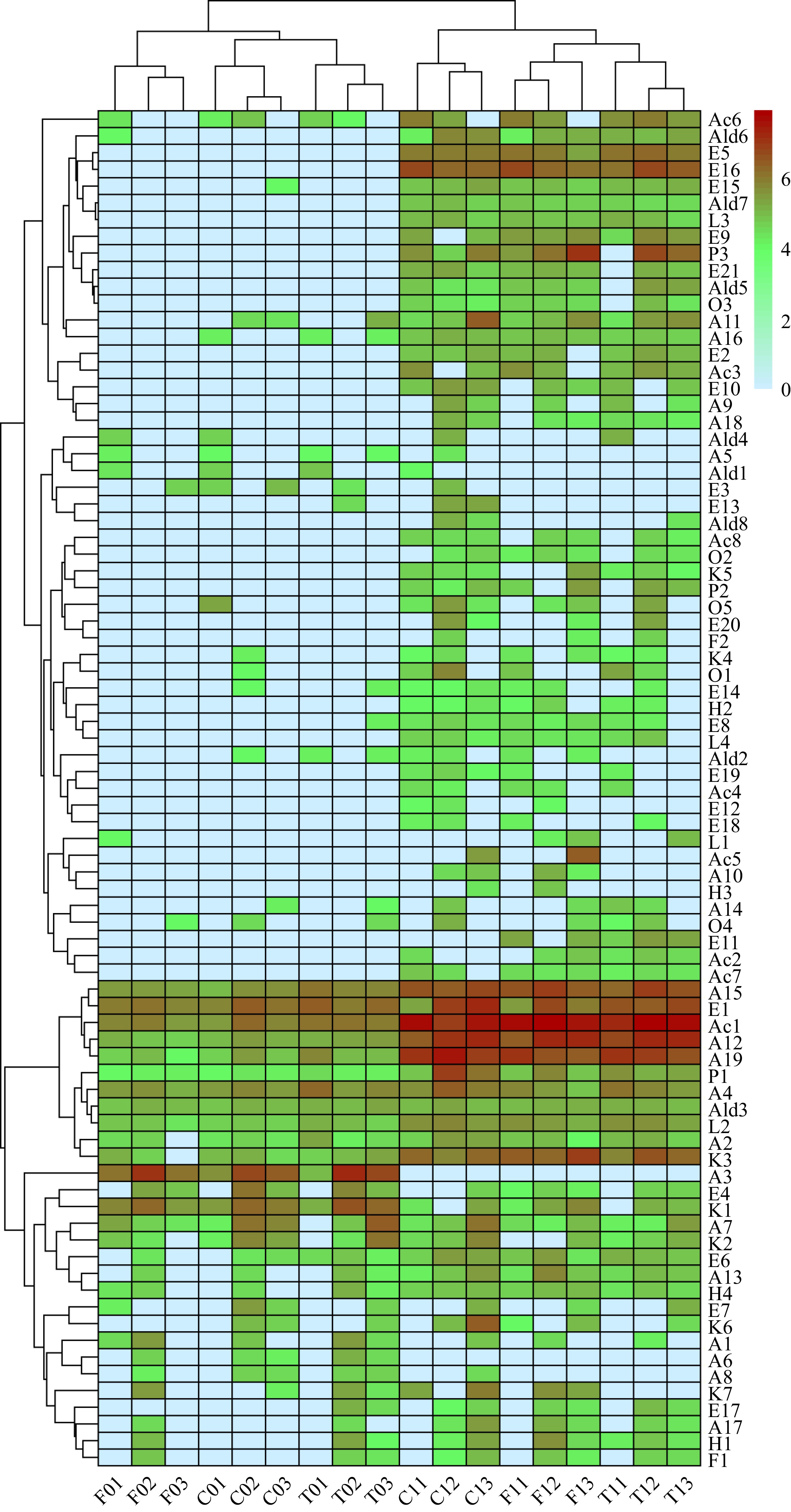
      Figure 6. 

      Volatile compounds cluster heat map. The code in the figure corresponds to the corresponding compound.

      The majority of quality traits were positively correlated with volatile components in unfermented beans (Supplemental Fig. S1a), revealing that substance accumulation was conducted with volatile components in synchronization. During the fermentation process, aldehydes inhibit the production of pyrazines[35], a significant negative correlation was determined between the two components (Supplemental Fig. S1b). Comprehensively, the phenotypic value of all tested quality traits was reduced in different magnitudes, contrarily, the content of volatile components was increased significantly, resulting in a negative correlation between quality traits and volatile components in fermented beans (Supplemental Fig. S1b).

    • The fermentation process has a significant impact on the test quality traits of unfermented and fermented Criollo, Forastero and Trinitario beans in Hainan Province, China. The phenotypic value of polyphenols, theobromine, caffeine and cocoa butter content is decreased to different degrees, of which polyphenols are shows to decrease by the largest decrease. For the unfermented beans, the Trinitario group exhibits the highest total content of volatile components, which include alcohols and esters, 2-pentanone, 2-heptanone, 2-heptanol and 1,3-butanedio. The Criollo group contains a higher content of ester components, such as 2-pentyl acetate and 2-heptyl acetate. Fermented beans of the Criollo group are transformed to a flavor profile with high levels of alcohols and esters, including 2-phenylethanol, isoamyl alcohol, ethyl acetate, and high levels of 2-nonanone, 2,3-dimethylpyrazine. Trinitario and Forastero groups contain large amounts of acidic compounds and tetramethylpyrazine, and ketone compounds, 2-methylbutanoic acid and acetoin are also enriched in the Forastero group of fermented beans. Nevertheless, the optimum fermentation time of cacao beans differ significantly, depending on varieties[4,22]. Therefore, in order to produce fermented cocoa beans with good flavor, different fermentation times should be set according to different varieties in production. The present results provide a baseline for a better understanding of the effect of the fermentation process for the three cacao groups cultivated in the Hainan region, principally according to the quality trait identification and profiles of volatile compounds, aiming to improve excellent cacao varieties with fine flavor.

    • The authors confirm contribution to the paper as follows: study conception and design: Yang D, Wu B, Wu G, Li F; data collection: Qin X, Zhao X, Zhu Z; analysis and interpretation of results: Yan L, Zhang F; draft manuscript preparation: Yang D, Wu B. All authors reviewed the results and approved the final version of the manuscript.

    • The datasets generated during and/or analyzed during the current study are available from the corresponding author on reasonable request.

      • This research was funded by the Hainan Province's Key Research and Development Project of China (No. ZDYF2021XDNY123) and Central Public-interest Scientific Institution Basal Research Fund (No.1630142022003).

      • The authors declare that they have no conflict of interest.

      • # These authors contributed equally: Dewei Yang, Baoduo Wu

      • Supplemental Table S1 Agronomic traits and pictures of each material.
      • Supplemental Table S2 Quality characteristics of fermented and unfermented beans for all cocoa materials.
      • Supplemental Fig. S1 Heat map of quality traits and volatile compound correlations. (a) unfermented beans. (b) fermented beans. The black diagonal separates them. The pie charts in the boxes indicate the strength of the correlations, red is positive, blue is negative, and "×" represents significance (< 0.05).
      • Copyright: © 2024 by the author(s). Published by Maximum Academic Press, Fayetteville, GA. This article is an open access article distributed under Creative Commons Attribution License (CC BY 4.0), visit https://creativecommons.org/licenses/by/4.0/.
    Figure (6)  Table (2) References (55)
  • About this article
    Cite this article
    Yang D, Wu B, Qin X, Zhao X, Zhu Z, et al. 2024. Quality differences and profiling of volatile components between fermented and unfermented cocoa seeds (Theobroma cacao L.) of Criollo, Forastero and Trinitario in China. Beverage Plant Research 4: e010 doi: 10.48130/bpr-0024-0002
    Yang D, Wu B, Qin X, Zhao X, Zhu Z, et al. 2024. Quality differences and profiling of volatile components between fermented and unfermented cocoa seeds (Theobroma cacao L.) of Criollo, Forastero and Trinitario in China. Beverage Plant Research 4: e010 doi: 10.48130/bpr-0024-0002

Catalog

  • About this article

/

DownLoad:  Full-Size Img  PowerPoint
Return
Return