











-
Lichenostigma was introduced by Hafellner in 1982 to accommodate the lichenicolous fungus L. maureri and was initially placed in Arthoniales based on ascocarp morphology[1]. The species grows on Usnea spp. and Pseudevernia furfuracea and has been reported from the Eastern Alps as well as other parts of Eurasia. Ertz et al.[2] provided the first phylogenetic study of Lichenostigma and synonymized two lichenicolous genera, Diederimyces and Phaeosporobolus, under Lichenostigma[2]. Based on the combined phylogenetic analyses of LSU and mtSSU sequence data, together with detailed morphological evidence, Lichenostigmatales was established within Arthoniomycetes to accommodate the family Phaeococcomycetaceae, with Lichenostigma designated as the type genus[2]. Phaeococcomycetaceae presently includes four genera, namely Antarctolichenia, Etayoa, Lichenostigma, and Phaeococcomyces[3].
Presently, Lichenostigma comprises approximately 34 species[3,4], with Lichenostigma maureri as the type species. The genus is polyphyletic, with its taxa distributed across both Lichenotheliaceae (Dothideomycetes) and Arthoniomycetes[2]. Ertz et al.[2] proposed that the genus comprises two subgenera: subgenus Lichenostigma, placed within Arthoniales, and corresponding to Lichenostigma sensu stricto, subgenus Lichenogramma, which is phylogenetically clustered within Dothideomycetes; these subgenera are primarily distinguished by differences in cell division. Lichenostigma sensu stricto divides by budding, whereas the latter produces cells through septation. The sexual morph lacks hamathecial filaments, with asci developing directly among stromatic cells; the ascus tholus may be I+ reddish and K/I+ blue, while ascospores are 1-septate, with the outer wall occasionally K/I+ blue. The asexual morph is characterized by conidiomata composed of similar globose stromatic cells, from which conidiogenous cells arise, producing multicellular, subspherical to ellipsoid conidia[2].
The first checklist of lichenicolous fungi in China was published in 2003, reporting the occurrence of Lichenostigma cosmopolites on Xanthoparmelia scabrosa from Hong Kong[5]. In the present study, a specimen collected in China, growing on Lecanora sp., was examined. Based on morphological characters and phylogenetic analyses of LSU and mtSSU sequence data, the specimen was identified as Lichenostigma chlaroterae, representing the first geographic record of this species from China.
-
A specimen of Lichenostigma was collected in 2018 from the bark of an unidentified tree hosting a Lecanora species in Lijiang (22°36′38.8′′ N, 100°10′33.5′′ E), Yunnan Province, China. The specimen was observed and examined using a stereo-microscope (SteREO Discovery V12, Carl Zeiss Microscopy GmbH, Germany) and a compound microscope (Nikon ECLIPSE 80i, Nikon, Japan), equipped with a Nikon DS-Ri2 digital camera (Nikon, Japan). The measurements were done with Tarosoft® Image Frame Work v0.9.7, and the morphological analysis was performed following Senanayake et al.[6]. The symbol '
$\overline {\rm x} $ DNA extraction, polymerase chain reaction amplification, and sequencing
-
Genomic DNA was extracted directly from fungal fruiting bodies using the Omega Bio-Tek Kit (Anoren [Beijing] Biotechnology Co., Ltd), following the manufacturer's protocol. PCR amplification of the LSU region was carried out with primers LR0R/LR5 following the conditions recommended by Vilgalys & Hester[8]. The mtSSU region was amplified using primers mrSSU1/mrSSU3R[9]. The PCR reaction mixture contained a total volume of 25 μL, consisting of 12.5 μL of 2 × PCR G013 Taq MasterMix with Dye (ABM, Canada), 1 μL of each primer (10 μM), 2 μL of genomic DNA, and 8.5 μL of sterile distilled water. The parameters of PCR amplifications are displayed in Table 1. The PCR products were visualized through electrophoresis on a 1% TAE gel and detected using TSJ003 GoldView nucleic acid dye (Qingke, Kunming, China). Sequencing was performed at Qingke Company, Kunming, China.
Table 1. Genes/loci used in this study with respective PCR primers and protocols.
Sequence alignment and phylogenetic analyses
-
All newly generated sequences were checked for quality using BioEdit v7.0.9[10]. Forward and reverse sequences were assembled using DNASTAR Lasergene SeqMan Pro v7.1.0[11]. The assembled sequences were then queried against the NCBI database[12] to identify close relatives. Preliminary identification of the newly generated sequences was performed through standard BLAST searches of GenBank. For phylogenetic analyses, LSU and mtSSU gene regions from representative species of Lichenostigmatales, as well as the outgroup taxa from Arthoniales, were retrieved from GenBank (Table 2). Each gene dataset was aligned using MAFFT v7 (
https://mafft.cbrc.jp/alignment/server/index.html ), subsequently trimmed with TrimAl v1.3 software, with a gap threshold set to 0.6 for both datasets[13]. The aligned LSU and mtSSU datasets were combined into a combined matrix using SequenceMatrix v1.8, and file formats were converted using AliView v1.19[14] for further analyses. The phylogenetic tree for maximum likelihood analysis was generated using the Phylip format, while the Nexus format was used for Bayesian inference analysis[15]. The ML analysis was performed using IQ-TREE with the ML+ rapid bootstrap setting and 1,000 replicates[16,17]. Bayesian inference was performed with MrBayes v3.1.2 to estimate Bayesian posterior probabilities (BYPP)[18]. The resulting trees were visualized using FigTree v1.4.0 (https://tree.bio.ed.ac.uk/software/figtree ). The identification of potential species followed previously proposed species delineation approaches[19,20].Table 2. Taxa used in the phylogenetic analyses and their GenBank accession numbers.
Taxa Strain Genbank accession number LSU mtSSU Antarctolichenia onofrii CCFEE 6564 MZ005697 – Antarctolichenia onofrii CCFEE 6574 MZ005692 – Arthonia didyma Ertz 7587 (BR) EU704083 EU704047 Arthonia dispersa UPSC2583 AY571381 AY571383 Briancoppinsia cytospora Ertz 15244 (BR) JF830770 JF830772 Cryptothecia candida Ertz 9260 (BR) HQ454520 EU704052 Etayoa trypethelii Common 9200-G KF176940 KF176967 Etayoa trypethelii Common 9215-P KF176941 KF176968 Etayoa trypethelii Common 9434-K KF176942 KF176969 Etayoa trypethelii Mukherjee s.n. (BR) KF176943 KF176970 Lichenostigma alpinum Ertz 17522 (BR) KF176945 KF176972 Lichenostigma alpinum Diederich 17379 KF176944 KF176971 Lichenostigma alpinum Ertz 17519 (BR) KF176946 KF176973 Lichenostigma chlaroterae Neuberg (h Diederich) KF176948 KF176975 Lichenostigma chlaroterae Goward 12-30 – KF176976 Lichenostigma chlaroterae Goward 12-42 KF176949 KF176977 Lichenostigma chlaroterae HKAS 148603 PX885106 PX896563 Lichenostigma maureri Diederich 17326 (h Diederich) KF176953 KF176979 Lichenostigma maureri Diederich 17337 KF176952 KF176980 Lichenostigma maureri Diederich 17306 KF176951 KF176978 Lichenostigma sp. Ertz 16122 (BR) KF176956 KF176982 Lichenostigma sp. Ertz 16340 (BR) KF176957 KF176983 Lichenostigma sp. Ertz 16455 (BR) KF176958 KF176984 Lichenostigma sp. Ertz 17457 (BR) KF176959 KF176985 Lichenostigma sp. Ertz 17540 (BR) KF176960 KF176986 Lichenostigmatales sp. LMCC0526 MH259005 MH259138 Lichenostigmatales sp. LMCC0528 MH259007 – Lichenostigmatales sp. LMCC0529 MH259008 – Lichenostigmatales sp. LMCC0530 MH259009 – Phaeococcomyces nigricans CBS 553.90 MH873917 – Phaeococcomyces sp. G506 OP600268 OP600250 Phaeococcomyces sp. G148 OP600267 OP600249 Newly generated sequences are in bold font and the unavailable sequences are indicated by '–'. -
The combined LSU and mtSSU dataset comprised 2,867 base pairs in total, with positions 1–1,826 corresponding to the LSU region and positions 1,827–2,867 representing the mtSSU region. The dataset consists of 1,231 distinct alignment patterns, with 39.97% of undetermined characters or gaps with Arthonia didyma, A. dispersa, Cryptothecia candida, and Briancoppinsia cytospora as outgroup taxa (Fig. 1). A total of 32 strains were included in the combined analyses. The best RAxML tree with a final likelihood value of −14109.293201 is presented in Fig. 1. Estimated base frequencies were as follows: A = 0.275194, C = 0.200804, G = 0.275049, T = 0.248953, substitution rates AC = 0.791749, AG = 2.569367, AT = 1.900859, CG = 1.275594, CT = 4.854511, and GT = 1.000000. Single-gene analyses were also performed to evaluate topological congruence and clade stability relative to the combined-gene analyses. The single-gene analyses produced tree topologies that were consistent with those inferred from the combined gene analyses. The ML analysis generated tree topologies consistent with those from the Bayesian inference (BI) analysis, supporting the reliability of our phylogenies generated herein.
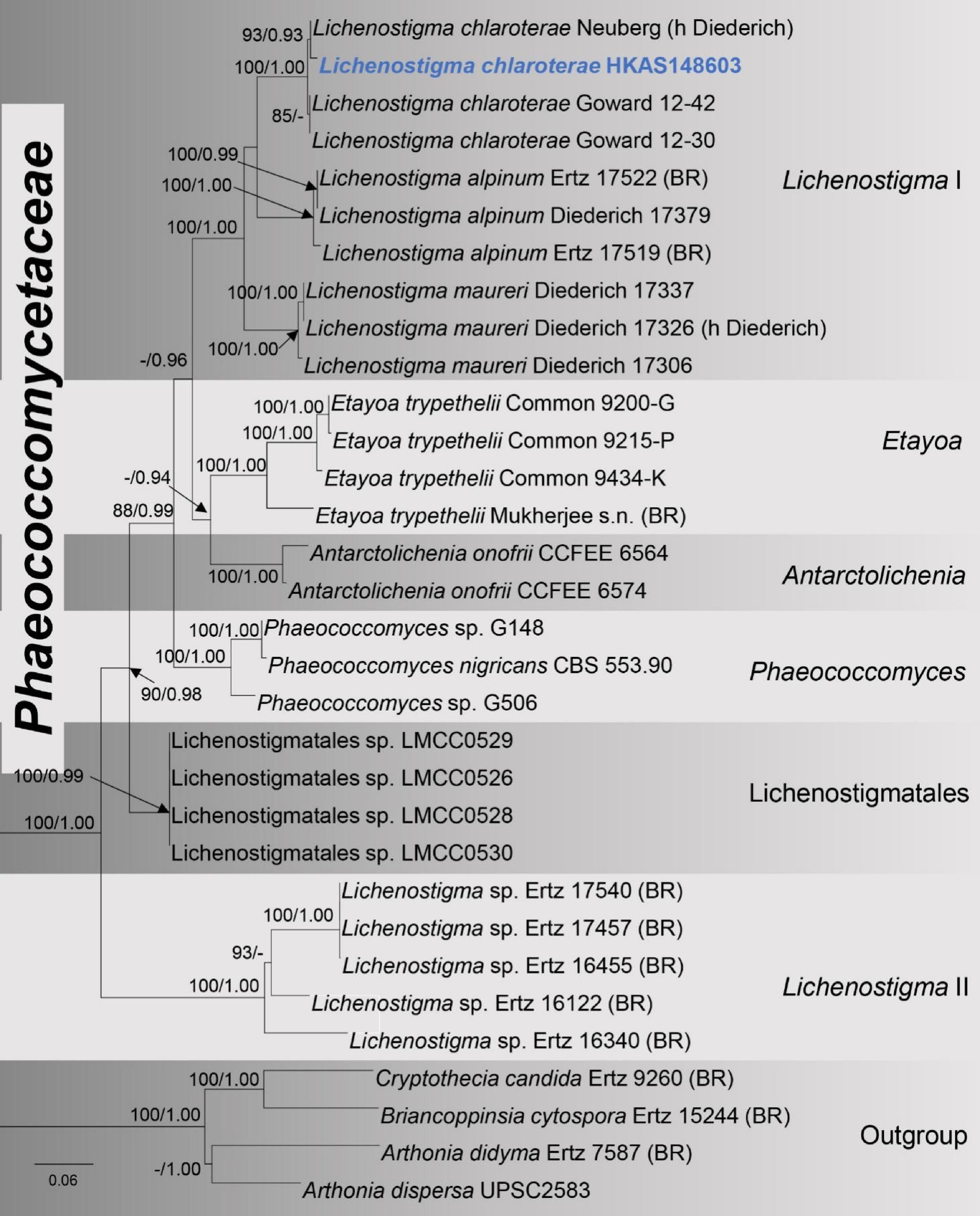
Figure 1.
Phylogram of RA × ML analysis based on the combined LSU and mtSSU sequence datasets. Bootstrap support values for Maximum Likelihood equal to or greater than 70%, and Bayesian posterior probabilities equal to or greater than 0.90 BYPP are indicated at the nodes as ML/BYPP. The newly generated strain in this study is indicated in blue bold text. The tree is rooted with Arthonia didyma (Ertz 7587 BR), A. dispersa (UPSC2583), Cryptothecia candida (Ertz 9260 BR), and Briancoppinsia cytospora (Ertz 15244 BR).
Taxonomy
-
Lichenostigma chlaroterae (F. Berger & Brackel) Ertz & Diederich, Fungal Diversity 66: 128 (2014) [MB#804672]. Fig. 2
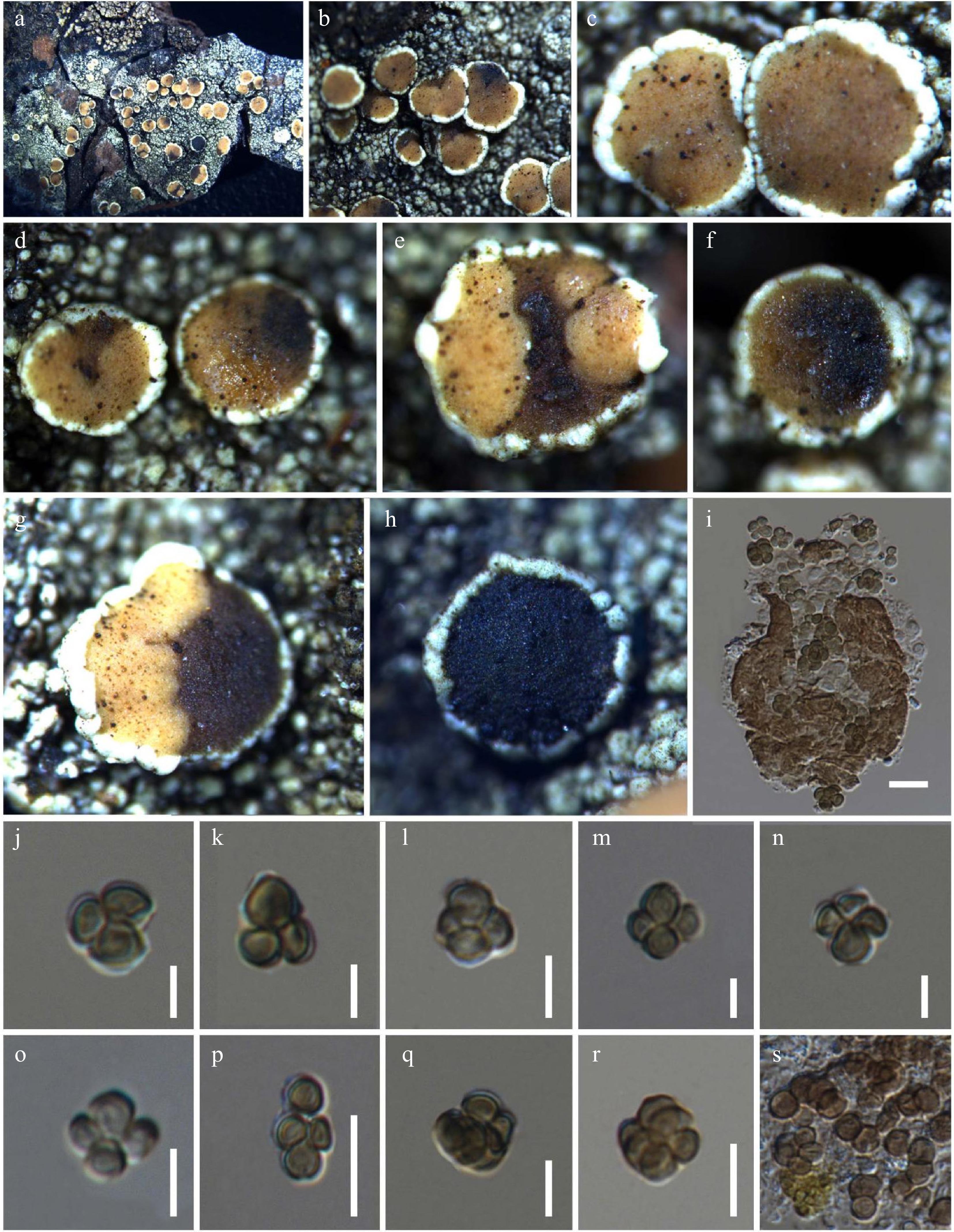
Figure 2.
Lichenostigma chlaroterae (HKAS148603). (a)–(h) Conidiomata on Lecanora sp. host. (i) Vertical section through conidioma. (j)–(r) Conidia. (s) Development of conidia. Scale bar: (i) = 20 μm, (j)–(r) = 10 μm.
=Phaeosporobolus chlaroterae F. Berger & Brackel, Herzogia 24(2): 351 (2011)
Index Fungorum: IF561182; Faces of Fungi Number: FoF 19647
The mycelium is extremely sparse, brown, and superficial. Asexual morph: Conidiomata macroscopically indistinguishable, 40–45 µm diam. × 60–70 µm high. (
$\overline x $ $\overline x $ $\overline x $ Material examined: China, Yunnan Province, Lijiang City, on a disc of Lecanora sp. on unidentified bark, 22°36' 38.8' N, 100°10' 33.5' E, elevation 3,351 m, 3 September 2018, V. Thiyagaraja, D1L18 (HKAS148603).
Distribution: China (This study). Europe, Macaronesia, Mongolia[21], North America[2], Russia[21], and South America (Bolivia).
Notes: the BLAST results of the LSU sequence data revealed the highest similarity to Lichenostigma chlaroterae (Goward 12–42) and L. chlaroterae (Neuberg [h Diederich]), with 99.10% and 100.00% similarity, respectively. The mtSSU sequence data showed the highest similarity to L. chlaroterae (Neuberg [h Diederich]) with 100.00%. In the multi-gene phylogenetic analyses, the new strain L. chlaroterae (HKAS148603) clustered with extant strains of L. chlaroterae (Neuberg [h Diederich], Goward 12–42 and Goward 12–30), with 93% ML bootstrap support and 0.93 BYPP. The strain L. chlaroterae (Diederich 17329) was excluded from the present analyses due to its unstable phylogenetic placement. The base pair comparison between our strain (HKAS 148603) and L. chlaroterae (Neuberg [h Diederich]) revealed no differences in either the LSU or mtSSU regions. In comparison with L. chlaroterae (Goward 12–42), sequence divergence was 0.12% (1/822 bp) in the LSU and 0.41% (3/715 bp) in the mtSSU, with no gaps detected. Similarly, comparison with L. chlaroterae (Goward 12–30) showed a divergence of 0.29% (2/675 bp) in the mtSSU, while LSU comparison was not performed due to the lack of molecular data for that strain. The pairwise sequence comparison between our strain and L. chlaroterae (Diederich 17329) revealed 1.26% (10/792) differences in the LSU and 1.25% (9/720) differences in the mtSSU without gaps.
Lichenostigma chlaroterae was established by Ertz et al.[2] and the species was originally introduced as Phaeosporobolus chlaroterae by Berger & von Brackel[22] based on material collected in Austria. It has also been reported from Germany and Portugal. In Central Europe, records range from colline to montane zones, and the species is probably more widespread[22]. The new specimen exhibits several characteristics similar to Lichenostigma chlaroterae, including superficial, smooth-walled brown hyphae, warty to granular ornamentation on the outer stromatal cells, and multicellular conidia composed of nearly spherical to ellipsoidal cells. Additionally, L. chlaroterae (Diederich 17329) occasionally produces a sexual stage with 1-septate brown ascospores, whereas no sexual structures have been observed in our sample[2]. Our specimen slightly differs from the L. chlaroterae (Diederich 17329) in the smaller conidia (4.8–6.6 × 4.4–7.1 µm vs [5.5–]7.1–10.2[–13] × [5–]6.3–8.8[–11] µm) and a smaller number of cells (3–6 vs [3–]4–9[–16])[2].
-
Since the beginning of the 21st century, research on lichenicolous fungi in China has gradually expanded. Aptroot & Sipman[23] reported three species in Hong Kong, including two lichenicolous fungi newly recorded from China (Lichenoconium lecanorae and Lichenostigma cosmopolites)[23]. In 2003, four additional lichenicolous fungi were reported in Yunnan[24]. In the same year, a checklist of 29 known lichenicolous fungi in China was systematically compiled[5]. In 2016, Polycoccum clauderouxii was recorded in Qinghai[25]. Subsequently, a total of 30 lichenicolous species were reported[26]. Thiyagaraja et al.[27] reported Sclerococcum simplex that was reported from Yunnan, China[27]. In 2023, nine species of Cercidospora were documented in China. However, as these fungi were isolated from the medullary tissues of lichen thalli and reported as "black lichenicolous fungi", their true lichenicolous nature remains uncertain[28]. Subsequently, a new record of Sclerococcum glaucomarioides was described from Yunnan, indicating that the diversity of lichenicolous fungi in China is far from being fully revealed, especially in Yunnan[29]. However, research on lichenicolous fungi in China is limited, with nearly 60 species reported in the past 25 years, indicating that the documented diversity remains remarkably low given China's vast area and habitat heterogeneity[5,23−29].
In the phylogenetic analyses, the genera within Lichenostigmales formed a monophyletic clade, with the exception of Lichenostigma, which appeared polyphyletic. This polyphyletic nature of Lichenostigma was also reported by Ertz et al.[2] highlighting the need for further studies, particularly due to the lack of molecular data for many species within the genus. Recently, Muggia et al.[30] introduced a new endolichenic genus, Antarctolichenia, within the order and the genus was collected from the Antarctic continent, which showed a close phylogenetic relationship with the genus Etayoa. A similar phylogenetic placement was observed in the present study. Further, our newly obtained strain of L. chlaroterae, together with previously reported strains of the same species, formed a well-supported clade with L. alpinum. This clade formed a sister clade to the type species, L. maureri, with strong statistical support (100/1.00).
Lichenostigma is a cosmopolitan genus that has been reported from a wide range of regions worldwide[2,21,31]. This study represents the second record of the genus Lichenostigma from China. Previously, L. cosmopolites was reported on Xanthoparmelia scabrosa from Hong Kong, China[23]. Lichenostigma cosmopolites is widely distributed in extratropical regions of both hemispheres (Africa, Asia, Australia, Europe, North America, and South America), occurring mainly in lowlands of temperate regions and at mid elevations or higher in tropical regions[32]. Lichenostigma chlaroterae, a relatively common species in Europe, has been reported from North America[2]. This species primarily colonizes Lecanora and other lichens, such as Buellia griseovirens, Fuscidea lightfootii, and Graphis pulverulenta[2,33,34]. Here, we report the first occurrence of L. chlaroterae in China. This record further expands the known distribution of the species in East Asia.
-
The authors confirm their contributions to the paper as follows: conceptualization, data curation, and formal analysis: Yuan L, Thilini Chethana KW, Meng Q, Thiyagaraja V; funding acquisition: Meng Q, Thiyagaraja V, Hyde KD; investigation, methodology, and writing – original draft: Yuan L, Thilini Chethana KW, Meng Q, Thiyagaraja V; project administration: Yuan L, Thiyagaraja V; supervision: Thilini Chethana KW, Meng Q, Thiyagaraja V; writing – review and editing: Thilini Chethana KW, Meng Q, Hyde KD, Zhao Q, Thiyagaraja V. All authors reviewed the results and approved the final version of the manuscript.
-
All the data used are included in the article. The sequences are cited by their accession numbers and are available from GenBank.
-
The authors declare that they have no conflict of interest.
- Copyright: © 2026 by the author(s). Published by Maximum Academic Press, Fayetteville, GA. This article is an open access article distributed under Creative Commons Attribution License (CC BY 4.0), visit https://creativecommons.org/licenses/by/4.0/.
-
About this article
Cite this article
Yuan L, Thilini Chethana KW, Meng Q, Hyde KD, Zhao Q, et al. 2026. A new geographical record of Lichenostigma chlaroterae from China. Studies in Fungi 11: e027 doi: 10.48130/sif-0026-0022 

A new geographical record of Lichenostigma chlaroterae from China
- Received: 05 November 2025
- Revised: 29 March 2026
- Accepted: 08 May 2026
- Published online: 18 September 2026
Abstract: This paper reports a new geographical record of a known Lichenostigma species from Lijiang, Yunnan Province, China. Based on morphological characteristics and phylogenetic analyses generated from multi-gene sequence data (LSU and mtSSU), the species is identified as Lichenostigma chlaroterae, a lichenicolous fungus growing on Lecanora sp. The mycelium is extremely sparse, brown, and superficial. The conidiomata are macroscopically indistinguishable, and they produce conidia that are ellipsoid, brown, smooth, but sometimes with a verrucose to echinulate ornamentation when over-mature. Each conidium comprises three to six conidial cells that are subspherical to ellipsoid. Detailed descriptions and photoplates along with molecular data of Lichenostigma chlaroterae are provided.
-
Key words:
- Ascomycetes /
- Lichenicolous /
- Morphology /
- Phylogeny /
- Taxonomy