










-
The morphologically similar species of Agaricomycetes fungi may potentially belong to separate phylogenetic lineages, as white-rot species of the polypore genus Fomes (Polyporaceae, Agaricomycetes)[1−5]. Meanwhile, the correct species identification of valuable and highly-priced edible and medicinal mushrooms, based on cultural characteristics, particularly during their biotechnological cultivation, is challenging[6−18].
The basidiomata of Fomes fomentarius (L.) Fr. do not reveal taxonomically significant variability[1]. However, recent studies have shown that this medicinal fungus is represented by a genetically heterogeneous species complex, Fomes fomentarius sensu lato, comprising two (A and B) lineages (clades), and four sublineages (A1, A2, and B1, B2) with different substrate confinement and distribution areas[2−6,19−21]. The sublineages А2 and В2 of the European lineages А and В correspond to the cryptic sympatric subspecies Fomes fomentarius sensu stricto and Fomes inzengae (Ces. & De Not.) Cooke[3,22,23] with various Angiosperm and Gymnosperm confinement, ecological characteristics, and geographical distribution patterns. However, F. fomentarius s.s. and F. inzengae are more frequently found in broadleaf (Acer, Aesculus, Alnus, Betula, Carpinus, Castanea, Cerasium, Chosenia, Festuca, Juglans, Fagus, Fraxinus, Platanus, Populus, Quercus, Salix, Sorbus, Tilia, and Ulmus), than pine trees (Abies, Larix, Picea, and Pinus)[2−5,20,21,24,25]. New Fomes species F. hengduanensis with a basal position to a potential sister taxon F. inzengae–Globifomes graveolens lineage has recently been reported in China[26]. Thus, four genetically distinct Fomes species (Fomes fasciatus, F. fomentarius s.s., F. inzengae, and F. hengduanensis) are taxonomically described.
The only Fomes species recorded in Armenia from Carpinus, Fagus, Fraxinus, and Quercus trees was F. fomentarius[27]. However, our recent phylogenetic studies described two new species from three floristic regions of Armenia with different substrate confinement: F. fomentarius s.s. (Carpinus, Fagus), and F. inzengae (Acer, Carpinus, Juglans, Fraxinus, Fagus, Ulmus, Populus, Platanus, Quercus, Salix)[4,5].
Previous cultural studies of Agaricomycetes fungi revealed taxonomically valuable mycelial traits useful in the identification of complex species, such as Fomes, Flammulina, and Ganoderma[6−15]. However, the detailed morpho-ecological, physiological, and growth characteristics of F. fomentarius s.s. and F. inzengae cultures have not been studied. Meanwhile, hyphal systems and morphology, the presence, occurrence, and form of clamp connections, chlamydospores, cuticular cells, asexual spores, crystals, mycelium growth patterns, including average growth rate (GRavr), growth temperature, and pH are taxonomically significant and may be used for taxonomic identification of fungal cultures during biotechnological cultivation[6−18,28−31].
Currently, very limited data are available on the morpho-ecological and physiological characteristics[3], as well as the bioactivity of F. inzengae mycelium[19,32,33]. Meanwhile, the mycelium of cultivated Agaricomycetes exceeds the basidiomata (mushrooms) in the content of proteins, amino acids, lipids, unsaturated fatty acids, polysaccharides, organic acids, vitamins, minerals and dietary fiber, as well as bioactive compounds (terpenoids, phenolics, polyketides, alkaloids, lectins, etc.), with potential dietary and medicinal (anti-inflammatory, antioxidant, anti-proliferative, antiviral, hypocholesterolaemic, hypoglycaemic, hypotensive, immunomodulatory, neuroprotective, wound-healing, etc.) properties[34−41]. Furthermore, the progress in biotechnological cultivation of mycelia (static, submerged)[42−44] of highly priced edible and medicinal mushrooms will further contribute to the production of environmentally friendly, innovative, and standardized mycelium-derived biotech products for medicinal (myco-pharmaceuticals), nutritional (nutraceuticals and nutriceuticals), cosmetic (nutricosmetics and nutracosmetics), and bioindustrial purposes[35−39,41,45−50].
The aim of this work is to study the cultural characteristics and biomass formation of genotyped mycelial collections of F. inzengae collected from Armenia for further biotechnological use.
-
Ten dikaryotic cultures (Fi/3, Fi/8, Fi/15, Fi/16, Fi/20, Fi/24, Fi/25, Fi/26, Fi/30, and Fi/31) of F. inzengae were isolated from basidiomata collected from walnut (Juglans regia), willow (Salix alba), beech (Fagus orientalis), poplar (Populus nigra), hornbeam (Carpinus betulus), and maple (Acer platanoides) trees from three floristic regions (Tavush, Aparan, and Yerevan) of Northern and Central Armenia during years 2005–2023[5,51] (Table 1). After morphological identification of basidiomata[24,25], the cultures were isolated by tissue method using 1.5% malt-extract agar (MEA, pH 6.0) medium[52]. Further genetic identification and verification of mycelial cultures was performed, as previously described[4,5].
Table 1. Metadata of dikaryotic collections of Fomes inzengae used in this study.
Strain Host tree Type of wood Origin and date of culture isolation, floristic region, and altitude (m a.s.l.) GenBank accession number Fi/3 Carpinus betulus L. Living tree Ijevan, Tavush province, July 2005; IJ, 750 m PP972742 Fi/8 Juglans regia L. Living tree Berdavan, Tavush province, April 2014; IJ, 680 m OL583665 Fi/15 Salix alba L. Stump Solak village, Kotayk province, July 2015; AP, 1,650 m OL583668 Fi/16 Salix alba L. Stump Solak village, Kotayk province, July 2015; AP, 1,650 m OL583669 Fi/20 Populus nigra L. Living tree Yeravan, July 2013; YR, 860 m PP972744 Fi/24 Fagus orientalis Lipsky Living tree Baghanis, Tavush province, July 2016; IJ, 920 m OL583671 Fi/25 Populus nigra L. Living tree Yerevan, July 2016; YR, 860 m OL583672 Fi/26 Carpinus betulus L. Living tree Dilijan, Lake Parz, Tavush province, July 2016; IJ, 1,220 m OL583673 Fi/30 Fagus orientalis Lipsky Trunk Jujevan, Tavush province, March 2023; IJ, 1,050 m PP972741 Fi/31 Acer platanoides L. Living tree Gomshavar, Tavush province, May 2023; IJ, 1,564 m PP972745 Legend: (AP) Aparan, (IJ) Ijevan, and (YR) Yerevan floristic regions of Armenia[5,51]. Observation of morphological and growth characteristics
-
The screening of micro- and macromorphological and growth characteristics of mycelial colonies of Fomes inzengae was performed on a 1.5% MEA at different temperatures. The inocula (ø 5 mm) of 6 d growing culture of each F. inzengae strain were transferred to the centre of МEА plates (ø 90 mm, three plates per isolate) and incubated at 26 and 30 °C in the dark for 14 d, except for daily checks and marking. The morphological observations, including fruiting bodies (FB) development, continued under a day/night regime for 45 d. The texture, density, and pigmentation of aerial mycelium, as well as the pigmentation of agar, were described according to Stalpers' scale[29].
Mycelium daily growth rate (GR, mm·d−1), as a measure of the colony growth under standardized culture conditions, was marked on lines drawn on the reverse side of Petri dishes through the inoculum until the colony completely covered the dishes. The GRavr was calculated from daily GR by the formula GRavr = S/Dn, where S (mm) is the distance of the constant mycelial growth over n days (Dn). Each strain was repeated three times with inocula from the same mother cultures. At least 12 consecutive measurements were performed from each strain.
The strains were also grown at 30 °C after 30 d pre-incubation at 4 °C. The morphological changes of colonies were described, and the GRavr suppression effect was calculated (in %).
Micromorphological observation
-
The cultures were grown on sterile microscope cover slips (2 cm × 2 cm, at least three for each plate) placed onto МEА next to the growing mycelia[10]. The plates were incubated at 26 and 30 °C for 10 d. When the mycelia overgrew the cover slips, they were carefully removed together with the mycelia for microscopic observation. The microscope (Omano OM157T, USA) was connected to a computer with the software program (OC View 7, v7.1) and a digital camera (OptixCam Summit, Ser. OCS 1.3MP, USA). The mycelial preparations were also made directly from the growing colonies. The microscopic studies were performed without staining. The measurements of microstructures, such as the diameter of skeletal and generative hyphae, the length and width of cuticular cells, chlamydospores and crystals, as well as the height and length of clamp connections, were performed. The taxonomic significance of the described mycelial characteristics was assessed.
The studied strains were incorporated into the Culture Collection of the Laboratory of Fungal Biology and Biotechnology, Yerevan State University[53].
Determination of mycelial biomass
-
The strains of Fomes inzengae were grown in a liquid malt-extract medium (MEM, pH 6.0) in Erlenmeyer flasks (five 10 mm3 inocula per 100 mL medium, three flasks per strain) and incubated in static conditions at 30 °C in the dark for 21 d. The inoculation was carried out with a 6-d growing culture of F. inzengae. The obtained mycelial biomass (MB) was separated from the cultural broth by a filtration method using a paper filter (FILTRAK FN 18), washed three times with distilled water, and air-dried at room temperature (22 ± 2 °C). The drying dynamics were observed over 7 d by daily weighing of fresh MB to a constant dry weight. The average amount of air-dried MB (g/100 mL) was calculated and expressed in g·L−1; high-yielding strains were selected.
The pH measurements of the cultural broth samples were performed in triplicate using a pH meter (HANNA Instruments, HI 8314) and compared with control (MEM, pH 6) values. The differences in pH values from the control (ΔpH) were calculated and expressed as a percentage. The morphological observation of aerial and substrate mycelia, hyphal microstructures, and pigmentation of the cultural broth was performed.
Statistical analysis
-
All studies were conducted in triplicate except for GR data (n = 12). Data of GR, pH, and biomass from each strain were expressed as mean ± standard deviation (± SD). Calculation of SD, Pearson's correlation coefficient, as well as data visualization and figure preparation, were performed using Microsoft Excel. The results were also subjected to statistical analysis of variance (ANOVA) using Analysis ToolPak. A p < 0.05 was considered statistically significant, calculated by a one-way ANOVA. Data from microscopic measurements of hyphal structures (n = 50–90) are given in a range from low to high values.
-
The mycelium of Fomes inzengae over 6–7 d of incubation in the dark on 1.5% MEA at 26 °C formed dense, cottony or cottony-felt, whitish to creamy (Fi/3, Fi/16, Fi/20, Fi/26), or light to dark cinnamon (Fi/15, Fi/24, Fi/25, Fi/30, Fi/31) colonies. The pigmentation started from the appressed center of the colony. The agar is light cinnamon to pinkish, or unchanged (Table 2; Fig. 1). The pigmentation of mycelia in strains isolated from a living Carpinus betulus tree appeared after 8 d of growth. The morphological characteristics, texture, and growth parameters of colonies of F. inzengae are strain-specific.
Table 2. The cultural characteristics of studied collections of Fomes inzengae.
Strain Host tree Colony morphology,
7 d, at 26 °CGenerative hyphae (µm), n = 90 Skeletal hyphae (µm), n = 80 Clamp connections (µm), n = 80 Cuticular cells (µm),
n = 70Chlamydospores (µm), n = 50 Crystals (µm), n = 50 FB (d) Fi/3 C. betulus Cottony-felt, whitish-yellowish in the center, agar light cinnamon 1.9–4.7 2.8–4.1 1.1(2.9) × 2.0(2.8) 8.2(14.5) × 8.6(18.2) 2.2(4.7) × 2.7(6.1) Rod-shaped (0.5[1.2] × 5.1
[8.8]); cuboid (3.1[3.4]
× 1.5[2.4]); tetragonal (2.6 × 2.8 × 1.3)13** Fi/8 J. regia Felt, cinnamon brown in the center, agar light cinnamon 1.6–1.9 1.9–2.9 1.2(1.7) × 2.3(3.9) 5.1(6.7) × 7.6(7.7) 2.6(3.1) × 2.8(3.5)
6.3(6.7) × 7.8(14.3) (in vitro FB)Hexagonal ([4.1 × 7.7] × 2.2), cuboid (4.3[5.5] × 1.4[2.3]),
rod-shaped (0.4[1.5] ×
5.2[6.8])20 Fi/15 S. alba Cottony-felt, cinnamon brown in the center, agar light cinnamon brown 1.4–2.9 1.4 – 2.9 0.9(1.6) × 2.0(2.9) 5.3(6.4) × 7.0(9.1) 1.8(3.1) × 2.7(4.0) Cuboid (0.8[1.0] × 0.96
2.2[2.9])21 Fi/16 S. alba Cottony, yellowish in the center, agar light cinnamon 1.3–2.5 1.6–2.7 1.2(1.4) × 2.0(2.5) 4.8(5.7) × 6.8(10.1)* 3.0(9.1) × 3.3(13.1) Rod-shaped (0.3[1.2] × 5.9[9.8]), cuboid (3.3[3.5] × 1.5[2.1]), tetragonal (6.2 × 9.5 × 3.2) NS Fi/20 P. nigra Cottony-felt, cinnamon in the center, agar light cinnamon brown 1.6–2.6 1.3–2.5 1.1(1.7) × 2.5(3.2) 7.8(13.4) × 8.3(13.8) 5.0(5.2) × 10(10.4)
4.2(4.6) × 5.2(5.6)Rod-shaped
(0.3[2.2] × 1.5[7.7]), cuboid (1[1.9] × 1.6[2.5]), octagonal (5.4[8.5] × 8.5[9.6] × 3.2[4.4]),
tetragonal (2.5 × 2.9 × 1.3)NS Fi/24 F. orientalis Cottony-felt, whitish-creamy in the center, agar pinkish 1.2–2.6 1.4–2.2 1.0(1.3) × 1.9(2.5) 4.9(6.0) × 6.7(9.5) 4.1(4.5) × 5.6(5.7) Rod-shaped [0.5(1.1) × 4.9(7.8)] 21 Fi/25 P. nigra Cottony-felt, cinnamon brown in the center, agar cinnamon brown 1.1–3.3 1.2–3.2 1.3(2.5) × 2.5(4.5) 2.1(2.3) × 5.7(5.8) 3.7(4.5) × 4.3(6.0) Rod-shaped (1.3[1.7] ×
9.2[9.8]), hexa- (4.9 × 11.7
× 3.2), tetragonal (4.3 × 5.4 × 2.4)21 Fi/26 C. betulus Cottony-felt, whitish-creamy, agar unchanged 1.4–2.4 1.6–2.8 1.2 (1.8) × 2.7(4.2) 6.0(9.0) × 8.2(12.0) 2.4(11.9) × 2.6(12.4) (in vitro FB) Rod-shaped (1.2[1.5] ×
7.2[8.8])14 Fi/30 F. orientalis Cottony-felt, yellowish in the center, agar light cinnamon brown 1.9–2.5 1.5–3.1 1.4(2.0) × 3.0(3.2) 4.5(4.6) × 19.8(21.5) 2.7(3.5) × 3.9(5.3) Tetra- (4.3 × 4.3 × 1.6)) and hexagonal (6.0 × 4.1 × 2.2) 17 Fi/31 A. platanoides Cottony-felt, light cinnamon brown
in the center, agar unchanged1.2–2.4 1.2–2.9 1.3(2.1) × 2.8(4.4) 2.2(2.4) × 8.1(10.0) 3.1(4.2) × 3.5(6.3) Rod-shaped (0.3[1.3] × 6.4
[7.0]), tetra- (4.5 × 5.5 ×
2.2), hexagonal (4.0 × 2.3
× 1.2)10** Legend: * [15]; ** during antagonistic interaction in dual culture on PDA (Badalyan et al., unpublished); day of FB initiation; (NS) not seen. 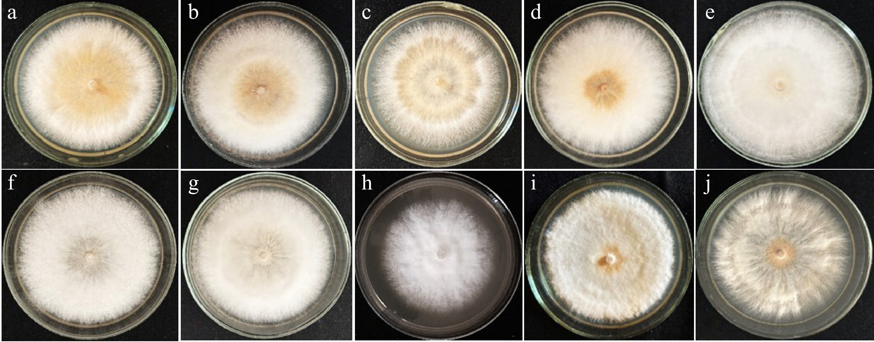
Figure 1.
The colony morphology in studied collections of Fomes inzengae on 1.5% MEA, at 26 °C: (a) Fi/8, (b) Fi/15, (c) Fi/20, (d) Fi/25, (e) Fi/3, (f) Fi/26, (g) Fi/24, (h) Fi/16 on the 7th d; (i) Fi/31, and (j) Fi/30 on the 6th d of growth.
The ageing colonies of F. inzengae become leathery and coriaceous with dense, cinnamon, or cinnamon-brown aerial mycelia (Supplementary Fig. S1). All strains, except Fi/3, Fi/16, Fi/20, and Fi/31, developed FB at a day/light regime after 14 d incubation in the dark over 2–3 weeks (Table 2; Supplementary Fig. S2). However, FB in the Fi/3 and Fi/31 strains were observed in dual culture experiments during antagonistic interaction with Microsporium gypseum, and M. gypseum and Trichophyton terrestre on potato-dextrose agar (PDA), respectively, at 26 °C, while FB were not seen in Fi/16 and Fi/20 strains (Badalyan et al., unpublished). The cultural studies of F. fomentarius s.s. strains from different eco-geographical origins showed that this fungus poorly develops FB in the same culture conditions compared with F. inzengae, which can be considered species-specific (Badalyan et al., unpublished).
Micromorphological characteristics of mycelia
-
The trimitic hyphal system, composed of thick-walled clampless, skeletal, and binding hyphae, and thin-walled generative hyphae with clamp connections, is typical for basidiomata and mycelia of polypore bracket fungi. The skeletal hyphae of polypores have significant structural function, providing stability and durability to basidiomata and assisting in the design and application of innovative mycelium-based bio-industrial materials[54].
The diameter of skeletal hyphae in pure cultures of polypores is shorter[29] than in wild basidiomata[25]. The skeletal hyphae in the basidiomata of Fomes inzengae and Fomes fomentarius s.s. are different: in F. inzengae, they are thicker compared with F. fomentarius s.s. In pure cultures of both species, the diameter of skeletal hyphae is temperature-dependent and reaches about half of its diameter in basidiomata[3].
In studied Armenian collections of F. inzengae, thick-walled, aseptate, rust-cinnamon-brown skeletal (1.2[2.8]–2.2[4.1] μm diam), and septate thin-walled generative hyaline hyphae (1.1[1.9]–1.9[4.7] μm diam) were observed. The single round-shaped clamp connections (0.9–1.5[1.3–2.9] × 1.9–3.0[2.5–4.5–6] μm), almost at each septum of generative hyphae, are characteristic taxonomic features for growing dikaryotic mycelium of F. inzengae (Table 2; Fig. 2a–g).
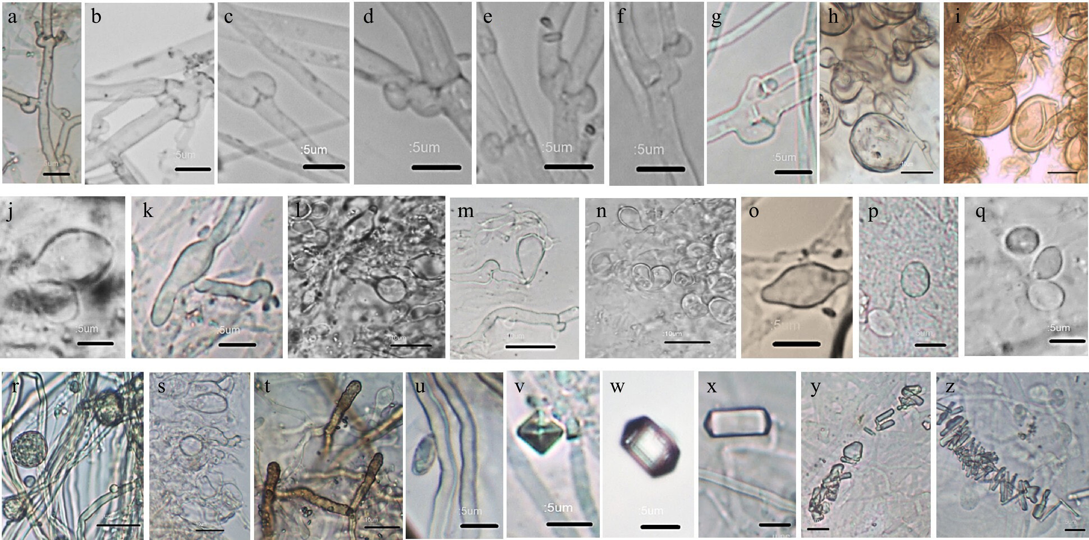
Figure 2.
Mircomorphological observation of studied cultures of Fomes inzengae: hyphal clamp connections in (a) Fi/15; (b) Fi/25; (c) Fi/31; (d), (e) Fi/20; and (f), (g) Fi/3; development of cuticular cells in (h), (i) Fi/30, (j), (k), (l) Fi/20, (m) Fi/24, and (n) Fi/8; chlamydospores in (o) Fi/20, (p) Fi/3, and (q) Fi/25; chlamydospores in (r) Fi/26, (s) Fi/8, (t), cystidiols in Fi/15, and (u) basidiospore in Fi/30 observed in an in vitro developed fruiting body; crystals in (v), (w), (y) Fi/20, (x) Fi/25, and (z) Fi/31 strains. Scale bars = (a)–(g), (j), (k), (o)–(q), (u)–(z) 5 µm, and (h), (i), (l)–(n), (r)–(t) 10 µm.
The formation of pseudoparenchyma or mycelial crust is a developmental process during the vegetative growth of many wood-decaying fungi (Piptoporus betulinus, Ganoderma adspersum, G. lucidum, and G. resinaceum), including Fomes species[14,31]. The swelling of cell walls of generative hyphae to develop compact globular pseudoparenchyma consisting of round to oval cuticular cells has been described in all studied strains[29,55]. The observation of Fomes fasciatus revealed that thin-walled generative hyphae lengthen in the colony margin to form white to yellowish-orange, ochraceous globular pseudoparenchyma[56]. However, this process was not described in the F. fasciatus strain from Brazil[57].
The hyaline or brownish, oval to round, thin-walled hyphal swelling or thick-walled chlamydospore-like cuticular cells in F. inzengae, as 'inflated roundish terminal and intercalary hyphal elements up to 10 μm diam' were described at 30 °C and above from the 5 d of mycelial growth[3]. The cuticular cells, as chlamydospore-like swellings, were previously described in the Fi/16 (former Ff/16) strain[15]. In the current study, the development of cuticular cells was observed in all studied strains starting from 6 d of incubation at 26 and 30 °C. The process was initiated by intense vacuolization and swelling of generative hyphae (Fig. 2h–n). The sizes of cuticular cells vary (2.2–6.0[2.3–14.5] × 5.7–19.8[5.8–21.5] µm) in different strains. Up to 21.5 µm giant cells were observed in the Fi/30 strain isolated from Fagus orientalis (Table 2; Fig. 2h, i).
The formation of globular pseudoparenchyma in polypores, including studied F. inzengae strains, preceded the development of FB. In our experimental conditions, the FB initially appeared at a day/night regime over 21 d after 14 d of incubation in the dark (Table 2; Supplementary Fig. S2). The mature basidiospores were observed in FB tissues of the Fi/26[15] and Fi/30 (this study) strains. (Table 2; Fig. 2u).
Our preliminary observation showed that the process of cuticular cell development in F. fomentarius s.s. cultures on MEA is weakly expressed, which is probably correlated with its poor ability to develop FB in comparison with F. inzengae strains (Badalyan et al., unpublished). Therefore, in vitro development of sexual structures at an earlier stage of mycelial growth of F. inzengae may be considered as a species-specific cultural characteristic and used in species delimitation of F. fomentarius s.s. and F. inzengae[4,5].
The chlamydospores are thick-walled, solitary, endogenously (terminal or intercalary) formed asexual spores developed by protoplast contraction in the aging hypha and are adapted for perennation rather than dissemination[17,18]. In a few Agaricomycetes species, the chlamydospores develop directly in FB tissue and may be considered as a species/strain-specific characteristic. The occurrence of chlamydospores is distributed throughout the order Agaricales; however, they are more common in Polyporales species, including F. inzengae[18]. The round to oval-shaped chlamydospores (1.8[9.1] × 2.7[13.1] µm) were observed in all studied strains of F. inzengae, while round- (4.2[4.6] × 5.2[5.6] µm) and lemon-shaped (5.0[5.2] × 10.0[10.4] µm) chlamydospores were only seen in Fi/20 strain from 5 d of growth at 26 and 30 °C (Table 2; Fig. 2o–q). The thick-walled chlamydospores previously reported in in vitro developed FB of the Fi/26 strain[18] were also observed in FB tissues of Fi/26 (2.4[11.9] × 2.6[12.4] µm) and Fi/8 (6.3[6.7] × 7.8[14.3] µm) strains during the current study (Fig. 2r, s). Thus, the formation and morphological characteristics of cuticular cells and chlamydospores in the life cycle of F. inzengae are a taxonomically important feature[14,17,18].
The cystidiols (3.2[3.7] × 18.0[23.4] µm) and basidiospores (2.8[3.0] × 7.5[7.7] µm) were also observed in FB developed in Fi/15 and Fi/30 strains, respectively (Table 2; Fig. 2t, u). However, the sizes of basidiospores of wild-grown basidiomata (S30), from which the Fi/30 strain was isolated, were significantly larger (3.5[3.8] × 11.5[11.8] µm).
The calcium oxalic crystals with different shapes (cuboid, rod-shaped, tetra-, hexa-, and octagonal, unformed, etc.) and sizes, single or in groups, were abundantly found in F. inzengae cultures from the first day of mycelial growth. They were particularly diverse in the Fi/20 strain isolated from a living P. nigra tree (Table 2; Fig. 2v–z). The tetra-, hexa-, and octagonal, as well as unformed crystals, were described in culture medium (Fig. 2v–y), while granular or rod-shaped crystals were mostly attached to the surface of hyphae (Fig. 2z).
Mycelial growth rate
-
The studied Fomes inzengae strains exhibited fast growth on 1.5% MEA. The significant differences were found among the GRavr indicators at 26 °C (7.5 ± 0.21–10.0 ± 0.04 mm·d−1) and 30 °C (8.7 ± 0.21–11.5 ± 0.22 mm·d−1). However, the highest GRavr indicators (≥ 10.0 mm·d−1) were detected at 30 °C in Fi/3, Fi/25, Fi/30, and Fi/31 strains isolated from C. betulus, P. nigra, F. orientalis, and A. platanoides, respectively (Table 3; Supplementary Fig. S3).
Table 3. The mycelial GRavr (mm·d−1) of Fomes inzengae strains on 1.5% MEA at different temperatures.
Strain Host tree 26 °C 30 °C 4/30 °C ΔGRavr ΔGRavr, % Fi/3 C. betulus 9.0 ± 0.3 10.0 ± 0.3 6.7 ± 0.2 3.3 ± 0.2 33.0 ± 2.1 Fi/26 C. betulus 9.0 ± 0.2 9.8 ± 0.3 7.4 ± 0.4 2.4 ± 0.5 24.5 ± 2.2 Fi/24 F. orientalis 8.9 ± 0.05 9.8 ± 0.4 7.4 ± 0.6 2.4 ± 0.5 24.5 ± 3.1 Fi/30 F. orientalis 9.2 ± 0.1 10.6 ± 0.3 8.2 ± 0.5 2.4 ± 0.5 22.6 ± 3.9 Fi/15 S. alba 8.1 ± 0.1 9.2 ± 0.4 7.1 ± 0.6 2.2 ± 0.5 23.9 ± 3.3 Fi/16 S. alba 7.5 ± 0.2 8.7 ± 0.3 6.8 ± 0.5 1.9 ± 0.5 21.8 ± 3.4 Fi/8 J. regia 8.4 ± 0.1 9.8 ± 0.2 6.9 ± 0.2 2.9 ± 0.3 29.6 ± 2.1 Fi/20 P. nigra 7.6 ± 0.1 9.2 ± 0.4 6.5 ± 0.2 2.7 ± 0.5 29.3 ± 1.9 Fi/25 P. nigra 8.8 ± 0.1 10.7 ± 0.3 7.8 ± 0.4 2.9 ± 0.4 27.1 ± 3.2 Fi/31 A. platanoides 10.0 ± 0.04 11.5 ± 0.2 8.4 ± 0.2 3.1 ± 0.3 26.9 ± 2.1 Legend: values are expressed in means ± SD, n = 12: ΔGRavr is the difference between GRavr values without stress, and with stress at 30 °C. The strains specificity among the GRavr indicators at 26 and 30 °C, as well as after stress at 30 °C are very highly significant (*** p < 0.001). The strains of F. inzengae after 30 d of pre-incubation at 4 °C, where none of cultures grew, start to grow at 30 °C with 21.8%–33% inhibited GRavr (ΔGRavr) and form sparse colonies, almost without pigmentation. The strains' specificity among the GRavr indicators at 26 and 30 °C, as well as after stress (4 °C) at 30 °C are very highly significant (*** p < 0.001) (Table 3; Supplementary Fig. S4).
Biomass formation
-
The strains of Fomes inzengae formed well-developed, white, cottony or cottony-felt, later leathery aerial mycelium with light cinnamon-brown pigmentation ascending the flasks' walls after 21 d of static growth on liquid MEM (pH 6) at 30 °C (Supplementary Fig. S5). The pigmentation of mycelium was almost absent in the Fi/16, Fi/24, and Fi/26 strains. The yellowish-brown mycelial exudate was only observed in the Fi/30 strain, isolated from Fagus orientalis. The color of the cultural broth samples remained almost unchanged compared to the control. An abundant presence of round- and club-shaped cuticular cells and thick-wall chlamydospores, as well as single and grouped crystals of various shapes and sizes were observed.
The formation of biomass is species/strains specific and depends on mycelial GR and density, cultivation methods (static, submerged) and conditions (nutrient medium, temperature, pH). The higher the amount of accumulated biomass, the more suitable is the species/strains for cultivation. The fast growing (GRavr = 8.7–11.5 mm·d−1) on MEA strains of F. inzengae accumulated on average up to 99.5 g·L−1 of wet (Wt) and 4.0–5.5 g·L−1 of dry (Dr) biomass in a static culture over 21 d of cultivation at 30 °C (Supplementary Table S1).
Three strains (Fi/20, Fi/25, and Fi/31) accumulated more than 95.5 g·L−1, while the other seven strains accumulated 80.5–88.0 g·L−1 Wt biomass (Supplementary Table S1). The lowest amount of Wt biomass (76.5 g·L−1) was detected in the Fi/8 strain from a living Juglans regia tree with moderately hard wood, while the highest amount (98.5–99.5 g·L−1) was in Fi/20 and Fi/25 strains from living Populus nigra with the softest hardwood; the common host of F. inzengae from which the fungus was originally described[3]. Thus, the correlation between host tree species and its wood type, as well as mycelium GRavr and biomass amount was revealed. The differences among strains according to average amounts of Wt and Dr biomass are highly significant (** p < 0.01). The correlation between GRavr and Dr (r = 0.4) as well as GRavr and Wt (r = 0.3) biomass was moderately positive.
The Wt/Dr biomass ratio of studied F. inzengae strains after the static growth on MEM was about 20:1 which has a reference value for their biotechnological cultivation (Fig. 3). The drying dynamics of biomass samples at room temperature lasted 4 d (Supplementary Table S2; Fig. 4).
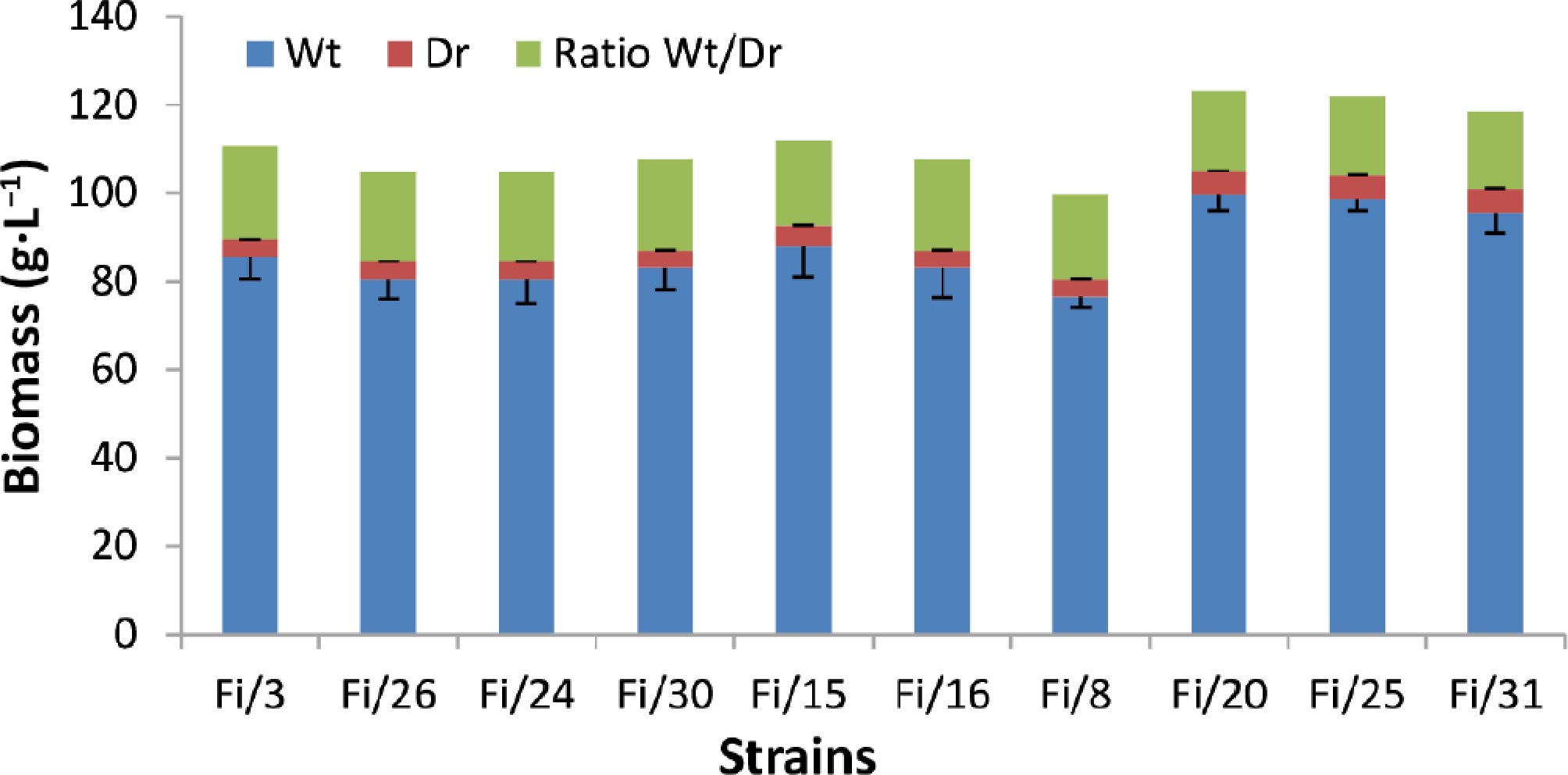
Figure 3.
The biomass formation in Fomes inzengae strains after 21 d of static cultivation on MEM.
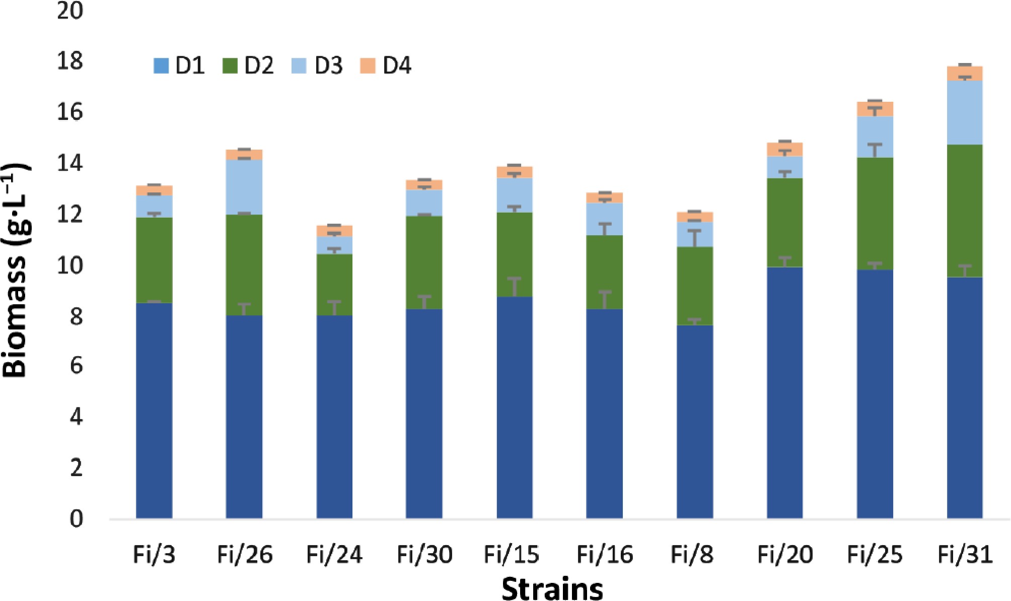
Figure 4.
The drying dynamics of wet biomass in Fomes inzengae strains at room temperature over 4 d.
The strains of F. inzengae, particularly Fi/20, Fi/25, and Fi/26 isolated from P. nigra and Carpinus betulus, decreased the medium pHavr by 11.7%–18.3% during static growth (Table 4; Supplementary Fig. S6). The correlation between GRavr, the average amount of Dr and Wt biomass, and ΔpH was revealed. A very weak positive correlation between GRavr and ΔpH (r = 0.12); moderate positive correlation between Dr (r = 0.53), and Wt (r = 0.6) biomass and ΔpH were detected. The strain specificity of pHavr was highly significant (** p < 0.01).
Table 4. The pHavr of cultural broth samples after 21 d of static cultivation of Fomes inzengae at 30 °C on MEM (pH 6).
Strain Host tree pHavr ΔpH ΔpH, % Fi/3 C. betulus 5.2 ± 0.05 0.8 ± 0.04 13.3 ± 0.8 Fi/26 C. betulus 4.9 ± 0.01 1.1 ± 0.01 18.3 ± 0.04 Fi/24 F. orientalis 5.3 ± 0.04 0.8 ± 0.04 13.3 ± 0.8 Fi/30 F. orientalis 5.1 ± 0.01 0.9 ± 0.01 15.0 ± 0.1 Fi/15 S. alba 5.2 ± 0.04 0.8 ± 0.04 13.3 ± 0.7 Fi/16 S. alba 5.0 ± 0.05 1.0 ± 0.8 16.7 ± 0.8 Fi/8 J. regia 5.3 ± 0.04 0.7 ± 0.1 11.7 ± 0.8 Fi/20 P. nigra 5.0 ± 0.04 1.0 ± 0.04 16.7± 0.7 Fi/25 P. nigra 4.9 ± 0.01 1.1 ± 0.01 18.3 ± 0.1 Fi/31 A. platanoides 5.0 ± 0.01 1.0 ± 0.01 16.7 ± 0.1 Legend: (ΔpH) difference between pHavr of MEM (control) and cultural broth. The values are expressed as mean ± SD, n = 3. The strain specificity of pHavr was highly significant (** p < 0.01). A positive correlation was very weak between GRavr and ΔpH (r = 0.12) and moderate between Dr (r = 0.53) and Wt (r = 0.6) biomass and ΔpH. -
Recently, the study of genetic resources of medicinal Agaricomycetes mushrooms, including wood decaying polypores (order Polyporales), as a valuable source of bioactive compounds, minerals, and vitamins, with a high exploitation potential in biotechnology, biomedicine, and bio-industry, has gained significant interest[37,39,40,58,59]. The polypores are easily isolated, grown, and sexually reproduced in culture conditions, which makes them promising organisms for biotechnological cultivation to obtain mycelial biomass, desired compounds, and biotech products[6,16,37]. However, the correct taxonomic identification of fungal cultures to control their purity during cultivation is required[7−18]. The original studies related to the taxonomy of wood-decaying mushrooms (the former order Aphyllophorales) based on taxonomically relevant mycelial characteristics were performed by Nobles[28] and Stalpers[29]. Currently, a number of taxonomically significant morphological, ecological, biochemical, and physiological characteristics of mycelia (hyphal morphology, presence, occurrence and form of clamp connections, asexual spores and mode of their formation, development of cuticular cells and chlamydospores, daily GR, growth temperature and pH, biosynthetic pathways of bioactive and volatile compounds, etc.), are used for correct taxonomic identification of valuable industrial polypore species/strains[8−11,13−15,17,18,60]. Moreover, the mycelial characteristics of mushrooms are less polymorphic between recently diverged taxa than those of the basidiomata[61]. The results of culture studies are also necessary to perform the phylogenetic analysis of complex Agaricomycetes taxa[1,3], particularly biotechnologically important species from many genera, such as Fomes, Ganoderma, Pleurotus, and Flammulina[53].
The white rot fungus Fomes inzengae has been recently described as a taxonomically distinct Mediterranean species[3,22,23], reported also in Armenia[4,5]. A very limited number of cultural studies of F. inzengae were conducted[3], therefore, comprehensive morphological, ecological, and physiological observation of geographically different mycelial collections with the assessment of taxonomic significance of revealed vegetative structures and traits is needed. In the current work, the study of morphological and growth characteristics, biomass formation, and acidification levels of medium pH during the cultivation of 10 genotyped dikaryotic strains of F. inzengae collected in Armenia from Angiosperm host trees (Acer platanoides, Carpinus betulus, Fagus orientalis, Juglans regia, Populus nigra, and Salix alba) has been discussed.
The morphological screening of studied F. inzengae strains has shown that they formed cottony or cottony-felt, later leathery colonies with whitish-creamy, yellowish, or dark cinnamon-brown pigmentation, and light pinkish-cinnamon or unchanged agar (Table 2; Fig. 1, Supplementary Figs S1 and S2).
In our experimental conditions, the development of cuticular cells, globular pseudoparenchyma, FB (except Fi/16 and Fi/20 strains), and asexual chlamydospores was observed in all strains of F. inzengae (Table 2; Fig. 2s–u, Supplementary Fig. S2). Single, round-shaped clamp connections occur almost at each septum of generative hyphae, while non-septate skeletal hyphae are clampless (Table 2; Fig. 2a–g). The cuticular cells were developed by swelling of intensively vacuolated generative hyphae starting from the 5th day, while the round- and lemon-shaped chlamydospores were formed from the 6th day of mycelial growth (Fig. 2h–q). The chlamydospores were also described in FB tissue of Fi/8 and Fi/26 strains developed in Petri dishes (Fig. 2r, s). The formation of chlamydospores in FB tissue of the Fi/26 strain was also reported in our previous work[18].
In natural conditions, the white rot fungi decay the wood faster than the brown rot fungi. The softwood pine trees decay more slowly compared to injured hardwood. The living hardwood trees support mycelium growth due to high nutrient composition, and therefore may promote the wood decaying process. Thus, mycelium GR depends on the host tree species (Gymnosperm or Angiosperm), wood type (soft or hard), and wood density, which varies significantly between the stump, trunk, and living tree. The studied F. inzengae strains from hardwood stumps (one), trunk (one), and living trees (seven) are fast-growing (GRavr = 8.7–11.5 mm·d−1) on MEA at 30 °C (Table 3). Among these, a relatively higher GRavr revealed strains isolated from living trees with a high nutrient availability (Tables 1, 2).
The low temperature is a stress factor for mycelial growth of polypores, which mainly prefer optimal growth temperatures ranging from 30 to 32 °C. One month incubation at 4 °C decreased the mycelium GRavr of F. inzengae by 21.8%–33%. The difference between strains is very highly significant (*** p < 0.001) (Table 3; Supplementary Figs S3, S4). Several morphological changes, such as low density and weakly pigmented aerial mycelium as a reaction to cold stress, were also observed. Different physiological mechanisms, including cold shock protein (CSP) responses, are activated at lower temperatures, and strain specificity of organic acid metabolism can also occur in this process. The ecological adaptability and geographical distribution (altitude) of Armenian strains of F. inzengae, as a southern phylogenetic lineage of Fomes fomentarius s.l., may significantly affect the mycelial GRavr, as a response to cold stress.
Mushroom cultivation significantly contributes to the protection of natural genetic resources, while mycelium cultivation (static, submerged) is suitable for obtaining innovative biotech products[37−45]. In static liquid cultures, the ratio of Wt/Dr biomass in Agaricomycetes fungi usually ranges from 10:1 to 20:1. The Wt/Dr ratio in F. inzengae strains, on average, was 20:1, which could serve as a reference value for their further biotechnological cultivation. The differences in Wt and Dr biomass production among F. inzengae strains are highly significant (** p < 0.01), while a moderate positive correlation was revealed between GRavr and Dr (r = 0.4), and Wt (r = 0.3) biomass (Supplementary Table S1). The drying process of Wt biomass samples at room temperature lasted 4 d (Supplementary Table S2; Fig. 4).
Comparing the GRavr (up to 11.5 mm·d−1) and average biomass amount (4–5.5 g·L−1) of studied F. inzengae strains with other white rot medicinal polypores from our Agaricomycetes collections[53], such as Laetiporus sulphureus (GRavr = 4.8–6.1 mm·d−1; dry biomass 3.5–5.1 g·L−1), and Ganoderma lucidum (4.8–6.1 mm·d−1; 1.3–3.7 g·L−1) obtained from the same culture conditions showed that F. inzengae is suitable for biotechnological cultivation. Xylotrophic agaricoid species, such as Flammulina velutipes (5.5–7.0 mm·d−1; 7–9 g·L−1) and Schizophillum commune (6.5–7.5 mm·d−1; 6.3–7.6 g·L−1) (unpublished biomass data)[53], as well as ascomycetous saprobic Morchella species[52] formed a higher amount of biomass (11.5–18.4 mm·d−1; 4.5–9.2 g·L−1) in the same experimental conditions. Further studies are required to reveal optimal growth conditions of the selected species/strains to receive a high amount of mycelial biomass for biotechnological and bio-industrial exploitation.
During late exponential and stationary phases of mycelial growth, the medium pH decreased due to excretion of extracellular metabolites, including organic acids (oxalic, lactic, malic, citric, etc.) to enhance nutrient acquisition and regulate the microenvironmental conditions[6,7]. Moreover, these organic acids possess antimicrobial and antioxidant activities[42,62]. The polypores, including Fomes species, are producers of valuable oxalic acid, mainly stored in the form of calcium oxalate crystals[63,64]. The crystals of different sizes and shapes were found in agar and liquid media of young cultures of F. inzengae (Table 2; Fig. 2v–z).
The polypore fungi preferably grow on extremely acidic (pH 3.0) to strongly alkaline (pH 9.0) environments, with the best suitable pH 4.5−7.5, which, along with medium C/N ratio, temperature, and other factors, affect the accumulation of mycelial biomass[6,65]. Over 21 d of static cultivation, the studied strains, particularly those isolated from a living P. nigra tree, the common host of F. inzengae, decreased the medium pH by up to 18.3% (Table 4, Supplementary Table S1; Fig. 3, Supplementary Fig. S6). A very weak positive correlation between GRavr and ΔpH (r = 0.12), as well as a moderate positive correlation between Dr (r = 0.53) and Wt (r = 0.6) biomass and ΔpH, were detected. The strain specificity of pHavr was highly significant (** p < 0.01). Thus, the described mycelial characteristics of F. inzengae strains are taxonomically valuable and may be used to control the culture purity during biotechnological cultivation.
Previous work[3,19,23] and our recent studies[2,4,5] on phylogenetic analysis of rDNA ITS1–5.8S–ITS2 sequence variability in Fomes fomentarius s.l. in Armenia showed moderate interspecific and lower intraspecific variability among two Fomes species, F. fomentarius s.s., and F. inzengae. The minimum intraspecific differences were also detected in the studied F. inzengae strains[5], which correspond to the results obtained from cultural studies. However, further studies of eco-geographically different collections of F. inzengae are needed for detailed discussions between genetic backgrounds and the phenotypic differences of strains.
-
The aim of the current work was to study mycelial morphology, biomass formation, and acidification level of medium pH during static growth of 10 genotyped strains of medicinal polypore fungus Fomes inzengae from Armenia. The revealed taxonomically valuable cultural characteristics (colony texture, hyphal micromorphology, including clamp connections, cuticular cells and chlamydospores, mycelial GR, growth temperature and pH, biomass accumulation, formation of crystals and fruiting bodies) have contributed to the limited data on mycelium morphology and growth characteristics of F. inzengae. The obtained data can be used to control the purity of culture and regulate growth conditions during biotechnological cultivation of F. inzengae, as well as to select the high-yielding productive strains for further use to develop mycelium-derived innovative biotech products with growing demand in biomedicine and bio-industry. The minimum intraspecific differences detected in the studied F. inzengae strains[5] correspond to the results obtained from current cultural observation. However, further studies of bio-ecological and genetic characteristics of geographically different F. inzengae collections are warranted.
-
The authors confirm their contributions to the paper as follows: study conception and design, methodology, draft manuscript preparation and review: Badalyan SB; observation and data collection, analysis and interpretation of results: Badalyan SB, Gharibyan NG. Both authors reviewed the results and approved the final version of the manuscript.
-
All data generated or analysed during this study are included in the current published article. The datasets analyzed statistically during the current study are available from the corresponding author on reasonable request.
-
The authors declare no conflict of interest.
-
accompanies this paper online at: https://doi.org/10.48130/sif-0026-0025.
- Supplementary Table S1 The average weight (g/L) of wet and dry biomass after 21 d of static cultivation of Fomes inzengae on MEM at 30 °C.
- Supplementary Table S2 Drying dynamics of wet mycelial biomass (g) of Fomes inzengae strains at room temperature to constant weight during four days (D1–D4).
- Supplementary Fig. S1 The pigmentation of colonies of Fomes inzengae strains on the 10 d of growth on MEA at 26 °C (first row) and 30 °C (second row): Fi/3 (A, a); Fi/15 (B, b); Fi/16 (C, c); Fi/20 (D, d), Fi/26 (E, e); Fi/30 (F, f), Fi/31 (G, g), Fi/18 (H, h), Fi/24 (I, i) and Fi/25 (J, j).
- Supplementary Fig. S2 Formation of fruiting bodies in Fomes inzengae strains on MEA at day/light regime after 14 d incubation in the dark: Fi/15 (a); Fi/26 (b) and Fi/30 (c) on 26 d (a, b) and 38 d (c) of growth.
- Supplementary Fig. S3 The mycelial GRavr (mm·d−1) of Fomes inzengae strains on MEA at different temperatures.
- Supplementary Fig. S4 The GRavr suppression effect (ΔGRavr, %) of Fomes inzengae strains at 30 °C on MEA after 30 d preincubation at 4 °C.
- Supplementary Fig. S5 Thick, leather-like mycelium mat after 21 d static cultivation of Fomes inzengae strains on MEM at 30 °C, pH 6: (a) Fi/3; (b) Fi/8; (c) Fi/15; (d) Fi/16; (e) Fi/20; (f) Fi/24; (g) Fi/25; (h) Fi/26; (i) Fi/30 and (j) Fi/31.
- Supplementary Fig. S6 The acidification level (%) of liquid malt-extract medium (1.5% MEM, pH 6) by Fomes inzengae strains.
- Copyright: © 2026 by the author(s). Published by Maximum Academic Press, Fayetteville, GA. This article is an open access article distributed under Creative Commons Attribution License (CC BY 4.0), visit https://creativecommons.org/licenses/by/4.0/.
-
About this article
Cite this article
Badalyan SM, Gharibyan NG. 2026. Тhe cultural characteristics and biomass formation of medicinal polypore fungus Fomes inzengae (Polyporaceae, Agaricomycetes) collected in Armenia. Studies in Fungi 11: e028 doi: 10.48130/sif-0026-0025 

Тhe cultural characteristics and biomass formation of medicinal polypore fungus Fomes inzengae (Polyporaceae, Agaricomycetes) collected in Armenia
- Received: 24 January 2026
- Revised: 26 April 2026
- Accepted: 09 May 2026
- Published online: 23 September 2026
Abstract: The cultural characteristics and biomass formation of 10 genotyped dikaryotic strains of medicinal polypore fungus Fomes inzengae (Polyporaceae, Agaricomycetes) isolated from basidiomata collected from the Ijevan, Aparan, and Yerevan floristic regions of Armenia from Acer platanoides, Carpinus betulus, Fagus orientalis, Juglans regia, Populus nigra, and Salix alba trees have been studied. All strains formed cottony and cottony-felt, whitish-creamy and cinnamon-brown colonies on a 1.5% malt-extract agar (MEA) medium with an average growth rate (GRavr) of 7.5–10.0 mm·d−1 and 8.7–11.5 mm·d−1 at 26 and 30 °C, respectively. The GRavr of mycelia at 30 °C after 30 d pre-incubation at 4 °C decreased by 21.8%–33.0%. Taxonomically significant single round-shaped hyphal clamp connections, cuticular cells, chlamydospores, and crystals were observed in all strains. The fruiting bodies were developed in all cultures in a day/night regime, except the Fi/16 and Fi/20 strains, over 2–3 weeks after 14 d of incubation in the dark. The studied strains accumulated 4–5.5 g·L−1 of air-dried biomass over 21 d of static growth on a liquid malt-extract medium (pH 6), decreasing the medium pH by 11.7%–18.3%. The drying dynamics of wet biomass at room temperature lasted 4 d. The ratio of wet to dry biomass was about 20:1. The revealed mycelial characteristics are required to control the purity of cultures during biotechnological cultivation of F. inzengae. Further studies of eco-geographically different collections of F. inzengae will reveal the optimal growth conditions of selected productive strains to develop mycelium-derived innovative biotech products with high demand in biomedicine and bio-industry.
-
Key words:
- Biomass /
- Clamp connections /
- Chlamydospores /
- Crystals /
- Cuticular cells /
- Fomes inzengae /
- Growth rate /
- Medium pH /
- Mycelium