ARTICLE   Open Access    

First record of Eutypella vitis causing branch dieback on new host trees in Canada

More Information
  • Eutypella vitis is reported for the first time on several new host trees viz. Fagus grandifolia, Fraxinus pennsylvanica and Syringa reticulata with associated branch dieback symptoms in the Niagara Region of Southern Ontario, Canada. Morphological characteristics and phylogenetic analyses of ITS sequence data confirmed the species identification. Eutypella vitis is known as a woody plant pathogen causing dieback of grapevines and other economically important fruit trees widely cultivated in Ontario. The fungus ability to infect several alternate new hosts can promote its faster and wider spread across vineyards and consequently increase a dieback hazard to viticulture in Canada.
  • Wild mushrooms have been consumed since ancient times due to their good taste and nutritional values. Also, mushrooms have the advantage of being rich in vital components such as proteins, vitamins, chitin, fibers, iron, zinc, selenium, sodium, etc. (Breene 1990, Chang & Miles 1992, Elkhateeb et al. 2019, 2020a, b, Daba et al. 2020, El-Hagrassi et al. 2020). Beside these nutritional values, many mushrooms have gained importance due to their therapeutical potentials (Bobek et al. 1995, Bobek & Galbavý 1999, Vaz et al. 2011, Khatun et al. 2012, Elkhateeb & Daba 2020, 2021a, b). The extracts of fruit bodies and mycelium obtained from various fungi have shown a crucial role in the prevention and/or the treatment of many diseases (Wasser & Weis 1999, Sharma et al. 2015). Nowadays, some immunomodulating molecules that have been obtained from different medicinal mushrooms have found great importance in improving the immune function in cancer patients while they are receiving chemo or radiotherapy (Mizuno 1995). Other edible mushrooms have gained medicinal importance due to their effectiveness in adjusting the blood pressure and reducing the free plasma cholesterol, as well as due to antimicrobial, antioxidant, and antitumor properties (Kabir & Kimura 1989, Smânia et al. 1995, Mau et al. 2005, Elkhateeb 2020, Thomas et al. 2020). The genus Ramaria is found all over the world, in which various species of Ramaria have been consumed by people for nutritional purposes. However, more studies are required to be conducted on these genera in order to detect the nutraceutical and nutritional compounds of these mushrooms. The genus Ramaria belonging to Basidiomycota, Class; Agaricomycetes, Order; Gomphales, Family; Gomphaceae, which includes approximately 200 species of coral fungi (Sharma et al. 2015). Several taxa are edible and picked in Europe and also they are easily confused with several mildly poisonous species capable of causing nausea, vomiting and diarrhea, in which among the most unusually shaped mushrooms of all, Ramaria are appropriately referred to as the coral mushrooms. Ramaria are recognizable by their variably thickly branched fruit bodies, fleshy and tough textures sometimes partly gelatinous or jellylike. Although many Ramaria species are very brightly colored, just as many are dull, and all have a tendency to become rather drab in age. Similar genera include Phaeoclavulina, Lentaria, Artomyces, and Ramariopsis, which differ in various aspects of color, consistency, substrate, microscopic features, or ecology (Sharma & Gautam 2017).

    Ramaria basidiocarps are either lignicolous or terricolous (white, yellow, orange, red, brilliant purple, brown and sometimes green hues) with many species forming a characteristic mycelial mat in the soil beneath the sporocarps (Nouhra et al. 2004). The colour may fade or change after collection in the field. Species of this genus often show a color change on bruising. The flesh may change to green (virescent), red brown (rubribrunnescent), brown to yellow brown (brunnescent), or a red wine color (vinescent) due to age or environmental conditions (Exeter et al. 2006).

    Clavaria is belonging to Basidiomycota, class Agaricomycetes, order Agaricales, and family Clavariaceae. Species of Clavaria produce basidiocarps (fruitbodies) that are cylindrical to club-shaped, branched and coral-like. They are often grouped with similar looking species from other genera, and they are collectively known as the clavarioid fungi (Olariaga et al. 2015).

    The most common species of this genus is Ramaria stricta (Kuo 2009), which grows on wood debris, which features branches that are usually strictly oriented, so that they are mostly straight and ascending. When fresh, its branch tips are yellow and its branches are dull yellowish buff, but its surfaces bruise and discolor purplish brown. Under the microscope it features roughened spores, clamp connections, and thick-walled hyphae. Several very similar species have been separated by mycologists, and the name Ramaria stricta should probably represent a group of potential species awaiting contemporary study. Ecology: Uncertain; while most ramarias are thought to be mycorrhizal, the wood-inhabiting species could be mycorrhizal or saprobic; growing from the dead (but sometimes buried) wood of conifers; appearing alone, scattered, or gregariously; early summer through fall; apparently widely distributed in North America, but more common from the Rocky Mountains westward. Fruitbody: 4−14 cm high; 4−10 cm wide; base well developed or nearly absent; branching repeatedly. Branches: Vertically oriented and elongated; often flattened; smooth; yellowish buff, becoming orangish buff as the spores mature; bruising and discoloring purplish brown; tips yellow when fresh and young. Base: Nearly absent, or fairly well developed; to 2 cm wide; white below; colored like the branches above; attached to numerous white rhizomorphs. Flesh: Whitish; fairly tough. Odor and Taste: Odor not distinctive, or sweet and fragrant; taste bitter. Spore color: Rusty yellowish. Microscopic Features: Spores are yellow-brown to rusty-brown in mass deposit 7.5−10.5 × 3.5−5µm; stretched-elliptical; smooth to roughened. Clamp connections present. Thick-walled hyphae present (Kuo 2009) (Figs 1, 2).

    Figure 1.  Fruitbody of Ramaria stricta in the field. The photograph was taken at the Sewickley Heights Borough Park, PA, USA by Fluffberger (https://www.inaturalist.org/observations/3596638). The photo was used under the CC BY-NC 4.0 non-commercial use license. Scale bar = 10 cm.
    Figure 2.  Fruitbody of Ramaria araiospora in the field. The photograph was taken at the Millersylvania State Park, Olympia, WA, USA by Yay4john (https://www.inaturalist.org/observations/62355314). The photo was used under the CC BY-NC 4.0 non-commercial use license. Scale bar = 10 cm.

    Few reports have been found about the discovery of these genera in the Northwestern Himalayas. The current surveys to Northwestern Himalayas have shown the existence of six species of coral mushrooms belonging to the genus Ramaria viz., R. aurea, R. botrytis, R. flava, R. flavescens, R. rubripermanens, and R. stricta (Sharma & Gautam 2017). Details about their culinary status were obtained from native inhabitants. Moreover, studies have been conducted on these species to get detailed biochemical profiling as well as exploring their various biological activities (Sharma & Gautam 2017).

    Coral mushrooms gain great therapeutical and nutritional importance due to their valuable components. Sharma & Gautam (2017), reported that 12 edible species of Ramaria occurring in Northwestern Himalayas contain proximal components, amino acids, and minerals, fatty acids. Ramaria aurea is an edible mushroom that is considered a promising dietary supplement as it consists of carbohydrates, amino acids, proteins, fibers, and minerals (Rai et al. 2013). Ramaria botrytis, another edible mushroom, consists of unsaturated fatty acids, tocopherol as well as other bioactive compounds (e.g. ascorbic acid, carotene, flavonoids, β- and lycopene) (Barros et al. 2008). Ramaria subalpina also contains significant amounts of ascorbic acid, β-carotene, flavonoids, and lycopene (Acharya et al. 2017). Also, some Ramaria species show the presence of alkaloids, saponins, terpenoids, coumarins, and cardiac glycosides (Aldred 2008, Dattaraj et al. 2020). Due to the existence of these bioactive molecules, these mushrooms possess various medicinal importance against various diseases. Among these medicinal benefits are antimicrobial, antiviral, anti-parasitic, antioxidant, radical scavenger, anticancer, anti-inflammatory, immune system enhancer, and anti-hyperlipidemia (Wasser 2017).

    Although there is a vast diversity of synthetic antibacterial compounds, the development of bacterial resistance has been substantially increasing (Alves et al. 2012). Thus, discovering novel antibiotics especially from natural sources is urgently required. Since ancient times, several natural resources have exerted potent antimicrobial activities. Among these natural resources are mushrooms that might be valuable alternative sources of novel antimicrobials (Gebreyohannes et al. 2019, Ghosh et al. 2020). In this context, various Ramaria sp. have gained great importance due to their potential antimicrobial activities against various pathogens (Table 1). The ethanolic extract of Ramaria flava was detected by the agar-well diffusion method, and the result showed positive activity against some Gram-positive bacteria such as Staphylococcus aureus ATCC 25923, Staphylococcus aureus Cowan I, Micrococcus luteus NRRL B-4375, Micrococcus flavus, Bacillus subtilis ATCC 6633, Bacillus cereus RSKK 863. Also, R. flava ethanolic extract showed potent activity against the tested Gram-negative bacteria including Salmonella enteritidis RSKK 171, Yersinia enterecolitica RSKK 1501, Klebsiella pneumoniae ATCC 27736 (Gezer et al. 2006). Additionally, Ramaria cystidiophora showed antimicrobial activity against Mycobacterium smegmatis (MIC = 8 µg/mL), and against Mycobacterium tuberculosis (MIC 64−128 µg/mL) (Hassan et al. 2019).

    Table 1.  Antimicrobial activities of various Ramaria species
    Ramaria species Tested extract Employed methods Tested bacteria Tested fungi References
    Gram-positive Gram-negative
    R. formosa Ethyl acetate, methanol, and water Percentage of inhibition B. subtili, S. aureus E. coli, K. pneumonia, Proteus vulgaris, P. aeruginosa Not tested Pala et al. 2019
    R. formosa Methanol Inhibition zone assay S. aureus P. aeruginosa Candida albicans Ramesh & Pattar 2010
    R. zippellii Ethanol, water 96-well microplate bioassay S. aureus E. coli No activity Bala et al. 2011
    R. aurea Ethanol Inhibition zone assay S. aureus E. coli, P. aeruginosa, P. vulgari Candida albicans Rai et al. 2013
    R. flava Ethanol Inhibition zone assay B. subtili, S. aureus E. coli Fusarium auenaceum, F. graminearum and Cercosporella albomaculans Liu et al. 2013
    R. flava Ethanol Agar-well diffusion method S. aureus, Micrococcus luteus, M. flavus, B. subtilis, B. cereus Salmonella enteritidis, Yersinia enterecolitica and K. pneumoniae No activity Gezer et al. 2006
    R. botrytis Ethanol Methanol Acetone Ethyl acetate Inhibition zone diameter S. aureus P. aeruginosa, E. coli, Enterobacter cloacae No activity Han et al. 2016
     | Show Table
    DownLoad: CSV

    The studies also reported that the ethanolic extract of Ramaria flava exhibited antifungal activities against three pathogenic fungi which are C. albomaculans, Fusarium graminearum and Fusarium auenaceum. The extract showed the highest efficacy against F. auenaceum, where 36.64% reduction in fungal growth was observed at the concentration of 2 mg/mL. The extract also inhibited the growth of F. graminearum and C. albomaculans by 19.99 and 30.03% respectively, at the same concentration of 2 mg/mL (Liu et al. 2013). Additionally, Ramaria mushrooms show their effectiveness to cure various viral infections. Zhang et al. (2015) isolated a novel ribonuclease from Ramaria formosa fruiting bodies and tested its antiviral activity against HIV-1 reverse transcriptase enzyme. The results showed that the ribonuclease showed 93% inhibition at a concentration of 30 µM (maximum tested concentration) and with an IC50 value of 3 µM. It was interesting to report that this enzyme exhibits unique features; these include its unique N-terminal sequences, optimum acidic pH value, and temperature resistance. Taken together these features suggest that this ribonuclease could play a vital role in HIV diseases prevention.

    Many reports regarding the antioxidative properties of various Ramaria sp. have been published, which showed the activity of these fungi as effective antioxidants (Table 2). Linoleic acid oxidation was compared with those of α- tocopherol, BHA, and R. flava ethanol extract. The results revealed that the inhibition values of both the standards and R. flava ethanol extract increased gradually with increasing the concentrations. 80 µg/ml BHA, α -tocopherol, and R. flava ethanolic extract exhibited 96.4, 98.6, and 73.3% inhibition, respectively. Whereas increasing the concentrations to 160 µg/ml concentrations resulted in 98.9, 99.2 and 94.7% inhibition, respectively (Gezer et al. 2006). Also, the ethanolic extract of that mushroom exhibited promising OH and DPPH radical-scavenging activities with low IC50 values of 18.08 and 5.86 μg/mL, respectively (Liu et al. 2013). The high inhibition value of R. flava ethanolic extract could be related to the high concentration of phenolic compounds (Komali et al. 1999).

    Table 2.  Antioxidant activities of various Ramaria species isolated from different places
    Ramaria species Place of collection Methods used Activity (EC 50 % of inhibition) References
    R. flava Turkey DPPH scavenging assay 94.78% at 12 mg/mL Gursoy et al. 2010
    β-carotene linoleic acid assay 95.02% at 20 mg/mL
    Reducing power 95.02% at 20 mg/mL
    Metal chelating effect 96.75 at 2 mg/mL
    R. flava Turkey DPPH scavenging assay 276 µg/mL Gezer et al. 2006
    β-carotene linoleic acid assay 94.7% at 160 µg/mL
    R. formosa India DPPH radical scavenging activity 5.8 mg/ml Ramesh & Pattar 2010
    R. patagonica Argentina DPPH scavenging assay 770 µg/mL Toledo et al. 2016
    β-carotene linoleic acid assay 610 µg/mL
    Reducing power 170 µg/mL
    TBARS inhibition activity 60 µg/mL
    R. Formosa Korea DPPH scavenging assay 117 AsA/mg/mL at 500 µg/mL Kim et al. 2016
    Reducing power 36% copper ion inhibition at 20 µg/mL concentration
    Peroxyl radical scavenging activity 7.8 µM trolox equivalent at 20 µg/mL
    R. stricta Ukraine Total antioxidant status 4.223±0.054 mmol/l Krupodorova & Sevindik 2020
    Oxidative stress index 0.194±0.001
    Total oxidant status 8.201 ± 0.095 μmol/l
     | Show Table
    DownLoad: CSV

    ABTS assay is another assay employed to evaluate the free radical scavenging ability resulted from hydrogen-donating ability (Re et al. 1999). A study reported that R. largentii extract was able to scavenge ABTS radical cation in a concentration-dependent manner. At an extract concentration of 250 mg/mL the scavenging activity was recorded to be 93.53 ± 0.03% (Aprotosoaie et al. 2017). A polyphenol-rich extract of R. aurea exhibited superoxide and DPPH radicals scavenging ability with EC50 of about 0.283 and 0.384 mg/mL, respectively (Khatua et al. 2015). Thus, Ramaria species extract seems to be a reasonably free radical scavenger agent.

    Cancer is one of the main causes of death worldwide. Thus, finding new molecules to prevent and/or treat cancer, especially from natural sources with lower toxicity, is considered a goal for scientists nowadays (Wang et al. 2012). As mentioned previously, mushrooms are sources of powerful pharmaceutical products. About 651 higher basidiomycetes species exhibit antitumor activities (Fan et al. 2006). In general, fungal anticancer substances are divided into two main groups which are low and high molecular-weight compounds. The low-molecular-weight secondary compounds comprise mainly sesquiterpenes, steroids and sterols, triterpenes, and polyketides (Mahajna et al. 2008). These molecules can penetrate the cell membrane and work on the targeted signal-transduction pathways (Zaidman et al. 2005). However, the high-molecular-weight compounds include polysaccharides or protein-bound polysaccharides (Kidd 2000). In 1982, a study reported by Yoo et al. (1982), examined the antitumor activity of a high molecular weight protein-bound polysaccharide fraction obtained from Ramaria formosa. These molecules were able to inhibit 66% of a tumor when administered at the dosage of 50 mg/kg/day and the tumor was wholly degenerated in two out of eight mice.

    The growth inhibitory effect of the ethanol extract obtained from Ramaria flava was tested on human cancer cell lines (BGC-803, NCI-H520, and MDA-MB-231) and the results showed a promising inhibition activity on the three human cancer cells with inhibition percentages of 33.83%, 54.63%, and 71.66% respectively, at mushroom extract concentration of 200 µg/mL. A major antitumor sterol found in many edible mushrooms is ergosterol peroxide is (Lindequist et al. 2005). Previous studies reported the antitumor properties of ergosterol peroxide in various cancer cells including SCC4 (head and neck squamous cell carcinoma), U266 (multiple myeloma), DU145 (prostate cancer), as well as MDA-MB-231 (breast cancer) cells (Rhee et al. 2012). Ergosterol peroxide inhibited cell growth and induced apoptosis in human prostate cancer cells DU-145 and LNCaP (Russo et al. 2010). Moreover, ergosterol peroxide showed a cytotoxic effect against Hep 3B cells (Chen et al. 2009). Six sterols comprising ergosterol peroxide were isolated from R. flava (Liu et al. 2012). And interestingly, their ethanolic extract exerted promising growth inhibitory activity on MDA-MB-231(human breast cancer cell line) which suggests that R. flava could find a great application as an antitumor agent.

    To date, a few numbers of other biological activities have been determined in Ramaria species. In a previous study, the hepatoprotective activity of methanol extract of R. botrytis towards liver toxicity induced by benzo(α)pyrene in mice was evaluated (Kim & Lee 2003). The results reported that the methanolic extract significantly reduced the elevated enzyme activities including glutathione S-transferase and r-glutamylcysteine synthetase that resulted from the induction of benzo(α)pyrene. The hepatoprotective activity of this mushroom could be related to its high antioxidant potential which was revealed by the low EC50 value (0.109 mg/ml) for DPPH radical scavenging assay. Ramaria botrytis collected from hilly areas of Darjeeling exerted potential immunostimulatory effect in a murine macrophage cell line (RAW264.7 cell) as well as in thymocyte cells and splenocytes. This activity was related to a water-soluble glucan obtained from fresh fruiting bodies of such mushrooms and which consisted of (1→6)-linked-β-D glucopyranosyl residues with the branching of (1→3)-linked-β-D-glucopyranosyl at O-3 position. This glucan enhanced the nitric oxide level and also resulted in the stimulation of thymocyte and splenocyte proliferation rates. Thus, this glucan can be employed as a potent immunostimulatory (Bhanja et al. 2013).

    Ramarin A and B are two novel sesquiterpene derivatives that were obtained and purified from the methanol extract of Ramaria formosa that is a very rare mushroom and that inhibits human neutrophil elastase, and thus can be employed for the treatment of skin aging (Kim et al. 2016). A recent study reported by Bhanja et al. (2020), prepared bio metallic composite nanoparticles from polysaccharides isolated from Ramaria botrytis. The nanoparticles exerted antibacterial activity against Pseudomonas aeruginosa. Moreover, these nanoparticles showed potential antioxidant activities towards DPPH radical, hydrogen peroxide, and nitric oxide radicals. They also catalyzed the p-nitrophenol reduction, indicating a new direction in the field of biomedicine mediated by nanotechnology (Bhanja et al. 2020).

    All Clavaria species are terrestrial and most are believed to be saprotrophic (Decaying dead plant material). In North America and elsewhere, they are more commonly found in woodlands. Clavaria fruit bodies are simple (cylindrical to club-shaped) or more rarely branched, sometimes with a distinct stem. Several of the species with simple fruit bodies form them in dense clusters. The fruit bodies themselves are smooth to grooved and typically brittle. Depending on species, they vary in color from white or cream to yellow, pink, violet, brown, or black. The hyphal system of Clavaria species is always monomitic. The context hyphae are inflated, thin-walled, and lack clamp connections (Olariaga et al. 2015). The basidia are two to four spored, in some species with an open, loop-like clamp connection at the base. Spores are smooth or spiny and color is white. Most Clavaria species are thought to be saprotrophic, decomposing leaf litter and other organic materials on the woodland floor. In Europe, species are more frequently found in old, unimproved grasslands, where they are presumed to be decomposers of dead grass and moss. Species of Clavaria occur in suitable habitats throughout the temperate regions and the tropics (Figs 3, 4) (Acharya 2012, Kautmanová et al. 2012).

    Figure 3.  Fruitbody of Clavaria zollingeri in the field. The photograph was taken at Ohio, USA by Rcurtis (https://www.inaturalist.org/observations/1693040). The photo was used under the CC BY-NC 4.0 non-commercial use license. Scale bar = 10 cm.
    Figure 4.  Fruitbodies of Clavaria rubicundula in the field. The photograph was taken at Hewitt, West Milford, NJ 07421, USA by Tombigelow (https://www.inaturalist.org/observations/16044756). The photo was used under the CC BY-NC 4.0 non-commercial use license. Scale bar = 10 cm.

    Ramaria and Clavaria are the two major genera of coral mushrooms within families Gomphaceae and Clavariaceae, respectively. Besides having important role in forest ecology, some species of these are reported to possess high nutraceutical and bioactive potential (Sharma & Gautam 2017). Many current studies describe the detailed biochemical profiling and antioxidant, and antibacterial activities of twelve coral mushroom (Ramaria and Clavaria) species (Vidović et al. 2014). Antioxidant activities were calculated using EC50 values from mushroom extracts. Antibacterial activities were checked on six pathogenic bacterial strains through minimum inhibition concentrations. All the species were found to be rich in protein, macro and micro minerals, carbohydrates, unsaturated fatty acids, essential amino acids, phenolics, tocopherols, anthocynadins and carotenoids. All the species showed significant antioxidant and antibacterial activities. These species are reported to free from heavy toxic metals. Sharma & Gautam (2017), reported that these Ramaria and Clavaria species will open the way for their large-scale commercial exploitations and use in pharmaceutical industries as antioxidant, antibacterial and nutraceutical constituents.

    Many studies conducted on Ramaria and Clavaria species are represented in the current review and showed that coral mushrooms exhibit the potential as a vital therapeutic food. However, more studies for profound exploration are still required. Ramaria and Clavaria species exerted some vital biological activities such as antimicrobial, antioxidant, antitumor, hepatoprotective, and anti-skin aging activity, etc. Further investigation is needed to explain the different mechanisms of action of these wild mushrooms. Although the nutritional values of the genus Ramaria and Clavaria have been also studied, there is a lack of information about their bioavailability, vitamin and mineral, and compositions. Thus, the current review recommends further exploration to get a full profile of the active components obtained from various Ramaria species and Clavaria species in nutrition and mycomedicine fields.

    This research does not receive any external funding.

    The authors declare no conflict of interest.

  • Carter MV. 1991 – The status of Eutypa lata as a pathogen. Monograph, hytopathological paper 32. Commonwealth Agric. Bureau, International Mycological Inst., Oxon, UK.

    Google Scholar

    Catal M, Jordan SA, Butterworth SC, Schilder AMC. 2007 – Detection of Eutypa lata and Eutypella vitis in grapevine by nested multiplex polymerase chain reaction. Phytopathology 97, 737–747. Doi 10.1094/PHYTO-97-6-0737 doi: 10.1094/PHYTO-97-6-0737

    CrossRef    Google Scholar

    Ellis JB, Everhart BM. 1892 – The North American Pyrenomycetes. Newfield, 793 p.

    Google Scholar

    Farr DF, Bills GF, Chamuris GF, Rossman AY. 1989 – Fungi on plants and plant products in the United States. St. Paul: APS Press, 1251 p.

    Google Scholar

    Firoozjah MJ, Mohammadi H, Banihashemi Z. 2015 – First report of Eutypella vitis associated with persimmon trees in Iran. Disease Notes. 99, 1181. Doi 10.1094/PDIS-01-15-0013-PDN doi: 10.1094/PDIS-01-15-0013-PDN

    CrossRef    Google Scholar

    Glawe DA, Jacobs KA. 1987 – Taxonomic notes on Eutypella vitis, Cryptosphaeria populina, and Diatrype stigma. Mycologia 79: 135–139. Doi 10.1080/00275514.1987.12025379 doi: 10.1080/00275514.1987.12025379

    CrossRef    Google Scholar

    Grassi E, Belen Pildain M, Levin L, Carmaran C. 2014 – Studies in Diatrypaceae: the new species Eutypa microasca and investigation of ligninolytic enzyme production. Sydowia 66, 99–114. Doi 10.12905/0380. sydowia66(1)2014-0099 doi: 10.12905/0380.sydowia66(1)2014-0099

    CrossRef    Google Scholar

    Huelsenbeck JP, Ronquist F. 2001 – MRBAYES: Bayesian inference of phylogenetic trees. Bioinformatics 17, 754–755. Doi 10.1093/bioinformatics/17.8.754 doi: 10.1093/bioinformatics/17.8.754

    CrossRef    Google Scholar

    Jayasiri SC, Hyde KD, Ariyawansa HA, Bhat J et al. 2015 – The Faces of Fungi database: fungal names linked with morphology, phylogeny and human impacts. Fungal Diversity 74(1): 3– 18. Doi 10.1007/s13225-015-0351-8. doi: 10.1007/s13225-015-0351-8

    CrossRef    Google Scholar

    Jayawadena RS, Purahong W, Zhang W, Wubet T et al. 2018 – Biodiversity of fungi on Vitis vinifera L. revealed by traditional and high-resolution culture-independent approaches. Fungal Diversity 90, 1–84. Doi 10.1007/s13225-018-0398-4 doi: 10.1007/s13225-018-0398-4

    CrossRef    Google Scholar

    Jordan S, Schilder A 2005 – Eutypella vitis, a potential pathogen of grapevines in Michigan. (Abstr. ). Phytopathology 95, S51.

    Google Scholar

    Kumar S, Stecher G, Li M, Knyaz C, Tamura K. 2018 – MEGA X: Molecular Evolutionary Genetics Analysis across computing platforms. Molecular Biology and Evolution 35, 1547– 1549. Doi 10.1093/molbev/msy096 doi: 10.1093/molbev/msy096

    CrossRef    Google Scholar

    Luque J, Martos S, Phillips AJL. 2005 – Botryosphaeria viticola sp. nov. on grapevines: A new species with a Dothiorella anamorph. Mycologia 97, 1111–1121. Doi 10.3852/mycologia. 97.5.1111 doi: 10.3852/mycologia.97.5.1111

    CrossRef    Google Scholar

    Moller WJ, Kasimatis AN. 1981 – Further evidence that Eutypa armeniacae – not Phomopsis viticola – incites dead arm symptoms on grape. Plant Dis. 65, 429–431. Doi 10.1094/PD-65-429 doi: 10.1094/PD-65-429

    CrossRef    Google Scholar

    O'Gorman D, Haag P, Úrbez-Torres J. 2013 – Grape Vine Trunk Disease in the Okanagan Valley. Pacific Agriculture Research Centre, Summerland BC, 47/49 https://brocku.ca/ccovi/wp-content/uploads/sites/125/O%E2%80%99Gorman-2013-dna.pdf

    Google Scholar

    OMAFRA. 2020 – (Ontario Ministry of Agriculture, Food and Rural Affairs). Statistics from http://www.omafra.gov.on.ca/english/stats/hort/grape.xls (Retrieved on 14 August 2020)

    Google Scholar

    Phookamsak R, Norphanphoun C, Tanaka K, Dai D et al. 2015 – Towards a natural classification of Astrosphaeriella-like species; introducing Astrosphaeriellaceae and Pseudoastrosphaeriellaceae fam. nov. and Astrosphaeriellopsis, gen. nov. Fungal Diversity 74, 143–197. Doi 10.1007/s13225-015-0352-7 doi: 10.1007/s13225-015-0352-7

    CrossRef    Google Scholar

    Rappaz F. 1987 – Taxonomie et nomenclature des Diatrypacees a asques octospores. Mycol. Helvetica 2: 285–648.

    Google Scholar

    Rolshausen PE, Mahoney NE, Moulynex RJ, Gubler WD. 2006 – A reassessment of the species concept in Eutypa lata, the causal agent of Eutypa dieback of grapes. Phytopathology 96, 369–377. Doi 10.1094/PHYTO-96-0369 doi: 10.1094/PHYTO-96-0369

    CrossRef    Google Scholar

    Silvestro D, Michalak I. 2011 – raxmlGUI: a graphical front-end for RAxML. Organisms Diversity & Evolution 12, 335–337. Doi 10.1007/s13127-011-0056-0 doi: 10.1007/s13127-011-0056-0

    CrossRef    Google Scholar

    Sohrabi M, Mohammadi H, Leon M, Armengol J, Banihashemi Z. 2020 – Fungal pathogens associated with branch and trunk cankers of nut crops in Iran. European Journal of Plant Pathology 157(2), 327–351. Doi 10.1007/s10658-020-01996-w doi: 10.1007/s10658-020-01996-w

    CrossRef    Google Scholar

    Stamatakis A, Hoover P, Rougemont J. 2008 – A rapid bootstrap algorithm for the raxml web servers. Systematic Biology 57, 758–771. Doi 10.1080/10635150802429642 doi: 10.1080/10635150802429642

    CrossRef    Google Scholar

    Thompson JD, Gibson TJ, Plewniak F, Jeanmougin F et al. 1997 – The Clustal-X windows interface: flexible strategies for multiple sequence alignment aided by quality analysis tools. Nucleic Acids Res 25: 4876–4882. Doi 10.1093/nar/25.24.4876 doi: 10.1093/nar/25.24.4876

    CrossRef    Google Scholar

    Trouillas F, Gubler WD. 2004 – Identification and characterization of Eutypa leptoplaca, a new pathogen of grapevine in northern California. Mycol. Res. 108, 1195–1204. Doi 10.1017/S0953756204000863 doi: 10.1017/S0953756204000863

    CrossRef    Google Scholar

    Trouillas FP, Pitt WM, Sosnowski MR, Huang R et al. 2011 – Taxonomy and DNA phylogeny of Diatrypaceae associated with Vitis vinifera and other woody plants in Australia. Fungal Diversity 49, 203–223. Doi 10.1007/s13225-011-0094-0 doi: 10.1007/s13225-011-0094-0

    CrossRef    Google Scholar

    Trouillas FP, Úrbez-Torres JR, Gubler WD. 2010 – Diversity of diatrypaceous fungi associated with grapevine canker diseases in California. Mycologia 102, 319–336. Doi 10.3852/08-185 doi: 10.3852/08-185

    CrossRef    Google Scholar

    Thiyagaraja V, Senanayake IC, Wanasinghe DN, Karunarathna SC et al. 2019 – Phylogenetic and morphological appraisal of Diatrype lijiangensis sp. nov. (Diatrypaceae, Xylariales) from China. Asian Journal of Mycology 2, 198–208. Doi 10.5943/ajom/2/1/10 doi: 10.5943/ajom/2/1/10

    CrossRef    Google Scholar

    Úrbez-Torres JR, Adams P, Kamas J, Gubler WD. 2009 – Identification, incidence, and pathogenicity of fungal species associated with grapevine dieback in Texas. American Journal of Enology and Viticulture 60, 497–507.

    Google Scholar

    Úrbez-Torres JR, Haag P, Bowen P, O'Gorman DT. 2014 – Grapevine trunk diseases in British Columbia: Incidence and characterization of the fungal pathogens associated with esca and Petri diseases of grapevine. Plant Dis. 98: 469–482. Doi 10.1094/PDIS-05-13-0523-RE doi: 10.1094/PDIS-05-13-0523-RE

    CrossRef    Google Scholar

    Vasilyeva LN, Stephenson SL. 2006 – Pyrenomycetes of the Great Smoky Mountains National Park. III. Cryptosphaeria, Eutypa and Eutypella (Diatrypaceae). Fungal Diversity 22: 243– 254.

    Google Scholar

    White TJ, Bruns T, Lee S, Taylor J. 1990 – Amplification and direct sequencing of fungal ribosomal RNA genes for phylogenetics. PCR protocols: A guide to methods and applications 18, 315–322. Doi 10.1016/B978-0-12-372180-8.50042-1 doi: 10.1016/B978-0-12-372180-8.50042-1

    CrossRef    Google Scholar

  • Cite this article

    E Ilyukhin, T Bulgakov, S Markovskaja. 2021. First record of Eutypella vitis causing branch dieback on new host trees in Canada. Studies in Fungi 6(1):71−77 doi: 10.5943/sif/6/1/3
    E Ilyukhin, T Bulgakov, S Markovskaja. 2021. First record of Eutypella vitis causing branch dieback on new host trees in Canada. Studies in Fungi 6(1):71−77 doi: 10.5943/sif/6/1/3

Figures(2)  /  Tables(1)

Article Metrics

Article views(4110) PDF downloads(359)

Other Articles By Authors

ARTICLE   Open Access    

First record of Eutypella vitis causing branch dieback on new host trees in Canada

Studies in Fungi  6 Article number: 3  (2021)  |  Cite this article

Abstract: Eutypella vitis is reported for the first time on several new host trees viz. Fagus grandifolia, Fraxinus pennsylvanica and Syringa reticulata with associated branch dieback symptoms in the Niagara Region of Southern Ontario, Canada. Morphological characteristics and phylogenetic analyses of ITS sequence data confirmed the species identification. Eutypella vitis is known as a woody plant pathogen causing dieback of grapevines and other economically important fruit trees widely cultivated in Ontario. The fungus ability to infect several alternate new hosts can promote its faster and wider spread across vineyards and consequently increase a dieback hazard to viticulture in Canada.

  • Grapevine is considered as one of the economically important horticultural crops in Ontario. The provincial growers produced 77, 775 tons of grapes with farm value of $111.2 Mil in 2019 (OMAFRA). Some important fungal diseases and pathogens affecting grapevine yield are still poorly known in Canada, including trunk diseases.

    The recent studies of grapevine diseases in British Columbia (Okanagan, Similkameen, Fraser valleys, and the Vancouver and Gulf islands) have revealed that the grapevine trunk diseases (GTD) are associated with fungal pathogens of Cadophora, Phaeomoniella, Phaeoacremonium and Togninia genera as well as with the cancer and dieback diseases caused by Botryosphaeria, Diaporthe and Eutypa species. These pathogenic fungi cause the diseases limiting both vineyard longevity and productivity in British Columbia (O'Gorman et al. 2013, Úrbez-Torres et al. 2014). The common dieback symptoms appear as branch stunting, leaf distortion and perennial cankers that can finely result in grapevine decline (Carter 1991, Úrbez-Torres et al. 2009).

    One of the grapevine cankers and consequent dieback diseases, Eutypa dieback, is usually associated with Eutypa lata (E. lata) (Rolshausen et al. 2006). However, the recent reports show that different diatrypaceous fungi can cause Eutypa dieback: Eutypa leptoplaca (Trouillas & Gubler 2004), Cryptovalsa ampelina (Luque et al. 2006) and E. vitis (Catal et al. 2007). The pathogen, E. vitis, was not previously reported from Ontario on grapevines as well as on other host plants. However, the fungus has been located in North America (Vasilyeva & Stephenson 2006) and is known in adjacent Michigan State of the USA (Jordan & Schilder 2005). E. vitis and other diatrypaceous fungi have been found to be pathogenic to grapevines in Texas, USA (Úrbez-Torres et al. 2009). It has also reported that the species is associated with branch dieback of Japanese persimmon trees (Diospyros kaki Thunb.) (Firoozjah et al. 2015) and Persian walnut trees (Juglans regia L.) in Iran (Sohrabi at al. 2020) indicating that these tree species as alternative hosts play the role of fungal inoculum's reservoirs that may contribute to the epidemiology of grapevine dieback.

    For this reason, we surveyed the local forest stands in the Niagara region of Southern Ontario (Canada) to find any trees and shrubs with symptoms similar to Eutypa dieback. This kind of branch dieback was observed on several tree species (Fagus grandifolia Ehrh., Fraxinus pennsylvanica Marshall and Syringa reticulata (Blume) H. Hara)) in the studied area.

    The main aim of this study was to isolate and identify the causative agent of the dieback on the trees growing in the region.

  • The samples of affected branches were collected and single spore isolates were obtained following the method described in Phookamsak et al. (2015). The isolates were plated on malt extract agar (MEA, Seaweed Solution Laboratories, USA; 35 g/l litre of sterile water) and incubated at room temperature (20–25ºC) in the dark. Germinating ascospores were aseptically transferred onto fresh MEA plates. Microscopic examinations, photomicrographs and measurements of 50 randomly selected spores from 3 different samples were taken with an AmScope B120C-E5 stereo microscope and AmScope SE306R-PZ dissecting microscope with a 5 MP digital AmScope camera MD500 supplied with AmScopeX software for Windows v4.8.15934 (AmScope, Irvine, USA). The voucher specimens were deposited into the Herbarium of the Nature Research Centre (BILAS), Institute of Botany, Vilnius, Lithuania. Facesoffungi number was registered for E. vitis as mentioned in Jayasiri et al. (2015).

    DNA was extracted by performing a crude NaOH lysis on each colony (EI-13-ON, EI-109-ON) picked. The internal transcribed spacer region (ITS) was amplified using primers ITS1/ITS4 (White et al. 1990) followed by enzymatic cleanup and sequenced using the BigDye Terminator v3.1 on an Applied Biosystems 3730XL (Applied Biosystems, Foster City, California, United States) high throughput capillary sequencer according to the GENEWIZ SOP at GENEWIZ, Inc. (South Plainfield, New Jersey, United States).

    The newly obtained and retrieved based the recently published studies on Diatrypaceae (Trouillas et al. 2010, 2011, Grassi et al. 2014, Thiyagaraja et al. 2019) ITS sequences (Table 1) were aligned in CLUSTAL-X2 (Thompson et al. 1997) and edited manually in MEGA-X (Kumar et al. 2018). Some characters were trimmed from both ends of the alignment to approximate the size of the obtained sequences to those included in the dataset. Phylogenetic analysis of the aligned data was performed employing maximum-likelihood (ML) and Bayesian inference (BI) analysis. Maximum likelihood analysis was executed by Randomized accelerated maximum likelihood (RAxML) method using raxmlGUI v. 2.0. A 3-parameter model with unequal base frequencies (TPM3uf) was applied with discrete gamma distribution complemented for each substitution model with four rate classes (Silvestro & Michalak 2011). One thousand bootstrap analysis and searches for the best-scoring ML tree were executed (Stamatakis et al. 2008). Bayesian posterior probabilities (BP) were defined by Markov Chain Monte Carlo sampling (MCMC) with GTR model in MrBayes v.3.2.7 (Huelsenbeck & Ronquist 2001). Six simultaneous Markov Chains were run for 100, 000 generations. Trees were sampled every 100 generation. The first 1000 trees were discarded, and the remaining trees were used for calculating posterior probabilities in the majority rule consensus tree.

    Table 1.  Fungal taxa with GenBank accession numbers of ITS sequences used in phylogenetic analyses. The strains and GenBank accession numbers of newly generated sequences from this study are in black bold

    Taxon Strain GenBank Accession Numbers of ITS Sequences
    Allocryptovalsa rabenhorstii WA07CO HQ692620
    Allocryptovalsa rabenhorstii WA08CB HQ692619
    Anthostoma decipiens IPVFW349 AM399021
    Anthostoma decipiens JL567 JN975370
    Cryptosphaeria ligniota CBS 273.87 KT425233
    Cryptosphaeria pullmanensis HBPF24 KT425202
    Cryptosphaeria pullmanensis ATCC 52655 KT425235
    Cryptosphaeria subcutanea DSUB100A KT425189
    Cryptosphaeria subcutanea CBS 240.87 KT425232
    Cryptovalsa ampelina A001 GQ293901
    Cryptovalsa ampelina DRO101 GQ293902
    Eutypella citricola HVVIT07 HQ692579
    Eutypella citricola HVGRF01 HQ692589
    Eutypella vitis UCD2484TX FJ790854
    Eutypella vitis 59 KU320620
    Eutypella vitis EI-13-ON MW145141
    Eutypella vitis EI-109-ON MW145154
    Peroneutypa alsophila EL58C AJ302467
    Peroneutypa diminutispora MFLUCC 17-2144 MG873479
    Peroneutypa kochiana EL53M AJ302462
    Peroneutypa rubiformis MFLUCC 17-2142 MG873477
    Peroneutypa scoparia MFLUCC 11-0478 KU940151
    Quaternaria quaternate GNF13 KR605645
    Quaternaria quaternate CBS 278.87 AJ302469
    Xylaria hypoxylon CBS 122620 AM993141
  • Eutypella vitis (Schwein.) Ellis & Everh., N. Amer. Pyren. (Newfield): 490 (1892)                    Fig. 1

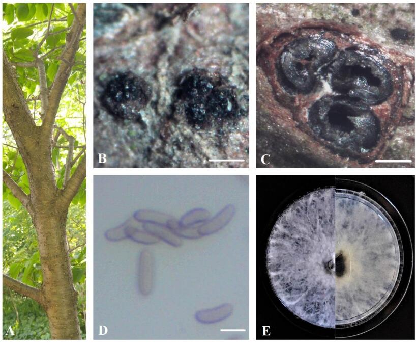
    Figure 1.  A Eutypa dieback symptoms on Syringa reticulata. B Stromata. C Transverse section through stroma. D Ascospores. E Seven-day-old culture on MEA. Scale bars: B = 0.5 mm, C = 100 μm, D = 5 μm.

    Index Fungorum number: IF 123583; Facesoffungi number: FoF 09536

    Sexual morph: Ascostromata 0.7-1.3 mm diam., black, superficial, subglobose or ellipsoidal, carbonaceous. Ascomata 120–350 μm diam., perithecial, black, clustered, semi-immersed, ovoid, with 2–4 locules. Asci 35–45 × 8–15 μm (n = 10), 8-spored, thin-walled, cylindric or slightly clavate, some rounded at apex. Ascospores 8.5–10 × 2–3.0 μm (x = 9.2 × 2.6 μm, n = 50), numerous, light brown to brown, allantoid, aseptate, slightly curved, smooth-walled, some with guttules.

    Culture characteristics – Colonies on MEA reach 5 cm diam. after 7 days of growth at room temperature in the dark, irregular, slightly raised, cottony, white, becoming beige, margin rough.

    Material examined – Canada, Ontario, Niagara Region: EI-13-ON, on dying branches of Fagus grandifolia, 43°11'59.9"N 79°13'37.6"W, 14.05.2020 (BILAS 51392); EI-21-ON, on dying twigs of Syringa reticulata, 43°13'12.3"N 79°13'35.7"W, 26.05.2020 (BILAS 51393); EI-109-ON, on dying branches of Fraxinus pennsylvanica, 43°05'55.2"N 79°16'15.5"W, 22.06.2020 (BILAS 51394).

  • The dataset included the representatives of the family Diatrypaceae and the strains obtained from the study. Based on BLASTn search of the NCBI database using ITS sequences for both EI-13-ON (MW145141) and EI-109-ON (MW145154), the top hits were E. vitis specimens collected in USA [GenBank KU320620; Identities = 579/581(99%), Gaps = 0/581(0%)], [GenBank AY462566; Identities = 579/581(99%), Gaps = 0/581(0%)], [GenBank FJ790854; Identities = 562/563(99%), Gaps = 0/563(0%)].

    The RAxML and Bayesian inference analysis showed the similar tree topologies. The tree with the highest log likelihood (-3331.95) is shown in Fig. 2. The new strains from the Niagara Region, Southern Ontario, Canada were grouped with E. vitis specimens from USA and formed a distinct clade with high bootstrap support values (ML/BP=95/1.00).

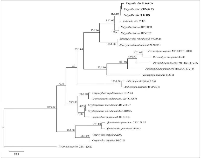
    Figure 2.  Phylogram generated from the best scoring RAxML tree based on ITS sequence data. Bootstrap support values for ML ≥ 60%, and Bayesian posterior probabilities (BP) ≥ 0.90 are given as ML/BP above or below nodes. Xylaria hypoxylon (CBS 122620) is used as outgroup. The specimens found in this study are in black bold.

  • Eutypa dieback in Canada on Vitis spp. have been previously detected in vineyards surveyed in Okanagan Valley, Vancouver and Gulf Islands and Lower Mainland during the 2011 growing season; the causal agents were identified as E. lata, Eutypa flavovirens and Eutypa laevata (O'Gorman et al. 2013). E. vitis was not found in Canada, however, this species was isolated from diseased grapevines (Vitis labrusca and V. vinifera) with the Eutypa dieback symptoms in several states of USA (Farr et al. 1989, Jordan & Schilder 2005, Vasilyeva & Stephenson 2006, Úrbez-Torres et al. 2009, Jayawadena et al. 2018). This study firstly reports E. vitis on multiply tree hosts (Fagus grandifolia, Fraxinus pennsylvanica and Syringa reticulata) in the Niagara region of Southern Ontario, Canada. The pathogen species identity was confirmed by both morphological identification and phylogenetic analyses of ITS gene region.

    According to many researchers, there are two main morphotypes within E. vitis: the first one has shorter ascospores, up to 10.5 µm long (Rappaz 1987), while the second one is characterized by larger ascospores with an average length of more than 10 µm, sometimes up to 14 µm long (Ellis & Everhart 1892, Glawe & Jacobs 1987, Vasilyeva & Stephenson 2006). Our isolates from Canada represent the morphotype with the shorter ascospores (8.5–10 µm). The low virulence of E. vitis in the USA was confirmed based on the results of the pathogenicity tests controlled in the laboratory over a 28-day period (Úrbez-Torres et al. 2009). It is known E. lata slowly colonizes wood while the fungus appears to be a highly virulent pathogen on grapevines (Moller & Kasimatis 1981). Therefore, we can assume that a pathogenicity of diatrypaceous fungi (including E. vitis) has been still underestimated. That can be a critical factor in the field conditions when trees are more likely to be stressed. More surveys are needed to find out if E. vitis has infected grapevines grown in the Niagara region of Southern Ontario. The ability of E. vitis to infect several alternate hosts can contribute to faster and wider spread of the fungus across vineyards in Canada.

Figure (2)  Table (1) References (31)
  • About this article
    Cite this article
    E Ilyukhin, T Bulgakov, S Markovskaja. 2021. First record of Eutypella vitis causing branch dieback on new host trees in Canada. Studies in Fungi 6(1):71−77 doi: 10.5943/sif/6/1/3
    E Ilyukhin, T Bulgakov, S Markovskaja. 2021. First record of Eutypella vitis causing branch dieback on new host trees in Canada. Studies in Fungi 6(1):71−77 doi: 10.5943/sif/6/1/3
  • Catalog

    • About this article

    /

    DownLoad:  Full-Size Img  PowerPoint
    Return
    Return