ARTICLE   Open Access    

Endophytic mycobiota of wild medicinal plants from New Valley Governorate, Egypt and quantitative assessment of their cell wall degrading enzymes

More Information
  • The present study isolated and identified 32 species of endophytic mycobiota belonging to 18 genera associated with 8 wild medicinal plants collected from El-Kharga Oasis, New Valley Governorate, Egypt. Fusarium was the most common genus followed by Alternaria and Aspergillus. Convolvulus arvensis was the plant with the highest number of endophytes over the other plant species, while Moringa oleifera reported the lowest number of endophytes. In addition, the entomopathogenic fungus Beauveria bassiana; was recorded for the first time from leaves of Portulaca oleracea. One hundred and twenty-three isolates representing 32 species were screened for their abilities to produce pectinase, carboxy methyl cellulase (CMCase) and avicellase enzymes on sucrose free-Cz supplemented, individually with 1% pectin or 1% CMC or 1% avicel as a sole carbon source, respectively. Ninety-four isolates produced pectinase while 66 isolates produced cellulases. The quantitative assays of the three enzymes for high-producers were performed in submerged fermentation using sucrose-free Cz broth. Aspergillus was the superior in the production of the three enzymes with the potent strains were A. terreus AUMC 14287 for CMCase (22.0 IU/ml/min) and avicellase (47.868 IU/ml/min) and A. terreus AUMC 14278 for pectinase (225.43 IU/ml/min).
  • Wild mushrooms have been consumed since ancient times due to their good taste and nutritional values. Also, mushrooms have the advantage of being rich in vital components such as proteins, vitamins, chitin, fibers, iron, zinc, selenium, sodium, etc. (Breene 1990, Chang & Miles 1992, Elkhateeb et al. 2019, 2020a, b, Daba et al. 2020, El-Hagrassi et al. 2020). Beside these nutritional values, many mushrooms have gained importance due to their therapeutical potentials (Bobek et al. 1995, Bobek & Galbavý 1999, Vaz et al. 2011, Khatun et al. 2012, Elkhateeb & Daba 2020, 2021a, b). The extracts of fruit bodies and mycelium obtained from various fungi have shown a crucial role in the prevention and/or the treatment of many diseases (Wasser & Weis 1999, Sharma et al. 2015). Nowadays, some immunomodulating molecules that have been obtained from different medicinal mushrooms have found great importance in improving the immune function in cancer patients while they are receiving chemo or radiotherapy (Mizuno 1995). Other edible mushrooms have gained medicinal importance due to their effectiveness in adjusting the blood pressure and reducing the free plasma cholesterol, as well as due to antimicrobial, antioxidant, and antitumor properties (Kabir & Kimura 1989, Smânia et al. 1995, Mau et al. 2005, Elkhateeb 2020, Thomas et al. 2020). The genus Ramaria is found all over the world, in which various species of Ramaria have been consumed by people for nutritional purposes. However, more studies are required to be conducted on these genera in order to detect the nutraceutical and nutritional compounds of these mushrooms. The genus Ramaria belonging to Basidiomycota, Class; Agaricomycetes, Order; Gomphales, Family; Gomphaceae, which includes approximately 200 species of coral fungi (Sharma et al. 2015). Several taxa are edible and picked in Europe and also they are easily confused with several mildly poisonous species capable of causing nausea, vomiting and diarrhea, in which among the most unusually shaped mushrooms of all, Ramaria are appropriately referred to as the coral mushrooms. Ramaria are recognizable by their variably thickly branched fruit bodies, fleshy and tough textures sometimes partly gelatinous or jellylike. Although many Ramaria species are very brightly colored, just as many are dull, and all have a tendency to become rather drab in age. Similar genera include Phaeoclavulina, Lentaria, Artomyces, and Ramariopsis, which differ in various aspects of color, consistency, substrate, microscopic features, or ecology (Sharma & Gautam 2017).

    Ramaria basidiocarps are either lignicolous or terricolous (white, yellow, orange, red, brilliant purple, brown and sometimes green hues) with many species forming a characteristic mycelial mat in the soil beneath the sporocarps (Nouhra et al. 2004). The colour may fade or change after collection in the field. Species of this genus often show a color change on bruising. The flesh may change to green (virescent), red brown (rubribrunnescent), brown to yellow brown (brunnescent), or a red wine color (vinescent) due to age or environmental conditions (Exeter et al. 2006).

    Clavaria is belonging to Basidiomycota, class Agaricomycetes, order Agaricales, and family Clavariaceae. Species of Clavaria produce basidiocarps (fruitbodies) that are cylindrical to club-shaped, branched and coral-like. They are often grouped with similar looking species from other genera, and they are collectively known as the clavarioid fungi (Olariaga et al. 2015).

    The most common species of this genus is Ramaria stricta (Kuo 2009), which grows on wood debris, which features branches that are usually strictly oriented, so that they are mostly straight and ascending. When fresh, its branch tips are yellow and its branches are dull yellowish buff, but its surfaces bruise and discolor purplish brown. Under the microscope it features roughened spores, clamp connections, and thick-walled hyphae. Several very similar species have been separated by mycologists, and the name Ramaria stricta should probably represent a group of potential species awaiting contemporary study. Ecology: Uncertain; while most ramarias are thought to be mycorrhizal, the wood-inhabiting species could be mycorrhizal or saprobic; growing from the dead (but sometimes buried) wood of conifers; appearing alone, scattered, or gregariously; early summer through fall; apparently widely distributed in North America, but more common from the Rocky Mountains westward. Fruitbody: 4−14 cm high; 4−10 cm wide; base well developed or nearly absent; branching repeatedly. Branches: Vertically oriented and elongated; often flattened; smooth; yellowish buff, becoming orangish buff as the spores mature; bruising and discoloring purplish brown; tips yellow when fresh and young. Base: Nearly absent, or fairly well developed; to 2 cm wide; white below; colored like the branches above; attached to numerous white rhizomorphs. Flesh: Whitish; fairly tough. Odor and Taste: Odor not distinctive, or sweet and fragrant; taste bitter. Spore color: Rusty yellowish. Microscopic Features: Spores are yellow-brown to rusty-brown in mass deposit 7.5−10.5 × 3.5−5µm; stretched-elliptical; smooth to roughened. Clamp connections present. Thick-walled hyphae present (Kuo 2009) (Figs 1, 2).

    Figure 1.  Fruitbody of Ramaria stricta in the field. The photograph was taken at the Sewickley Heights Borough Park, PA, USA by Fluffberger (https://www.inaturalist.org/observations/3596638). The photo was used under the CC BY-NC 4.0 non-commercial use license. Scale bar = 10 cm.
    Figure 2.  Fruitbody of Ramaria araiospora in the field. The photograph was taken at the Millersylvania State Park, Olympia, WA, USA by Yay4john (https://www.inaturalist.org/observations/62355314). The photo was used under the CC BY-NC 4.0 non-commercial use license. Scale bar = 10 cm.

    Few reports have been found about the discovery of these genera in the Northwestern Himalayas. The current surveys to Northwestern Himalayas have shown the existence of six species of coral mushrooms belonging to the genus Ramaria viz., R. aurea, R. botrytis, R. flava, R. flavescens, R. rubripermanens, and R. stricta (Sharma & Gautam 2017). Details about their culinary status were obtained from native inhabitants. Moreover, studies have been conducted on these species to get detailed biochemical profiling as well as exploring their various biological activities (Sharma & Gautam 2017).

    Coral mushrooms gain great therapeutical and nutritional importance due to their valuable components. Sharma & Gautam (2017), reported that 12 edible species of Ramaria occurring in Northwestern Himalayas contain proximal components, amino acids, and minerals, fatty acids. Ramaria aurea is an edible mushroom that is considered a promising dietary supplement as it consists of carbohydrates, amino acids, proteins, fibers, and minerals (Rai et al. 2013). Ramaria botrytis, another edible mushroom, consists of unsaturated fatty acids, tocopherol as well as other bioactive compounds (e.g. ascorbic acid, carotene, flavonoids, β- and lycopene) (Barros et al. 2008). Ramaria subalpina also contains significant amounts of ascorbic acid, β-carotene, flavonoids, and lycopene (Acharya et al. 2017). Also, some Ramaria species show the presence of alkaloids, saponins, terpenoids, coumarins, and cardiac glycosides (Aldred 2008, Dattaraj et al. 2020). Due to the existence of these bioactive molecules, these mushrooms possess various medicinal importance against various diseases. Among these medicinal benefits are antimicrobial, antiviral, anti-parasitic, antioxidant, radical scavenger, anticancer, anti-inflammatory, immune system enhancer, and anti-hyperlipidemia (Wasser 2017).

    Although there is a vast diversity of synthetic antibacterial compounds, the development of bacterial resistance has been substantially increasing (Alves et al. 2012). Thus, discovering novel antibiotics especially from natural sources is urgently required. Since ancient times, several natural resources have exerted potent antimicrobial activities. Among these natural resources are mushrooms that might be valuable alternative sources of novel antimicrobials (Gebreyohannes et al. 2019, Ghosh et al. 2020). In this context, various Ramaria sp. have gained great importance due to their potential antimicrobial activities against various pathogens (Table 1). The ethanolic extract of Ramaria flava was detected by the agar-well diffusion method, and the result showed positive activity against some Gram-positive bacteria such as Staphylococcus aureus ATCC 25923, Staphylococcus aureus Cowan I, Micrococcus luteus NRRL B-4375, Micrococcus flavus, Bacillus subtilis ATCC 6633, Bacillus cereus RSKK 863. Also, R. flava ethanolic extract showed potent activity against the tested Gram-negative bacteria including Salmonella enteritidis RSKK 171, Yersinia enterecolitica RSKK 1501, Klebsiella pneumoniae ATCC 27736 (Gezer et al. 2006). Additionally, Ramaria cystidiophora showed antimicrobial activity against Mycobacterium smegmatis (MIC = 8 µg/mL), and against Mycobacterium tuberculosis (MIC 64−128 µg/mL) (Hassan et al. 2019).

    Table 1.  Antimicrobial activities of various Ramaria species
    Ramaria species Tested extract Employed methods Tested bacteria Tested fungi References
    Gram-positive Gram-negative
    R. formosa Ethyl acetate, methanol, and water Percentage of inhibition B. subtili, S. aureus E. coli, K. pneumonia, Proteus vulgaris, P. aeruginosa Not tested Pala et al. 2019
    R. formosa Methanol Inhibition zone assay S. aureus P. aeruginosa Candida albicans Ramesh & Pattar 2010
    R. zippellii Ethanol, water 96-well microplate bioassay S. aureus E. coli No activity Bala et al. 2011
    R. aurea Ethanol Inhibition zone assay S. aureus E. coli, P. aeruginosa, P. vulgari Candida albicans Rai et al. 2013
    R. flava Ethanol Inhibition zone assay B. subtili, S. aureus E. coli Fusarium auenaceum, F. graminearum and Cercosporella albomaculans Liu et al. 2013
    R. flava Ethanol Agar-well diffusion method S. aureus, Micrococcus luteus, M. flavus, B. subtilis, B. cereus Salmonella enteritidis, Yersinia enterecolitica and K. pneumoniae No activity Gezer et al. 2006
    R. botrytis Ethanol Methanol Acetone Ethyl acetate Inhibition zone diameter S. aureus P. aeruginosa, E. coli, Enterobacter cloacae No activity Han et al. 2016
     | Show Table
    DownLoad: CSV

    The studies also reported that the ethanolic extract of Ramaria flava exhibited antifungal activities against three pathogenic fungi which are C. albomaculans, Fusarium graminearum and Fusarium auenaceum. The extract showed the highest efficacy against F. auenaceum, where 36.64% reduction in fungal growth was observed at the concentration of 2 mg/mL. The extract also inhibited the growth of F. graminearum and C. albomaculans by 19.99 and 30.03% respectively, at the same concentration of 2 mg/mL (Liu et al. 2013). Additionally, Ramaria mushrooms show their effectiveness to cure various viral infections. Zhang et al. (2015) isolated a novel ribonuclease from Ramaria formosa fruiting bodies and tested its antiviral activity against HIV-1 reverse transcriptase enzyme. The results showed that the ribonuclease showed 93% inhibition at a concentration of 30 µM (maximum tested concentration) and with an IC50 value of 3 µM. It was interesting to report that this enzyme exhibits unique features; these include its unique N-terminal sequences, optimum acidic pH value, and temperature resistance. Taken together these features suggest that this ribonuclease could play a vital role in HIV diseases prevention.

    Many reports regarding the antioxidative properties of various Ramaria sp. have been published, which showed the activity of these fungi as effective antioxidants (Table 2). Linoleic acid oxidation was compared with those of α- tocopherol, BHA, and R. flava ethanol extract. The results revealed that the inhibition values of both the standards and R. flava ethanol extract increased gradually with increasing the concentrations. 80 µg/ml BHA, α -tocopherol, and R. flava ethanolic extract exhibited 96.4, 98.6, and 73.3% inhibition, respectively. Whereas increasing the concentrations to 160 µg/ml concentrations resulted in 98.9, 99.2 and 94.7% inhibition, respectively (Gezer et al. 2006). Also, the ethanolic extract of that mushroom exhibited promising OH and DPPH radical-scavenging activities with low IC50 values of 18.08 and 5.86 μg/mL, respectively (Liu et al. 2013). The high inhibition value of R. flava ethanolic extract could be related to the high concentration of phenolic compounds (Komali et al. 1999).

    Table 2.  Antioxidant activities of various Ramaria species isolated from different places
    Ramaria species Place of collection Methods used Activity (EC 50 % of inhibition) References
    R. flava Turkey DPPH scavenging assay 94.78% at 12 mg/mL Gursoy et al. 2010
    β-carotene linoleic acid assay 95.02% at 20 mg/mL
    Reducing power 95.02% at 20 mg/mL
    Metal chelating effect 96.75 at 2 mg/mL
    R. flava Turkey DPPH scavenging assay 276 µg/mL Gezer et al. 2006
    β-carotene linoleic acid assay 94.7% at 160 µg/mL
    R. formosa India DPPH radical scavenging activity 5.8 mg/ml Ramesh & Pattar 2010
    R. patagonica Argentina DPPH scavenging assay 770 µg/mL Toledo et al. 2016
    β-carotene linoleic acid assay 610 µg/mL
    Reducing power 170 µg/mL
    TBARS inhibition activity 60 µg/mL
    R. Formosa Korea DPPH scavenging assay 117 AsA/mg/mL at 500 µg/mL Kim et al. 2016
    Reducing power 36% copper ion inhibition at 20 µg/mL concentration
    Peroxyl radical scavenging activity 7.8 µM trolox equivalent at 20 µg/mL
    R. stricta Ukraine Total antioxidant status 4.223±0.054 mmol/l Krupodorova & Sevindik 2020
    Oxidative stress index 0.194±0.001
    Total oxidant status 8.201 ± 0.095 μmol/l
     | Show Table
    DownLoad: CSV

    ABTS assay is another assay employed to evaluate the free radical scavenging ability resulted from hydrogen-donating ability (Re et al. 1999). A study reported that R. largentii extract was able to scavenge ABTS radical cation in a concentration-dependent manner. At an extract concentration of 250 mg/mL the scavenging activity was recorded to be 93.53 ± 0.03% (Aprotosoaie et al. 2017). A polyphenol-rich extract of R. aurea exhibited superoxide and DPPH radicals scavenging ability with EC50 of about 0.283 and 0.384 mg/mL, respectively (Khatua et al. 2015). Thus, Ramaria species extract seems to be a reasonably free radical scavenger agent.

    Cancer is one of the main causes of death worldwide. Thus, finding new molecules to prevent and/or treat cancer, especially from natural sources with lower toxicity, is considered a goal for scientists nowadays (Wang et al. 2012). As mentioned previously, mushrooms are sources of powerful pharmaceutical products. About 651 higher basidiomycetes species exhibit antitumor activities (Fan et al. 2006). In general, fungal anticancer substances are divided into two main groups which are low and high molecular-weight compounds. The low-molecular-weight secondary compounds comprise mainly sesquiterpenes, steroids and sterols, triterpenes, and polyketides (Mahajna et al. 2008). These molecules can penetrate the cell membrane and work on the targeted signal-transduction pathways (Zaidman et al. 2005). However, the high-molecular-weight compounds include polysaccharides or protein-bound polysaccharides (Kidd 2000). In 1982, a study reported by Yoo et al. (1982), examined the antitumor activity of a high molecular weight protein-bound polysaccharide fraction obtained from Ramaria formosa. These molecules were able to inhibit 66% of a tumor when administered at the dosage of 50 mg/kg/day and the tumor was wholly degenerated in two out of eight mice.

    The growth inhibitory effect of the ethanol extract obtained from Ramaria flava was tested on human cancer cell lines (BGC-803, NCI-H520, and MDA-MB-231) and the results showed a promising inhibition activity on the three human cancer cells with inhibition percentages of 33.83%, 54.63%, and 71.66% respectively, at mushroom extract concentration of 200 µg/mL. A major antitumor sterol found in many edible mushrooms is ergosterol peroxide is (Lindequist et al. 2005). Previous studies reported the antitumor properties of ergosterol peroxide in various cancer cells including SCC4 (head and neck squamous cell carcinoma), U266 (multiple myeloma), DU145 (prostate cancer), as well as MDA-MB-231 (breast cancer) cells (Rhee et al. 2012). Ergosterol peroxide inhibited cell growth and induced apoptosis in human prostate cancer cells DU-145 and LNCaP (Russo et al. 2010). Moreover, ergosterol peroxide showed a cytotoxic effect against Hep 3B cells (Chen et al. 2009). Six sterols comprising ergosterol peroxide were isolated from R. flava (Liu et al. 2012). And interestingly, their ethanolic extract exerted promising growth inhibitory activity on MDA-MB-231(human breast cancer cell line) which suggests that R. flava could find a great application as an antitumor agent.

    To date, a few numbers of other biological activities have been determined in Ramaria species. In a previous study, the hepatoprotective activity of methanol extract of R. botrytis towards liver toxicity induced by benzo(α)pyrene in mice was evaluated (Kim & Lee 2003). The results reported that the methanolic extract significantly reduced the elevated enzyme activities including glutathione S-transferase and r-glutamylcysteine synthetase that resulted from the induction of benzo(α)pyrene. The hepatoprotective activity of this mushroom could be related to its high antioxidant potential which was revealed by the low EC50 value (0.109 mg/ml) for DPPH radical scavenging assay. Ramaria botrytis collected from hilly areas of Darjeeling exerted potential immunostimulatory effect in a murine macrophage cell line (RAW264.7 cell) as well as in thymocyte cells and splenocytes. This activity was related to a water-soluble glucan obtained from fresh fruiting bodies of such mushrooms and which consisted of (1→6)-linked-β-D glucopyranosyl residues with the branching of (1→3)-linked-β-D-glucopyranosyl at O-3 position. This glucan enhanced the nitric oxide level and also resulted in the stimulation of thymocyte and splenocyte proliferation rates. Thus, this glucan can be employed as a potent immunostimulatory (Bhanja et al. 2013).

    Ramarin A and B are two novel sesquiterpene derivatives that were obtained and purified from the methanol extract of Ramaria formosa that is a very rare mushroom and that inhibits human neutrophil elastase, and thus can be employed for the treatment of skin aging (Kim et al. 2016). A recent study reported by Bhanja et al. (2020), prepared bio metallic composite nanoparticles from polysaccharides isolated from Ramaria botrytis. The nanoparticles exerted antibacterial activity against Pseudomonas aeruginosa. Moreover, these nanoparticles showed potential antioxidant activities towards DPPH radical, hydrogen peroxide, and nitric oxide radicals. They also catalyzed the p-nitrophenol reduction, indicating a new direction in the field of biomedicine mediated by nanotechnology (Bhanja et al. 2020).

    All Clavaria species are terrestrial and most are believed to be saprotrophic (Decaying dead plant material). In North America and elsewhere, they are more commonly found in woodlands. Clavaria fruit bodies are simple (cylindrical to club-shaped) or more rarely branched, sometimes with a distinct stem. Several of the species with simple fruit bodies form them in dense clusters. The fruit bodies themselves are smooth to grooved and typically brittle. Depending on species, they vary in color from white or cream to yellow, pink, violet, brown, or black. The hyphal system of Clavaria species is always monomitic. The context hyphae are inflated, thin-walled, and lack clamp connections (Olariaga et al. 2015). The basidia are two to four spored, in some species with an open, loop-like clamp connection at the base. Spores are smooth or spiny and color is white. Most Clavaria species are thought to be saprotrophic, decomposing leaf litter and other organic materials on the woodland floor. In Europe, species are more frequently found in old, unimproved grasslands, where they are presumed to be decomposers of dead grass and moss. Species of Clavaria occur in suitable habitats throughout the temperate regions and the tropics (Figs 3, 4) (Acharya 2012, Kautmanová et al. 2012).

    Figure 3.  Fruitbody of Clavaria zollingeri in the field. The photograph was taken at Ohio, USA by Rcurtis (https://www.inaturalist.org/observations/1693040). The photo was used under the CC BY-NC 4.0 non-commercial use license. Scale bar = 10 cm.
    Figure 4.  Fruitbodies of Clavaria rubicundula in the field. The photograph was taken at Hewitt, West Milford, NJ 07421, USA by Tombigelow (https://www.inaturalist.org/observations/16044756). The photo was used under the CC BY-NC 4.0 non-commercial use license. Scale bar = 10 cm.

    Ramaria and Clavaria are the two major genera of coral mushrooms within families Gomphaceae and Clavariaceae, respectively. Besides having important role in forest ecology, some species of these are reported to possess high nutraceutical and bioactive potential (Sharma & Gautam 2017). Many current studies describe the detailed biochemical profiling and antioxidant, and antibacterial activities of twelve coral mushroom (Ramaria and Clavaria) species (Vidović et al. 2014). Antioxidant activities were calculated using EC50 values from mushroom extracts. Antibacterial activities were checked on six pathogenic bacterial strains through minimum inhibition concentrations. All the species were found to be rich in protein, macro and micro minerals, carbohydrates, unsaturated fatty acids, essential amino acids, phenolics, tocopherols, anthocynadins and carotenoids. All the species showed significant antioxidant and antibacterial activities. These species are reported to free from heavy toxic metals. Sharma & Gautam (2017), reported that these Ramaria and Clavaria species will open the way for their large-scale commercial exploitations and use in pharmaceutical industries as antioxidant, antibacterial and nutraceutical constituents.

    Many studies conducted on Ramaria and Clavaria species are represented in the current review and showed that coral mushrooms exhibit the potential as a vital therapeutic food. However, more studies for profound exploration are still required. Ramaria and Clavaria species exerted some vital biological activities such as antimicrobial, antioxidant, antitumor, hepatoprotective, and anti-skin aging activity, etc. Further investigation is needed to explain the different mechanisms of action of these wild mushrooms. Although the nutritional values of the genus Ramaria and Clavaria have been also studied, there is a lack of information about their bioavailability, vitamin and mineral, and compositions. Thus, the current review recommends further exploration to get a full profile of the active components obtained from various Ramaria species and Clavaria species in nutrition and mycomedicine fields.

    This research does not receive any external funding.

    The authors declare no conflict of interest.

  • Al-Bedak OA, Abdel-Sater MA, Abdel-Latif AMA, Abdel-Wahab D. 2020 – Aspergillus creber and A. keveii, two new records as endophytes from wild medicinal plants in Egypt. Journal of Multidisciplinary Sciences 2, 1–9.

    Google Scholar

    Al-Bedak OA, Sayed RM, Hassan SH. 2019 – A new low-cost method for long-term preservation of filamentous fungi. Biocatalysis and Agricultural Biotechnology 22, 101417. doi: 10.1016/j.bcab.2019.101417

    CrossRef    Google Scholar

    Al-Snafi A. 2015 – The chemical contents and pharmacological effects of Anagallis arvensis-A review. International Journal of Pharmacy 5, 37–41.

    Google Scholar

    Anwar F, Latif S, Ashraf M, Gilani AH. 2007 – Moringa oleifera: a food plant with multiple medicinal uses. Phytotherapy Research: An International Journal Devoted to Pharmacological and Toxicological Evaluation of Natural Product Derivatives 21, 17–25.

    Google Scholar

    Bailey MJ, Biely P, Poutanen K. 1992 – Interlaboratory testing of methods for assay of xylanase activity. Journal of biotechnology 23, 257–270. doi: 10.1016/0168-1656(92)90074-J

    CrossRef    Google Scholar

    Booth C. 1971 – The genus Fusarium. Commonwealth Mycological Institute. Kew, Surrey 237 p.

    Google Scholar

    Carbungco E, Pedroche N, Panes V, De la Cruz T. 2015 – Identification and characterization of endophytic fungi associated with the leaves of Moringa oleifera Lam, I International Symposium on Moringa 1158, 373–380.

    Google Scholar

    Carroll G, Petrini O. 1983 – Patterns of substrate utilization by some fungal endophytes from coniferous foliage. Mycologia 75, 53–63. doi: 10.1080/00275514.1983.12021637

    CrossRef    Google Scholar

    Choi Y, Hodgkiss I, Hyde K. 2005 – Enzyme production by endophytes of Brucea javanica. J Agric Technol 1, 55–66.

    Google Scholar

    De Aldana BRV, Bills G, Zabalgogeazcoa I. 2013 – Are endophytes an important link between airborne spores and allergen exposure? Fungal Diversity 60, 33–42. doi: 10.1007/s13225-013-0223-z

    CrossRef    Google Scholar

    de Hoog GS, Guarro J, Gené J, Figueras M. 2000 – Atlas of clinical fungi. Centraalbureau voor Schimmelcultures (CBS) 1126 p.

    Google Scholar

    Domsch K, Gams W, Anderson T. 2007– Compendium of soil fungi. 1–672. IHW-Verlag, Eching, Germany 672 p.

    Google Scholar

    Edor SP, Edogbanya OP, Kutshik JR. 2018 – Cellulase activity of Aspergillus niger in the biodegradation of rice husk. MOJ Biology and Medicine 3(3), 49–51

    Google Scholar

    Ellis M. 1976 – More Dematiaceous Hyphomycetes., (Commonwealth Mycological Institute: Kew, Surrey) 507 p.

    Google Scholar

    Gessner R. 1980 – Degradative enzyme production by salt-marsh fungi. Botanica Marina 23, 133–139.

    Google Scholar

    Gherbawy Y, Gashgari R. 2014 – Molecular characterization of fungal endophytes from Calotropis procera plants in Taif region (Saudi Arabia) and their antifungal activities. Plant Biosystems-An International Journal Dealing with all Aspects of Plant Biology 148, 1085–1092. doi: 10.1080/11263504.2013.819043

    CrossRef    Google Scholar

    Hamayun M, Khan SA, Khan AL, Rehman G et al. 2010 – Gibberellin production and plant growth promotion from pure cultures of Cladosporium sp. MH-6 isolated from cucumber (Cucumis sativus L. ). Mycologia 102, 989–995. doi: 10.3852/09-261

    CrossRef    Google Scholar

    Ismail MA, Moubasher AH, Mohamed RA, Al-Bedak OA. 2017 – Extremophilic fungi and chemical analysis of hypersaline, alkaline lakes of Wadi-El-Natrun, Egypt. International Journal of Technical Research and Science 1, 345–363.

    Google Scholar

    Ismail MA, Moubasher AH, Mohamed RA, Al-Beddak OA. 2018 – Agro–industrial residues as alternative sources for cellulases and xylanases production and purification of xylanase produced by Aspergillus flavus AUMC 10331 isolated from extreme habitat. Current Research in Environmental & Applied Mycology 8, 313–322.

    Google Scholar

    Jalis H, Ahmad A, Khan S, Sohail M. 2014 – Utilization of apple peels for the production of plant cell-wall degrading enzymes by Aspergillus fumigatus MS16. J Anim Plant Sci 24, 64–67.

    Google Scholar

    Kaur M, Kalia A. 2012 – Convolvulus arvensis: A useful weed. Int J Pharm Pharm Sci 4, 38–40.

    Google Scholar

    KC S, Upadhyaya J, Joshi DR, Lekhak B et al. 2020 – Production, Characterization, and Industrial Application of Pectinase Enzyme Isolated from Fungal Strains. Fermentation 6(59), 2–10.

    Google Scholar

    Khairnar AK, Bhamare S, Bhamare H. 2012 – Calotropis procera: an ethnopharmacological update. Advance Research in Pharmaceuticals and Biologicals 2, 142–156.

    Google Scholar

    Khan AL, Waqas M, Hussain J, Al-Harrasi A, Lee I-J. 2014 – Fungal endophyte Penicillium janthinellum LK5 can reduce cadmium toxicity in Solanum lycopersicum (Sitiens and Rhe). Biology and fertility of soils 50, 75–85. doi: 10.1007/s00374-013-0833-3

    CrossRef    Google Scholar

    Khan Marwat S, Khan EA, Baloch MS, Sadiq M et al. 2017 – Ricinus cmmunis: Ethnomedicinal uses and pharmacological activities. Pakistan journal of pharmaceutical sciences 30(5), 1815–1827.

    Google Scholar

    Khan R, Shahzad S, Choudhary MI, Khan SA, Ahmad A. 2007 – Biodiversity of the endophytic fungi isolated from Calotropis procera (Ait. ) R. Br. Pakistan Journal of Botany 39, e2239.

    Google Scholar

    Krishnamurthy YL, Naik SB, Jayaram S. 2008 – Fungal communities in herbaceous medicinal plants from the Malnad region, Southern India. Microbes and environments 23, 24–28. doi: 10.1264/jsme2.23.24

    CrossRef    Google Scholar

    Kuhad RC, Gupta R, Singh A. 2011 – Microbial cellulases and their industrial applications. Enzyme research 2011, 1–10.

    Google Scholar

    Leslie J, Summerell B. 2006 – The Fusarium Laboratory Manual Blackwell Publishing. Ames, Iowa 382 p.

    Google Scholar

    Li J-X, Zhang F, Jiang D-D, Li J et al. 2020 – Diversity of Cellulase-Producing Filamentous Fungi From Tibet and Transcriptomic Analysis of a Superior Cellulase Producer Trichoderma harzianum LZ117. Frontiers in Microbiology 11, 1617. doi: 10.3389/fmicb.2020.01617

    CrossRef    Google Scholar

    Miller GL. 1959 – Use of dinitrosalicylic acid reagent for determination of reducing sugar. Analytical chemistry 31, 426–428. doi: 10.1021/ac60147a030

    CrossRef    Google Scholar

    Moubasher AH. 1993 – Soil fungi in Qatar and other Arab countries. The Centre for Scientific and Applied Research, University of Qatar 568 p.

    Google Scholar

    Moubasher AH, Ismail MA, Mohamed RA, Al-Bedak OA. 2016 – Xylanase and cellulase production under extreme conditions in submerged fermentation by some fungi isolated from hypersaline, alkaline lakes of Wadi-El-Natrun, Egypt. Journal of Basic & Applied Mycology (Egypt) 7, 19–32.

    Google Scholar

    Muhammad G, Hussain MA, Anwar F, Ashraf M, Gilani AH. 2015 – Alhagi: a plant genus rich in bioactives for pharmaceuticals. Phytotherapy research 29, 1–13. doi: 10.1002/ptr.5222

    CrossRef    Google Scholar

    Oumer OJ. 2017 – Pectinase: substrate, production and their biotechnological applications. International Journal of Environment, Agriculture and Biotechnology 2, 238761.

    Google Scholar

    Oumer OJ, Abate D. 2018 – Screening and molecular identification of pectinase producing microbes from coffee pulp. BioMed research international 2018, 1–7.

    Google Scholar

    Patil MG, Pagare J, Patil SN, Sidhu AK. 2015 – Extracellular enzymatic activities of endophytic fungi isolated from various medicinal plants. Int J Curr Microbiol App Sci 4, 1035–1042.

    Google Scholar

    Petrini O. 1991 – Fungal Endophytes of Tree Leaves. In: Andrews J.H., Hirano S.S. (eds) Microbial Ecology of Leaves. Brock/Springer Series in Contemporary Bioscience. Springer, New York, NY. 179–197.

    Google Scholar

    Pitt JI. 1979 – The genus Penicillium and its teleomorphic states Eupenicillium and Talaromyces.

    Google Scholar

    Prabavathy D, Valli NC. 2012 – Screening for extracellular enzymes and production of cellulase by an endophytic Aspergillus sp., using cauliflower stalk as substrate. International Journal on Applied Bioengineering 6(2), 40–45.

    Google Scholar

    Raviraja N. 2005 – Fungal endophytes in five medicinal plant species from Kudremukh Range, Western Ghats of India. Journal of Basic Microbiology: An International Journal on Biochemistry, Physiology, Genetics, Morphology, and Ecology of Microorganisms 45, 230–235.

    Google Scholar

    Rodriguez R, White Jr J, Arnold AE, Redman aRa. 2009 – Fungal endophytes: diversity and functional roles. New phytologist 182, 314–330. doi: 10.1111/j.1469-8137.2009.02773.x

    CrossRef    Google Scholar

    Samson R, Hoekstra E, Frisvad J, Filtenborg O. 2004 – Introduction to food and airborne fungi 7th ed. Utrecht: Centraal Bureau voor Schimmelcultures 389 p.

    Google Scholar

    Schardl CL, Leuchtmann A, Spiering MJ. 2004 – Symbioses of grasses with seedborne fungal endophytes. Annu. Rev. Plant Biol. 55, 315–340. doi: 10.1146/annurev.arplant.55.031903.141735

    CrossRef    Google Scholar

    Shubha J, Srinivas C. 2017 – Diversity and extracellular enzymes of endophytic fungi associated with Cymbidium aloifolium L. African Journal of Biotechnology 16, 2248–2258.

    Google Scholar

    Simmons EG. 2007 – Alternaria: An Indentification Manual. Utrecht, The Netherlands: CBS Fungal Biodiversity Centre. 775 p.

    Google Scholar

    Singh LP, Gill SS, Tuteja N. 2011 – Unraveling the role of fungal symbionts in plant abiotic stress tolerance. Plant signaling & behavior 6, 175–191.

    Google Scholar

    Sopalun K, Iamtham S. 2020 – Isolation and screening of extracellular enzymatic activity of endophytic fungi isolated from Thai orchids. South African Journal of Botany (2020), 1–7.

    Google Scholar

    Strobel G, Daisy B, Castillo U, Harper J. 2004 – Natural products from endophytic microorganisms. Journal of Natural products 67, 257–268. doi: 10.1021/np030397v

    CrossRef    Google Scholar

    Sunitha V, Nirmala Devi D, Srinivas C. 2013 – Extracellular enzymatic activity of endophytic fungal strains isolated from medicinal plants. World Journal of Agricultural Sciences 9, 1–9.

    Google Scholar

    Syed S, Fatima N, Kabeer G. 2016 – Portulaca oleracea L. : a mini review on phytochemistry and pharmacology. International journal of Biology and Biotechnology 13, 637–641.

    Google Scholar

    Xue Y, Han J, Li Y, Liu J et al. 2020 – Promoting cellulase and hemicellulase production from Trichoderma orientalis EU7-22 by overexpression of transcription factors Xyr1 and Ace3. Bioresource Technology 296, 122355. doi: 10.1016/j.biortech.2019.122355

    CrossRef    Google Scholar

    Zou W, Meng J, Lu H, Chen G et al. 2000 – Metabolites of Colletotrichum gloeosporioides, an endophytic fungus in Artemisia mongolica. Journal of Natural Products 63, 1529–1530. doi: 10.1021/np000204t

    CrossRef    Google Scholar

  • Cite this article

    MA Abdel-Sater, AMA Abdel-Latif, DA Abdel-Wahab, OA Al-Bedak. 2021. Endophytic mycobiota of wild medicinal plants from New Valley Governorate, Egypt and quantitative assessment of their cell wall degrading enzymes. Studies in Fungi 6(1):78−91 doi: 10.5943/sif/6/1/4
    MA Abdel-Sater, AMA Abdel-Latif, DA Abdel-Wahab, OA Al-Bedak. 2021. Endophytic mycobiota of wild medicinal plants from New Valley Governorate, Egypt and quantitative assessment of their cell wall degrading enzymes. Studies in Fungi 6(1):78−91 doi: 10.5943/sif/6/1/4

Figures(2)  /  Tables(5)

Article Metrics

Article views(3804) PDF downloads(590)

ARTICLE   Open Access    

Endophytic mycobiota of wild medicinal plants from New Valley Governorate, Egypt and quantitative assessment of their cell wall degrading enzymes

Studies in Fungi  6 Article number: 4  (2021)  |  Cite this article

Abstract: The present study isolated and identified 32 species of endophytic mycobiota belonging to 18 genera associated with 8 wild medicinal plants collected from El-Kharga Oasis, New Valley Governorate, Egypt. Fusarium was the most common genus followed by Alternaria and Aspergillus. Convolvulus arvensis was the plant with the highest number of endophytes over the other plant species, while Moringa oleifera reported the lowest number of endophytes. In addition, the entomopathogenic fungus Beauveria bassiana; was recorded for the first time from leaves of Portulaca oleracea. One hundred and twenty-three isolates representing 32 species were screened for their abilities to produce pectinase, carboxy methyl cellulase (CMCase) and avicellase enzymes on sucrose free-Cz supplemented, individually with 1% pectin or 1% CMC or 1% avicel as a sole carbon source, respectively. Ninety-four isolates produced pectinase while 66 isolates produced cellulases. The quantitative assays of the three enzymes for high-producers were performed in submerged fermentation using sucrose-free Cz broth. Aspergillus was the superior in the production of the three enzymes with the potent strains were A. terreus AUMC 14287 for CMCase (22.0 IU/ml/min) and avicellase (47.868 IU/ml/min) and A. terreus AUMC 14278 for pectinase (225.43 IU/ml/min).

  • Endophytic microorganisms colonize in plant tissues in which they spend part or all their life cycle without causing disease symptoms in the host (Petrini 1991). Fungal endophytes may inhabit in different organs of the host including leaves, stems, bark, roots, fruits, flowers and seeds (Rodriguez et al. 2009). Generally, in this symbiotic relationship fungal endophytes receive shelter and nutrients from the host, while the host plant may benefit from an array of attributes which include protection against natural enemies such as pathogens and herbivores (Schardl et al. 2004, Singh et al. 2011), plant growth promotion (Hamayun et al. 2010) and increasing the resistance of plants to abiotic stresses such as salinity and heavy metal toxicity in soil (Khan et al. 2014). Some medicinal plants are known for harboring endophytic fungi, which are important sources of various bioactive secondary metabolites and enzymes valuable for the pharmaceutical industry (Zou et al. 2000, Strobel et al. 2004, Krishnamurthy et al. 2008).

    Endophytic fungi are relatively unexplored producers of metabolites useful in pharmaceutical and agricultural industries. A single endophyte can produce several bioactive metabolites. As a result, the role of endophytes in the production of various natural products with greater bioactivity have received increased attention (Prabavathy & Valli 2012). Pectinases and cellulases, besides other enzymes, are the most important enzymes produced by endophytic fungi as a resistance mechanism against pathogenic invasion and to obtain nutrition from the host. These enzymes have various industrial applications, thus of major interest. Increasing efforts are being taken to characterize and identify endophytic fungi from medicinal plants. Therefore, the present work was designed to study the biodiversity of endophytic fungi in some wild medicinal plants from the New Valley Governorate, Egypt, and to evaluate their ability to produce extracellular pectinases and cellulases.

  • The New Valley Governorate is located at the Western Desert of Egypt. It encompasses 440, 098 km2, which is approximately 44% of the total area of Egypt and 66% of the area of Western Sahara. It is demarcated by the Governorates of Minya, Assiut, Sohag, Qena and Aswan from the east, by Libya and the Governorates of Matrouh and the Marine Oasis of the 6th of October City from the West and by Sudan from the South. The New Valley includes four large Oases namely El-Kharga (the sampling sites), El-Dakhla, El-Bahariya and El-Farafra, and the capital is El-Kharga (Fig. 1).

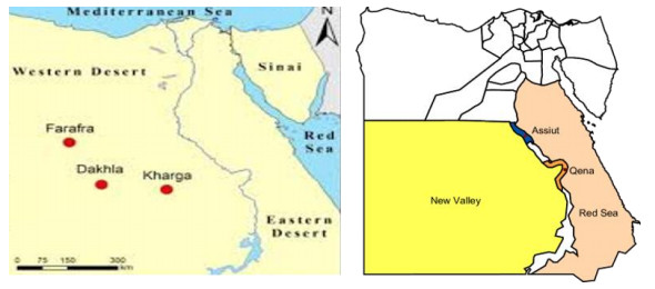
    Figure 1.  Location of the New Valley Governorate showing study site.

  • Healthy and mature plant leaves and roots of eight wild medicinal plants were collected from El-Kharga Oasis, New Valley Governorate once during April 2018. Ten replicates from each of Alhagi graecorum, Anagallis arvensis, Calotropis procera, Chenopodium ambrosioides, Convolvulus arvensis, Moringa oleifera, Portulaca oleracea, and Ricinus communis plants were collected in sterile polyethylene bags and promptly brought to the laboratory for isolation of fungi. The plant species collected in the current investigation were identified according to morphological features and taxonomical characters at the Assiut University Herbarium, Department of Botany and Microbiology, Faculty of Science, Assiut University, Assiut, Egypt (Fig. 2).

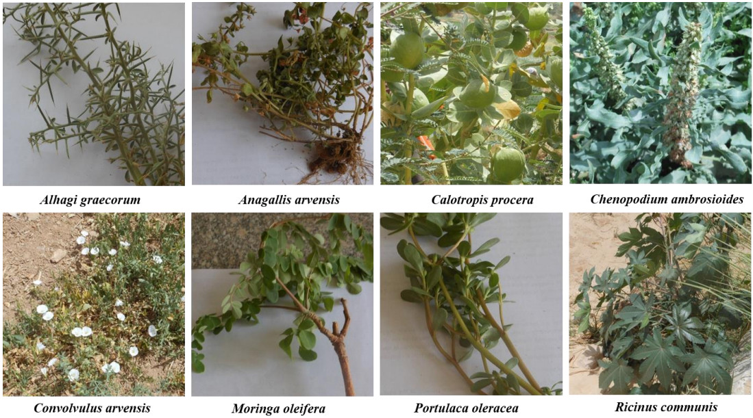
    Figure 2.  Wild medicinal plant species collected from El-Kharga Oasis, the New Valley Governorate, Egypt, during April 2018.

  • Prior to surface sterilization, leaves and roots of each sample were thoroughly washed with tap water to remove the dust followed by distilled water. The samples were then cut into 5-cm segments. The samples were surface sterilized using the following sequence; 5% sodium hypochlorite for 3 min, 70% ethanol for 1 min, and washing with sterile distilled water 3 times each for 1 min. In aseptic conditions, both ends of each segment (1 cm) was cut off to produce a 3-cm segments (Al-Bedak et al. 2020).

  • Segments of each sample were plated on Petri-dishes containing 1% glucose-Cz with the following composition (g/l): Glucose, 10; Na2NO3, 2; K2HPO4, 1; KCl, 0.5; MgSO4.7H2O, 0.5; FeSO4, 0.01; ZnSO4, 0.01; CuSO4, 0.005; Rose Bengal, 0.05; chloramphenicol, 0.25; agar, 15 and the final pH 7.3 (Ismail et al. 2017). The plates were incubated for 7-21 days at 25℃. Counts of CFUs of each fungal isolate were calculated per 25 segments in every sample. The obtained fungi were identified morphologically to the species level at the Assiut University Mycological Centre according to their macroscopic and microscopic characteristics. Pure cultures of the fungal strains were preserved for further investigations on PDA slants, as well as on cotton balls (Al-Bedak et al. 2019) at 4℃ in the culture collection of the Assiut University Mycological Centre.

  • The obtained fungi in this study were identified morphologically to the species level at the Assiut University Mycological Centre according to their macroscopic and microscopic characteristics. The following references were used for the identification of fungal genera and species (purely morphologically, based on macroscopic and microscopic features): Booth (1971), Ellis (1976), Pitt (1979), Domsch et al. (2007), Moubasher (1993), de Hoog et al. (2000), Samson et al. (2004), Leslie & Summerell (2006), Simmons (2007) and Al-Bedak et al. (2020).

  • Production of pectinase and endoglucanase was detected on sucrose-free Czapek's agar medium amended with pectin (from citrus peel) and CMC as a sole carbon source, respectively. 50 µl of spore suspension from 7-day-old culture of each fungal strain was individually added to each 5-mm diameter well on the agar plate (Moubasher et al. 2016). The inoculated plates were incubated for 2 days at 30ºC. The clear zones formed around the wells were more visible when the plates were flooded with 0.25% (w/v) aqueous iodine solution. The diameters of the clear zones were measured (in mm) against the brown color of the test medium indicating enzyme production.

  • All positive fungal strains were grown, individually in 250-ml Erlenmeyer conical flasks each containing 50 ml sucrose-free Czapek's broth medium supplemented with 1% pectin or 1% CMC as sole carbon source. The flasks were then inoculated individually with 1 ml spore suspension containing 1 x 10⁷ spore/ml of 7-day-old culture of the tested strains. The inoculated flasks were then incubated at 30ºC in shaking condition of 150 rpm for 7 days.

  • After incubation period, the flasks contents were individually filtered through filter papers (Whatman No. 1) and the filtrate was then centrifuged at 10000 xg for 10 min at 4ºC. The clear supernatants were used as a source for CMCase or pectinase enzyme.

  • The enzyme production was determined by mixing 0.9 ml of 1% pectin (prepared in 50 mM Na-citrate buffer, pH 5.0) with 0.1 ml of filtered crude enzyme, and the mixture was incubated at 50℃ for 15 min in a water bath (Bailey et al. 1992). The reaction was stopped by the addition of 2 ml of 3, 5-dinitrosalicylic acid (DNS) and the contents were boiled in water bath for 10 min (Miller 1959). After cooling, absorbance was measured at 540 nm using Cary 60 UV-Vis spectrophotometer. The amount of reducing sugar liberated was quantified using calibration curve of glucose. One unit of pectinase is defined as the amount of enzyme that liberates 1 µmol of glucose equivalents per minute under the standard assay conditions.

  • The cellulases activity was determined by mixing 0.9 ml of 1% CMC or 1% avicel (prepared in 50 mM Na-citrate buffer, pH 5.0) with 0.1 ml of filtered crude enzyme, and the mixture was incubated at 50℃ for 15 min in a water bath (Bailey et al. 1992). The reaction was stopped by the addition of 2 ml of 3, 5-dinitrosalicylic acid (DNS) and the contents were boiled in water bath for 10 min (Miller 1959). After cooling, absorbance of the developed color was measured at 540 nm using Cary 60 UV-Vis spectrophotometer. The amount of reducing sugar liberated was quantified using calibration curve of glucose. One unit of CMCase or avicellase is defined as the amount of enzyme that liberates 1 µmol of glucose equivalents per minute under the standard assay conditions. Glucose concentration was calculated using the calibration curve.

    Glucoseconcentration= Absorbance  slope (=1.0472)mg/ml(=g/L)Enzymeconcentration= Glucose concentration (g/L)0.00018IU/L

    The enzyme activity (pectinase or CMCase or avicellase) was calculated according to the following formula (Moubasher et al. 2016)

    Enzymeactivity=AbsorbancexDFx(1x)(1y)(1t)(1slope)

    Where: DF = the dilution factor for enzyme, x = the volume of enzyme used, y = the volume of hydrolysate used for assay of reducing sugars, t = the time of hydrolysis, slope is determined from a standard curve

  • A total of 32 species related to 18 genera of endophytic mycobiota were recovered on 1% glucose-Cz at 25℃ from healthy and mature plant leaves and roots of eight wild medicinal plants, collected from El-Kharga Oasis, the New Valley Governorate. The high incidence in genera were recorded in Alternaria, Aspergillus and Fusarium. Fusarium (represented by 2 species) was the most common and encountered total CFU constituting 37.0% of total fungi. It was recovered from 7 plants out of 8. F. oxysporum was the most prevalent species encountering 23.1% of total fungi, however it was recorded from 3 plants only, followed by F. solani giving rise to 13.9% of total fungi and it was the most frequent recovered from 6 plants. Alternaria (7 species) came next to Fusarium and it was comprised 24.0% of total fungi with A. alternata being the most common Alternaria species recorded from 4 plants and was comprised 10.33% of total fungi followed by A. tenuissima (from 4 plants) comprising 7.42% of total fungi. Aspergillus (5 species in addition to 2 unknown species) was the runner of Alternaria comprising 17.5% of total fungi. It was the most frequent genus isolated from all the studied plants. The most prevalent Aspergillus species were A. terreus followed by A. flavus constituting 7.42% and 5.84% of total fungi respectively (Table 1).

    Table 1.  CFUs (calculated to the total CFUs of each fungus per 25 segments of leaves (L) or roots (R) of each plant sample), Gross total CFUs and % gross total CFUs of fungi isolated from 8 wild medicinal plants collected from El-Kharga Oasis, New Valley Governorate on 1 % glucose-Cz at 25℃ during April 2018.

    Fungal genera & species Plant species Gross total
    Alhagi graecorum Convolvulus arvensis Chenopodium ambrosioides Calotropis procera Ricinus communis Angallis arvensis Moringa oleifera Portulaca oleracea
    L R L R L R L R L R L R L R L R CFU %CFU
    Acremonium 4 4 0.89
    A. rutilum 1 1 0.22
    A. sclerotigenum 3 3 0.67
    Alternaria 1 34 36 1 20 15 107 24
    A. alternata 1 21 20 4 46 10.33
    A. brassicicola 1 1 0.22
    A. chlamydospora 8 8 1.79
    A. citri 1 7 8 1.79
    A. citri macularis 4 4 0.89
    A. longipes 3 3 1 7 1.57
    A. tenuissima 10 8 9 6 33 7.42
    Aspergillus 1 10 3 2 1 1 20 10 21 2 2 2 1 2 78 17.5
    A. creber 2 2 0.44
    A. flavus 1 1 1 19 2 1 1 26 5.84
    A. fumigatus 1 1 2 0.44
    A. keveii 1 1 0.22
    A. parasiticus 1 8 1 1 1 12 2.70
    A. terreus 1 9 1 20 1 1 33 7.42
    A. tubingensis 2 2 0.44
    Beauveria bassiana 1 1 0.22
    Chaetomium senegalense 1 1 0.22
    Cladosporium exile 1 1 0.22
    Clonostachys solani 1 1 0.22
    Curvularia spicifera 19 1 20 4.49
    Fusarium 7 33 37 14 12 14 22 6 20 165 37
    F. oxysporum 6 33 36 22 6 103 23.1
    F. solani 1 1 14 12 14 20 62 13.9
    Macrophomina phaseolina 1 4 1 1 7 1.57
    Penicillium olsonii 21 21 4.72
    Pseudoallescheria boydii 2 2 0.44
    Rhizoctonia solani 1 2 3 0.66
    Rhizopus microspores 1 1 0.22
    Sarocladium kiliense 2 2 0.44
    Scopulariopsis fimicola 1 1 0.22
    Stemphylium botryosum 6 1 6 13 2.92
    Verticillium fungicola 2 2 0.44
    Yeast spp. 1 1 1 1 1 1 8 1 15 3.37
    CFUs 1 37 43 44 36 20 62 23 45 21 52 9 18 0 10 24 445 100
    No. of genera 1 4 5 6 3 4 3 3 5 6 6 3 3 0 2 4
    No. of species 1 6 7 8 5 4 7 4 7 6 9 3 6 0 2 5
    Total CFUs 38 87 56 85 66 61 18 34 445
    Total genera (18) 4 9 6 5 10 7 3 5
    Total species (32) 6 14 9 11 12 10 6 6

    Aspergillus parasiticus, Macrophomina phaseolina were found in 4 plant species, A. longipes and Stemphylium botryosum in 3 plants, A. citri, A. fumigatus, and C. spicifera in 2 plants while Acremonium rutilum, Beauveria bassiana, Chaetomium senegalense, Cladosporium exile, Clonostachys rosea, C. solani, Pseudoallescheria boydii, Rhizoctonia solani, Rhizopus microsporus, Scopulariopsis fimicola, Stemphylium botryosum and Verticillium fungicola were recorded each in one plant species. Convolvulus arvensis was the richest plant with endophytes containing 14 species belonged to 8 genera and recording the highest CFUs of 79 per 25 segments over the remaining plant species, while Moringa oleifera was the poorest in endophytes with 5 species belonging to 2 genera and the lowest CFUs of 13 per 25 segments. It is worth mentioning that Beauveria bassiana; the known entomopathogenic fungus was recorded for the first time from leaves of Portulaca oleracea as an endophyte (Table 1).

  • One-hundred and twenty fungal isolates representing 31 species related to 17 genera of endophytic fungi were screened for their abilities to produce pectinase and endoglucanase on sucrose free-Cz supplemented with 1% pectin or 1% CMC as a sole carbon source, respectively. Ninety-four isolates could produce pectinase enzyme, of which 18 were high producers, 25 moderate and 51 low. 66 isolates could produce cellulase, of which 13 were high producers, 16 moderate and 37 low (Appendix 1).

    Table Appendix 1.  Preliminary screening of pectinases and cellulases production by endophytic fungi recovered from leaves and roots of eight wild medicinal plants collected from El-Kharga Oasis, the New Valley Governorate, Egypt, during April 2018.

    Fungal species Number of isolates tested Preliminary screening
    Pectinases Cellulases
    Positive L M H Positive L M H
    Acremonium 2 2 1 1 2 1 1
    Acremonium rutilum 1 1 1 1 1
    Acremonium sclerotigenum 1 1 1 1 1
    Alternaria 26 20 14 6 6 4 2
    A. alternata 8 7 5 2 2 2
    A. brassicicola 1 1 1
    A. chlamydospora 1 1 1 1 1
    A. citri 3 2 2
    A. citri macularis 2 2 2
    A. longipes 3 3 1 2 1 1
    A. tenuissima 8 6 6
    Aspergillus 35 27 11 9 7 21 6 7 8
    A. flavus 9 7 3 2 2 4 1 2 1
    A. fumigatus 2 1 1 2 1 1
    A. parasiticus 6 3 2 1 2 2
    A. terreus 15 14 5 5 4 12 4 2 6
    A. tubingensis 1
    Aspergillus AY-1 1 1 1 1 1
    Aspergillus AY-2 1 1 1
    Beauveria bassiana 1 1 1
    Chaetomium senegalense 1 1 1
    Cladosporium exile 1 1 1 1 1
    Clonostachys solani 1 1 1 1 1
    Curvularia spicifera 2 1 1 2 2
    Fusarium 23 20 14 3 3 16 13 1 2
    F. oxysporum 13 11 8 2 1 11 10 1
    F. solani 10 9 6 1 2 5 3 1 1
    Macrophomina phaseolina 7 3 3 3 2 1
    Penicillium olsonii 1 1 1 1 1
    Pseudo allescheria boydii 1 1 1 1 1
    Rhizopus microsporus 1 1 1 1 1
    Sarocladium kiliense 1 1 1 1 1
    Scopulariopsis fimicola 1
    Stemphylium botryosum 5 5 3 2 3 3
    Verticillium fungicola 1 1 1 1 1
    Yeast spp. 10 7 3 1 3 6 4 2
    Total isolates 120 94 51 25 18 66 37 16 13
    No. of genera 17 16 10 8 8 14 9 6 5
    No. of species 31 28 17 15 9 23 14 9 8
    Note: H = high producers: ≥ 20 mm, M = moderate: 11-19 mm, L = < 11 mm
  • The quantitative assay of pectinase, CMCase and avicellase for high-producing isolates were performed in submerged fermentation using sucrose-free Cz broth medium amended with 1% pectin or CMC or avicel as the sole carbon source. Of these, 17 isolates could produce pectinase enzyme with a relative activity ranged from 147.84 IU/ml/min to 225.43 IU/ml/min while 14 could produce CMCase (1.84 IU/ml/min – 22.0 IU/ml/min) and avicellase (26.0 IU/ml/min – 47.87 IU/ml/min). Six isolates were found to have the abilities to produce the three enzymes, of which Aspergillus was the superior with the potent strains were A. terreus AUMC 14278 for pectinase activity giving 225.43 IU/ml/min and A. terreus AUMC 14287 for CMCase producing 22.0 IU/ml/min and avicellase recording 47.868 IU/ml/min (Tables 2-4).

    Table 2.  Pectinase production and activity of some endophytic fungi.

    Fungal species AUMC no. Pectinase
    Glucose g/l Production IU/ml Activity IU/ml/min
    Aspergillus flavus 14274 19.27 107.063 147.8
    A. flavus 14289 27.82 154.555 213.4
    A. fumigatus 14283 27.44 152.438 210.5
    A. terreus* 14278 29.4 163.244 225.4
    A. terreus 14287 26.34 146.326 202.0
    A. terreus 14293 26.0 144.814 200.0
    A. terreus 14279 24.14 134.103 185.2
    Cladosporium exile 14294 23.91 132.846 183.4
    Curvularia spicifera 14276 23.6 131.016 180.9
    C. spicifera 14273 29.0 161.255 222.7
    Fusarium solani 14277 23.3 129.472 178.8
    F. solani 14292 21.6 119.954 165.6
    Macrophomina phaseolina 14272 23.253 129.185 178.4
    M. phaseolina 14275 23.0 127.854 176.5
    Penicillium olsonii 14295 23.73 131.843 182.0
    Yeast sp. 14289 24.85 138.066 190.7
    Yeast sp. 14281 22.9 127.275 175.8
    * The highest producer showed in bold

    Table 3.  Endoglucanase (CMCase) production and activity of some endophytic fungi.

    Fungal species AUMC no. Endoglucanase (CMCase)
    Glucose g/l Production IU/ml Activity IU/ml/min
    Aspergillus flavus 14274 0.41 2.282 3.15
    A. fumigatus 14283 0.3 1.684 2.32
    A. terreus 14278 1.9 10.623 14.7
    A. terreus* 14287 2.874 15.966 22.0
    A. terreus 14280 2.68 14.895 20.6
    A. terreus 14282 1.47 8.164 11.3
    A. terreus 14284 1.9 10.579 14.6
    A. terreus 14285 1.5 8.328 11.5
    A. terreus 14288 1.0 5.575 7.7
    Clonostachys rosea 14291 0.24 1.332 1.84
    Curvularia spicifera 14276 1.3 7.167 9.9
    C. spicifera 14273 1.39 7.711 10.65
    Fusarium oxysporum 14290 0.757 4.206 5.8
    F. solani 14286 0.7 3.910 5.4
    * The highest producer showed in bold

    Table 4.  Avicellase production and activity of some endophytic fungi.

    Fungal species AUMC no. Avicellase
    Glucose g/l Production IU/ml Activity IU/ml/min
    Aspergillus flavus 14274 4.213 23.405.5 35.55
    A. fumigatus 14283 4.232 23.512 35.71
    A. terreus 14278 4.049 22.496 34.17
    A. terreus* 14287 5.672 31.51 47.87
    A. terreus 14280 4.418 24.51 37.23
    A. terreus 14282 3.706 20.588 31.3
    A. terreus 14284 3.671 20.395 30.98
    A. terreus 14285 3.876 21.53 32.71
    A. terreus 14288 4.907 27.258 41.41
    Clonostachys rosea 14291 3.466 19.258 29.25
    Curvularia spicifera 14276 3.592 19.958 30.32
    C. spicifera 14273 3.782 21.008 31.91
    Fusarium oxysporum 14290 3.085 17.139 26.0
    F. solani 14286 3.709 20605 31.3
    * The highest producer showed in bold
  • In the current study, endophytic mycobiota in healthy and mature leaves and roots of eight wild medicinal plants were isolated on 1% glucose-Cz at 25℃ from sample collected once in April 2018. This study is considered as the first in the New Valley Governorate, Egypt for evaluation of endophytic fungi from these medicinal plants. There is a growing body of literatures that recognize the importance of endophytic fungi across a number of disciplines in recent years as biological sources of a wide range of valuable compounds including plant growth regulatory, antibacterial, antifungal, antiviral, insecticidal substances to enhance the growth and competitiveness of the host in nature (Anwar et al. 2007, Kaur & Kalia 2012, Khairnar et al. 2012, Al-Snafi 2015, Muhammad et al. 2015, Syed et al. 2016, Khan Marwat et al. 2017).

    The current results revealed that endophytic fungal assemblages were obtained from all plant species examined and some plants were occupying by the same fungal genera and species, indicating that endophytic fungi can be the same in plants belonging to different families. Altogether, 32 species related to 18 genera were recovered from the leaves and roots of all tested plants.

    The high occurrence genera where described by Fusarium, Aspergillus and Alternaria. Fusarium was the most widespread genus retrieved from 7 plants. F. oxysporum is the most dominant led by F. solani. Such latest observations have, to some degree, been compatible with the reports of Raviraja (2005) who researched endophytic fungi in five Brazilian medicinal plants and found that Aspergillus and Penicillium were isolated at high frequencies, however, Fusarium oxysporum was reported at low levels from leaves of two plants tested. Previous studies on plants of the same size as ours have previously been conducted with the genera Fusarium, Aspergillus, Nigrospora, Stachybotrys, Rhizoctonia and Macrophomina from Moringa leaves (Carbungco et al. 2015). Almost similar results were obtained in other studies on Calotropis procera in Karachi (Khan et al. 2007) or in Saudi Arabia (Gherbawy & Gashgari 2014).

    Endophytic fungi produce enzymes such as amylases, cellulase, lipases and proteases, as part of their mechanism to overcome the defense of the host against microbial invasion and to obtain nutrients for their development (Patil et al. 2015). In addition, these enzymes are essential for endophytic fungi to colonize in the plant tissue (Sunitha et al. 2013). The array of enzymes produced differs between fungi and often depends on the host and their ecological factors (Sunitha et al. 2013). In the current study, 120 fungal isolates were screened for their ability to produce pectinase and cellulase. The results obtained revealed that 78.0% of the total isolates tested could produce pectinase enzyme and 55.0% could produce cellulase enzyme.

    The quantitative assay of the three enzymes for high-producers were performed in submerged fermentation using sucrose-free Cz broth. Aspergillus was superior in the production of the three enzymes with the potent strains were A. terreus AUMC 14287 for CMCase and avicellase, and A. terreus AUMC 14278 for pectinase. Almost similar results were reported by (Sunitha et al. 2013) who found that 62.0% and 32.0% of their tested endophytic isolates were positive for pectinase and cellulase respectively, however, their tested fungi were isolated from plants differ from ours. In another study of cellulase activity of fungi inhabiting salt marshes, 100% of the tested isolates showed cellulolytic activity (Gessner 1980), while 66.0 % of fungi isolated from Brucea javanica could produce cellulase enzyme (Choi et al. 2005). The main endophytic fungi work in literature involves screening for secondary metabolites of antimicrobial and antioxidant activity. Not many explored the possibility of endophytic fungi as industrially essential biotechnological reservoirs of enzymes.

    Cellulases have been widely used in agricultural, biofuel, detergent, fermentation, food, paper pulp, and textile industries (Kuhad et al. 2011). Screening of the isolates for cellulase activity was attempted with a view of endophytes penetrating the plant tissue through the lignocellulosic wall with the help of the hydrolytic enzymes, cellulases being predominant among them (Carroll & Petrini 1983). In addition, it was reported that some endophytes might behave as latent saprophytes, and when the host dies, they use these enzymes for tissue degradation to obtain nutrients (De Aldana et al. 2013). Studies also estimated that microbial pectinase accounts for 25% of global food and industrial enzymes revenues and is increasingly growing in the market (Oumer 2017). In addition, enzymes are a well-established global industry that is expected to hit USD 6.3 billion in 2021 (Oumer & Abate 2018).

    The current results revealed that 78.3% of total isolates could hydrolyze pectin in submerged fermentation, of which 77.14% were Aspergillus isolates, 76.9% Alternaria, and 86.95% Fusarium showed positive results. The present findings were in concurrence with those of Sunitha et al. (2013) who reported that 62% of their tested fungi were pectinase producers, and better than results obtained by Shubha & Srinivas (2017) who found that 30% of their tested fungi had the pectinolytic activity. However, Choi et al. (2005) have reported that pectinase production was absent in all the endophytic fungi of Brucea javanica.

    Aspergillus species was superior in pectinase activity with A. terreus being the potent strain giving rise to 163.244 IU/ml which is more than the result of pectinase production (106.7 IU/ml) produced by Aspergillus sp. Gm (KC et al. 2020) and much more the outcome of pectinase production (1.524 IU/ml) stated by (Sopalun & Iamtham 2020) from endophytic fungi isolated from Thai Orchids.

    The production of plant cell-wall digestive enzymes is now a focus of current research. Many such researches have been done into the production of cellulase and pectinase due to the huge number of application scenarios of these enzymes (Jalis et al. 2014, Edor et al. 2018, Ismail et al. 2018, Li et al. 2020, Xue et al. 2020).

  • The current research investigates the ecology of endophytic fungi in wild medicinal plants in the New Valley Governorate, Egypt and determines their ability to produce hydrolyzing enzymes. The study managed to retrieve a total of 120 fungal isolates from just eight plants, indicating their widespread distribution. The study also confirmed the ability of these fungal isolates to produce pectinase and cellulase. The potent strains of Aspergillus was the superior in enzymes production with A. terreus AUMC 14287 for CMCase and avicellase, and A. terreus AUMC 14278 for pectinase. The study further highlights the promising ability to produce extracellular enzymes by endophytic fungi, thus showing the importance of further analysis to resolve key issues in this area.

  • The authors have not declared any conflict of interests.

Figure (2)  Table (5) References (53)
  • About this article
    Cite this article
    MA Abdel-Sater, AMA Abdel-Latif, DA Abdel-Wahab, OA Al-Bedak. 2021. Endophytic mycobiota of wild medicinal plants from New Valley Governorate, Egypt and quantitative assessment of their cell wall degrading enzymes. Studies in Fungi 6(1):78−91 doi: 10.5943/sif/6/1/4
    MA Abdel-Sater, AMA Abdel-Latif, DA Abdel-Wahab, OA Al-Bedak. 2021. Endophytic mycobiota of wild medicinal plants from New Valley Governorate, Egypt and quantitative assessment of their cell wall degrading enzymes. Studies in Fungi 6(1):78−91 doi: 10.5943/sif/6/1/4
  • Catalog

    • About this article

    /

    DownLoad:  Full-Size Img  PowerPoint
    Return
    Return