PERSPECTIVE   Open Access    

China requires region-specific manure treatment and recycling technologies

More Information
  • Scale insects (Hemiptera: Coccomorpha) with hypogeic habits are considered of high phytosanitary relevance for coffee crops (Rubiaceae: Coffea spp.) in Colombia[1]. A total of 65 species of scale insects associated with coffee roots have been recorded in Colombia[24]. The most species-rich family is the Pseudococcidae with 28 species distributed in nine genera: Dysmicoccus Ferris, 1950 (13 spp.), followed by Pseudococcus Westwood, 1840 (four spp.), Phenacoccus Cockerell, 1893 (three spp.), Planococcus Ferris, 1950, and Spilococcus Ferris, 1950 (two spp. each), and Chorizococcus McKenzie, 1960, Distichlicoccus Ferris, 1950, Ferrisia Fullaway, 1923, and Paraputo Laing, 1929 (one sp. each). For the family Rhizoecidae, 19 species have been recorded in six genera, namely, Rhizoecus Kunckel d'Herculais, 1878 (13 spp.), Pseudorhizoecus Green, 1933 (two spp.), and Capitisetella Hambleton, 1977, Coccidella Hambleton, 1946, Geococcus Green, 1902, and Ripersiella Tinsley, 1899 (one sp. each). Other minor families include Coccidae and Ortheziidae (five spp. in each family), Xenococcidae (three spp.), and Putoidae and Diaspididae (two spp. in each family) and Margarodidae (one sp.). For this study all previous records were re-analysed with the purpose of providing an accurate list of species

    The taxonomic identification of scale insects by a morphological approach is particularly difficult, mainly for two reasons. First, they are small insects (usually < 5 mm) that require the preparation of slide-mounted specimens. Second, the taxonomic keys needed for morphological identifications are primarily designed for adult female specimens[5]. Differing from other insect orders (e.g., Coleoptera, Diptera and Hymenoptera), female scale insects lack well-defined tagmata, as well as sclerites, sutures, or discernible areas. Characters of taxonomic value in scale insects include cuticular processes, such as pores, ducts and, setae[5]. Recognizing these cuticular structures on such small bodies poses a difficult task for non-expert entomologists. To facilitate accessible identification, this manuscript offers an illustrated taxonomic key to scale insect species associated with coffee roots in Colombia and is aimed at users with basic knowledge of scale insect morphology.

    A careful revision of the specimens studied by Caballero et al.[2], preserved in the Scale Insect Collection at the Entomological Museum 'Universidad Nacional Agronomía Bogota' UNAB (Bogotá, Colombia), was carried out to exclude species that are doubtfully recorded from coffee roots in Colombia. This re-assessment allowed the compilation of an accurate list of species that could be included in the taxonomic key. Additional species and information from Caballero[3] and Caballero et al.[4] also were used for construction of the key. List of species recorded for Colombia and c ollection data of specimens analized are in Supplemmental Table S1 and S2 respectivately.

    The illustrated taxonomic key (Table 1) is based on the external morphology of the adult female with a dichotomous structure. Each couplet after the first one is numbered followed by the number of the preceding couplet in parenthesis, e.g. 12(7) means that couplet 12 is derived from couplet 7; the numbers at the end of the couplet indicate the next couplet in order to arrive at the species name that best matches the character states selected by the user. It is illustrated in most of the steps using microphotographs. Acquisition and analysis of images were done with a Lumenera 1-5C camera and the software Image Pro Insight 8.0. Designs were performed with Affinity Photo V 2.1 and Affinity Designer V 2.1 software. The taxonomic keys were structured with some adaptations of published taxonomic keys[615]. The general morphological terminology follows Kondo & Watson[5] with specific terminology for Coccidae[6,16], Margarodidae[17], Ortheziidae[7], Diaspididae[18], Pseudococcidae, Putoidae[8,9], and Rhizoecidae[10,11]. The abdominal segmentation is given as SabdI for abdominal segment 1 to SabdVIII for abdominal segment 8. All microphotographs are of adult female scale insects or their taxonomically important morphological structures.

    Table 1.  Illustrated taxonomic key.
    No.DetailsRef.
    1Abdominal spiracles present (Fig. 1a)2
    Abdominal spiracles absent (Fig. 1b)7
    Fig. 1 Abdominal spiracles (sp) on margin (a) present on Eurhizococcus colombianus, (b) absent on Distichlicoccus takumasai.
    2(1)Anal aperture without pores and setae (Fig. 2a); legs shorter than half of the transversal diameter of body (Fig. 2b); eyespots and mouthparts absentEurhizococcus colombianus

    Jakubski, 1965
    Anal aperture forming a well-developed anal ring with pores and setae (Fig. 2c); legs longer than transversal diameter of body; eyespots and mouthparts present (Fig. 2d)
    3
    Fig. 2 Eurhizococcus colombianus: (a) Anal aperture without pores and setae in the border, (b) section of mid body showing the length of hind leg (lel) and transversal body line (btl). Insignorthezia insignis: (c) Anal aperture with pores (po) and setae (st), (d) section of head with protruding eyespot (es) and labium (lb).
    3(2)Antennae each with eight segments (Fig. 3a)4
    Antennae each with fewer than five segments (Fig. 3b)5
    Fig. 3 (a) Eight-segmented antenna. (b) Four-segmented antenna.
    4(3)Transversal bands of spines absent in ventral region surrounded by an ovisac band (Fig. 4a); dorsal interantennal area without sclerosis (Fig. 4b)Insignorthezia insignis (Browne, 1887)
    Transversal bands of spine plates present in ventral region surrounded by an ovisac band (Fig. 4c); longitudinal sclerosis on dorsum in interantennal area (Fig. 4d)Praelongorthezia praelonga (Douglas, 1891)
    Fig. 4 Insignorthezia insignis: (a) Abdomen without transversal clusters of wax plates, (b) Dorsal interantennal area without sclerosis. Praelongorthezia praelonga: (c) Abdomen with transversal clusters of wax plates marked by dash lines, (d) dorsal interantennal area with a longitudinal sclerotic plate (ep).
    5(3)Antennae each with three segments (Fig. 5a)Newsteadia andreae Caballero, 2021
    Antennae each with four segments (Fig. 5b)6
    Fig. 5 (a) Three-segmented antenna of Newsteadia andreae. Note the presence of pseudosegmentation which gives the appearance of additional segments in the last antennal segment. (b) Four-segmented antenna of Mixorthezia minima.
    6(5)Dorsal area anterior to anal ring with simple pores on protuberances (Fig. 6a); ventral areas surrounding each coxa with a row of wax plate spines (Fig. 6b)Mixorthezia minima Koczné Benedicty & Kozár, 2004
    Dorsal area anterior to anal ring without simple pores or protuberances (Fig. 6c); ventral areas posterior to each coxa without wax plate spines (Fig. 6d)Mixorthezia neotropicalis (Silvestri, 1924)
    Fig. 6 Mixorthezia minima: (a) Dorsum of area anterior to anal ring with close-up of simple pores on protuberances (dash box); (b) ventral area posterior to each coxa with a row of wax plate spines (dash box). Mixorthezia neotropicalis: (c) Close-up of dorsum of area anterior to anal ring lacking simple pores on protuberances (dash box); (d) ventral area posterior to each coxa without associated wax plate spines.
    7(1)Anal plates present (Fig. 7a)8
    Anal plates absent (Fig. 7b)12
    Fig. 7 (a) Anal apparatus of Saissetia coffeae with anal plates (ap) covering the anal aperture (aa). (b) Anal apparatus of Pseudococcus sp. with anal aperture lacking anal plates.
    8(7)Antennae and legs with length similar to or shorter than spiracles (Fig. 8a)9
    Antennae and legs with length at least twice as long as spiracles (Fig. 8b)11
    Fig. 8 (a) Antenna (an) and foreleg (lg) (green lines), and anterior spiracle (sp) (yellow line) of Toumeyella coffeae showing their relative length. Note the similar size of the limbs and spiracle. (b) Antenna (an) and leg (lg) (green lines), and anterior spiracle (sp) (yellow line) of Coccus viridis showing their relative length. Note the relatively smaller size of the spiracle.
    9(8)Ventral tubular macroducts present (Fig. 9)Toumeyella coffeae
    Kondo, 2013
    Ventral tubular macroducts absent10
    Fig. 9 Ventral tubular macroducts (dash box) and close-up of macroducts (photo on right side).
    10(9)Orbicular pores (Fig. 10a) and cribriform platelets present (Fig. 10b); dorsal setae absent; opercular pores absentCryptostigma urichi (Cockerell, 1894)
    Orbicular pores and cribriform platelets absent; dorsal setae present (Fig. 10c); numerous opercular pores present throughout mid areas of dorsum (Fig. 10d)Akermes colombiensis Kondo & Williams, 2004
    Fig. 10 Cryptostigma urichi: (a) Orbicular pore and (b) close-up of a cribriform platelet. Akermes colombiensis: (c) Close-up of a dorsal body setae (dash box) and (d) close-up of opercular pores (arrows).
    11(8)Band of ventral tubular ducts in lateral and submarginal regions absent, ventral tubular ducts of one type; anal plates without discal setae (Fig. 11a); dorsal body setae capitate or clavate (Fig. 11b); perivulvar pores with seven or eight loculi, rarely with 10 loculi (Fig. 11c)Coccus viridis
    (Green, 1889)
    Band of ventral tubular ducts in lateral and submarginal regions present, submarginal region with two types of tubular ducts (Fig. 11d); anal plates with discal setae (Fig. 11e); dorsal body setae spine-like, apically pointed (Fig. 11f); perivulvar pores mostly with 10 loculi (Fig. 11g)Saissetia coffeae
    (Walker, 1852)
    Fig. 11 Coccus viridis: (a) Anal plates without discal setae; (b) dorsal body setae capitate (top) or clavate (below); (c) multilocular disc pores mostly with eight loculi. Saissetia coffeae: (d) Ventral submarginal region with two types of tubular ducts; (e) each anal plate with a discal seta; (f) dorsal body setae acute; (g) multilocular disc pores with mostly 10 loculi.
    12(7)Cerarii present on body margin, at least a pair on each anal lobe (Fig. 12a)13
    Cerarii absent on body margin (Fig. 12b)38
    Fig. 12 Abdominal body margin of (a) Pseudococcus sp. with three cerarii (dash box) and (b) Rhizoecus sp. (dash box) without cerarii.
    13(12)Enlarged oral collar tubular ducts composed of a sclerotized area surrounding the border and a set of flagellated setae (Ferrisia-type oral collar tubular ducts) (Fig. 13a)Ferrisia uzinuri
    Kaydan & Gullan, 2012
    Oral collar tubular ducts simple, not as above (Fig. 13b) or absent14
    Fig. 13 (a) Ferrisia-type oral collar tubular ducts with aperture of tubular duct (ad) surrounded by a sclerotized area (sa) and associated flagellate setae (fs). (b) Oral collar tubular ducts simple (arrows).
    14(12)Antenna with nine segments (Fig. 14a)15
    Antenna with eight segments (Fig. 14b) or fewer (Fig. 14c)19
    Fig. 14 Antenna with (a) nine segments, (b) eight segments and (c) seven segments.
    15(14)Cerarii with more than five conical setae (Fig. 15a); hind trochanter with six sensilla, three on each surface (Fig. 15b)16
    Cerarii with two lanceolate setae (Fig. 15c); hind trochanter with four sensilla, two on each surface (Fig. 15d)17
    Fig. 15 Puto barberi: (a) upper and lateral view of a cerarius, (b) close-up of the surface of trochanter with three sensilla (arrows). Phenacoccus sisalanus: (c) cerarius, (d) trochanter with two sensilla (arrows) on single surface.
    16(15)Cerarii with tubular ducts (Fig. 16a)Puto antioquensis
    (Murillo, 1931)
    Cerarii without tubular ducts (Fig. 16b)Puto barberi
    (Cockerell, 1895)
    Fig. 16 (a) Cerarius associated with tubular ducts (arrows). (b) Cerarius without tubular ducts.
    17(15)Oral collar tubular ducts absentPhenacoccus sisalanus Granara de Willink, 2007
    Oral collar tubular ducts present, at least on venter (Fig. 17)18
    Fig. 17 Ventral surface with oral collar tubular ducts (dash circles).
    18(17)Oral collar tubular ducts restricted to venterPhenacoccus solani
    Ferris, 1918
    Oral collar tubular ducts present on dorsum and venterPhenacoccus parvus Morrison, 1924
    19(14)Oral rim tubular ducts present (Fig. 18)20
    Oral rim tubular ducts absent26
    Fig. 18 Oral rim tubular ducts in upper view (dash circles) and close-up of lateral view.
    20(19)Oral rim tubular ducts present on venter onlyPseudococcus landoi (Balachowsky, 1959)
    Oral rim tubular ducts present on both dorsum and venter21
    21(20)Cerarii restricted to anal lobes (Fig. 19a)Chorizococcus caribaeus Williams & Granara de Willink, 1992
    Cerarii present, at least on the last five abdominal segments (Fig. 19b)22
    Fig. 19 Location of cerarii (dash boxes) on abdominal margin with close-up of cerarius (a) restricted to anal lobes (dash boxes) and (b) cerarii present on the last five abdominal segments.
    22(21)Circulus absent (Fig. 20a)23
    Circulus present (Fig. 20b)24
    Fig. 20 Ventral mid area of abdominal segments III and IV (dash box) of (a) Distichlicoccus takumasai without circulus and (b) Pseudococcus jackbeardsleyi with circulus.
    23(22)Multilocular disc pores present on venter of SabdIV and posterior segments (Fig. 21a); hind coxa with translucent pores and hind femur without translucent pores (Fig. 21b)Spilococcus pressus
    Ferris, 1950
    Multilocular disc pores absent, if some present, not more than three around vulvar opening (i.e. venter of SabdVII or SabdVIII); hind coxa without translucent pores (Fig. 21c) and hind femur with translucent pores (Fig. 21d)Distichlicoccus takumasai Caballero, 2021
    Fig. 21 Spilococcus pressus: (a) Ventral section of abdomen with multilocular disc pores (arrows); (b) hind leg with close-up of coxa with translucent pores (arrows). Distichlicoccus takumasai: (c) Hind coxa without translucent pores; (d) hind femur with translucent pores (arrows).
    24(22)Eyes without discoidal pores nor sclerotized surrounding area (Fig. 22a); circulus with transversal diameter 40 to
    60 µm (Fig. 22b)
    Pseudococcus luciae Caballero, 2021
    Eyes with discoidal pores and sclerotized surrounding area (Fig. 22c); circulus diameter 100 to 200 µm (Fig. 22d)26
    25(24)Oral rim tubular ducts on dorsal abdominal segments numbering three to eight; area between posterior ostiole and cerarius of SabdVII without oral rim tubular ducts (Fig. 23a)Pseudococcus elisae Borchsenius, 1947
    Oral rim tubular ducts on dorsal abdominal segments numbering 14 to 27; area between posterior ostiole and cerarius of SabdVII with an oral rim tubular duct (Fig. 23b)Pseudococcus jackbeardsleyi Gimpel & Miller, 1996
    Fig. 22 Pseudococcus luciae: (a) Eyespot without surrounding sclerotized area nor associated pores; (b) circulus ca. 58 µm wide. Pseudococcus jackbeardsleyi: (a) Eyespot with sclerotized area (sa) and associated pores (po); (d) circulus ca. 154 µm wide.
    Fig. 23 (a) Dorsal margin of abdominal segments VI to VIII, between cerarius of anal lobe (C1), cerarius of SabdVII (C2) and posterior ostiole (os) without oral rim tubular ducts. (b) Dorsal margin of abdominal segments VI to VIII, between cerarius of anal lobe (C1), cerarius of SabdVII (C2) and posterior ostiole (os) with an oral rim tubular duct and/or cerarius adjacent to SabdVII.
    26(19)Oral collar tubular ducts (Fig. 24) on both dorsum and venter27
    Oral collar tubular ducts restricted to venter28
    Fig. 24 Oral collar tubular duct in lateral view.
    27(26)Hind coxa with translucent pores (Fig. 25a); anal lobe with sclerotized bar, not on a sclerotized area (Fig. 25b); multilocular disc pores present posterior to fore coxaPlanococcus citri-minor complex
    Hind coxa without translucent pores (Fig. 25c); anal lobe without sclerotized bar, on a sclerotized area (Fig. 25d); multilocular disc pores absent posterior to fore coxaDysmicoccus quercicolus (Ferris, 1918)
    28(27)Oral collar tubular ducts absent on venter of both head and thorax.29
    Oral collar tubular ducts present on either head or thorax, but not on both areas (Fig. 26)30
    Fig. 25 Planococcus citri-minor complex: (a) Hind coxa with translucent pores (dash box) and (b) anal lobe with a sclerotization forming a bar (ab). Dysmicoccus quercicolus: (c) Hind coxa without translucent pores and (d) anal lobe with irregular broad sclerotized area (sa).
    Fig. 26 Marginal area of Dysmicoccus grassii, lateral to posterior spiracle (ps), with close-up of oral collar tubular ducts (oc) (left side).
    29(28)Translucent pores present on hind coxa, trochanter, femur and tibia (Fig. 27a); marginal clusters of oral collar tubular ducts on venter of SabdVI and SabdVIIDysmicoccus caribensis Granara de Willink, 2009
    Translucent pores restricted to hind femur and tibia (Fig. 27b); marginal clusters of oral collar tubular ducts present on venter of SabdIV to SabdVIIParaputo nasai
    Caballero, 2021
    Fig. 27 (a) Hind leg of Dysmicoccus caribensis with translucent pores on coxa (cx), trochanter (tr) and femur (fm), and tibia (tb). (b) Hind leg of Paraputo nasai with translucent pores restricted to femur (fm) and tibia (tb).
    30(28)Hind coxa with translucent pores (Fig. 28a)Dysmicoccus sylvarum
    Williams & Granara de Willink, 1992
    Hind coxa without translucent pores (Fig. 28b)31
    Fig. 28 (a) Translucent pores on hind coxa. (b) Translucent pores absent on hind coxa.
    31(30)Hind trochanter with translucent pores (Fig. 29a)Dysmicoccus varius
    Granara de Willink, 2009
    Hind trochanter without translucent pores (Fig. 29b)32
    Fig. 29 Translucent pores (a) on hind trochanter, (b) absent from hind trochanter.
    32(31)Oral collar tubular ducts present on margin of thorax (Fig. 30)33
    Oral collar tubular ducts absent from margin of thorax34
    Fig. 30 Prothorax margin of Dysmicoccus grassii with close-up of oral collar tubular ducts.
    33(32)Multilocular disc pores absent on SabdV; dorsal area immediately anterior to anal ring with tuft of flagellate setae; longest flagellate seta as long as diameter of anal ring (Fig. 31a), and discoidal pores larger than trilocular pores (Fig. 31b)Dysmicoccus radicis
    (Green, 1933)
    Multilocular disc pores present on SabdV; dorsal area immediately anterior to anal ring without a tuft of flagellate setae; flagellate setae much shorter than diameter of anal ring (Fig. 31c) and discoidal pores smaller than trilocular pores (Fig. 31d)Dysmicoccus grassii (Leonardi, 1913)
    34(32)Oral collar tubular ducts absent in interantennal area35
    Oral collar tubular ducts present in interantennal area (Fig. 32)36
    35(34)Translucent pores on hind leg restricted to tibia (Fig. 33a)Dysmicoccus perotensis
    Granara de Willink, 2009
    Translucent pores on hind leg present on tibia and femur (Fig. 33b)Dysmicoccus joannesiae-neobrevipes complex
    Fig. 31 Dysmicoccus radicis: (a) Area anterior to anal ring with a cluster of flagellate setae (fs) and anal ring (ar) showing the diameter of the different pores (dash box); (b) discoidal pores (dp) and trilocular pores (tp). Dysmicoccus grassii: (c) Area anterior to anal ring with scattered short flagellate setae (fs) contrasted with anal ring (ar) diameter (dash box); (d) discoidal pores (dp) and trilocular pores (tp) with similar diameter.
    Fig. 32 Interantennal area (dash box) of Dysmicoccus brevipes with close-up of oral collar tubular ducts.
    Fig. 33 (a) Hind leg of Dysmicoccus perotensis with close-up of femur and tibia with translucent pores on tibia only (arrows). (b) Hind leg of Dysmicoccus joannesiae-neobrevipes complex with close-up of femur and tibia with translucent pores (arrows).
    36(34)Hind coxa with translucent pores (see Fig. 28a)Dysmicoccus mackenziei
    Beardsleyi, 1965
    Hind coxa without translucent pores (see Fig. 28b)37
    37(36)Dorsal SabdVIII setae forming a tuft-like group, each seta conspicuously longer than remaining dorsal abdominal setae (Fig. 34a) and setal length similar to anal ring diameter (60–80 µm long)Dysmicoccus brevipes (Cockerell, 1893)
    Dorsal SabdVIII setae evenly distributed, each setae as long as remaining dorsal abdominal setae (Fig. 34b) and length less than half diameter of anal ringDysmicoccus texensis-neobrevipes complex
    38(12)Tritubular ducts absent39
    Tritubular ducts present (Fig. 35a-b)46
    Fig. 34 (a) Abdomen of Dysmicoccus brevipes with dorsal setae on SabdVIII (lfs) longer than setae on anterior segments (sfs). (b) Abdomen of Dysmicoccus texensis-neobrevipes complex with dorsal setae (ufs) along the abdominal segments of uniform length and scattered distribution.
    Fig. 35 (a) Tritubular duct in upper (left) and lateral view (right) with the border of the cuticular ring attached to tubules. (b) Tritubular duct with the border of the cuticular ring widely separated from tubules (arrows).
    39(38)Anal lobes strongly protruded, bulbiform (Fig. 36a) jutting out from margin for a distance equivalent to diameter of anal ring40
    Anal lobes shallow, if protruded, their length never more than half of diameter of anal ring (Fig. 36b)42
    Fig. 36 (a) Abdomen of Neochavesia caldasiae with anal lobes (al) protruding beyond the anal aperture (aa). (b) Abdomen of Ripersiella sp. with anal lobes (al) at the same level as the anal aperture (aa).
    40(39)Anal aperture located at the same level as the base of anal lobes (Fig. 37a); antennae located on ventral margin of headNeochavesia caldasiae (Balachowsky, 1957)
    Anal aperture located anterior to bases of anal lobes (Fig. 37b); antennae located on dorsum of head41
    Fig. 37 (a) Abdomen of Neochavesia caldasiae with anal aperture (aa) positioned between the anal lobes (al), at the same level as the bases of anal lobes (dash line). (b) Abdomen of Neochavesia eversi with anal aperture (aa) situated anterior to the bases of the anal lobes (al) (dash line).
    41(40)Antennae each with five segments, situated on a membranous base (Fig. 38a); length of hind claw less than length of hind tarsus (Fig. 38b)Neochavesia trinidadensis (Beardsley, 1970)
    Antennae each with four segments, situated on a sclerotized base (Fig. 38c); hind claw longer than hind tarsus (Fig. 38d)Neochavesia eversi (Beardsley, 1970)
    Fig. 38 (a) Antenna with four segments and a membranous base (mb). (b) Hind tarsus (green line) longer than the hind claw (red line). (c) Antenna with four segments and a sclerotized base (sb). (d) Hind tarsus (green line) shorter than hind claw (red line).
    42(39)Body setae capitate, at least on one surface (Fig. 39a)43
    Body setae never capitate (Fig. 39b)44
    Fig. 39 (a) Capitate setae. (b) Flagellate setae.
    43(42)Anal aperture without associated cells (Fig. 40a); three-segmented antennae (Fig. 40b); ventral setae in median
    and submedian regions capitate
    Capitisitella migrans
    (Green, 1933)
    Anal aperture surrounded by cells (Fig. 40c); six-segmented antennae (Fig. 40d); ventral setae in medial and submedial regions flagellateWilliamsrhizoecus coffeae
    Caballero & Ramos, 2018
    44(42)Three-segmented antennae (Fig. 41a); circulus present (Fig. 41b)Pseudorhizoecus bari
    Caballero & Ramos, 2018
    Five-segmented antennae (Fig. 41c); circulus absent45
    Fig. 40 Capitisitella migrans: (a) Anal aperture of surrounded only by setae; (b) antenna composed of three segments. Williamsrhizoecus coffeae: (c) Anal aperture of surrounded by setae and cells (flesh); (d) antenna composed of six segments.
    Fig. 41 Pseudorhizoecus bari: (a) Antenna composed of three segments and (b) circulus. (c) Antenna of Pseudorhizoecus proximus composed of five segments.
    45(44)Multilocular disc pores absent; anal aperture ornamented with small protuberances and two to five short setae, each seta never longer than 1/3 diameter of anal aperture, without cells (Fig. 42a)Pseudorhizoecus proximus
    Green, 1933
    Multilocular disc pores present (Fig. 42b); anal aperture not ornamented with small protruberances, ring with well-developed cells and six long setae, each seta as long as diameter of anal ring (Fig. 42c)Ripersiella andensis (Hambleton,
    1946)
    Fig. 42 (a) Anal aperture of Pseudorhizoecus proximus surrounded by protuberances (pr) and a few short setae (st). Ripersiella andensis: (b) Ventral section of abdomen with multilocular disc pores (mp); (c) anal aperture with a ring of cells and six long setae (se).
    46(38)Anal lobes strongly protruded, conical, each one with a stout spine at apex (Fig. 43a)Geococcus coffeae
    Green, 1933
    Anal lobes flat or barely protruded, without spines at apex (Fig. 43b)47
    47(46)Venter of abdomen with clusters of trilocular pores in medial region (Fig. 44a)Coccidella ecuadorina Konczné Benedicty & Foldi, 2004
    Venter of abdomen with trilocular pores evenly dispersed, never forming clusters in medial region (Fig. 44b)48
    Fig. 43 (a) Abdomen of Geococcus coffeae with protruding anal lobe (al) with a stout spine at the apex (sp). (b) Abdomen of Rhizoecus sp. with anal lobe (al) flat, with numerous flagellate setae (fs) at the apex.
    Fig. 44 (a) Ventral surface of Coccidella ecuadorina with clusters of trilocular pores (tc) (dash box) on medial region of abdomen. (b) Ventral surface of Rhizoecus sp. with trilocular pores (tr) scattered on venter of abdomen.
    48(47)Antennae with six well-developed segments (Fig. 45a)51
    Antennae with five well-developed segments (Fig. 45b), apical segment sometimes partially divided (Fig. 45c)49
    Fig. 45 (a) Six-segmented antenna. (b) Five-segmented antenna. (c) Five-segmented antenna with partially divided apical segment (pd). Note: antennal segments numbered in Roman numerals.
    49(48)Antennae length more than 140 µm (Fig. 46a); tritubular ducts of similar diameter to trilocular pores (± 2 µm variation) (Fig. 46b); tritubular ducts with space between ductules and edge as wide as the ductules (Fig. 46c); slender ductule, width/length ratio 1:6Rhizoecus coffeae
    Laing, 1925
    Antennae length less than 130 µm (Fig. 46d); tritubular ducts of diameter nearly twice diameter of trilocular pores (Fig. 46e); tritubular ducts with reduced space or without space between ductules and edge (Fig. 46f); stout ductule, width/length ratio 1:350
    50(49)Tubular ducts present (Fig. 47a); each anal lobe with around 28 dorsal setae of similar length, greater than 30 µm (Fig. 47b, al); and dorsal marginal clusters of setae on SabdVII 20–30 µm long (Fig. 47b, SabdVII)Rhizoecus setosus (Hambleton, 1946)
    Tubular ducts absent; each anal lobe with around 14 dorsal setae, with length less than 15 µm (Fig. 47c, al); dorsal marginal clusters of setae on SabdVII with length less 15 µm (Fig. 47c, SabdVII)Rhizoecus compotor
    Williams & Granara de Willink, 1992
    Fig. 46 (a) Antenna ca. 207 µm long. (b) Tritubular ducts (td) and trilocular pores (tp) with similar diameter. (c) Close-up of a tritubular duct indicating the space between the cuticular ring (mg) and the ductule (dt). (d) Antenna ca. 105 µm long. (e) Each tritubular duct (td) twice the diameter of a trilocular pore (tp). (f) Close-up of tritubular duct without a space between the cuticular ring (mg) and the ductule (dt).
    Fig. 47 Rhizoecus setosus: (a) Tubular ducts (td); (b) anal lobe (al) and abdominal segment (SabdVII) with marginal clusters of setae longer than 30 µm. (c) Abdomen of Rhizoecus compotor with marginal cluster of setae shorter than 20 µm on anal lobe (al) and abdominal segment (SabdVII).
    51(48)Fore tibia with at least one of two internal preapical setae spine-like (Fig. 48a-b)52
    Fore tibia with both internal preapical setae flagellate (Fig. 48c)56
    Fig. 48 Fore legs with preapical setae on tibia (ft): (a) one flagellate (fs) and one spine seta (ss), (b) with a pair of spine setae (ss), (c) with a pair of flagellate setae (fs).
    52(51)Fore tibia with one internal preapical spine-like setae and other seta flagellate (Fig. 48a); anal ring composed of spine-like setae (Fig. 49a); circulus absentRhizoecus spinipes (Hambleton, 1946)
    Fore tibia with both internal preapical setae spine-like (Fig. 48b); anal ring composed of flagellate-like setae (Fig. 49b); at least, one circulus present (Fig. 49c)53
    Fig. 49 (a) Anal ring (ar) of Rhizoecus spinipes with spine-like setae (ss). (b) Anal ring (ar) of Rhizoecus arabicus with flagellate setae (fs). (c) Circulus of Rhizoecus cacticans.
    53(52)Claw digitules setose and short, length less than half length of claw (Fig. 50a)54
    Claw digitules capitate and long, as long as claw (Fig. 50b)55
    Fig. 50 Claw with claw digitule: (a) setose (sd), (b) flagellate (fd).
    54(53)Anal ring with external row composed of 35 cells or more (Fig. 51a, ext); anal ring with external and internal rows separated by a space as wide as a cell of the external row (Fig. 51a, spc); anal ring cells without spicules (Fig. 51a, sp)Rhizoecus variabilis Hambleton, 1978
    Anal ring with external row composed of less than 30 cells (Fig. 51b, ext); anal ring with external and internal rows separated by a narrow space, as wide as half (or less) a cell of the external row (Fig. 51b, spc); anal ring cells with spicules (Fig. 51b, sp)Rhizoecus arabicus Hambleton, 1976
    Fig. 51 (a) Anal ring of Rhizoecus variabilis with external row (ext) of anal ring consisting of over 35 cells; external row separated from the internal row (int) by a similar width as the diameter of a cell (spc). (b) Anal ring of Rhizoecus arabicus with external row (ext) of anal ring with less than 30 cells; external row separated from the internal row (int) by a width less than half the diameter of a cell (spc); cells of the external row with spicules (sp).
    55(53)More than 80 tritubular ducts; circulus with basal diameter at least five times greater than apical diameter (Fig. 52a); stick-like genital chamber, parallel borders and all of similar width and structure, length across about two abdominal segments (169–175 µm long) (Fig. 52b)Rhizoecus atlanticus (Hambleton, 1946)
    Less than 50 tritubular ducts; circulus with basal diameter less than three times the apical diameter (Fig. 52c); genital chamber with basal third two times wider than anterior two-thirds, length across one abdominal segment (43–52 µm long) (Fig. 52d)Rhizoecus cacticans (Hambleton, 1946)
    Fig. 52 Rhizoecus atlanticus: (a) Circulus with diameter at base five times the apical diameter, (b) genital chamber tubular shape, length ca. 150 µm long. Rhizoecus cacticans: (c) Circulus with diameter at base about two times the apical diameter, (d) genital chamber with proximal section basiform and distal section tubular, with arms, length ca. 45 µm long.
    56(51)Multilocular disc pores absent on dorsumRhizoecus mayanus (Hambleton, 1946)
    Multilocular disc pores present on dorsum57
    57(56)Marginal prothoracic setae length greater than 50 µm (Fig. 53a); marginal SabdVII setae length greater than 45 µm (Fig 53b)Rhizoecus colombiensis Ramos-Portilla & Caballero, 2016
    Marginal prothoracic setae length less than 25 µm (Fig. 53c); marginal SabdVII setae length less than 30 µm (Fig. 53d)58
    Fig. 53 Rhizoecus colombiensis: (a) Body margin with a long seta (pts) (> 40 µm), longer than remaining setae in prothorax; (b) margin of abdominal segment VII (SabdVII) (st). with a long seta (pts) (> 40 µm), longer than remaining setae in abdomen. Rhizoecus americanus: (c) Margin of prothorax (pts) with setae of uniform length, shorter than 30 µm; (d) margin of abdominal segment VII (SabdVII) with setae (st) shorter than 30 µm.
    58(57)Tritubular ducts of two sizesRhizoecus caladii
    Green, 1933
    Tritubular ducts of three sizesRhizoecus americanus (Hambleton, 1946)
     | Show Table
    DownLoad: CSV

    The following illustrated taxonomic key (Table 1) is a tool for the identification of adult female scale insects (Hemiptera : Sternorrhyncha : Coccomorpha) associated with coffee roots in Colombia, which includes 59 species from seven families (see Supplemental Table S1).

    The taxonomic key includes 59 species associated with coffee roots. Hemiberlesia sp., Odonaspis sp., Rhizoecus stangei McKenzie, 1962, Spilococcus mamillariae (Bouche, 1844), Planococcus citri (Risso, 1813) and Planococcus minor (Maskell, 1897) were excluded from the key. In the case of the two armoured scale insects, the specimens were found in Berlese funnel samples associated with coffee roots[2], however, there is no evidence of these species feeding on the roots and there are no previous records of association of Hemiberlesia nor Odonaspis species with coffee roots.

    Previous records of single specimens of R. stangei and S. mamillariae by Caballero et al.[2] were determined as misidentifications of Rhizoecus caladii Green, 1933 and Spilococcus pressus Ferris, 1950, respectively. Spilococcus mamillariae is considered as an oligophagous species, but mainly associated with Cactaceae plants and feeding on the aerial parts of plants[19,20]. There are no records of S. mamillariae being found on any plant species of the family Rubiaceae, and hence we have removed this species from the list of species associated with coffee roots. The species R. stangei, which has been recorded only from Mexico and lacks information on its host plant[21] has apparently not been found since its original description[8].

    Planococcus citri and Pl. minor were listed also by Caballero et al.[2] as literature records. the morphological identification of P. citri and P. minor needs to be complemented with molecular and geographical analysis to be more accurate[22]. Therefore, the present key considers only identification to the Planococcus citri-minor complex.

    Furthermore, many specimens of Dysmicoccus collected from coffee roots in Colombia have morphological character states that overlap with Dysmicoccus neobrevipes Beardsley, 1959, Dysmicoccus joannesiae (Costa Lima, 1939) and Dysmicoccus texensis (Tinsley, 1900). The first case is a mix of character states of D. texensis and D. neobrevipes. The number of setae in the abdominal cerarii and the size of oral collar tubular ducts are the most important characters used to differentiate the adult females of Dysmicoccus species[8,23]. Adult females of D. texensis have a consistent pattern of only two setae in all thoracic and abdominal cerarii, along with a uniform size of oral collar tubular ducts (OC). On the other hand, D. neobrevipes varies in the number of setae in the cerarii, ranging from two to seven, accompanied by two distinct sizes of OC. These character states are generally constant among specimens found on the aerial parts of plants. However, among the specimens examined here, while the anal lobe cerarii consistently have two setae on the specimens of D. texensis found on the roots, the remaining cerarii display a variable number of setae, notably ranging from two to five, particularly within the abdominal cerarii. Furthermore, the OC of these specimens all are the same size. Regarding the differences in number of setae in the cerarii, Granara de Willink[23] underlined the need of more comprehensive studies to definitively separate these species.

    The second case involves D. joannesiae and D. neobrevipes. These species exhibit similarities in the number of setae on each cerarius (ranging from two to seven setae per cerarius) and differences in the number of clusters of OC along the abdominal margin; D. joannesiae has more than 25 clusters of OC and D. neobrevipes has fewer than 10 clusters of OC[8]. Granara de Willink also separated these two species by the presence of OC on the thorax and head[23] (present in D. neobrevipes and absent in D. joannesiae). Within the specimens of putative D. neobrevipes studied here, a few had clusters of OC numbering 15 to 20 along the abdominal margin and OC on the thorax and head. The primary challenge with addressing this dilemma lies in the fact that D. joannesiae has only been reported on Joannesia princeps Vell., 1798 (Euphorbiaceae) in Brazil and on Annona muricata (Annonaceae) intercepted in London from Saint Lucia[8,24]. Moreover, there has been no additional morphological variations recorded in the new records of D. joannesiae since its initial description in 1932 by Costa Lima. Therefore, the character states defining D. joannesiae are based on six type specimens. Based on these arguments, the following taxonomic key considers two species complex groups, namely the Dysmicoccus texensis-neobrevipes complex and the D. joannesiae-neobrevipes complex.

    Following article 31.1.2 of the International Commission of Nomenclature (ICZN), herein we make a change in nomenclature for Distichlicoccus takumasae Caballero, 2021. The ending -ae for takumasae is incorrect because the species was dedicated to Dr. Takumasa Kondo (a male coccidologist), and thus the correct ending is -i, hence the species epithet is herein amended to 'takumasai'. The corrected name is Distichlicoccus takumasai Caballero, 2021.

    After reviewing the species of scale insects associated with coffee roots in Colombia, we have compiled a list of 59 species (Supplemental Table S1). Although this study did not focus on the effect of habit (aerial vs underground) or host plant on the morphology of scale insects, we detected significant morphological variation within facultative hypogeal species. Until further studies allow an understanding of the overlap of character states between D. texensisD. neobrevipes and D. joannesiaeD. neobrevipes, we suggest considering these species as a morphological complex for hypogeal specimens. Further ecomorphological studies should be conducted to determine whether the morphology of a species may differ when feeding on the aerial parts compared when feeding on the underground parts of a host and to try to elucidate what factors trigger those changes, especially in species associated with coffee plants. As for the species complex, further collecting, morphological, and molecular studies should help elucidate these taxonomic problems.

    During the literature review performed for this study, we realized that most of the records of species are limited to mentioning the host but not the plant part on which collections were made, however, it is suspected that most species are normally collected from the aerial parts of the plant host. Although this taxonomic key is limited to root-associated species recorded in Colombia, this key could be useful for identifying scale insects associated with coffee in other tropical regions, extending also to species collected from the aerial parts of the hosts.

    The authors confirm contributions to the paper as follows: study conception and design: Caballero A, Kondo T; data collection: Caballero A, Kondo T; analysis and interpretation of results: Caballero A, Kondo T; draft manuscript preparation: Caballero A, Kondo T. Both authors reviewed the results and approved the final version of the manuscript.

    The data (microscopy slides of specimens) that support the findings of this study are available in the Scale insect repository of the entomological museum Universidad Nacional Agronomia Bogota – UNAB, Facultad de Ciencias Agrarias, Colombia. All data generated or analyzed during this study are included in this published article and its supplementary information files.

    The authors thank Dr. Andrea Ramos-Portilla for clarifying some aspects of the morphological variations of Rhizoecus species and Dr. Penny Gullan (Australian National University, Canberra, Australia) for reviewing an earlier version of the manuscript. Many thanks to Erika Valentina Vergara (AGROSAVIA) and Dr. Francisco Serna (Universidad Nacional de Colombia) for their help to access the Museum UNAB. Special thanks to Dr Giuseppina Pellizzari (University of Padova, Italy) for advice on scientific nomenclature. This study was financed by Colciencias (Programa Nacional de Ciencias Básicas [National Program on Basic Sciences]), code 110165843233, contract FP44842-004-2015), by the entomological museum UNAB (Facultad Ciencias Agrarias, Universidad Nacional de Colombia, sede Bogotá) and by Federación Nacional de Cafeteros.

  • The authors declare that they have no conflict of interest.

  • [1]

    Zhang C, Liu S, Wu S, Jin S, Reis S, et al. 2019. Rebuilding the linkage between livestock and cropland to mitigate agricultural pollution in China. Resources, Conservation and Recycling 144:65−73

    doi: 10.1016/j.resconrec.2019.01.011

    CrossRef   Google Scholar

    [2]

    Jin X, Bai Z, Oenema O, Winiwarter W, Velthof G, et al. 2020. Spatial planning needed to drastically reduce nitrogen and phosphorus surpluses in China's agriculture. Environmental Science & Technology 54:11894−904

    doi: 10.1021/acs.est.0c00781

    CrossRef   Google Scholar

    [3]

    Bai Z, Schmidt-Traub G, Xu J, Liu L, Jin X, et al. 2020. A food system revolution for China in the post-pandemic world. Resources, Environment and Sustainability 2:100013

    doi: 10.1016/j.resenv.2020.100013

    CrossRef   Google Scholar

    [4]

    Yu C, Huang X, Chen H, Godfray HCJ, Wright JS, et al. 2019. Managing nitrogen to restore water quality in China. Nature 567:516−20

    doi: 10.1038/s41586-019-1001-1

    CrossRef   Google Scholar

    [5]

    Bai Z, Jin S, Wu Y, Ermgassen Ez, Oenema O, et al. 2019. China’s pig relocation in balance. Nature Sustainability 2:888

    doi: 10.1038/s41893-019-0391-2

    CrossRef   Google Scholar

    [6]

    Zhao ZQ, Bai ZH, Winiwarter W, Kiesewetter G, Heyes C, et al. 2017. Mitigating ammonia emission from agriculture reduces PM2.5 pollution in the Hai River Basin in China. Science of the Total Environment 609:1152−60

    doi: 10.1016/j.scitotenv.2017.07.240

    CrossRef   Google Scholar

    [7]

    Liu M, Huang X, Song Y, Tang J, Cao J, et al. 2019. Ammonia emission control in China would mitigate haze pollution and nitrogen deposition, but worsen acid rain. Proceedings of the National Academy of Sciences of the United States of America 116:7760−5

    doi: 10.1073/pnas.1814880116

    CrossRef   Google Scholar

    [8]

    The Ministry of Agriculture and Rural Affairs (MARA). 2020. http://www.gov.cn/zhengce/zhengceku/2020-06/17/content_5520019.htm

    [9]

    Schmidt-Traub G, Locke H, Gao J, Ouyang Z, Adams J, et al. 2020. Integrating climate, biodiversity, and sustainable land-use strategies: innovations from China. National Science Review 0:1−5

    doi: 10.1093/nsr/nwaa139

    CrossRef   Google Scholar

    [10]

    Yan J, de Buisonjé FE, Melse RW. 2017. Livestock manure treatment technology of the Netherlands and situation of China. Report 1048. https://library.wur.nl/WebQuery/wurpubs/fulltext/423982

    [11]

    Willems J, van Grinsven HJM, Jacobsen BH, Jensen T, Dalgaard T, et al. 2016. Why Danish pig farms have far more land and pigs than Dutch farms? Implications for feed supply, manure recycling and production costs. Agricultural Systems 144:122−32

    doi: 10.1016/j.agsy.2016.02.002

    CrossRef   Google Scholar

    [12]

    Stokstad E. 2019. Nitrogen crisis threatens Dutch environment−and economy. Science 366:1180−1

    doi: 10.1126/science.366.6470.1180

    CrossRef   Google Scholar

    [13]

    Liu Z, Wang X, Wang F, Bai Z, Chadwick D, et al. 2020. The progress of composting technologies from static heap to intelligent reactor: Benefits and limitations. Journal of Cleaner Production 270

    doi: 10.1016/j.jclepro.2020.122328

    CrossRef   Google Scholar

    [14]

    Yan J, Wang S, Wu L, Li S, Li H, et al. 2020. Long-term ammonia gas biofiltration through simultaneous nitrification, anammox and denitrification process with limited N2O emission and negligible leachate production. Journal of Cleaner Production 270:122406

    doi: 10.1016/j.jclepro.2020.122406

    CrossRef   Google Scholar

    [15]

    Yoon H, Song MJ, Kim DD, Sabba F, Yoon S. 2019. A serial biofiltration system for effective removal of low-concentration nitrous oxide in oxic gas streams: mathematical modeling of reactor performance and experimental validation. Environmental Science & Technology 53:2063−74

    doi: 10.1021/acs.est.8b05924

    CrossRef   Google Scholar

    [16]

    Shi L, Hu Y, Xie S, Wu G, Hu Z, et al. 2018. Recovery of nutrients and volatile fatty acids from pig manure hydrolysate using two-stage bipolar membrane electrodialysis. Chemical Engineering Journal 334:134−42

    doi: 10.1016/j.cej.2017.10.010

    CrossRef   Google Scholar

    [17]

    Hoeksma P, de Buisonjé FE. 2015. Production of mineral concentrates from animal manure using reverse osmosis; Monitoring of pilot plants in 2012−2014. Livestock Research Report 858, Wageningen UR (University Research centre) Livestock Research, Wageningen

    [18]

    Newton L, Sheppard C, Watson DW, Burtle G, Robert D. 2005. Using the black soldier fly, Hermetia illucens, as a value-added tool for the management of swine manure. Animal and Poultry Waste Management Center, North Carolina State University, Raleigh, NC, 17. https://www.researchgate.net/publication/267377822

    [19]

    Liu C, Yao H, Chapman SJ, Su J, Wang C. 2020. Changes in gut bacterial communities and the incidence of antibiotic resistance genes during degradation of antibiotics by black soldier fly larvae. Environment International 142:105834

    doi: 10.1016/j.envint.2020.105834

    CrossRef   Google Scholar

    [20]

    Awasthi MK, Liu T, Awasthi SK, Duan Y, Pandey A, et al. 2020. Manure pretreatments with black soldier fly Hermetia illucens L. (Diptera: Stratiomyidae): A study to reduce pathogen content. Science of the Total Environment 737:139842

    doi: 10.1016/j.scitotenv.2020.139842

    CrossRef   Google Scholar

    [21]

    Beskin KV, Holcomb CD, Cammack JA, Crippen TL, Knap AH, et al. 2018. Larval digestion of different manure types by the black soldier fly (Diptera: Stratiomyidae) impacts associated volatile emissions. Waste Manag. 74:213−20

    doi: 10.1016/j.wasman.2018.01.019

    CrossRef   Google Scholar

    [22]

    Elhag O, Zhou D, Song Q, Soomro AA, Cai M, et al. 2017. Screening, Expression, Purification and Functional Characterization of Novel Antimicrobial Peptide Genes from Hermetia illucens (L.). PloS One 12:e0169582

    doi: 10.1371/journal.pone.0169582

    CrossRef   Google Scholar

    [23]

    En S. 2019. http://www.enzme.com/page16?article_id=250

    [24]

    Abd El-Hack M, Shafi M, Alghamdi W, Abdelnour S, Shehata A, et al. 2020. Black Soldier Fly (Hermetia illucens) meal as a promising feed ingredient for Poultry: A comprehensive review. Agriculture 10:339

    doi: 10.3390/agriculture10080339

    CrossRef   Google Scholar

    [25]

    Maurer V, Holinger M, Amsler Z, Früh B, Wohlfahrt J, et al. 2016. Replacement of soybean cake by Hermetia illucens meal in diets for layers. Journal of Insects as Food and Feed 2:83−90

    doi: 10.3920/jiff2015.0071

    CrossRef   Google Scholar

    [26]

    Wang L, Liu J, Zhao Q, Wei W, Sun Y. 2016. Comparative study of wastewater treatment and nutrient recycle via activated sludge, microalgae and combination systems. Bioresource Technology 211:1−5

    doi: 10.1016/j.biortech.2016.03.048

    CrossRef   Google Scholar

    [27]

    Altiner M. 2019. Recovery of apatite from ore slimes using centrifugal heavy liquid separation. Journal of the Minerals, Metals and Materials Society (JOM) 71:3202−8

    doi: 10.1007/s11837-019-03591-9

    CrossRef   Google Scholar

    [28]

    Jiang T, Wen Z, Ma X, Yang J, Chen M, et al. 2016. Characteristics and efficiency evaluation of livestock slurry separation technologies. Transactions of the Chinese Society of Agricultural Engineering 32:218−25

    doi: 10.11975/j.issn.1002-6819.2016.z2.030

    CrossRef   Google Scholar

    [29]

    Carmen S, Traian C, Nicoleta M, Bogdan R. 2018. Liquid fertilizers with organic substances − agrochemical effects obtained by application. Rev. Chim. (Bucharest) 69:1478−84

    doi: 10.37358/RC.18.6.6350

    CrossRef   Google Scholar

    [30]

    Kim M-S, Lee Y-S, Min H-G, Kim J-G. 2020. Applicability of the dynamic chamber-capture system (DCS) for estimating the flux of ammonia emission during liquid fertilizer spreading. Atmospheric Pollution Research 11:723−9

    doi: 10.1016/j.apr.2020.01.001

    CrossRef   Google Scholar

    [31]

    Christensen ML, Christensen KV, Sommer SG. 2013. Solid–liquid separation of animal slurry. In Animal manure recycling: Treatment and Management, ed. Sommer SG, Christensen ML, Chapter 7. US: John Wiley & Sons Ltd. pp. 105-30

    [32]

    Da Silva MJ, Magalhães PSG. 2018. Modeling and design of an injection dosing system for site-specific management using liquid fertilizer. Precision Agriculture 20:649−62

    doi: 10.1007/s11119-018-9602-5

    CrossRef   Google Scholar

    [33]

    Li S, Wu J, Wang X, Ma L. 2020. Economic and environmental sustainability of maize-wheat rotation production when substituting mineral fertilizers with manure in the North China Plain. Journal of Cleaner Production 271:122683

    doi: 10.1016/j.jclepro.2020.122683

    CrossRef   Google Scholar

    [34]

    Adegbeye MJ, Ravi Kanth Reddy P, Obaisi AI, Elghandour MMMY, Oyebamiji KJ, et al. 2020. Sustainable agriculture options for production, greenhouse gasses and pollution alleviation, and nutrient recycling in emerging and transitional nations − An overview. Journal of Cleaner Production 242:118319

    doi: 10.1016/j.jclepro.2019.118319

    CrossRef   Google Scholar

    [35]

    Ali B, Shah GA, Traore B, Shah SAA, Shah SU, et al. 2019. Manure storage operations mitigate nutrient losses and their products can sustain soil fertility and enhance wheat productivity. Journal of Environmental Management 241:468−78

    doi: 10.1016/j.jenvman.2019.02.081

    CrossRef   Google Scholar

    [36]

    Liu J, Bai Z, Cao Y, Zhang N, Zhao Z, et al. 2019. Impacts of surface acidification of manure on ammonia emission in animal housing. Chinese Journal of Eco-Agriculture 27:677−85

    doi: 10.13930/j.cnki.cjea.181086

    CrossRef   Google Scholar

    [37]

    Li T, Zhang X, Gao H, Li B, Wang H, et al. 2019. Exploring optimal nitrogen management practices within site-specific ecological and socioeconomic conditions. Journal of Cleaner Production 241:11829

    doi: 10.1016/j.jclepro.2019.118295

    CrossRef   Google Scholar

    [38]

    Sharpley AN, Bergstrom L, Aronsson H, Bechmann M, Bolster CH, et al. 2015. Future agriculture with minimized phosphorus losses to waters: Research needs and direction. Ambio 44(Suppl 2):S163−S179

    doi: 10.1007/s13280-014-0612-x

    CrossRef   Google Scholar

    [39]

    The Ministry of Agriculture and Rural Affairs (MARA). 2018. http://www.moa.gov.cn/ztzl/xczx/xczxzlgh/201811/t20181129_6163953.htm

    [40]

    State Council of the People's Republic of China. 2018. https://www.gov.cn/zhengce/content/2018-07/03/content_5303158.htm

    [41]

    Jin S, Zhang B, Wu B, Han D, Hu Y, et al. 2000. Decoupling livestock and crop production at the household level in China. Nature Sustainability 4:48−55

    doi: 10.1038/s41893-020-00596-0

    CrossRef   Google Scholar

    [42]

    Zhang T, Hou Y, Meng T, Ma Y, Tan M, et al. 2000. Replacing synthetic fertilizer by manure requires adjusted technology and incentives: A farm survey across China. Resources, Conservation and Recycling In press:105301

    doi: 10.1016/j.resconrec.2020.105301

    CrossRef   Google Scholar

  • Cite this article

    Bai Z, Wang X, Wu X, Wang W, Liu L, et al. 2021. China requires region-specific manure treatment and recycling technologies. Circular Agricultural Systems 1: 1 doi: 10.48130/CAS-2021-0001
    Bai Z, Wang X, Wu X, Wang W, Liu L, et al. 2021. China requires region-specific manure treatment and recycling technologies. Circular Agricultural Systems 1: 1 doi: 10.48130/CAS-2021-0001

Figures(4)

Article Metrics

Article views(7518) PDF downloads(1687)

PERSPECTIVE   Open Access    

China requires region-specific manure treatment and recycling technologies

Circular Agricultural Systems  1 Article number: 1  (2021)  |  Cite this article
  • Heterogenous distribution of crops, feed and livestock across China has halted the circulation of nutrients within the agricultural system and is responsible for massive nutrient losses[1, 2]. Generated livestock manure exceeded optimal crop requirements in 30% and 50% of over 2 300 studied counties when there was improved recycling of nitrogen (N) and phosphorus (P) in the food chain, repectively[2]. Most of these counties are located in southern and coastal areas, whereas there is a deficit of livestock manure in northern and western China. Such heterogenous distribution of crop-livestock production led to 4.0 Tg manure N and 0.9 Tg manure P[2], which are economically impossible to recycle and will end up in the surrounding environment. In addition, about 40% of feed protein consumed by domestic livestock production relied on importation, putting China’s livestock production supply at high risk in the post pandemic world[3]. Hence, China is facing the twin issues of too many manure nutrients but too little feed nutrients simultaneously. Such mismatch of feed protein demand and manure nutrient production is more severe at the regional level due to the heterogenous distribution of crops, feed and livestock within China, which may further impact sustainable livestock production.

    Heterogenous distribution of crop-livestock production sites has also led to region-specific conservation activities. For example, southern China has suffered from severe water pollution, resulting from intensive watercourse and livestock production, leading to lower capacities for crop nutrient uptake[4]. Hence, the central government initiated the south-to-north pig transfer project in which southern farms were closed and northern ones established to effectively manage water pollution[5]. This will, however, increase ammonia emissions in northern China, a region already suffering from high PM2.5 levels that are in part due to ammonia emissions generated from livestock production[6, 7].

    Recently, treatment and the recycling of manure have received greater attention from the public and policymakers. The central government has tightened environmental regulations on livestock manure management, aiming to promote the recycling of manure to reduce losses[8]. New regulations require manure to be treated for parasites, e. coli, flies and mosquitos prior to field application. However, this policy has overlooked the possible increase of ammonia emissions from this mandatory manure processing. Increasing ammonia emissions may have large impacts on the quality and biodiversity richness of plants in some protected and ecologically sensitive regions, contradicting the newly released ecological protection and restoration polices by the National Development and Reform Commission[9].

    Current environmental regulations on livestock manure treatment overlook environmental risk as well as region-specific requirements and conditions. Treatment and circular manure systems across different regions are both necessary, but China lacks technologies and relevant system designs, despite a long history of manure application. Lessons learned abroad, where there is oftentimes less heterogeneity of crop-livestock production, may be difficult to adapt to China. For example, in the Netherlands, a country with high livestock density and a surplus of nutrients, manure recycling and processing is far-reaching and well developed. Around 25% of its annually produced manure is exported to neighboring countries after being heated at 70 °C for one hour[10]. However, such transregional transportation costs could contribute up to 10% of total production costs in livestock farms in the Netherlands[11]. In China, the lower profitability of livestock production and longer transportation distances limit the possibility of transnational or trans-provincial transportation of manure, especially when large surpluses of manure are located in South China with deficits more common in the Northeast[2]. Lessons and technology systems from the Netherlands are difficult to adapt to China, particularly given the recent controversial ban of agricultural production due to ammonia emissions in protected regions in the Netherlands[12].

  • We argue the need for designing new region-specific manure recycling and treatment strategies to combat environmental pollution, reduce reliance on imported feed and protect vulnerable ecosystems in China. The criteria for the design of such a system should seriously consider the following elements: (i) recycling nutrients in the food chain; (ii) local livestock feed self-sufficiency; and (iii) county-level human health and ecosystem vulnerability to ammonia emission.

    All regions are categorized into three groups according their status: regions (Region I) with an excess of manure nutrients, increased sensitivity to ammonia emissions and lack of feed that should implement new modern technologies to treat manure with low ammonia emissions, high efficiency and rapid manure dewatering, liquid manure nutrient concentration and feed protein production technologies, which will allow exporting excess manure nutrients to other regions while increasing internal recycling of manure nutrients as feed protein (Fig 1, 2); regions (Region II) with moderate soil nutrient carrying capacities featuring sensitivity to ammonia emissions that should more focus on solid-liquid manure separation and solid manure dewatering to allow exporting solid manure and in-situ application of liquid manure within region (Fig 1, 3); and regions (Region III) with a deficit of manure nutrients featuring no sensitivity to ammonia emissions and feed protein supply that should implement little or no treatment technologies to allow the full potential recycling of manure nutrients on farms (Fig 1, 4).
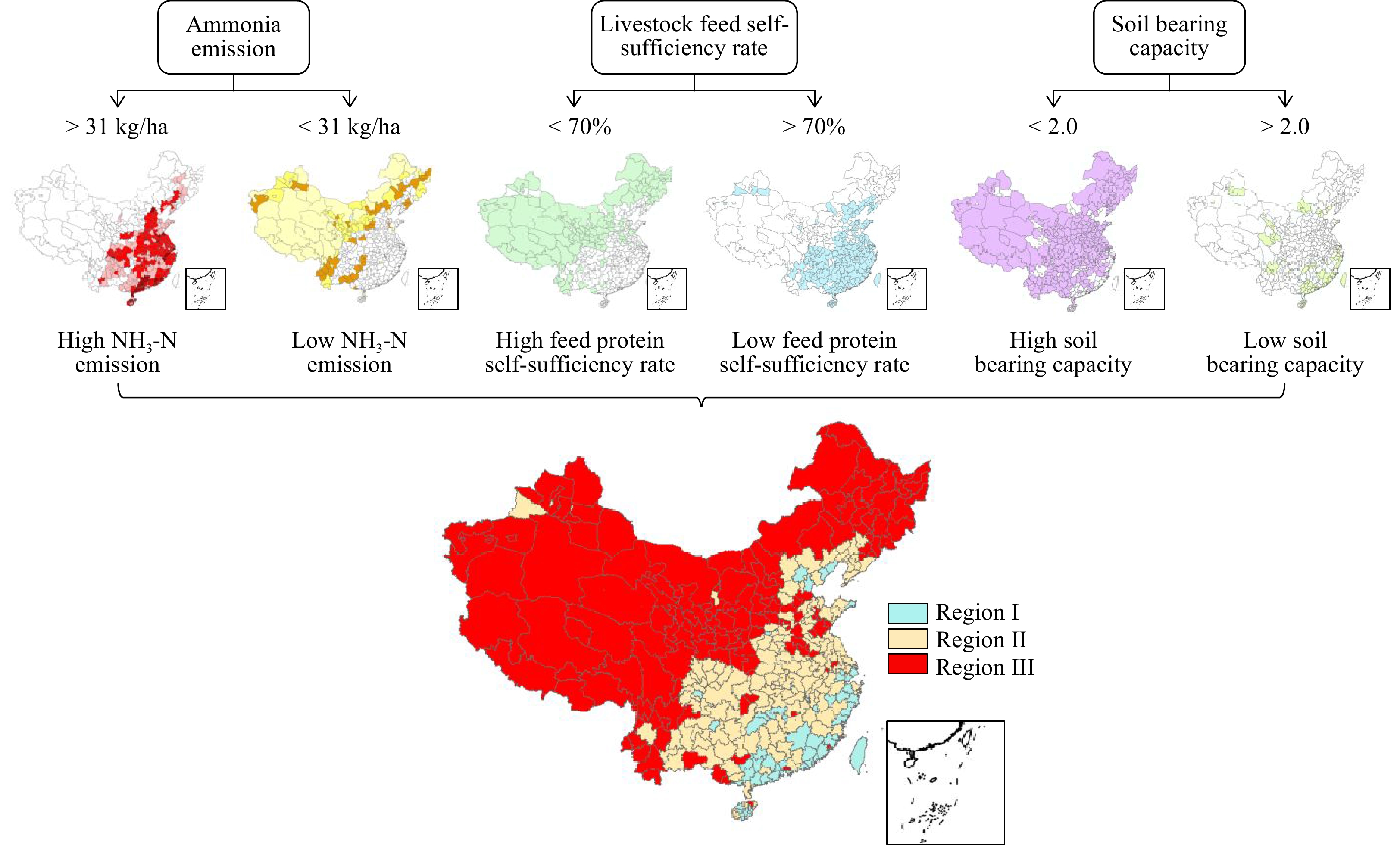
    Figure 1. 

    Flow chart dividing all cities into three different manure treatment and recycle regions.
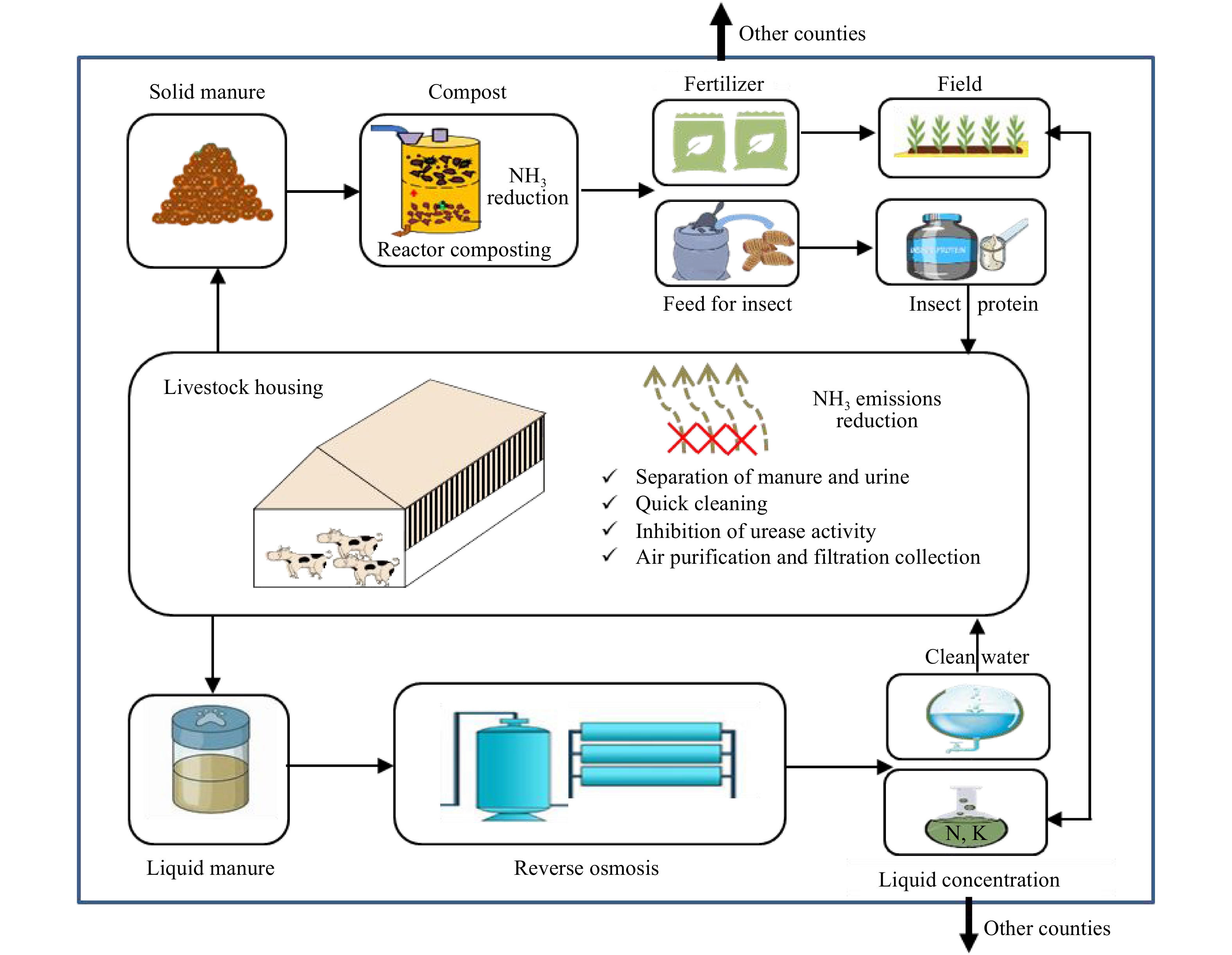
    Figure 2. 

    Technical model diagram of region I, including: reactor composting, liquid manure concentration technologies and freed protein production.
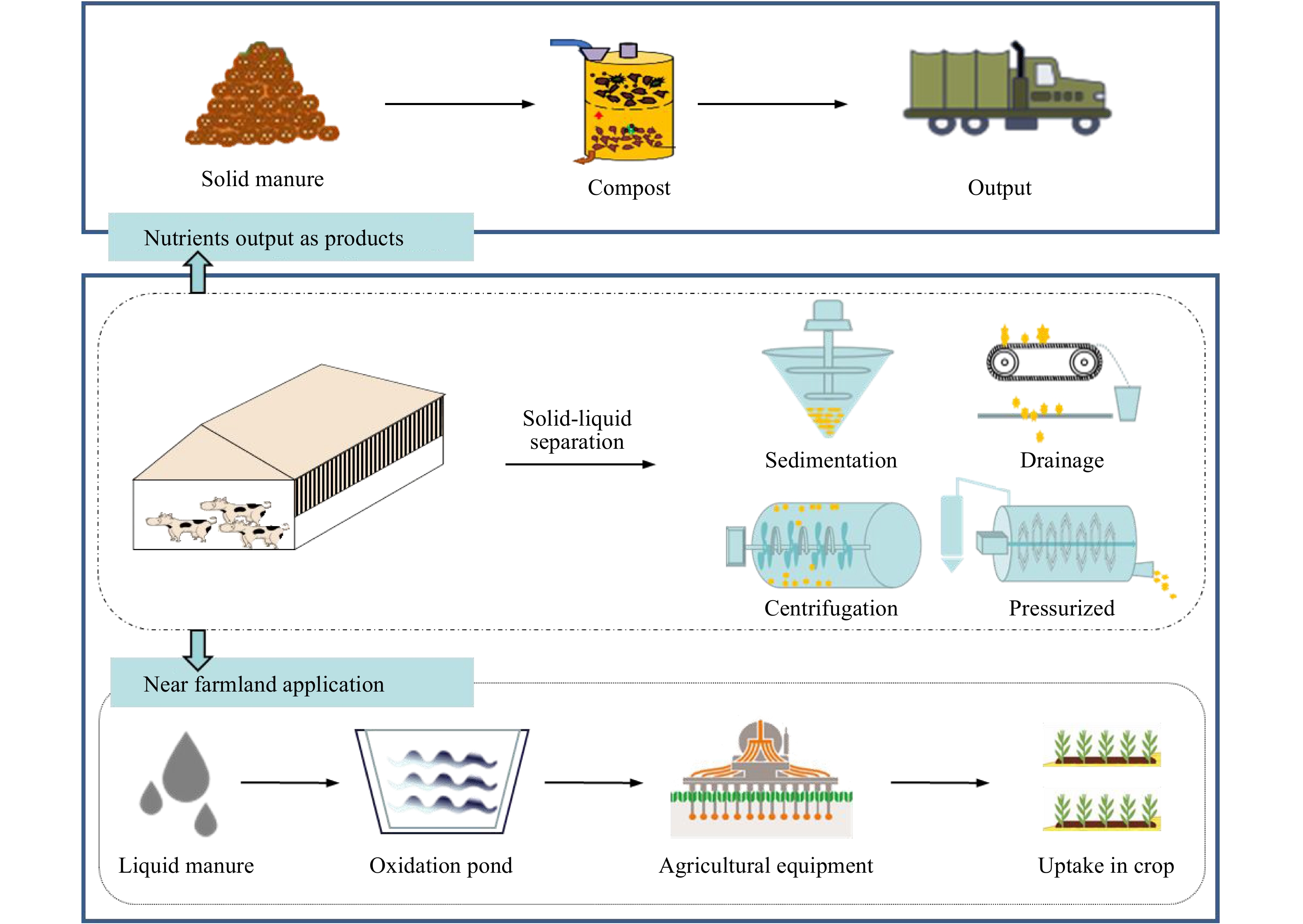
    Figure 3. 

    Technical model diagram of region II, including: efficient solid-liquid separation and liquid returned to field.
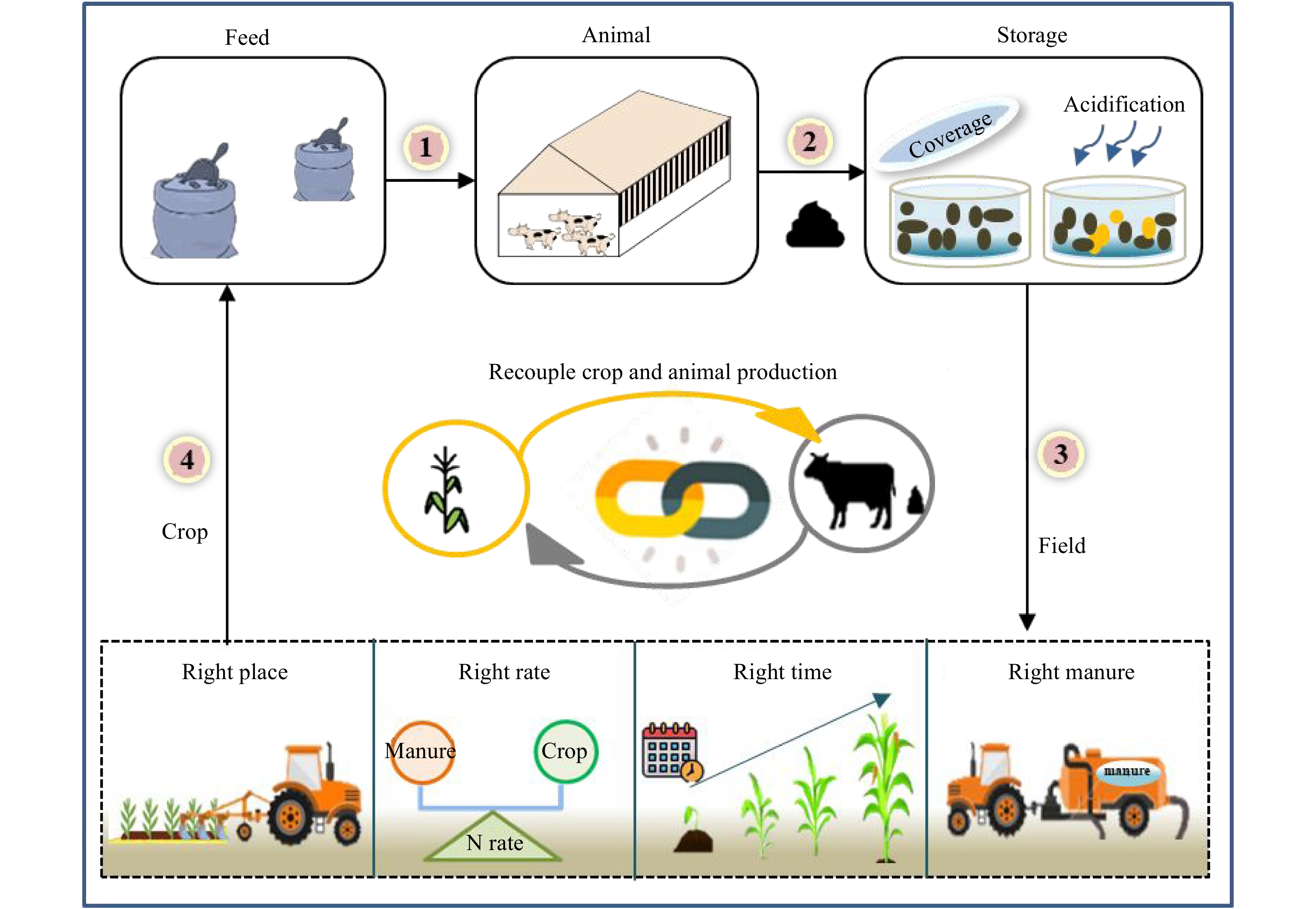
    Figure 4. 

    Technical model diagram of region III, including: manure storage and in-situ field application.

    Through using information from over 2,300 counties, including crop nutrient uptake, whole food chain nutrient management, ammonia emissions and self-sufficiency of feed protein, we have developed the first region-specific designation of manure treatment technologies in China (Fig 1). Categorization was carried out at the city level with over 360 cities included due to the strong role of city-level governments in providing subsidies to technology companies and farms. This illustrative example shows that the total area of Region I covers around 14.3 million ha cropland (12% of the manure N production), and Region II and III cover the remaining 97% and 88% of cropland and manure N production, respectively (Fig 1).

  • Region I is generally located in southeast China, mainly in Guangdong, Fujian and Hainan Provinces as well as the cities of Beijing and Tianjin (Fig 1). Technology system concepts are shown in Fig 2. Generally, livestock farms in Region I need to implement strict ammonia mitigation options in animal housing, such as frequent cleaning, air purification and filtration. Region I has a lower capacity to recycle manure, hence there is less need for storing large amounts of manure as most of it is regularly treated. Solid manure should be dewatered via advanced reactor composting technology, most of which is either directly exported outside of the region or used to produce insect feed to feed animals (Fig 2). The liquid part of manure needs to be treated via reverse osmosis technology to concentrate nutrients, allowing for long-distance transportation. Summarized, the three main proper treatments of manure in Region I are as follows: i) advanced reactor composting technology; ii) efficient liquid manure nutrient concentration technologies; and iii) feed protein production technologies.

    Reactor composting. Reactor composting technology is an effective and environmentally friendly method[13]. The well-controlled temperature and aeration in closed vessels achieves the elimination of pathogens, parasites and weed seeds within 7−10 days, with more than 90% of antibiotics and their resistance genes undergoing degradation[13]. Combined with a serial exhaust gas bio-filtration system, zero emission of ammonia, GHG and odor could be achieved[14, 15]. Meanwhile, fresh manure generated in livestock farms could be feeding into reactors continuously, combined with manure cleaning systems in animal housing. Closed composting reactors play the role of both storage and treatment for manure that shortens the manure management chain. Its high efficiency and small size make it an ideal in-situ manure treatment method for intensive livestock farms. It could thus be used both in Region I and II to achieve NH3 mitigation and convert manure into high-quality organic fertilizer that benefits manure nutrient transportation and epidemic prevention.

    Liquid manure concentration. Reversed osmosis (RO) is based on the ability of RO-membranes to let water pass and block salt ions. The technique is widely used for the desalination of sea water. Recently, the application of selective electrodialysis with monovalent exchange membranes on the recovery phosphate or ammonium from sewage water and livestock slurry has been investigated as a promising technique, and the technology has performed well in the Netherlands. Recovery efficiency of ammonia could reach 78% and 75% of phosphate and 87% of volatile fatty acids via using a bipolar membrane electro-dialysis system[16]. After treatment, 20% of mass was retained in the solid fraction, while 30% and 50% was retained in the concentrate and permeate material. The volume of concentrate, which contains higher concentration of ammonium-N, K and other elements, could be further reduced via use ventilating heat[17]. The effectiveness of nutrients in the concentrate was comparable to granulate chemical fertilizer (calcium-ammonium-nitrate), a common fertilizer in European countries. The Netherlands began pioneering experiments in 2009, and in total, eight large pilot plants have successfully treated slurry[17]. These could serve as good examples for Region I in China as nutrients in liquid slurry are effectively concentrated, allowing long-distance transportation to get rid of excess manure nutrients.

    Feed protein production. The use of insects for animal manure management is a sustainable and low-cost technology that effectively reduces the volume and nutrient concentration of manure residue, thereby reducing potential pollution. The black soldier fly larva (BSFL), one of the most powerful recyclers, can reduce the bulk of manure residue by 56% and nutrient concentrations by 40%−55% within 14 days of manure breeding[18]. Besides effectively degrading antibiotics in the manure[19], BSFL can also greatly reduce the abundance of pathogenic bacteria[20], decrease the offensive odor[21] and inhibit the breeding of house flies[22]. Few pilot plants have initiated commercial level production. Henan Enzyme Company has developed a pilot-scale automated BSFL breeding facility to treat pig manure and produce insect proteins[23]. Through cloud technology, the device can upload data in real-time, and the controller can adjust and monitor the parameters of the breeding workshop through mobile application or computer. The facility can completely decompose 3.24 tonnes (78% moisture) of pig manure per day and produce 480 kg of insect biomass. This effectively reduces the volume and nutrient concentration of the manure residue, thereby reducing potential pollution by at least 50%−60%[18]. BSFL biomass, containing about 40% protein and 30% fat[24], can be used as a substitute for soybean and fish meal for feeding poultry and fish without causing adverse reactions[25]. Through the recycling of insects, the twin problems of excessive manure nutrients and shortage of feed in Region I can be addressed simultaneously. An alternative choice is microalgae protein production. Recently, microalgae have been considered as a promising solution for wastewater management owing to their high capacity to deplete inorganic nutrients (N and P) from a wide range of wastewater[26]. Microalgae could use wastewater effluent as a source of carbon and nitrogen to support their rapid growth and be converted to microbial protein as animal feed after proper filtration, concentration and drying.

  • Region II covers major cropland production areas in China, excluding the northeast and Region I (Fig 1). These area shows a moderate level of ammonia emission intensity, manure loading capacity and feed self-sufficiency; accordingly, attention should focus on the trans-regional recycling of manure with less emphasis on complex and high-cost treatment technologies. The core recommended technology system is solid-liquid separation in which solid manure is dewatered via closed reactor composting, transported between regions and liquid manure injected to fields adjacent livestock farms.

    Efficient solid-liquid separation system. Solid-liquid separation can be divided into sedimentation, drainage, centrifugation and pressure filtration (Fig 3). Centrifugal separation is the most effective method for slurry separation, with the highest removal rate of total solids reaching up to 60%[27]. However, centrifugal equipment is used relatively less in China due to its high price, high energy consumption and demanding maintenance. Pressurized filtration, including screw press and press auger, is usually more efficient than screening technology. Improving pressure can increase the removal rate of solids and accelerate the removal of nitrogen, phosphorus, potassium and other nutrient elements. This technology is suitable for widespread application in China due to lower costs[28]. In addition, flocculent can be used before separation to improve separation efficiency.

    Liquid manure injection or irrigation. Liquid manure is rich in N after separation, which can be connected to the irrigation system through the pipe network or transported to the farmland and applied with supporting agricultural machinery as liquid fertilizer. Liquid fertilizer can be diluted directly into irrigation water for field application, but the spread of nutrients on the field surface may be uneven[29]. Liquid fertilizers can be applied to fields by spraying vehicles, but it may cause the evaporation of ammonia and odors[30]. Deep soil injection could reduce ammonia emissions. Fecal injection could be combined with seeding, reducing costs and improving seeding rates, especially in dry regions and seasons. In the Netherlands, strict regulations of manure application have promoted the development and manufacturing of equipment related to liquid fertilizer. The disc-type fertilizer applicator is an injection machine with disc harrow capable of simultaneous stubble crushing, land overturning and manure injecting[31, 32]. In China, use of the liquid injection machine is relatively unexplored. However, injecting liquid fertilizer can greatly improve the utilization rate of fertilizer and reduce pollution. Li et al. (2020) showed that integrating seedings with liquid manure injection could replace 50% of mineral N fertilizer, reduce ammonia emissions by 27%−49% and increase corn grain yield by 17%−33%[33].

  • Region III, largely found across Xinjiang, Inner Mongolia and Northeast China, has an abundance of feed resources, with higher manure nutrient loading capacity and lower sensitivity to ammonia emissions. These regions usually feature larger farms, which require fewer manure treatment technologies but better manure storage facilities. This is because these regions have a single cropping system that is different from Region I and II. This indicates manure could only be applied once per year in these regions. An ideal model for Region III is shown in Fig 4. This model is characterized by manure storage and in-situ field applications. Manure needs to be stored in belowground concrete tanks for almost one year. Coverage, which reduces the timing for manure exposure in the air and resulted in reduced emissions of NH3, odour[34, 35] as well as surface acidification, further decreasing NH3 emissions with lower cost[36], are two favorable technologies to preserve N in manure during long-term storage. Applying the slurry in accordance with the principles of 4R nutrient stewardship, that is, applying the slurry at the right place, rate, time and type[37, 38], could also be used to help treat storage manure.

  • We used current data to demonstrate an approach to designing region-specific technology systems for the treatment and recycling of manure in China, and we provided an illustrative example in Fig 1. This is a preliminary analysis based on limited data and broad assumptions, and more research is needed to improve the granularity of such designs by quantifying water and airborne pollution as well as landscape and transportation costs at the regional level. Furthermore, region-specific manure management designs could become part of the recently implemented rural revitalization[39] and Blue-Sky actions[40].

    The ultimate goal of manure treatment is to reduce environment impacts by recycling it as either fertilizer for crop production or feed protein for livestock production. However, in China, livestock has been largely decoupled from crop production in terms of exchange between feed and manure at both the farm household and regional scale. The share of rural households with both crop and livestock production has declined from 71% to 12% in the period between 1986 to 2017[41]. A recent nation-wide survey revealed that only 1/3 of farmers were willing to use manure as fertilizer in cereal crop production[42] due to concerns of cost, odor, antibiotics and heavy metal issues. Recycling of manure-based insect feed protein to livestock production is also a controversial issue in terms of consumer acceptance. These obstacles could be alleviated via strict feed quality control regulations and public education campaigns.

  • The criteria used in this study included soil-bearing capacity, local livestock feed self-sufficiency rate and ecosystem vulnerability. The soil-bearing rate refers to the ratio of total excretion of N by livestock and humans as well as the N withdrawal of harvested crops. In the present study, the soil-bearing capacity was estimated based on the NUFER (NUtrient flow in Food chains, Environment and Resources use) model, which calculates all nutrients for each city. The equation used to calculate soil bearing capacity was:

    Nsoil=Nhumanmanure+NlivestockmanureNplantuptake+Ngrass (1)

    Where N soil is soil bearing capacity, N human manure is the N content from human manure (tonnes N yr−1), N livestock manure is the N content from livestock manure (tonnes N yr−1), N plant uptake is N content taken by plants (tonnes N yr−1) and N grass is the N content taken by grass. The estimated soil bearing capacity was summarized in Fig 1. Jin et al. (2020) claimed that 2 was the threshold value for the soil-bearing capacity in China, which means areas with values higher than 2 were considered low soil-bearing capacity[2]. Across China, 19% of the total area was lower than 2 and considered as having high soil-bearing capacity. Conversely, areas higher than 2 were considered as having low soil-bearing capacity.

    The feed self-sufficiency rate refers to the ratio of domestically consumed feed supplied by domestic producers. Local livestock feed self-sufficiency rate was estimated based on livestock consumption and feed production. The equation used was:

    Nratio=NtotalconsNimported+NexportedNtotalcons (2)

    Where N ratio is livestock feed self-sufficiency rate and N total cons is the total N consumption for each category of livestock (sheep, cattle, pig, poultry, horse, rabbit, mule and donkey). N imported is feed N imported from other areas. N exported is feed N exported to other areas. The distribution of livestock feed self-sufficiency rate across China is shown in Fig 2. The present study designated 0.7 as the threshold value, and values higher than 0.7 were defined as high livestock feed self-sufficiency rates. As seen in Fig 2, high livestock feed self-sufficiency rates (exceeding 0.7) were mainly located in northern and western China China.

    Ammonia emissions considered here include NH3 from crop and animal production. The amount of total ammonia emissions was estimated by the NUFER model (Fig 3). The SDGs report, EU SDG index scores and ammonia data are reported for each country. Using this data, the relationship between SDG index scores and ammonia emissions data was established through multiple linear regression analysis. The statistical model (R2 = 0.91, n = 23) used was:

    Ammonia=1.4Score+104.9 (3)

    Where Ammonia is ammonia emission per agricultural land (kg ha−1) and Score is the SDG index score. SDGs report designated a score of 60 as the threshold for European countries, and this present study assumes the European standard as the corresponding limit for China. Therefore, this statistical modeling can provide the ammonia threshold value (31 kg ha−1) for the designated 60 score. As seen in Fig 3, higher ammonia emissions (higher than 31 kg ha−1) were found in southeastern regions.

    The total land area of China was divided into 3 regions, each of which in turn contains two or more ecosystem statuses (Fig 4): regions (Region I) with high ammonia emissions, low soil-bearing capacity and low livestock feed self-sufficiency rates; regions (Region II) with low ammonia emissions, low soil-bearing capacity and low livestock feed self-sufficiency rates; and regions (Region III) with low ammonia emissions and high soil-bearing capacity. This process was compiled in ArcGIS 10.6 in which areas were selected by overlaying different criteria layer (ecosystem status layers).

    • This work was supported by the National Key R&D Program of China (2016YFD0800106); the National Natural Science Foundation of China (31572210, 31872403, 71961137011); Key Research Program of Frontier Sciences-CAS (QYZDY-SSW-SMC014); Key Laboratory of Agricultural Water Resources-CAS (ZD201802); the Key Research Program-CAS (KFJ-STS-ZDTP-053); Hebei Dairy Cattle Innovation Team of Modern Agro-industry Technology Research System, China (HBCT2018120206); the Youth Innovation Promotion Association, CAS (2019101) and Outstanding Young Scientists Project of Natural Science Foundation of Hebei (C2019503054).

    • The authors declare that they have no conflict of interest.

    • Copyright: © 2023 by the author(s). Published by Maximum Academic Press, Fayetteville, GA. This article is an open access article distributed under Creative Commons Attribution License (CC BY 4.0), visit https://creativecommons.org/licenses/by/4.0/.
Figure (4)  References (42)
  • About this article
    Cite this article
    Bai Z, Wang X, Wu X, Wang W, Liu L, et al. 2021. China requires region-specific manure treatment and recycling technologies. Circular Agricultural Systems 1: 1 doi: 10.48130/CAS-2021-0001
    Bai Z, Wang X, Wu X, Wang W, Liu L, et al. 2021. China requires region-specific manure treatment and recycling technologies. Circular Agricultural Systems 1: 1 doi: 10.48130/CAS-2021-0001
  • Catalog

    • About this article

    /

    DownLoad:  Full-Size Img  PowerPoint
    Return
    Return