ARTICLE   Open Access    

Molecular identification and in vitro interaction of molds associated with dry rot of potato (Solanum tuberosum L.) collected in La Trinidad, Benguet, Philippines

More Information
  • Solanum tuberosum L. from the Solanaceae family is one of the most important agricultural crops grown in the Philippines next to rice. Unfortunately, harvested potatoes were plagued with various fungal diseases resulting to occurrence of dry rot affecting the production and harvest of potatoes. Dry rot in potatoes is caused by several fungal species in the genus Fusarium. Hence, this study aimed to isolate and identify fungal species associated with dry rot disease of potato. Potatoes showing diseased symptoms of dry rot were randomly selected and collected from various traders in La Trinidad, Benguet and were subjected for fungal isolation through serial dilution and plating. Identification was done through observation of cultural and morphological characteristics as well as DNA barcoding using ITS region. A total of six fungal species were subjected into BLAST which revealed the following species: Aspergillus flavus (100.00%), A. fumigatus (100.00%), A. niger (99.82%), Fusarium oxysporum (95.06%), F. solani (100.00%) and Mucor velutinosus (96.45%). To establish possible fungal species as potential biocontrol agent that would lessen the use of harmful chemicals, the identified fungal organisms were interacted with one another through in vitro interaction. Results showed that fungi exhibited antagonistic interaction against each other. Hyphal denaturation, hyphal penetration, hyphal coiling and cell lysis, were observed through microscopic observation. Meanwhile, M. velutinosus showed the ability to penetrate the host hyphae of the F. solani and F. oxysporum. Therefore, Mucor velutinosus could be a potential biological control agent against Fusarium species, which can reduce the use of harmful fungicides in controlling fungal diseases in potato that causes dry rot.
  • Bananas (Genus Musa, family Musacaea) are herbaceous perennial monocots grown in more than 150 countries worldwide[1]. In the Philippines, banana accounts for around 17.2% of the total agricultural exports[2]. Cavendish bananas remain the primary cultivar grown commercially, accounting for 53.2% of the total production in the Philippines, followed by Lakatan (16.8%) [1] and Cardaba (14%)[2]. Commercial bananas, including the Cavendish group, are generally seedless and sterile[3]. Large-scale propagation of banana is therefore highly dependent on using vegetative planting materials- sword suckers, rhizomes, and bits- that potentially carry disease-causing microorganisms[4]. Throughout the years, various methods have been explored for banana production via plant tissue culture. The process allows the propagation of thousands of plantlets from a small amount of planting material. Shoot tip cultures and sword suckers are used primarily for in vitro propagation of true-to-type and disease-free plantlets. However, increased production of off-types has been observed using these methods[5].

    Somatic embryogenesis is another important means of plant production. It is defined as the asexual reproduction of plants from somatic embryos[6]. The success of the technology relies on the potential of cells for totipotency: the ability of a single cell to divide and undergo differentiation[7]. Somatic embryo formation is based on dedifferentiation in plants and the plants ability to reinitiate cell division. Somatic embryos may be induced using direct or indirect methods. Indirect embryogenesis, unlike direct, involves an intermediate callus phase from organized tissues[8]. Studies have reported the establishment of direct somatic embryogenesis, but low plant conversion rates were observed (for example, Remakanthan et al.[9]). Panis et al. reported direct somatic embryogenesis from protoplast cultures[10]. Recently, the use of shoot-tip cultures has been reported[9]. Here, the indirect production of somatic embryos from callus cultures were the focus.

    Somatic embryogenesis was first described in carrot (Daucus carota) cells in culture[11]. Although initially investigated for micropropagation of plants, somatic embryogenesis is also utilized for gene expression programs and genetic transformation to improve quality and disease resistance[12]. Genetic transformation using somatic embryos has been proven to minimize the formation of chimeric plantlets[13]. In plant breeding, somatic embryogenesis shortens the breeding cycle[14]. The protocol is also primarily used for cryopreservation of Musa germplasms[15].

    Several cultivars of banana, especially those belonging to the Cavendish subgroup, have been propagated from somatic embryogenesis (Table 1). The protocol for somatic embryogenesis in bananas is standardized using different types of explants. However, low embryo germination and plant conversion rates remain a concern[13]. Other issues include the labor-intensive optimization of culture medium, high production costs, and the formation of off-types[16]. Nevertheless, somatic embryogenesis has been exploited to generate planting materials that are of value and disease-free. Several of these methods have been scaled up to commercial laboratories and some for the protection and preservation of commercial banana cultivars that are under threat of extinction[17,18]. Studies have reported the use of somatic embryogenesis in banana but few have focused on the different culture conditions for growth. This review explored the different culture conditions used for somatic embryogenesis in banana and some of their advantages and constraints.

    Table 1.  Cultivars with successful embryogenic callus (EC) and cell suspension (ECS) protocols.
    Cultivar Genetic group Explants used EC ECS Ref.
    Calcutta 4 AA Scalps
    Axillary buds
    x x [110]
    Lakatan AA Shoot tips x x [30]
    Highgate AAA Scalps x [47]
    Yangambi km5 AAA Immature flowers x [90]
    Williams AAA Scalps
    Immature flowers
    x x [47,109,16,
    92,113]
    Grand Nain AAA Scalps
    Immature flowers
    Shoot tips
    x x [37,47,111,
    112,39,92,9]
    Nanicão AAA Leaf sheath disks x [27]
    Gros Michel AAA Immature flowers x x [90,113]
    Lady finger AAB Scalps x [47]
    Prata AAB Scalps x [47,114]
    Saba ABB Immature flowers
    Scalps
    x x [106,115]
    Cardaba ABB Scalps, shoot tips x [106,116,117]
    Bluggoe ABB Shoot tips
    Scalps
    x x [26,118]
     | Show Table
    DownLoad: CSV

    Somatic embryogenesis is an elaborate and complex process involving the production of a whole new plant from unorganized cells. The process is generally comprised of five stages: selection of suitable explant, production of embryogenic callus, development of somatic embryos from cell suspensions, regeneration of viable cells into plantlets, and field monitoring of acclimatized plants (Fig. 1). Each developmental stage requires different nutritional and environmental conditions for growth and is controlled by several factors including endogenous hormones, proteins, and transcription factors[19].

    Figure 1.  Flowchart showing the different stages of somatic embryogenesis in banana.

    The quality and volume of embryogenic callus are crucial for implementing the subsequent steps in somatic embryogenesis[16]. A callus is a mass of unorganized cells naturally found in plants that form in response to stress and wounding[6]. Callus formation in plants is highly controlled by abiotic (light condition, pH and osmotic pressure, sugar content) and biotic (explant age and size, genotype, phytohormones) stimuli[20]. Callus formation differs between monocots and dicots and between diploid and triploid species[21,22]. Pathogen infection also leads to callus formation in plants through auxin and cytokinin production[23].

    Callus forms may vary from one set-up to another and can be differentiated based on macroscopic characteristics[20]. Generally, four types of calli can be observed in banana cultures: white and compact (Fig. 2a), clear and friable (Fig. 2b), yellow nodular (Fig. 2c), and ideal callus with translucent proembryos (Fig. 2d). Out of these four, only the ideal callus with translucent proembryos can regenerate and develop into a whole new plant[20]. The translucent proembryos contain differentiated and competent cells that enable plant organogenesis and regeneration[24]. Meanwhile, the white and compact, clear and friable, and yellow nodular calli are all non-embryogenic and non-regenerative types that may be used for further biotechnological studies such as metabolite production and cell suspensions[24,25]. In some cases, shoots and roots may form alongside these non-embryogenic calli that also have the potential to develop into new plants[20].

    Figure 2.  Types of callus formed in banana: (a) white and compact (non-embryogenic), (b) clear and friable, (c) non-embryogenic yellow nodular, and (d) ideal callus with translucent proembryos.

    Scalps (meristematic tissues with cauliflower-like structure) and immature flowers (male and female inflorescence) are the two most commonly used explants in banana[16]. However, shoot-tips[26], leaf sheaths[27], sword suckers[28], and protoplasts[29] from tissue-cultured plantlets have also been reported. Callus induction may take from 8 weeks to 8 months, depending on the type of explant used. The formation of callus cultures from scalps take the longest, with 6 months average induction time[16]. Induction of embryogenic callus in 12 weeks has been observed from shoot tips[30], sword suckers[28], and immature flowers[20,31]. Callus induction from protoplast cultures are initiated in about three weeks[3,32]. However, it is usually derived from established cell suspensions[29].

    Somatic embryogenesis relies on the exogenous application of auxins and cytokinins to promote in-vitro callus induction in plants[16,33]. The combination of callus induction hormones differs from the type of explant used (Table 2). Commonly used auxins for callus initiation are 2,4-dichloro phenoxy acetic acid (2,4-D), indoleacetic acid (IAA), naphthalene acetic acid (NAA), 3,6 dichloro-2 methoxybenzoic acid (Dicamba) and picloram. These may be prepared with cytokinins such as kinetin (KIN), 6-benzyl amino purine (BAP), and zeatin. Brassinosteroids and abscisic acid (ABA) also induce callus formation in some plant species[34,35]. Thidiazuron (TDZ), a hormone with both cytokinin and auxin effects on plants, was also found to induce callus formation in banana[36].

    Table 2.  Synthetic hormones commonly used for embryogenic callus induction in Musa spp.
    Explant used Hormones tested (mg/L) Ref.
    2,4-D IAA NAA KIN 2iP BAP TDZ 4-CPPU ZEATIN Picloram Dicamba
    Immature flowers 2−6 1 1 [107,121,16,111,
    92,82,119,120,
    122,52,123,39]
    2−9 [62,37,125,16,112,13]
    2 [126]
    2 0.5−1 [111]
    1 0.22 [54]
    Scalps 1 0.22 [106,118,127,115,109,
    114,54,113,110,48]
    2−2.9 2.2−3.2 [128]
    6.4 [128]
    5.7 [128]
    Shoot tips 0.05 1 [9]
    0.1−4 [9]
    Leaf sheaths & rhizomes 6.63 [129]
    Protoplasts 2 [29]
    Leaf sheath disks 1.1 6.64 [22]
    100 100 [27]
    Sword suckers 0.5-2 0.5 [28]
     | Show Table
    DownLoad: CSV

    Optimum hormone levels for callus induction in banana vary from one genotype to another. For auxins, concentrations range from 0.2 to 4 mg/L when used alongside cytokinins and 4 to 9 mg/L if treated alone. Cytokinins, at 0.5 to 1.0 mg/L, are combined with auxins for callus induction. In addition, culture additives such as amino acids (e.g. proline, glutamine, methionine, tryptophan), sugars (e.g., sucrose, maltose, myo-inositol), and vitamins (e.g., biotin) also support callus induction in banana[22,3740].

    Light exposure also affects callus formation in banana. In numerous studies, callus formation was frequently performed under dark conditions. One study found that light exposure is positively correlated with tissue browning due to increased physiological activity[22]. Hence, the dark treatment seems to prevent necrosis caused by photooxidative stress[41]. Color change of medium is also frequently encountered and can be resolved using gerlite as a gelling agent[22]. Blackening or browning of tissues due to the wounding of explant can be minimized by subculture every two weeks[16]. The addition of antioxidants such as ascorbic acid[42], citric acid[43], cysteine[44], activated charcoal[43], polyvinylpyrrolidone (PVP)[45], potassium citrate, and citrate[46] have been proven to prevent explant browning in banana. It is challenging to optimize culture conditions and culture medium composition due to the extremely low amount of good embryogenic material available for use. Usually, young banana suspensions require a high inoculum density and frequent transfer to a new medium (every three to seven days) during the first few months[47]. In Grand Nain, only 3% to 10% of embryogenic calli (EC) were formed from scalps and 8% from immature flowers[16]. But for other species, % EC can reach up to 97%[48]. The embryogenic potential of callus is also expected to decrease over long periods of incubation[9,21].

    Somatic embryos are clones of the parent material formed in response to the changing culture conditions of the explant[49]. Unlike sexual structures (zygotic embryos), somatic embryos form in response to the drastic reduction of auxin levels after exposure to callus cultures[7]. Somatic embryos possess a bipolar structure that allows the formation of both apical and radical meristems where shoot and root structures initiate, respectively[13]. Depending on the cultivar, embryos generally form in 3 to 8 months[47].

    Complex processes are known to affect somatic embryogenesis in banana. Kumaravel et al. have characterized 25 endogenous proteins in banana associated with somatic embryo formation[40]. Several studies have further explored the involvement of genetic transcription factors in growth[21,5052]. The addition of cytokinins, alternation of physiological state (pH), and heat shock are known drivers of somatic embryogenesis[21,53]. Reduction of MS salts to half strength and exposure to dark conditions to reduce osmotic pressure and prevent phenolic oxidation, respectively have also been frequently performed in established ECS protocols but the underlying principle remains poorly understood[47,48,54]. In cassava (Manihot esculenta), the use of half and quarter-strength MS resulted in enhanced viability and formation of somatic embryos compared to full-strength MS medium[55]. On the other hand, Groll and co-workers reported a higher formation of mature somatic embryos in full-strength MS[56].

    There are four main stages in the formation of somatic embryos- globule stage, oblong stage, heart stage, and torpedo stage- a developmental process shared with zygotic embryos that can be differentiated through distinct cell shape formation[12,57,58]. The first stage, the globular stage, is achieved through the establishment of embryogenic cell suspensions (ECS). Banana ECS protocols vary with the explant used for callus formation (Table 3) and are established by transferring the embryogenic callus into a (liquid) medium with reduced auxin levels or callus-induction medium devoid of agar; most with added amino acids (e.g. L-glutamine and malt extract) that function for metabolism and protein synthesis (Table 4)[13,16,59,60]. For instance, L-glutamine and proline were found to enhance the plant regeneration efficiency of banana (Musa acuminata cv. Berangan)[61]. Scalp-derived ECS utilizes a uniform concentration of exogenous growth regulators (e.g., 2,4-D and zeatin) during induction and multiplication phases[16]. For the immature flower method, somatic embryo expression is enhanced by reducing auxin concentration[59,60,62]. The continued presence of auxin drives the synthesis of gene products necessary to complete the globular stage through increased DNA demethylation[63,64].

    Table 3.  Culture media used for formation of somatic embryos in banana.
    ComponentsMA2ZZ1M2bECS1BM2SK4SS2IM1
    Macro-elementsMS1/2MS1/2MS1/2MSMSMSMSSH
    MicroelementsMSMSMSMSMSMSMSSH
    VitaminsMAMSDhed'aMSMSMSMSMS
    FeEDTA+
    2,4-D (mg/L)1111
    Picloram0.11
    Zeatin (mg/L)0.2190.2190.219
    BAP (mg/L)0.05
    Coconut water (%)10
    Biotin (mg/L)11
    Casein hydrolysate200
    Ascorbic acid (mg/L)101010
    Malt extract (mg/L)100100100100100
    Amino acidsGlutamine
    100 g/L
    Glutamine
    100 mg/L
    Proline
    4 mg/L
    Glutamine
    100 mg/L
    Glutamine
    100 mg/L
    Glutamine
    100 mg/L
    SugarSaccharose
    45 g/L
    Sucrose
    30 g/L
    Sucrose
    20 g/L
    Sucrose
    30 g/L
    Sucrose
    45 g/L
    Sucrose
    30 g/L
    Sucrose
    30 g/L
    Sucrose
    30 g/L
    pH5.35.85.85.85.35.85.85.8
    Cultivars testedGrand Nain, Tropical, Rasthali,Somrani monthan,
    High gate, Williams,
    Gros Michel, Lady
    finger, Prata
    Mas, Bluggoe,
    Saba, Cardaba
    Calcutta 4RajeliDwarf BrazilianGrand NainGrand Nain, Ardhapuri, Basrai, Shrimanti, Mutheli, Lalkela and
    Safed Velchi
    Ref.[39,107,90,131,52][39,54,113,48][118,130,115][110][82][121][9][111]
    Ma2, M2b, BM2, SK4, IM1-immature flower method; ECS1, ZZI-scalps method; SS2-split shoot tips.
     | Show Table
    DownLoad: CSV
    Table 4.  Culture media used for somatic embryo maturation in banana.
    ComponentsMA3RD1BM3SK8MMSS3IM2M3b
    Macro-elementsSH1/2MSSH1/2MSSHMSSHMS
    MicroelementsSHMSSHMSMSMSSHMS
    VitaminsMAMSMSMSMSMSMSMS
    FeEDTA+
    2,4-D (mg/L)1
    BAP (mg/L)50.050.05
    IAA (mg/L)0.2
    NAA (mg/L)0.20.2
    Zeatin (mg/L)0.050.05
    Kinetin (mg/L)0.10.1
    2iP (mg/L)0.2
    Picloram (mg/L)0.1
    Myo-inositol (mg/L)100100
    Biotin (mg/L)1
    Ascorbic acid (mg/L)10
    Malt extract (mg/L)100100100100
    Amino acidsGlutamine
    100 mg/L
    Proline
    230 mg/L
    Glutamine
    100 mg/L
    Glutamine
    100 mg/L
    SugarSaccharose
    45 g/L
    Sucrose
    30 g/L
    Sucrose
    45 g/L
    Sucrose
    30 g/L
    Saccharose
    45 g/L
    Sucrose
    30 g/L
    Sucrose
    30 g/L
    Sucrose
    30 g/L
    Gelling agent (g/L)Phytagel
    4 g/L
    Gelrite
    3 g/L
    Gelrite
    2 g/L
    Phytagel
    2.6 g/L
    Gelrite
    2 g/L
    Gelrite
    3 g/L
    pH5.85.85.85.85.85.85.85.8
    Cultivars testedGrand Nain, Gros Michel, WilliamsGrand Nain, Calcutta 4,
    Somrani monthan,
    High gate, Williams,
    Lady finger, Prata
    Rajeli, Grand
    Nain, Tropical
    Dwarf BrazilianGrand Nain; RasthaliGrand NainGrand Nain, Ardhapuri, Basrai, Shrimanti,
    Mutheli, Lalkela,
    Safed Velchi
    Bluggoe, Saba,
    Cardaba
    Ref.[39,107,125][47,110,54,113][82,123,124][121,132][125,16,52][9][111][118,115]
    Ma3, BM3, SK8, MM , IM2, M3b-immature flower method; RDI-scalps method; SS3-split shoot tips.
     | Show Table
    DownLoad: CSV

    At the globular stage, the pro-embryos also contain other mRNAs and proteins that generally inhibit the continuation of embryogenesis[11]. The removal of auxin is believed to result in the inactivation of these genes necessary to enter the next embryogenic growth stage[50]. Guzzo et al. proposed a model linking auxin response, asymmetric division, and totipotency: upon environmental stimuli, cells can be made morpho-genetically totipotent in response to auxin if the cells contain inducible receptors to complete embryogenesis; but only organogenesis or unorganized proliferation will occur otherwise[65]. Cytokinins, in minute concentrations, may also affect the sensitivity of somatic embryogenesis and cell division, but their molecular basis remains unknown[66,67].

    The globular embryo then enters the oblong stage, signaling the shift from isodiametric to bilaterally symmetrical growth, followed by the beginning of the heart stage[68]. This globular-to-heart embryot ransition is pronounced by the outgrowth of the two cotyledons, hypocotyl elongation, and radicle initiation[11]. Finally, the embryo enters the torpedo stage, a stage with a distinct increase in size, before reaching full maturity[68]. Sometimes, immature embryos formed from callus cultures may undergo differentiation, and this can be prevented through high osmotic pressure and the addition of abscisic acid[21]. Removal of bigger aggregates of, more developed, somatic embryos is recommended because they have the tendency to accumulate starch and produce high amounts of polyphenols[47].

    Water stress is one of the most important factors for somatic embryo maturation[69]. During maturation, embryos undergo gradual loss of water and initiate desiccation tolerance to survive[7072]. Available ECS protocols regulate water availability to the developing somatic embryos through high concentrations of gelling gum or overlaid filter paper[13]. Studies suggest the involvement of early response to dehydration proteins (ERDs) in embryo maturation[73]. Oxygen availability and pH of the culture medium also affect embryo maturation. High levels of oxygen have been shown to promote somatic embryo multiplication while low levels result in histodifferentiation[74]. The optimum pH for embryo development is pH 5.8, but relatively lower quality and irregular embryos may also form at pH 4.5-5.5 and at pH 6.0 to 7.0[7577].

    The germination of the somatic embryo into normal shoots, termed regeneration, is achieved primarily on culture medium in a genotype-dependent manner. Plants derived from embryogenic cell suspensions (ECS), called emblings, are highly dependent on ECS density and quality[50]. High cell density (105 cells/mL) is for embryogenic cell clusters formation from and lower cell density (2 × 10⁴ cells/mL) for embryo development originating from embryogenic cells[78,79]. Embling conversion rates vary within banana genotypes. For instance, 13% in the edible (AA) Pisang Mas and 13% to 25% for Grand Nain of the Cavendish subgroup (AAA)[80]. High regeneration rates (90% to 95%) from ECS cultures have been recorded for some triploid and diploid species such as cv. Dwarf Brasilian (AAB) and M. a. ssp. malaccensis (AA), both of which passed through a differentiation–maturation phase[13,81].

    Most commonly, BAP, at 0.2 to 3 mg/L concentrations, is used for plant regeneration[47,54,82]. Sometimes, BAP is complemented with other cytokinins (at 0.2 to 0.5 mg/L) for embryo germination (Table 5). These are supplemented with antioxidants such as activated charcoal and ascorbic acid to prevent browning and further support the regeneration of tissues[83]. Kumaravel and co-workers further investigated different concentrations of NAA (2.68, 5.37, and 10.74 μM) for the regeneration of banana somatic embryos with three (100 and 200 μM) and methionine (335.09, 670.19, and 1 mM) as additives[40]. They also tested various concentrations of CaCl2 (5, 10, and 15 mM) and gibberellic acid (GA3) (1.44, 2.88, and 5.77 μM) with 11.41 μM IAA and 2.21 μM BAP. In 'Grand Nain', media supplemented with 5.37 μM NAA + 1.44 μM GA3 showed the highest regeneration efficiency (91.0%). The lowest regeneration was recorded in the medium supplemented with 1 mM methionine in 'Rasthali', whereas 'Grand Nain' media with 200 μM showed the least germination. It was found that in 'Grand Nain', an increased concentration of IAA recorded the highest regeneration (24.28%), but relatively lower (showed 18.96%) in 'Red Banana' in kinetin-supplemented media. These results demonstrate that in banana, regeneration is not only genome-dependent but also cultivar-dependent. The observed overexpression of IAA monooxygenase in the emblings also showed that tryptophan-dependent auxin biosynthesis plays a key role in somatic embryo formation. El-Kereamy et al. previously reported the overexpression of these proteins in rice resulted in enhanced shoot formation due to increased biosynthesis of GA and cytokinin, whereas Patterson et al. reported the role of germination-related proteins for root hormone regulation in Arabidopsis[84,85]. These results suggested that the endogenous hormones stimulated the formation of pro-embryonic roots and shoots of somatic embryos. Furthermore, scientists discovered important genes affecting the morphogenesis of somatic embryos. Boutilier and co-workers described the role of the BABY BOOM1 (BBM1) gene for morphogenesis in coffee (Coffea canephora) embryogenesis, while the LEAFY COTYLEDON1 (LEC1) and WUSCHEL-RELATED HOMEOBOX4 (WOX4) genes are crucial in the initial phase of cell differentiation[8689]. Elhiti and co-workers further identified 12 candidate genes that play key roles in the early stages of somatic embryogenesis[6]. According to their study, epigenetic regulation occurs among the candidate genes involved.

    Table 5.  Culture conditions used for plant regeneration from somatic embryos of banana.
    ComponentsMA4RD2BM5SK10M4SS4IM3SB4
    Macro-elementsMS1/2 MSSHMSMSMS1/2 MSMS
    MicroelementsMSMSSHMSMSMSMSMS
    VitaminsMorelMSMSMorelMorelMorelMSMorel
    FeEDTA++
    IAA (mg/L)2.02
    BAP (mg/L)0.50.2270.520.4
    NAA (mg/L)0.5
    Zeatin (mg/L)2
    Myo-inositol (mg/L)100
    Ascorbic acid (mg/L)10
    Activated charcoal (%)0.5
    Lactose (g/L)0.1
    SugarSaccharose
    30 g/L
    Sucrose
    30 g/L
    Sucrose
    30 g/L
    Sucrose
    30 g/L
    Saccharose
    30 g/L
    Sucrose
    30 g/L
    Sucrose
    30 g/L
    Sucrose
    30 g/L
    Gelling agent (g/L)Phytagel
    3 g/L
    Gelrite
    3 g/L
    Gelrite
    3 g/L
    Phytagel
    2.6 g/L
    Gelrite
    2 g/L
    Gelrite
    3 g/L
    Gelrite
    2 g/L
    Gelrite
    2 g/L
    pH5.85.85.85.85.75.85.85.8
    Cultivars testedGrand Nain, Tropical, Rasthali, Calcutta 4Somrani monthan,
    High gate, Williams,
    Lady finger, Prata
    RajeliDwarf BrazilianMasGrand NainGrand Nain, Ardhapuri,Basrai, Shrimanti, Mutheli, Lalkela and
    Safed Velchi
    Bluggoe, Saba,
    Cardaba
    Ref.[39,107,125,110,52,123][47,54,113,48][82][121,130][13]
    [9][111][118,115]
    Ma4, BM5, SK10, IM3, SB4-immature flower method; RD2-scalps method; SS4-split shoot tips.
     | Show Table
    DownLoad: CSV

    A common method for quantitative and qualitative assessment of callus induction is obtaining the percent formation of ideal callus (IC) calculated using the formula: %IC = the number of IC/number of inoculated explants. The %IC values obtained for 'Grand Nain' range between 3% to 10%, using the scalping method, and 8% on average, using the immature flower method[16]. But a higher callus induction percentage of 70% has been reported using sword suckers[28]. Qualitative assessment of IC can be performed by physical examination of the type of callus formed as previously mentioned above (Fig. 2).

    According to Strosse and co-workers, the quality of an embryogenic cell suspension (ECS) can be primarily assessed according to the number of embryos/mL of plated cells[16]. It can be conveniently applied for analysis since it only requires a very small aliquot (1 mL) of the cell material[47]. The number of embryos/mL can yield between 100 to 300,000[60,90]. But only one out of two to one out of five embryonic calli will lead to a good quality ECS, characterized by bright to light yellow color with a high proportion of homogeneous embryogenic cell aggregates[91]. On the other hand, pale white suspensions are indicative of a high proportion of starch-rich and non-regenerable cells[16].

    ECS establishment can further be measured using the formula: % of ECS initiated = number of ECSs/number of IC placed in liquid medium or by counting the number of embryos formed per IC[13,39,92]. A cell viability test using fluorescein diacetate (FDA) is usually accompanied to determine ECS quality[93]. To perform the FDA test, add a few drops of fluorescein diacetate (FDA) stock (−20 °C, dissolved in acetone water) to distilled water until a blue shine is observed. Add 1 to 2 drops of this diluted stock to a suspension sample. Viable tissue fluorescence is brightly green when observed under ultra-violet light. Somatic embryos with an FDA score over 80% are considered to be viable and acceptable for regeneration.

    ECS quality declines with increased subcultures[18]. Subsequently, higher rates of subculture result in an increased probability of contamination and a decreased growth rate, regeneration capacity, and higher risk of somaclonal variation[13]. The increased contamination and regeneration can be owed to the fast-growing, dense, and starch-rich cells taking over the cultures[91]. To reduce these problems, cryopreservation protocols have been developed which allow the storage of ECSs for longer periods[10]. In addition, early detection of undesirable genetic variation in suspensions can be assessed using the flow cytometry method[94].

    The regeneration rate of somatic embryos often describes the success of a somatic embryogenesis protocol. Hence, proper evaluation of a regeneration process is crucial for somatic embryogenesis. Strosse and co-workers suggested the following criteria for evaluation: % of germination (number of plantlets obtained/number of embryos in medium) and regeneration capacity (Regeneration capacity = number of in vitro plants produced/mL of plated cells)[16]. According to their study, the regeneration capacity of an ECS may further be assessed using the following morphometric assays: total weight of the regenerated embryos, the average number of green shoots 1.5 months after shoot emergence, and the average amount of rooted shoots 1.5 to 2 months after root initiation. The settled cell volume (SCV) (precipitation by gravity forces), packed cell volume (PCV) (precipitation by centrifugation), and fresh and dry weights were also described as determinants of regeneration capacity and growth rate.

    Subculture of regenerants (somaclones) is an important part of the regeneration stage to prevent the production of somaclonal variants[95]. The required number of cycles for the subculture of regenerated embryos (clones) depends on the genotype but usually ranges from 2 to 10 cycles[13]. The subcultured clones are then transferred to a rooting medium followed by acclimatization under greenhouse conditions before planting in the field[90]. Regenerated plantlets should be 6−8 cm tall before transplanting in the greenhouse[96]. High relative humidity (> 80%) and a temperature ranging from 19 to 30 °C are also required for growth under greenhouse conditions[97].

    Somatic embryogenesis (SE) is essential in the development of in vitro regeneration systems which are critical steps for the development of resistant varieties[98]. Despite extensive studies in SE, low embryo regeneration rates, and somaclonal variation continue to be the bottlenecks of SE procedures in various banana embryogenic systems[90]. In 'Grand Nain', regeneration values reach as low as 8% under optimal conditions and less than 1% under non-optimal conditions[99]. Embryogenic responses of over 30% could be obtained, from scalps, for some plantain types and cooking bananas[47]. Recently, Youssef and co-workers recorded a high regeneration rate (80%) of 'Grand Nain' from male flower buds[92].

    The in vitro culture environment, the type (and concentration) of plant growth regulators (PGRs), the plant's genetic background and the number and duration of subcultures can also affect the properties of plants regenerated by somatic embryos, contributing to the generation of genetic and epigenetic variation[100]. This variation is apparent in the culture's phenotype, more popularly known as somaclonal variation was thought to be a pre-existent genetic variation in the explant due to changes in chromosome structure, chromosome numbers such as polyploidy and aneuploidy, or induced during in vitro culture[101104]. These genetic variations may be detected based on plant morphology (e.g. plant height, size, and number of hands) and using advanced DNA markers (e.g. ISSR, SSR, RAPD, SNP)[105].

    Dhed'a observed 5%−10% abnormal somatic embryos recovered from a 'Bluggoe' (ABB, cooking banana) suspension derived from the scalp with only one off-type (0.7%) found with phenotypic changes[106]. Grapin and co-workers reported 16%−22% somaclonal variants regenerated from a 'French Sombre' (AAB, plantain) male flower-derived suspension[90]. Côte and co-workers reported 'variegated' plants with 'double' leaves (two parts coalescing at the central vein) in 'Grand Nain' plants due to somaclonal variation[107]. But all 500 tested plants showed later an agronomical behavior similar to that of plants produced by in vitro budding method. Contrastingly, Uma and co-workers evaluated genetic fidelity in banana cv. 'Grand Nain' and 'Rasthali' were produced from embryogenic cell suspensions using ISSR markers[3]. The overall variation was found to be 3.34% and 2.09%, respectively. Field evaluation further showed no negative effects of vegetative and yield, with no off-types produced.

    Somaclonal variation in banana has been reported to be associated with long-term cultures or cultures that involve a callus phase or high rates of multiplication treatments[96,108]. The decline in the regeneration capacity of ECS cultures has also been associated with cytogenetic instabilities in triploid (AAA, genome) Cavendish bananas, off-type regenerants from long-term Bluggoe suspension cultures (ABB, cooking banana), and the subsequent loss of regeneration potential[13,95,100]. For example, a four-year-old Three Han Planty' (AAB, plantain) suspension was found to have very high regeneration potential with normal ploidy levels, but a nine-year-old 'Bluggoe' (ABB, cooking banana) suspension was found to lack 4−5 chromosomes[47].

    This paper reviews the current protocols used for somatic embryogenesis in banana, with a focus on the commercial Cavendish group. Due to the various factors affecting somatic embryogenesis and the laborious aspect of optimization, protocols are usually standardized based on the explant source. Much attention was given to the alteration of culture media conditions such as the concentration of plant growth regulators and additives for the formation of desirable clones. However, the particular effect of these alterations on the genetic aspect and the formation of somaclonal variants is lacking. Understanding the physiological, biochemical, and molecular processes involved in each stage of growth is therefore essential for the proper optimization of somatic embryogenesis protocols. For example, determining the sensitivity of clones to changes in the exogenous hormone application, the subsequent levels of endogenous hormones, and gene regulation which miRNA-mediated gene silencing can offer. Functional characterization of key genes involved during somatic embryogenesis may lead to an enhanced understanding of the totipotency of plant cells and provide approaches to improve the efficiency of the process.

    The authors confirm contribution to the paper as follows: topic conception: Cruz MA, Alcasid C, Silvosa-Millado CS, Balendres MA; data collection: Cruz MA; data curation: Cruz MA; formal analysis: Cruz MA, Alcasid C, Silvosa-Millado CS, Balendres MA; writing - original draft: Cruz MA; writing - review & editing: Alcasid C, Silvosa-Millado CS, Balendres MA; supervision: Balendres MA. All authors reviewed the results and approved the final version of the manuscript.

    Data sharing not applicable to this article as no datasets were generated or analyzed during the current study.

    The authors thank the Department of Agriculture-Bureau of Agricultural Research and the University of the Philippines Los Baños.

  • The authors declare that they have no conflict of interest.

  • Adebola MO, Amadi JOE. 2010 -Screening three Aspergillus species for antagonistic activities against the cocoa black pod organisms (Phytophthora palmivora). Agriculture and Biology Journal of North 1, 362-365.

    Google Scholar

    Alvez MH, Takaki GM, Porto AL, Milanez AI. 2002 - Screening of Mucor spp. for the production of amylase, lipase, polygalacturonase and protease. Brazillian Journal of Microbiology 33.

    Google Scholar

    Arya AO, Parello AE. 2010 - Management of fungal plant pathogens. Center for Agriculture and Bioscience International Publishing, 403.

    Google Scholar

    Barnette HL. 1963 -The nature of mycoparasitism by fungi. Annual Review Microbiology 17, 1- 14. doi: 10.1146/annurev.mi.17.100163.000245

    CrossRef    Google Scholar

    Bellemain E, Carlsen T, Brochmann C, Coissac E, Taberlet P, Kauserud H. 2010 - ITS as an environmental DNA barcode for fungi: an in silico approach reveals potential PCR biases. BMC Microbiology 10, 189. doi: 10.1186/1471-2180-10-189

    CrossRef    Google Scholar

    Brimmer TA, Boland GJ. 2003 -A review of the non-target effects of fungi used to biologically control plant diseases. Agriculture, Ecosytem and Environment 100, 3-16. doi: 10.1016/S0167-8809(03)00200-7

    CrossRef    Google Scholar

    Camire ME, Kubow S, Donnely D. 2009 -Potatoes and human health. Critical Reviews in Food Science and Nutrition 49, 823-40. doi: 10.1080/10408390903041996

    CrossRef    Google Scholar

    Chamoun R, Jabaji S. 2011 -Expression of genes of Rhizoctonia solani and the biocontrol Stachybotrys elegans during mycoparasitism of hyphae and sclerotia. Mycologia Journal 103, 483-493. doi: 10.3852/10-235

    CrossRef    Google Scholar

    Chet I, Harman GE, Baker R. 1981 - Trichoderma hamatum: its hyphal interactions with Rhizoctonia solani and Pythium spp. Microbial Ecology 7, 29-38. doi: 10.1007/BF02010476

    CrossRef    Google Scholar

    Cullen DW, Toth IK, Pitkin Y, Boonham N et al. 2005 - Use of quantitative molecular diagnostic assays to investigates Fusarium dry rot in potato stocks and soil. Phytopathology 95, 1462-1471. doi: 10.1094/PHYTO-95-1462

    CrossRef    Google Scholar

    Dix NJ, Webster J. 1995 -Fungal ecology. Japanese Journal of Ecology 45, 332-333.

    Google Scholar

    Dubey RC, Dwivedi RS. 1986 -Destructive mycoparasitic behaviour of Fusarium solani (Mart. ) App. and Woll. against Mucor spinosus Van Tieghem. Microbios Letter 32, 123-127.

    Google Scholar

    Duffy B, Schouten A, Raaijmakers JM. 2003 -Pathogens cell defense: mechanisms to counteract microbial antagonisms. Annual Review Phytopathology 41, 501-538. doi: 10.1146/annurev.phyto.41.052002.095606

    CrossRef    Google Scholar

    El-Debaiky SA. 2017 -Antagonistic studies and hyphal interaction of the new antagonist Aspergillus piperis against some phytopathogenic fungi in-vitro in comparison with Trichoderma harzianum. Microbial Pathogenesis 113, 135-143. doi: 10.1016/j.micpath.2017.10.041

    CrossRef    Google Scholar

    Fakhrunnisa M, Hashmi H, Ghaffar A. 2006 -In vitro interaction of Fusarium spp. with other fungi. Parkish Journal of Botany 38, 1317-1322.

    Google Scholar

    Fernald M. 1970 - Gray's manual of botany: a hand book of the flowering plants and ferns of the central and northeast United States and adjacent Canada 8th edition. American Book Company, New York, 1251.

    Google Scholar

    Fones HN, Bebber DP, Chaloner TM, Kay WT et al. 2020-Threats to global food security from emerging fungal and oomycete crop pathogens. Nature Food 1, 332-342. doi: 10.1038/s43016-020-0075-0

    CrossRef    Google Scholar

    Fones H, Gurr S. 2015-The impact of Septoria tritici blotch disease on wheat: an EU perspective. Fungal Genetics Biology79, 3-7. doi: 10.1016/j.fgb.2015.04.004

    CrossRef    Google Scholar

    George PBL, Creer S, Griffiths RI, Emmett BA, Robinson DA, Jones DL. 2019 - Primer and data base choice affect fungal function but not biological diversity findings in a national soil survey. Frontiers in Environmental Science 7, 173. doi: 10.3389/fenvs.2019.00173

    CrossRef    Google Scholar

    Gibb GD, Strohl WR. 1987 - Physiological regulation of protease activity in Streptomyces peucetius. Canadian Journal of Microbiology 34, 187-190.

    Google Scholar

    Globe Newswire. 2018-Global fungicides market research report. https://www.globenewswire.com/en/news-release/2018/01/17/1295610/0/en/Global-Fungicides-Market-Noticeable-Shift-from-Inorganic-to-Organic-Fungicides-to-Propel-Growth-in-Market-finds-TMR.html (Accessed on May 15, 2021).

    Google Scholar

    Gomathi S, Ambikapathy V. 2011 - Antagonistic activity of fungi against Pythium debayanum (Hesse) isolated from chili field soil. Advances in Applied Science Research 2, 291-297.

    Google Scholar

    Hay WT, Fanta GF, Rich JO, Schisler DA, Selling GW. 2019 - Antifungal activity of a fatty ammonium chloride amylose inclusion complex against Fusarium sambucinum; control of dry rot on multiple potato varieties. American Journal of Potato Research 96, 79-85. doi: 10.1007/s12230-018-9683-8

    CrossRef    Google Scholar

    Hongsanan S, Sanchez S, Crous PW, Ariyawansa HA et al. 2016 -The evolution of fungal epiphytes. Mycosphere 7, 1690-1712. doi: 10.5943/mycosphere/7/11/6

    CrossRef    Google Scholar

    Ibrahim M, Tambuwal NI. 2015 -Mycoflora associated with post-harvest rots of tomato (Solanum lycopersiconL. ) fruits in Sokoto, Northwestern, Nigeria. Journal of Zoological and Bioscience 2(2), 42-46.

    Google Scholar

    Jayasiri SC, Hyde KD, Ariyawansa HA, Bhat J et al. 2015 - The Faces of Fungi database: fungal names linked with morphology, phylogeny and human impacts. Fungal Diversity 74(1): 3- 18. Doi10.1007/s13225-015-0351-8 doi: 10.1007/s13225-015-0351-8

    CrossRef    Google Scholar

    Jeffries P. 1995 -Biology and ecolgy of mycoparasitism. Canadian Journal of Botany 73, 1284- 1290. doi: 10.1139/b95-389

    CrossRef    Google Scholar

    Kredics L, Zsuzsanna A, Manczinger L, Szekeres A et al. 2003 - Influence of environmental parameters on Trichoderma strains with biocontrol potential. Food Technology Biotechnology 41, 37-42.

    Google Scholar

    Kumar D, Gouda A. 2018 - Evaluation of mycoparasitic efficacy of nematode-trapping fungi against Rhizoctonia solani inciting sheath blight disease in rice (Oryza sativa L. ). Science Direct 122, 31-40.

    Google Scholar

    Li KL. 1992 - The dry rot of potato in Hohhot. Acta Science National University Intramongolica23, 429-433.

    Google Scholar

    Matroudi S, Zamani MR, Motallebi M. 2009 - Antagonistic effect of three species of Trichoderma sp. on Sclerotinia sclerotiorum, the causal agent of canola stem rot. Egyptian Journal of Biology 11, 37-44.

    Google Scholar

    Nagano Y, Elborn JS, Millar BC, Goldsmith CE, Rendall J, Moore JE. 2008 - Development of novel PCR assay for the identification of the black yeast, Exophiala (Wangiella) dermatitidis from adult patients with cystic fibrosis (CF). Journal of Cystic Fibrosis 7, 576-580. doi: 10.1016/j.jcf.2008.05.004

    CrossRef    Google Scholar

    Okigbo RN, Ogbonnoya UO. 2006 -Antifungal effects of two tropical plant leaf extracts (Ocimum gratissimum and Aframomum melegueta) on postharvest yam (Dioscorea spp. ) rot. African Journal of Biotechnology 5, 721-731.

    Google Scholar

    Peng XW, Zhu JH. 2008 - Variety and distribution of potato fungi disease in Herbei Province. China Potato Journal 22, 31-33.

    Google Scholar

    Premalatha K, Gokul S, Amit K, Priyanka M et al. 2014 -Molecular profiling of fungal assemblages in the healthy and infected roots of Decalepis arayalpathra (J. Joseph & V. Chandras) Venter, an endemic and endangered ethnomedicinal plant from Western Ghats, India. Annals of Microbiology 65. doi: 10.1007/s13213-014-0919-7

    CrossRef    Google Scholar

    Raaijmakers J, Vlami M, De Souza J. 2002 - Antibiotic production by bacterial biocontrol agents. Kluwer Academic Publishers 81, 537-547.

    Google Scholar

    Shoresh M, Harman G, Mastouri F. 2010 -Induced systematic resistance and plant responses to fungal biocontrol agents. Annual Review of Phytopathology 48, 21-43. doi: 10.1146/annurev-phyto-073009-114450

    CrossRef    Google Scholar

    Spooner DM, Knapp S. 2013 - Solanum stipuloideum Rusby, the correct name for Solanum circaeifolium Bitter. American Journal Potato Research 90, 301-305. doi: 10.1007/s12230-013-9304-5

    CrossRef    Google Scholar

    Stefanczyk E, Sobkowiak SM, Brylinska M, Sliwka J. 2016 -Diversity of Fusarium spp. associated with dry rot of potato tubers in Poland. Europe Journal Plant Pathology 145, 871-884

    Google Scholar

    Verma M, Brar SK, Tyagi RD, Surampalli RY, Valero JR. 2007 -Antagonistic fungi, Trichoderma spp. : panoply of biological control. Biochemical Engineering Journal 37, 1-20. doi: 10.1016/j.bej.2007.05.012

    CrossRef    Google Scholar

    Waing KGD, Abella EA, Kalaw SP, Waing FP, Galvez CT. 2015 -Antagonistic interaction among different species of leaf litter fungi of Central Luzon State University. Plant Pathology & Quarantine 52, 122-130.

    Google Scholar

    Wharton P. 2015 - Potato disease Fusarium dry rot (E2992). Department of Plant Pathology, Michigan State University. https://www.canr.msu.edu/resources/potato_diseases_fusarium_dry_rot_e2992 (Accessed on November 15, 2018).

    Google Scholar

    Wharton P, Hammerschmidt R, Kirk W. 2007 -Department of Plant Pathology, Michigan State University. https://www.canr.msu.edu/uploads/files/fusarium-dry-rotbulletin.pdf (Accessed on November 15, 2018).

    Google Scholar

    Xing KY, Guo SX, Lee MW. 2005 -Morphological characteristics of hyphal interaction between Grifola umbellate and its companion fungus. Mycobiology Journal 33, 1-6. doi: 10.4489/MYCO.2005.33.1.001

    CrossRef    Google Scholar

    Ye Q, Wang, GC. 1994 - On Fusarium dry rot of potato in Zhejiang. Acta Phytopathology 5(14).

    Google Scholar

  • Cite this article

    GAD Carreon, EE Gandalera, KGD Waing. 2021. Molecular identification and in vitro interaction of molds associated with dry rot of potato (Solanum tuberosum L.) collected in La Trinidad, Benguet, Philippines. Studies in Fungi 6(1):315−326 doi: 10.5943/sif/6/1/22
    GAD Carreon, EE Gandalera, KGD Waing. 2021. Molecular identification and in vitro interaction of molds associated with dry rot of potato (Solanum tuberosum L.) collected in La Trinidad, Benguet, Philippines. Studies in Fungi 6(1):315−326 doi: 10.5943/sif/6/1/22

Figures(7)  /  Tables(2)

Article Metrics

Article views(4008) PDF downloads(569)

Other Articles By Authors

ARTICLE   Open Access    

Molecular identification and in vitro interaction of molds associated with dry rot of potato (Solanum tuberosum L.) collected in La Trinidad, Benguet, Philippines

Studies in Fungi  6 Article number: 22  (2021)  |  Cite this article

Abstract: Solanum tuberosum L. from the Solanaceae family is one of the most important agricultural crops grown in the Philippines next to rice. Unfortunately, harvested potatoes were plagued with various fungal diseases resulting to occurrence of dry rot affecting the production and harvest of potatoes. Dry rot in potatoes is caused by several fungal species in the genus Fusarium. Hence, this study aimed to isolate and identify fungal species associated with dry rot disease of potato. Potatoes showing diseased symptoms of dry rot were randomly selected and collected from various traders in La Trinidad, Benguet and were subjected for fungal isolation through serial dilution and plating. Identification was done through observation of cultural and morphological characteristics as well as DNA barcoding using ITS region. A total of six fungal species were subjected into BLAST which revealed the following species: Aspergillus flavus (100.00%), A. fumigatus (100.00%), A. niger (99.82%), Fusarium oxysporum (95.06%), F. solani (100.00%) and Mucor velutinosus (96.45%). To establish possible fungal species as potential biocontrol agent that would lessen the use of harmful chemicals, the identified fungal organisms were interacted with one another through in vitro interaction. Results showed that fungi exhibited antagonistic interaction against each other. Hyphal denaturation, hyphal penetration, hyphal coiling and cell lysis, were observed through microscopic observation. Meanwhile, M. velutinosus showed the ability to penetrate the host hyphae of the F. solani and F. oxysporum. Therefore, Mucor velutinosus could be a potential biological control agent against Fusarium species, which can reduce the use of harmful fungicides in controlling fungal diseases in potato that causes dry rot.

  • Potato (Solanum tuberosum L.), locally known as "patatas" is widely cultivated in 130 countries worldwide including the Philippines. According to Fernald (1970) and Spooner & Knapp (2013), Solanum species comprise more than 1000 species. Potato follows rice and wheat as one of the most important food crop for human consumption. The nutritional composition of potato has major impacts on population health such as the presence of vitamins and minerals thereby preventing malnutrition and slows down the accumulation of chronic diseases because of its antioxidant content (Camire et al. 2009).

    However, this staple food can be infected by post-harvest diseases caused by fungi such as dry rot, caused by several species of Fusarium causing great loss in crop production. Accordance to the earlier reports, there are 13 Fusarium species considered as causal agents of dry rot of potatoes worldwide (Cullen et al. 2005) and the most frequent and devastating are F. sambucinum, F. solani and F. oxysporum (Li 1992, Ye & Wang 1994, Peng & Zhu 2008, Hay et al. 2018). Infection starts when the pathogen enters the tuber, causing rotting out of the left, and once the pathogens penetrated the tuber skin, it begins to develop internally into the tissue (Wharton et al. 2007).

    To control fungal diseases, chemical fungicides are extensively used in agriculture but can cause problems such as environmental pollution and deterioration of human health (Gomathi & Ambikapathy 2011). Consequently, studies reveal that a fungi can be used as a biocontrol agent if interactions between other fungi can be determined (Duffy et al. 2003). Moreover, biological control agents have specific advantages over synthetic fungicides. It was proven to have effective control of pathogens and reduces pests with the use of natural enemies (Brimmer & Boland 2003, Adebola & Amadi 2010). Hence, the present study was conducted to isolate and identify molds associated with dry rot of potato. Interactions between the identified molds were carried out to determine the species of fungi that have potential as bio control agent.

  • Potato tubers showing diseased symptoms such as dark depressions on the surface, wrinkled skin in concentric rings, necrotic areas from light to dark brown or black in color and rotted cavities (Wharton 2015) were randomly selected and obtained from 5 traders of La Trinidad, Benguet Vegetable Trading Post. Tubers were kept in proper labeled paper bags and immediately transported to laboratory for isolation.

  • Collected potato tubers were rinsed with tap water to remove the dust and adhering debris. Then, the infected part of the sample was cut from the healthy part using sterilized blades under aseptic conditions. Infected part were then rinsed with sterile distilled water for 1 minute. Then, infected potato parts were then pulverized using a blender. Ten grams from the pulverized sample were added with 90 ml sterilized distilled water and was regarded as 10-1. Six test tubes were prepared having 9 ml of sterilized distilled water each and were labeled from 10-2 to 10-7. One ml from 10-1 dilution was transferred into 10-2 dilution and was shaken. Then, one ml from 10-2 was transferred to 10-3 dilution. This procedure was done in which one ml was being transferred from one dilution to another up to 10-7 dilution. Then, one ml of sample from 10-1, 10-3, 10-5 and 10-7 were poured on sterilized petri plate separately and pour plated with approximately 20 ml previously sterilized Potato Dextrose Agar (PDA). Triplicate was made for each plates. It was incubated at room temperature in alternating light and dark conditions for 4-5 days or until sufficient mycelial growth was available for isolation.

  • Distinct fungal colonies were purified into pure culture using three-point inoculation technique. Using a sterilized needle, mycelia were isolated from the mixed culture into a previously plated PDA. The plated isolates were incubated for 4-7 days at room temperature. Isolates were only considered as pure culture when colonies show consistent growth characteristics. Pure culture of the fungal isolates were deposited at the Biodiversity Conservation Laboratory, Department of Biological Sciences, College of Science, Central Luzon State University, Science City of Munoz, Nueva Ecija, Philippines, 3120. Facesoffungi numbers were registered as mentioned in Jayasiri et al. (2015).

  • Three-point inoculation was done in previously prepared plated PDA to observe the cultural characteristics of the six isolates. The re-growth, size and pigmentation of fungi were observed and recorded.

  • In a petri plate with moistened tissue paper, a clean glass slide was placed on top of a v-shaped foil and was sterilized. Agar block approximately 1 cm-thick was placed on the left of the slide. Fungal species were inoculated on the agar block and covered with sterilized cover slip. Inoculated slides were incubated for 3-5 days at room temperature. After incubation, morphological characteristics were observed under a compound microscope.

  • Seven-day old fungal isolates cultured on test tubes with PDA medium was sent to Philippine Genome Center in Quezon City, Manila for DNA extraction and sequencing following their standard procedures and protocols. In which DNA extraction was done by CTAB method using universal ITS1 (5'- TCCGTAGGTGAACCTGCGG-3') and ITS4 (5'-TCCTCCGCTTATTGATATGC-3') primers with 58'C annealing temperature for polymerase chain reaction. Capillary sequencing was carried out on the ABI 3730xl DNA Analyzer using a 50cm 96-capillary array, POP7TM Polymer, and 3730xl Data Collection Software v3.1 and base calling was done on Sequencing Analysis Software v5.4. Trimming and assembling of sequences were done using Codon Code Aligner V8.0.2

  • Two fungi were inoculated using one-point technique at the opposite sides of the previously plated PDA and then allowed to ramify at room temperature for seven days. After incubation, colony growth was observed and their interaction was determined (Waing et al. 2015).

  • Interactions of fungi were described as antagonism and mutual antagonism. Fungal isolates were classified as victim or aggressor in all antagonism tested (Dix & Webster 1995). Mutual inhibition or mutual slight inhibition is determined if there is mutual antagonism between fungi. In mutual inhibition, the fungus approached each other until almost contacted and a demarcation line of more than 2 mm was visible while mutual slight inhibition shows a distance of 0.1 to 2mm between two fungal colonies (Fakhrunnisa et al. 2006).

  • The procedure for interfungal parasitic relationship was adopted from Matroudi et al. (2009) with minor modifications, in which the slide culture technique determined the antagonistic interaction between fungal species. In a petri plate with moistened tissue paper, a clean glass slide was placed on v-shaped foil and was sterilized. The inocula of the two fungi were placed on both ends measuring 1 cm apart of sterilized glass slide separately. One end of the slide was coated with approximately 5.0 mm-thick layer of PDA while the other end was kept free for handling. The inoculated slides were incubated at room temperature for 3-5 days. After incubation, presence of coil formation and penetration structures, or wall disintegration where both hyphae meet were observed microscopically.

  • A total of six species of fungi were identified from infected potato with dry rot disease. The external part of diseased potato was seen (Fig. 1A) having dark depression on the surface with large lesions and the skin was dry and wrinkled (Fig. 1B). The internal diseased part of the potato was observed (Fig. 1C) with mycelial clumps of varying color from white to yellow to pink.

    Figure 1.  Observed symptoms of dry rot in samples collected in La Trinidad. A Benguet showing surface dark depression. B wrinkled and dry skin. C mycelial clumps in dead skin.

  • The cultural characteristics of isolated fungi described on the 7th day of incubation were as follows: A. flavus colonies (Fig. 2A) formed fastidious whitish mycelium at the margin and yellow green color at the left on obverse side of the plate. Mycelium at reverse side was dirty white in appearance with light brown color at the left with a diameter of 48.80 mm. Colonies of A. fumigatus (Fig. 2B) attained 58.18 mm in diameter. On its surface and margin, it appears blue green in color with white fluffy mycelium. Reverse side was dark blue to pinkish in appearance with light brown color at the left. The colonies of A. niger (Fig. 2C) in PDA plate produce white spreading mycelium at the margin with powdery pale yellow to black color mycelium on the left in both sides of the agar plate. Colony growth measures 51.15 mm. The colonies of F. oxysporum (Fig. 2D) were peach in color with velvetly aerial mycelium. Reversed side of the plate was yellowish to orange with dark violet mycelium on its left. Colony diameter was 57.29 mm. The colonies of F. solani (Fig. 2E) in obverse side of the plate were seen creamy white in appearance with cottony mycelium. Reversed side appears yellowish to pale brown mycelium, and colony diameter measures 46.87 mm. M. velutinosus (Fig. 2F) colonies at obverse side of the plate was seen powdery gray in color and white on the left. Reverse side of PDA plate show pale yellow in the left with white concentric rings. Colony diameter was 61.48 mm.
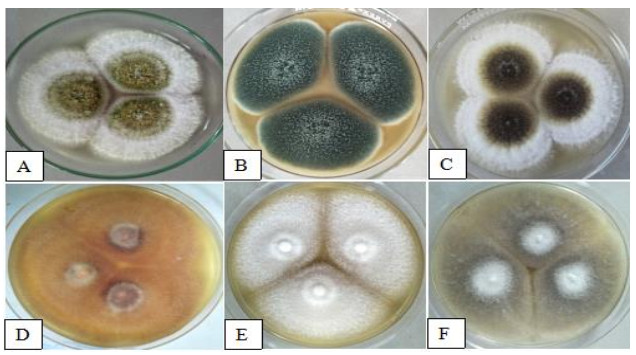
    Figure 2.  Cultural characteristics represented by A A. flavus. B A. fumigatus. C A. niger. D F. oxysporum. E F. solani. F M. velutinosus.

  • Observed morphological characteristics of the isolated fungi were described on the 7th day incubation were as follows: The stipes of A. flavus (Fig. 3A) appears rough and light brown, while the conidium was rough and have spherical surface. The conidia of A. fumigatus (Fig. 3B) is non septated and have globose shape with rough edges, conidiophores appears hyaline. Conidiophores of A. niger (Fig. 3C) are branched with lumped phialides. The conidiophores of F. oxysporum (Fig. 3D) were long with fusiform conidia, hypha is hyaline with branch monophialides. F. solani (Fig. 3E) shows narrow macro conidia, and long and branch monophialides. M. velutinosus (Fig. 3F) have globose sporangiospores, and formed septated hyphae.
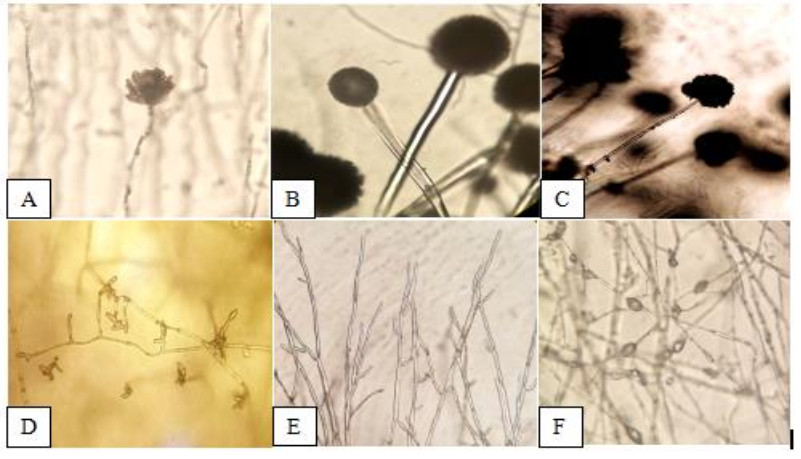
    Figure 3.  Morphological characteristics represented by A A. flavus. B A. fumigatus. C A. niger.D F. oxysporum. E F. solani. F M. velutinosus (400X).

  • The identities of fungal organisms isolated from the S. tuberosum infected with dry rot disease were confirmed through amplification and sequencing of the Internal Transcribed Spacer region (ITS) region using ITS1 and ITS4 primers. BLAST analysis showed the identities of fungal isolates, namely Aspergillus flavus FoF 09597 (100%), Aspergillus fumigatus FoF 10086 (100%), Aspergillus niger FoF 10087 (99.82%), Fusarium oxysporum FoF 03824 (95.06%) Fusarium solani FoF 01873 (100%) and Mucor velutinosus FoF 10088 (96.45%) as shown in Table 1.

    Table 1.  Identities of the identified fungi with Facaesoffungi number using BLAST with NCBI Genbank Accession number.

    Isolate No. FacesofFungi Number Species E-Value Identify Accession
    1 FoF 09597 Aspergillus flavus 0.0 100.00% MN511747.1
    2 FoF 10086 Aspergillus fumigatus 0.0 100.00% MF540309.1
    3 FoF 10087 Aspergillus niger 0.0 99.82% KY607770.1
    4 FoF 03824 Fusarium oxysporum 0.0 95.06% MG372014.1
    5 FoF 01873 Fusarium solani 0.0 100.00% MN202790.1
    6 FoF 10088 Mucor velutinosus 0.0 96.45% KY203942.1
  • Meanwhile, antagonistic interaction was observed in all fungal isolates (Table 2). Antagonism in all fungal isolates was identified as aggressor or victim. The observed interfungal parasitic relationships were hyphal penetration, hyphal coiling, hyphal denaturation, hyphal branching and lysed cells, all observed under the compound microscope (Figs 4-7) (Waing et al. 2015).

    Table 2.  Antagonistic interaction of isolated fungi in potato tuber infected with dry rot disease.

    Interacting Fungi Interaction
    M. velutinosus(+) A. flavus(-) Antagonism
    M. velutinosus(+) A. fumigatus(-) Antagonism
    M. velutinosus(+) F. solani(-) Antagonism
    M. velutinosus(+) F. oxysporum(-) Antagonism
    M. velutinosus(-) A. niger(+) Antagonism
    A. flavus(-) A. fumigatus(+) Antagonism
    A. flavus(+) F. solani(-) Antagonism
    A. flavus(-) F. oxysporum(+) Antagonism
    A. flavus(-) A. niger(+) Antagonism
    A. fumigatus(-) F. solani(+) Antagonism
    A. fumigatus(-) F. oxysporum(+) Antagonism
    A. fumigatus(-) A. niger(+) Antagonism
    F. solani(-) F. oxysporum(+) Antagonism
    F. solani(-) A. niger(-) Antagonism
    F. oxysporum F. oxysporum Antagonism
    F. oxysporum(+) A. niger(-) Antagonism
    (+) aggressor, (-) victim

    Figure 4.  Antagonistic interaction represented by; A M. velutinosus (left) and A. flavus (right). B hyphal denaturation and lysed cells of A. flavus (400x). C M. velutinosus (left) and A. fumigatus(right). D hyphal denaturation and lysed cells of A. fumigatus (400x). E M. velutinosus (left) and F. solani (right). F hyphal penetration of M. velutinosus to F. solani (400x). G M. velutinosus (left) and F. oxysporum (right). H hyphal penetration of M. velutinosus to F. oxysporum (400x). I M. velutinosus (left) and A. niger (right). J hyphal denaturation and lysed cells of M. velutinosus (400x).

    Figure 5.  Antagonistic interaction represented by; A A. flavus (left) and A. fumigatus (left). B lysed cells of conidiophores of A. flavus (400x). C A. flavus (left) and F. oxysporum (right). D hyphal coiling of F. oxysporum leading to hyphal denaturation of A. flavus (400x). E A. flavus (left) and A. niger (right). F hyphal denaturation of A. flavus (400x). G A. flavus (left) and F. solani (right). H hyphal denaturation and lysed cells of F. solani (400x).
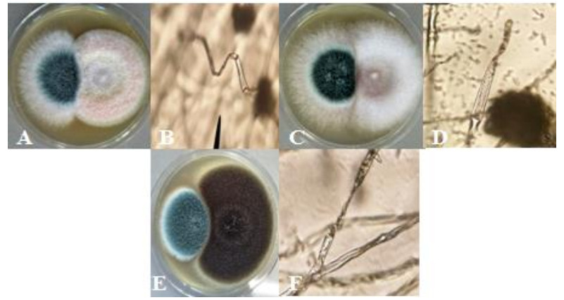
    Figure 6.  Antagonistic interaction represented by; A A. fumigatus (left) and F. solani (right).B hyphal denaturation of A. fumigatus (400x). CA. fumigatus (left) and F. oxysporum (right). D lysed cells of A. fumigatus (400x). EA. fumigatus (left) and A. niger (right). F lysed cells of A. fumigatus (400x).
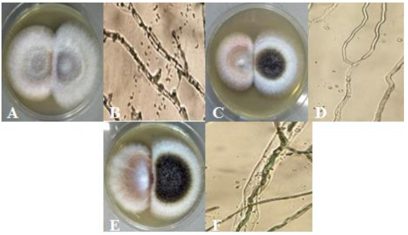
    Figure 7.  Antagonistic interaction represented by; A F. solani (left) and F. oxysporum (right). B hyphal penetration of F. oxysporum to F. solani (400x). C F. solani (left) and A. niger (right). D hyphal denaturation and lysed cell of F. solani (400x). E F. oxysporum (right) and A. niger (left). F hyphal penetration of F. oxysporum to A. niger (400x).

  • Observed characteristics of infected potato with dry rot disease are similar to the study of Wharton (2015). In addition, wounds caused by unavoidable process of harvesting potato allowed the manifestation of fungal organisms that later on led to dry rot disease (Wharton et al. 2007). The present study has isolated two species of Fusarium, F. solani and F. oxysporum in which based on the study conducted by Stefanczyk et al. (2016), Fusarium species have been associated with potato dry rot. Such, F. sambucinum, F. solani and F. oxysporum are the most frequent and devastating species. In addition, three species of Aspergillus namely: A. flavus, A. fumigatus and A. niger were also isolated. According to the study of Ibrahim & Tambuwal (2015), A. niger, and A. fumigatus were both pathogenic causing rotting of tomato fruit. Meanwhile, A. flavus was previously isolated from Dioscorea spp. rot (Okigbo & Ogbonnaya 2006). Moreover, M. velutinosus is one of the most frequent fungal endophyte identified from the rotted root samples of D. arayalpathra (Premalatha et al. 2014). Fungal epiphytes are found on the surface of the plants. Also, many of them are obligate parasites which penetrated the host plant and prevent the uptake of the nutrients as cited by Hongsanan et al. (2016). These isolated fungal species are confirmed through BLAST analysis. In which based on the previous study of Nagano et al. (2008) the use of ITS primer for fungal identification shows accurate and reliable result. Furthermore, ITS primer is the standard marker for fungal DNA barcoding (Bellemain et al. 2010) and is generally used for the assessment and characterization of fungal communities from environmental samples (George et al. 2019).

    The interaction study revealed antagonism between all identified fungi. Hyphal denaturation, hyphal coiling, hyphal penetration and lysed cells were the observed antagonistic interaction of hypha. In which, M. velutinosus showed the ability to penetrate the host hyphae of the F. solani and F. oxysporum which are considered as causative agents of potato dry rot. In microbial communities, there are different relationships and interactions characterized by mutualism to antagonism and parasitism (Duffy et al. 2003). These interactions happen when nutrients and space are limited (Arya & Perello 2010). Fungi that affects the other by harming them by means of suppressing the growth of other or killing them is mycoparasitism or hyperparasites (Jeffries 1995, Kumar & Gouda 2018). Its behavior which was observed in previous studies includes hyphal penetration, hyphal coiling, hyphal branching, hyphal denaturation, cell lysis, spore production, and barrier formation (Dubey & Dwivedi 1986, El-Debaiky 2017). As a result of these interactions, the host hyphae were vacuolated, shrank, collapsed, and disintegrated. Moreover, at the interface region of colonies, certain fungi were observed having irregular branched hyphae, cracked hyphae, ruptured cell wall, and dead cells at the left of antagonism line (Chet et al. 1981, Xing et al. 2005). The ability of mycoparasites to penetrate the host cell after coiling can lead to cell lysis because of the enzymatic interaction. Wherein, certain fungus penetrates internally and uses direct enzymatic lysis of hyphal cells. Hyphal coiling incorporated by means of hock between the hyphae and lysed cells can lead to fusion. In addition, hyphal cell wall lysis leads to penetration of the hyphae for obtaining nourishment to the host by means of haustoria (Dubey & Dwivede 1986, El-Debaiky 2017). However, certain host fungi counter attack by forming resting bodies. Also, host hypha accumulates cytoplasm to stop the infiltration of antagonist inside the cell. These host defense act as a barrier and survival measures against destruction in adverse condition and the over expression of pyridoxal reductase is a result of stress condition of a certain host fungi (Dubey & Dwivede 1986, Chamoun & Jabaji 2011, El-Debaiky 2017). According to the studies of Barnette (1963) and El-Debaiky (2017), the degree of damage by the parasites to the host cells depends to the resistance of host species. These studies show that certain parasites have direct contact to the host because of the absence of diffusible antibiotics which caused severe damage. In addition, some antagonist can produce spore inside the host hypha depending to the nutrition from the host fungi. Furthermore, according to Verma et al. (2007), there are several modes of actions that mycoparasitism exhibited which includes competition in space and nutrient, production of antibiosis and plant defense system induction. One evidence of this mode was observed by Waing et al. (2015) in which, the presence of zone of inhibition was observed when the two fungi are paired which indicates the production of antibiotics.

    Emerging fungal diseases in various agricultural crops due to rapid growth of new pathogens that are fungicide resistant posed a significant threat in agricultural productivity and risk to global food security (Fones et al. 2020). An example of which is the fungal disease known as Septoria tritici blotch (STB) that resulted to million per year in yield losses in UK growers alone (Fones & Gurr 2015). In order prevent loss due to fungal pathogens, fungicides were being applied. However, commercially available fungicides are expensive and became an added burden to farmers at the same time it is not environmental friendly (Gomathi & Ambikapathy 2011, Globe Newswire 2018). Thus, public concerns arise due to the hazardous effect of these harmful chemicals in the field of agriculture. Therefore, biocontrol microorganism has been stimulated as a substitute and answers for the pest control (Raaijmakers et al. 2002) and biocontrol pf pathogens. Several mycoparasites which belongs to the filamentous fungal genus were the best candidates for bio control agent against fungal pathogens (Kredics et al. 2003). Species exhibited extracellular enzyme such as polygalaturonase, amylase, protease and lipase which enable them to absorb nutrients (Gibb & Strohl 1987, Alvez et al. 2002). Thus, certain biocontrol agents not only function as pest control but can also enhance the uptake of nutrients in plants, slow down the occurrence of abiotic stresses and some can reduce physiological stresses (Shoresh et al. 2010).

Figure (7)  Table (2) References (45)
  • About this article
    Cite this article
    GAD Carreon, EE Gandalera, KGD Waing. 2021. Molecular identification and in vitro interaction of molds associated with dry rot of potato (Solanum tuberosum L.) collected in La Trinidad, Benguet, Philippines. Studies in Fungi 6(1):315−326 doi: 10.5943/sif/6/1/22
    GAD Carreon, EE Gandalera, KGD Waing. 2021. Molecular identification and in vitro interaction of molds associated with dry rot of potato (Solanum tuberosum L.) collected in La Trinidad, Benguet, Philippines. Studies in Fungi 6(1):315−326 doi: 10.5943/sif/6/1/22
  • Catalog

    • About this article

    /

    DownLoad:  Full-Size Img  PowerPoint
    Return
    Return