2021 Volume 6
Article Contents
About this article
ARTICLE   Open Access    

Diversity of wood – decaying fungi in Haikou Forestry Farm, Yunnan Province, P.R. China

More Information
  • Nine field trips carried out in Haikou Forestry Farm, Yunnan Province, P.R. China resulted in 681 specimens of wood-decaying fungi. The present paper summarizes 52 species collected that are distributed in 37 genera, 16 families, 6 orders including their hosts and substrates. A checklist of wood-decaying fungi in Haikou Forestry Farm is also given. Phylogenetic analysis of ITS nrRNA gene region was performed for all the collected samples with maximum likelihood, maximum parsimony and Bayesian inference methods. The phylogenetic tree showed that fifty-two species nested in sixteen families belonging to six orders in Agaricomycetes.
  • Aquaporins (AQPs) constitute a large family of transmembrane channel proteins that function as regulators of intracellular and intercellular water flow[1,2]. Since their first discovery in the 1990s, AQPs have been found not only in three domains of life, i.e., bacteria, eukaryotes, and archaea, but also in viruses[3,4]. Each AQP monomer is composed of an internal repeat of three transmembrane helices (i.e., TM1–TM6) as well as two half helixes that are formed by loop B (LB) and LE through dipping into the membrane[5]. The dual Asn-Pro-Ala (NPA) motifs that are located at the N-terminus of two half helixes act as a size barrier of the pore via creating an electrostatic repulsion of protons, whereas the so-called aromatic/arginine (ar/R) selectivity filter (i.e., H2, H5, LE1, and LE2) determines the substrate specificity by rendering the pore constriction site diverse in both size and hydrophobicity[59]. Based on sequence similarity, AQPs in higher plants could be divided into five subfamilies, i.e., plasma membrane intrinsic protein (PIP), tonoplast intrinsic protein (TIP), NOD26-like intrinsic protein (NIP), X intrinsic protein (XIP), and small basic intrinsic protein (SIP)[1017]. Among them, PIPs, which are typically localized in the cell membrane, are most conserved and play a central role in controlling plant water status[12,1822]. Among two phylogenetic groups present in the PIP subfamily, PIP1 possesses a relatively longer N-terminus and PIP2 features an extended C-terminus with one or more conserved S residues for phosphorylation modification[5,15,17].

    Tigernut (Cyperus esculentus L.), which belongs to the Cyperaceae family within Poales, is a novel and promising herbaceous C4 oil crop with wide adaptability, large biomass, and short life period[2327]. Tigernut is a unique species accumulating up to 35% oil in the underground tubers[2830], which are developed from stolons and the process includes three main stages, i.e., initiation, swelling, and maturation[3133]. Water is essential for tuber development and tuber moisture content maintains a relatively high level of approximately 85% until maturation when a significant drop to about 45% is observed[28,32]. Thereby, uncovering the mechanism of tuber water balance is of particular interest. Despite crucial roles of PIPs in the cell water balance, to date, their characterization in tigernut is still in the infancy[21]. The recently available genome and transcriptome datasets[31,33,34] provide an opportunity to address this issue.

    In this study, a global characterization of PIP genes was conducted in tigernut, including gene localizations, gene structures, sequence characteristics, and evolutionary patterns. Moreover, the correlation of CePIP mRNA/protein abundance with water content during tuber development as well as subcellular localizations were also investigated, which facilitated further elucidating the water balance mechanism in this special species.

    PIP genes reported in Arabidopsis (Arabidopsis thaliana)[10] and rice (Oryza sativa)[11] were respectively obtained from TAIR11 (www.arabidopsis.org) and RGAP7 (http://rice.uga.edu), and detailed information is shown in Supplemental Table S1. Their protein sequences were used as queries for tBLASTn[35] (E-value, 1e–10) search of the full-length tigernut transcriptome and genome sequences that were accessed from CNGBdb (https://db.cngb.org/search/assembly/CNA0051961)[31,34]. RNA sequencing (RNA-seq) reads that are available in NCBI (www.ncbi.nlm.nih.gov/sra) were also adopted for gene structure revision as described before[13], and presence of the conserved MIP (major intrinsic protein, Pfam accession number PF00230) domain in candidates was confirmed using MOTIF Search (www.genome.jp/tools/motif). To uncover the origin and evolution of CePIP genes, a similar approach was also employed to identify homologs from representative plant species, i.e., Carex cristatella (v1, Cyperaceae)[36], Rhynchospora breviuscula (v1, Cyperaceae)[37], and Juncus effusus (v1, Juncaceae)[37], whose genome sequences were accessed from NCBI (www.ncbi.nlm.nih.gov). Gene structure of candidates were displayed using GSDS 2.0 (http://gsds.gao-lab.org), whereas physiochemical parameters of deduced proteins were calculated using ProtParam (http://web.expasy.org/protparam). Subcellular localization prediction was conducted using WoLF PSORT (www.genscript.com/wolf-psort.html).

    Nucleotide and protein multiple sequence alignments were respectively conducted using ClustalW and MUSCLE implemented in MEGA6[38] with default parameters, and phylogenetic tree construction was carried out using MEGA6 with the maximum likelihood method and bootstrap of 1,000 replicates. Systematic names of PIP genes were assigned with two italic letters denoting the source organism and a progressive number based on sequence similarity. Conserved motifs were identified using MEME Suite 5.5.3 (https://meme-suite.org/tools/meme) with optimized parameters as follows: Any number of repetitions, maximum number of 15 motifs, and a width of 6 and 250 residues for each motif. TMs and conserved residues were identified using homology modeling and sequence alignment with the structure resolved spinach (Spinacia oleracea) SoPIP2;1[5].

    Synteny analysis was conducted using TBtools-II[39] as described previously[40], where the parameters were set as E-value of 1e-10 and BLAST hits of 5. Duplication modes were identified using the DupGen_finder pipeline[41], and Ks (synonymous substitution rate) and Ka (nonsynonymous substitution rate) of duplicate pairs were calculated using codeml in the PAML package[42]. Orthologs between different species were identified using InParanoid[43] and information from synteny analysis, and orthogroups (OGs) were assigned only when they were present in at least two species examined.

    Plant materials used for gene cloning, qRT-PCR analysis, and 4D-parallel reaction monitoring (4D-PRM)-based protein quantification were derived from a tigernut variety Reyan3[31], and plants were grown in a greenhouse as described previously[25]. For expression profiling during leaf development, three representative stages, i.e., young, mature, and senescing, were selected and the chlorophyll content was checked using SPAD-502Plus (Konica Minolta, Shanghai, China) as previously described[44]. Young and senescing leaves are yellow in appearance, and their chlorophyll contents are just half of that of mature leaves that are dark green. For diurnal fluctuation regulation, mature leaves were sampled every 4 h from the onset of light at 8 a.m. For gene regulation during tuber development, fresh tubers at 1, 5, 10, 15, 20, 25, and 35 d after tuber initiation (DAI) were collected as described previously[32]. All samples with three biological replicates were quickly frozen with liquid nitrogen and stored at −80 °C for further use. For subcellular localization analysis, tobacco (Nicotiana benthamiana) plants were grown as previously described[20].

    Tissue-specific expression profiles of CePIP genes were investigated using Illumina RNA-seq samples (150 bp paired-end reads) with three biological replicates for young leaf, mature leaf, sheath of mature leaf, shoot apex, root, rhizome, and three stages of developmental tuber (40, 85, and 120 d after sowing (DAS)), which are under the NCBI accession number of PRJNA703731. Raw sequence reads in the FASTQ format were obtained using fastq-dump, and quality control was performed using fastQC (www.bioinformatics.babraham.ac.uk/projects/fastqc). Read mapping was performed using HISAT2 (v2.2.1, https://daehwankimlab.github.io/hisat2), and relative gene expression level was presented as FPKM (fragments per kilobase of exon per million fragments mapped)[45].

    For qRT-PCR analysis, total RNA extraction and synthesis of the first-strand cDNA were conducted as previously described[24]. Primers used in this study are shown in Supplemental Table S2, where CeUCE2 and CeTIP41[25,33] were employed as two reference genes. PCR reaction in triplicate for each biological sample was carried out using the SYBR-green Mix (Takara) on a Real-time Thermal Cycler Type 5100 (Thermal Fisher Scientific Oy). Relative gene abundance was estimated with the 2−ΔΔCᴛ method and statistical analysis was performed using SPSS Statistics 20 as described previously[13].

    Raw proteomic data for tigernut roots, leaves, freshly harvested, dried, rehydrated for 48 h, and sprouted tubers were downloaded from ProteomeXchange/PRIDE (www.proteomexchange.org, PXD021894, PXD031123, and PXD035931), which were further analyzed using Maxquant (v1.6.15.0, www.maxquant.org). Three dominant members, i.e., CePIP1;1, -2;1, and -2;8, were selected for 4D-PRM quantification analysis, and related unique peptides are shown in Supplemental Table S3. Protein extraction, trypsin digestion, and LC-MS/MS analysis were conducted as described previously[46].

    For subcellular localization analysis, the coding region (CDS) of CePIP1;1, -2;1, and -2;8 were cloned into pNC-Cam1304-SubN via Nimble Cloning as described before[30]. Then, recombinant plasmids were introduced into Agrobacterium tumefaciens GV3101 with the helper plasmid pSoup-P19 and infiltration of 4-week-old tobacco leaves were performed as previously described[20]. For subcellular localization analysis, the plasma membrane marker HbPIP2;3-RFP[22] was co-transformed as a positive control. Fluorescence observation was conducted using confocal laser scanning microscopy imaging (Zeiss LMS880, Germany): The wavelength of laser-1 was set as 730 nm for RFP observation, where the fluorescence was excited at 561 nm; the wavelength of laser-2 was set as 750 nm for EGFP observation, where the fluorescence was excited at 488 nm; and the wavelength of laser-3 was set as 470 nm for chlorophyll autofluorescence observation, where the fluorescence was excited at 633 nm.

    As shown in Table 1, a total of 14 PIP genes were identified from eight tigernut scaffolds (Scfs). The CDS length varies from 831 to 882 bp, putatively encoding 276–293 amino acids (AA) with a molecular weight (MW) of 29.16–31.59 kilodalton (kDa). The theoretical isoelectric point (pI) varies from 7.04 to 9.46, implying that they are all alkaline. The grand average of hydropathicity (GRAVY) is between 0.344 and 0.577, and the aliphatic index (II) ranges from 94.57 to 106.90, which are consistent with the hydrophobic characteristic of AQPs[47]. As expected, like SoPIP2;1, all CePIPs include six TMs, two typical NPA motifs, the invariable ar/R filter F-H-T-R, five conserved Froger's positions Q/M-S-A-F-W, and two highly conserved residues corresponding to H193 and L197 in SoPIP2;1 that were proven to be involved in gating[5,48], though the H→F variation was found in CePIP2;9, -2;10, and -2;11 (Supplemental Fig. S1). Moreover, two S residues, corresponding to S115 and S274 in SoPIP2;1[5], respectively, were also found in the majority of CePIPs (Supplemental Fig. S1), implying their posttranslational regulation by phosphorylation.

    Table 1.  Fourteen PIP genes identified in C. esculentus.
    Gene name Locus Position Intron no. AA MW (kDa) pI GRAVY AI TM MIP
    CePIP1;1 CESC_15147 Scf9:2757378..2759502(–) 3 288 30.76 8.82 0.384 95.28 6 47..276
    CePIP1;2 CESC_04128 Scf4:3806361..3807726(–) 3 291 31.11 8.81 0.344 95.95 6 46..274
    CePIP1;3 CESC_15950 Scf54:5022493..5023820(+) 3 289 31.06 8.80 0.363 94.57 6 49..278
    CePIP2;1 CESC_15350 Scf9:879960..884243(+) 3 288 30.34 8.60 0.529 103.02 6 33..269
    CePIP2;2 CESC_00011 Scf30:4234620..4236549(+) 3 293 31.59 9.27 0.394 101.57 6 35..268
    CePIP2;3 CESC_00010 Scf30:4239406..4241658(+) 3 291 30.88 9.44 0.432 98.97 6 31..266
    CePIP2;4 CESC_05080 Scf46:307799..309544(+) 3 285 30.44 7.04 0.453 100.32 6 28..265
    CePIP2;5 CESC_05079 Scf46:312254..314388(+) 3 286 30.49 7.04 0.512 101.68 6 31..268
    CePIP2;6 CESC_05078 Scf46:316024..317780(+) 3 288 30.65 7.68 0.475 103.06 6 31..268
    CePIP2;7 CESC_05077 Scf46:320439..322184(+) 3 284 30.12 8.55 0.500 100.00 6 29..266
    CePIP2;8 CESC_14470 Scf2:4446409..4448999(+) 3 284 30.37 8.30 0.490 106.90 6 33..263
    CePIP2;9 CESC_02223 Scf1:2543928..2545778(–) 3 283 30.09 9.46 0.533 106.47 6 31..262
    CePIP2;10 CESC_10007 Scf27:1686032..1688010(–) 3 276 29.16 9.23 0.560 106.05 6 26..256
    CePIP2;11 CESC_10009 Scf27:1694196..1696175(–) 3 284 29.71 9.10 0.577 105.49 6 33..263
    AA: amino acid; AI: aliphatic index; GRAVY: grand average of hydropathicity; kDa: kilodalton; MIP: major intrinsic protein; MW: molecular weight; pI: isoelectric point; PIP: plasma membrane intrinsic protein; Scf: scaffold; TM: transmembrane helix.
     | Show Table
    DownLoad: CSV

    To uncover the evolutionary relationships, an unrooted phylogenetic tree was constructed using the full-length protein sequences of CePIPs together with 11 OsPIPs and 13 AtPIPs. As shown in Fig. 1a, these proteins were clustered into two main groups, corresponding to PIP1 and PIP2 as previously defined[10,49], and each appears to have evolved into several subgroups. Compared with PIP1s, PIP2s possess a relatively shorter N-terminal but an extended C-terminal with one conserved S residue (Supplemental Fig. S1). Interestingly, a high number of gene repeats were detected, most of which seem to be species-specific, i.e., AtPIP1;1/-1;2/-1;3/-1;4/-1;5, AtPIP2;1/-2;2/-2;3/-2;4/-2;5/-2;6, AtPIP2;7/-2;8, OsPIP1;1/-1;2/-1;3, OsPIP2;1/-2;4/-2;5, OsPIP2;2/-2;3, CePIP1;1/-1;2, CePIP2;2/-2;3, CePIP2;4/-2;5/-2;6/-2;7, and CePIP2;9/-2;10/-2;11, reflecting the occurrence of more than one lineage-specific whole-genome duplications (WGDs) after their divergence[50,51]. In Arabidopsis that experienced three WGDs (i.e. γ, β, and α) after the split with the monocot clade[52], AtPIP1;5 in the PIP1 group first gave rise to AtPIP1;1 via the γ WGD shared by all core eudicots[50], which latter resulted in AtPIP1;3, -1;4, and -1;2 via β and α WGDs; AtPIP2;1 in the PIP2 group first gave rise to AtPIP2;6 via the γ WGD, and they latter generated AtPIP2;2, and -2;5 via the α WGD (Supplemental Table S1). In rice, which also experienced three WGDs (i.e. τ, σ, and ρ) after the split with the eudicot clade[51], OsPIP1;2 and -2;3 generated OsPIP1;1 and -2;2 via the Poaceae-specific ρ WGD, respectively. Additionally, tandem, proximal, transposed and dispersed duplications also played a role on the gene expansion in these two species (Supplemental Table S1).

    Figure 1.  Structural and phylogenetic analysis of PIPs in C. esculentus, O. sativa, and A. thaliana. (a) Shown is an unrooted phylogenetic tree resulting from full-length PIPs with MEGA6 (maximum likelihood method and bootstrap of 1,000 replicates), where the distance scale denotes the number of amino acid substitutions per site. (b) Shown are the exon-intron structures. (c) Shown is the distribution of conserved motifs among PIPs, where different motifs are represented by different color blocks as indicated and the same color block in different proteins indicates a certain motif. (At: A. thaliana; Ce: C. esculentus; PIP: plasma membrane intrinsic protein; Os: O. sativa).

    Analysis of gene structures revealed that all CePIP and AtPIP genes possess three introns and four exons in the CDS, in contrast to the frequent loss of certain introns in rice, including OsPIP1;2, -1;3, -2;1, -2;3, -2;4, -2;5, -2;6, -2;7, and -2;8 (Fig. 1b). The positions of three introns are highly conserved, which are located in sequences encoding LB (three residues before the first NPA), LD (one residue before the conserved L involved in gating), and LE (18 residues after the second NPA), respectively (Supplemental Fig. S1). The intron length of CePIP genes is highly variable, i.e., 109–993 bp, 115–1745 bp, and 95–866 bp for three introns, respectively. By contrast, the exon length is relatively less variable: Exons 2 and 3 are invariable with 296 bp and 141 bp, respectively, whereas Exons 1 and 4 are of 277–343 bp and 93–132 bp, determining the length of N- and C-terminus of PIP1 and PIP2, respectively (Fig. 1b). Correspondingly, their protein structures were shown to be highly conserved, and six (i.e., Motifs 1–6) out of 15 motifs identified are broadly present. Among them, Motif 3, -2, -6, -1, and -4 constitute the conserved MIP domain. In contrast to a single Motif 5 present in most PIP2s, all PIP1s possess two sequential copies of Motif 5, where the first one is located at the extended N-terminal. In CePIP2;3 and OsPIP2;7, Motif 5 is replaced by Motif 13; in CePIP2;2, it is replaced by two copies of Motif 15; and no significant motif was detected in this region of CePIP2;10. PIP1s and PIP2s usually feature Motif 9 and -7 at the C-terminal, respectively, though it is replaced by Motif 12 in CePIP2;6 and OsPIP2;8. PIP2s usually feature Motif 8 at the N-terminal, though it is replaced by Motif 14 in CePIP2;2 and -2;3 or replaced by Motif 11 in CePIP2;10 and -2;11 (Fig. 1c).

    As shown in Fig. 2a, gene localization of CePIPs revealed three gene clusters, i.e., CePIP2;2/-2;3 on Scf30, CePIP2;4/-2;5/-2;6/-2;7 on Scf46, and CePIP2;10/-2;11 on Scf27, which were defined as tandem repeats for their high sequence similarities and neighboring locations. The nucleotide identities of these duplicate pairs vary from 70.5% to 91.2%, and the Ks values range from 0.0971 to 1.2778 (Table 2), implying different time of their birth. According to intra-species synteny analysis, two duplicate pairs, i.e., CePIP1;1/-1;2 and CePIP2;2/-2;4, were shown to be located within syntenic blocks (Fig. 2b) and thus were defined as WGD repeats. Among them, CePIP1;1/-1;2 possess a comparable Ks value to CePIP2;2/-2;3, CePIP1;1/-1;3, and CePIP2;4/-2;8 (1.2522 vs 1.2287–1.2778), whereas CePIP2;2/-2;4 harbor a relatively higher Ks value of 1.5474 (Table 2), implying early origin or fast evolution of the latter. While CePIP1;1/-1;3 and CePIP2;1/-2;8 were characterized as transposed repeats, CePIP2;1/-2;2, CePIP2;9/-2;10, and CePIP2;8/-2;10 were characterized as dispersed repeats (Fig. 2a). The Ks values of three dispersed repeats vary from 0.8591 to 3.0117 (Table 2), implying distinct times of origin.

    Figure 2.  Duplication events of CePIP genes and synteny analysis within and between C. esculentus, O. sativa, and A. thaliana. (a) Duplication events detected in tigernut. Serial numbers are indicated at the top of each scaffold, and the scale is in Mb. Duplicate pairs identified in this study are connected using lines in different colors, i.e., tandem (shown in green), transposed (shown in purple), dispersed (shown in gold), and WGD (shown in red). (b) Synteny analysis within and between C. esculentus, O. sativa, and A. thaliana. (c) Synteny analysis within and between C. esculentus, C. cristatella, R. breviuscula, and J. effusus. Shown are PIP-encoding chromosomes/scaffolds and only syntenic blocks that contain PIP genes are marked, i.e., red and purple for intra- and inter-species, respectively. (At: A. thaliana; Cc: C. cristatella; Ce: C. esculentus; Je: J. effusus; Mb: megabase; PIP: plasma membrane intrinsic protein; Os: O. sativa; Rb: R. breviuscula; Scf: scaffold; WGD: whole-genome duplication).
    Table 2.  Sequence identity and evolutionary rate of homologous PIP gene pairs identified in C. esculentus. Ks and Ka were calculated using PAML.
    Duplicate 1 Duplicate 2 Identity (%) Ka Ks Ka/Ks
    CePIP1;1 CePIP1;3 78.70 0.0750 1.2287 0.0610
    CePIP1;2 CePIP1;1 77.20 0.0894 1.2522 0.0714
    CePIP2;1 CePIP2;4 74.90 0.0965 1.7009 0.0567
    CePIP2;3 CePIP2;2 70.50 0.1819 1.2778 0.1424
    CePIP2;4 CePIP2;2 66.50 0.2094 1.5474 0.1353
    CePIP2;5 CePIP2;4 87.30 0.0225 0.4948 0.0455
    CePIP2;6 CePIP2;5 84.90 0.0545 0.5820 0.0937
    CePIP2;7 CePIP2;6 78.70 0.0894 1.0269 0.0871
    CePIP2;8 CePIP2;4 72.90 0.1401 1.2641 0.1109
    CePIP2;9 CePIP2;10 76.40 0.1290 0.8591 0.1502
    CePIP2;10 CePIP2;8 64.90 0.2432 3.0117 0.0807
    CePIP2;11 CePIP2;10 91.20 0.0562 0.0971 0.5783
    Ce: C. esculentus; Ka: nonsynonymous substitution rate; Ks: synonymous substitution rate; PIP: plasma membrane intrinsic protein.
     | Show Table
    DownLoad: CSV

    According to inter-species syntenic analysis, six out of 14 CePIP genes were shown to have syntelogs in rice, including 1:1, 1:2, and 2:2 (i.e. CePIP1;1 vs OsPIP1;3, CePIP1;3 vs OsPIP1;2/-1;1, CePIP2;1 vs OsPIP2;4, CePIP2;2/-2;4 vs OsPIP2;3/-2;2, and CePIP2;8 vs OsPIP2;6), in striking contrast to a single one found in Arabidopsis (i.e. CePIP1;2 vs AtPIP1;2). Correspondingly, only OsPIP1;2 in rice was shown to have syntelogs in Arabidopsis, i.e., AtPIP1;3 and -1;4 (Fig. 2b). These results are consistent with their taxonomic relationships that tigernut and rice are closely related[50,51], and also imply lineage-specific evolution after their divergence.

    As described above, phylogenetic and syntenic analyses showed that the last common ancestor of tigernut and rice is more likely to possess only two PIP1s and three PIP2s. However, it is not clear whether the gene expansion observed in tigernut is species-specific or Cyperaceae-specific. To address this issue, recently available genomes were used to identify PIP subfamily genes from C. cristatella, R. breviuscula, and J. effuses, resulting in 15, 13, and nine members, respectively. Interestingly, in contrast to a high number of tandem repeats found in Cyperaceae species, only one pair of tandem repeats (i.e., JePIP2;3 and -2;4) were identified in J. effusus, a close outgroup species to Cyperaceae in the Juncaceae family[36,37]. According to homologous analysis, a total of 12 orthogroups were identified, where JePIP genes belong to PIP1A (JePIP1;1), PIP1B (JePIP1;2), PIP1C (JePIP1;3), PIP2A (JePIP2;1), PIP2B (JePIP2;2), PIP2F (JePIP2;3 and -2;4), PIP2G (JePIP2;5), and PIP2H (JePIP2;6) (Table 3). Further intra-species syntenic analysis revealed that JePIP1;1/-1;2 and JePIP2;2/-2;3 are located within syntenic blocks, which is consistent with CePIP1;1/-1;2, CePIP2;2/-2;4, CcPIP1;1/-1;2, CcPIP2;3/-2;4, RbPIP1;1/-1;2, and RbPIP2;2/-2;5 (Fig. 2c), implying that PIP1A/PIP1B and PIP2B/PIP2D were derived from WGDs occurred sometime before Cyperaceae-Juncaceae divergence. After the split with Juncaceae, tandem duplications frequently occurred in Cyperaceae, where PIP2B/PIP2C and PIP2D/PIP2E/PIP2F retain in most Cyperaceae plants examined in this study. By contrast, species-specific expansion was also observed, i.e., CePIP2;4/-2;5, CePIP2;10/-2;11, CcPIP1;2/-1;3, CcPIP2;4/-2;5, CcPIP2;8/-2;9, CcPIP2;10/-2;11, RbPIP2;3/-2;4, and RbPIP2;9/-2;10 (Table 3 & Fig. 2c).

    Table 3.  Twelve proposed orthogroups based on comparison of representative plant species.
    Orthogroup C. esculentus C. cristatella R. breviuscula J. effusus O. sativa A. thaliana
    PIP1A CePIP1;1 CcPIP1;1 RbPIP1;1 JePIP1;1 OsPIP1;3 AtPIP1;1, AtPIP1;2,
    AtPIP1;3, AtPIP1;4,
    AtPIP1;5
    PIP1B CePIP1;2 CcPIP1;2, CcPIP1;3 RbPIP1;2 JePIP1;2
    PIP1C CePIP1;3 CcPIP1;4 RbPIP1;3 JePIP1;3 OsPIP1;1, OsPIP1;2
    PIP2A CePIP2;1 CcPIP2;1 RbPIP2;1 JePIP2;1 OsPIP2;1, OsPIP2;4,
    OsPIP2;5
    AtPIP2;1, AtPIP2;2,
    AtPIP2;3, AtPIP2;4,
    AtPIP2;5, AtPIP2;6
    PIP2B CePIP2;2 CcPIP2;2 RbPIP2;2 JePIP2;2 OsPIP2;2, OsPIP2;3
    PIP2C CePIP2;3 CcPIP2;3 RbPIP2;3, RbPIP2;4
    PIP2D CePIP2;4, CePIP2;5 CcPIP2;4, CcPIP2;5 RbPIP2;5
    PIP2E CePIP2;5 CcPIP2;5 RbPIP2;6
    PIP2F CePIP2;6 CcPIP2;6
    PIP2G CePIP2;7 CcPIP2;7 RbPIP2;7 JePIP2;3, JePIP2;4
    PIP2H CePIP2;8 CcPIP2;8, CcPIP2;9 RbPIP2;8 JePIP2;5 OsPIP2;6 AtPIP2;7, AtPIP2;8
    PIP2I CePIP2;9, CePIP2;10,
    CePIP2;11
    CcPIP2;10, CcPIP2;11 RbPIP2;9, RbPIP2;10 JePIP2;6 OsPIP2;7, OsPIP2;8
    At: A. thaliana; Cc: C. cristatella; Ce: C. esculentus; Je: J. effuses; Os: O. sativa; Rb: R. breviuscula; PIP: plasma membrane intrinsic protein.
     | Show Table
    DownLoad: CSV

    Tissue-specific expression profiles of CePIP genes were investigated using transcriptome data available for young leaf, mature leaf, sheath, root, rhizome, shoot apex, and tuber. As shown in Fig. 3a, CePIP genes were mostly expressed in roots, followed by sheaths, moderately in tubers, young leaves, rhizomes, and mature leaves, and lowly in shoot apexes. In most tissues, CePIP1;1, -2;1, and -2;8 represent three dominant members that contributed more than 90% of total transcripts. By contrast, in rhizome, these three members occupied about 80% of total transcripts, which together with CePIP1;3 and -2;4 contributed up to 96%; in root, CePIP1;1, -1;3, -2;4, and -2;7 occupied about 84% of total transcripts, which together with CePIP2;1 and -2;8 contributed up to 94%. According to their expression patterns, CePIP genes could be divided into five main clusters: Cluster I includes CePIP1;1, -2;1, and -2;8 that were constitutively and highly expressed in all tissues examined; Cluster II includes CePIP2;2, -2;9, and -2;10 that were lowly expressed in all tested tissues; Cluster III includes CePIP1;2 and -2;11 that were preferentially expressed in young leaf and sheath; Cluster IV includes CePIP1;3 and -2;4 that were predominantly expressed in root and rhizome; and Cluster V includes remains that were typically expressed in root (Fig. 3a). Collectively, these results imply expression divergence of most duplicate pairs and three members (i.e. CePIP1;1, -2;1, and -2;8) have evolved to be constitutively co-expressed in most tissues.

    Figure 3.  Expression profiles of CePIP genes in various tissues, different stages of leaf development, and mature leaves of diurnal fluctuation. (a) Tissue-specific expression profiles of 14 CePIP genes. The heatmap was generated using the R package implemented with a row-based standardization. Color scale represents FPKM normalized log2 transformed counts, where blue indicates low expression and red indicates high expression. (b) Expression profiles of CePIP1;1, -2;1, and -2;8 at different stages of leaf development. (c) Expression profiles of CePIP1;1, -2;1, and -2;8 in mature leaves of diurnal fluctuation. Bars indicate SD (N = 3) and uppercase letters indicate difference significance tested following Duncan's one-way multiple-range post hoc ANOVA (p< 0.01). (Ce: C. esculentus; FPKM: Fragments per kilobase of exon per million fragments mapped; PIP: plasma membrane intrinsic protein)

    As shown in Fig. 3a, compared with young leaves, transcriptome profiling showed that CePIP1;2, -2;3, -2;7, -2;8, and -2;11 were significantly down-regulated in mature leaves, whereas CePIP1;3 and -2;1 were up-regulated. To confirm the results, three dominant members, i.e., CePIP1;1, -2;1, and -2;8, were selected for qRT-PCR analysis, which includes three representative stages, i.e., young, mature, and senescing leaves. As shown in Fig. 3b, in contrast to CePIP2;1 that exhibited a bell-like expression pattern peaking in mature leaves, transcripts of both CePIP1;1 and -2;8 gradually decreased during leaf development. These results were largely consistent with transcriptome profiling, and the only difference is that CePIP1;1 was significantly down-regulated in mature leaves relative to young leaves. However, this may be due to different experiment conditions used, i.e., greenhouse vs natural conditions.

    Diurnal fluctuation expression patterns of CePIP1;1, -2;1, and -2;8 were also investigated in mature leaves and results are shown in Fig. 3c. Generally, transcripts of all three genes in the day (8, 12, 16, and 20 h) were higher than that in the night (24 and 4 h). During the day, both CePIP1;1 and -2;8 exhibited an unimodal expression pattern that peaked at 12 h, whereas CePIP2;1 possessed two peaks (8 and 16 h) and their difference was not significant. Nevertheless, transcripts of all three genes at 20 h (onset of night) were significantly lower than those at 8 h (onset of day) as well as 12 h. In the night, except for CePIP2;1, no significant difference was observed between the two stages for both CePIP1;1 and -2;8. Moreover, their transcripts were comparable to those at 20 h (Fig. 3c).

    To reveal the expression patterns of CePIP genes during tuber development, three representative stages, i.e., 40 DAS (early swelling stage), 85 DAS (late swelling stage), and 120 DAS (mature stage), were first profiled using transcriptome data. As shown in Fig. 4a, except for rare expression of CePIP1;2, -2;2, -2;9, and -2;10, most genes exhibited a bell-like expression pattern peaking at 85 DAS, in contrast to a gradual decrease of CePIP2;3 and -2;8. Notably, except for CePIP2;4, other genes were expressed considerably lower at 120 DAS than that at 40 DAS. For qRT-PCR confirmation of CePIP1;1, -2;1, and -2;8, seven stages were examined, i.e., 1, 5, 10, 15, 20, 25, and 35 DAI, which represent initiation, five stages of swelling, and maturation as described before[32]. As shown in Fig. 4b, two peaks were observed for all three genes, though their patterns were different. As for CePIP1;1, compared with the initiation stage (1 DAI), significant up-regulation was observed at the early swelling stage (5 DAI), followed by a gradual decrease except for the appearance of the second peak at 20 DAI, which is something different from transcriptome profiling. As for CePIP2;1, a sudden drop of transcripts first appeared at 5 DAI, then gradually increased until 20 DAI, which was followed by a gradual decrease at two late stages. The pattern of CePIP2;8 is similar to -1;1, two peaks appeared at 5 and 20 DAI and the second peak was significantly lower than the first. The difference is that the second peak of CePIP2;8 was significantly lower than the initiation stage. By contrast, the second peak (20 DAI) of CePIP2;1 was significantly higher than that of the first one (1 DAI). Nevertheless, the expression patterns of both CePIP2;1 and -2;8 are highly consistent with transcriptome profiling.

    Figure 4.  Transcript and protein abundances of CePIP genes during tuber development. (a) Transcriptome-based expression profiling of 14 CePIP genes during tuber development. The heatmap was generated using the R package implemented with a row-based standardization. Color scale represents FPKM normalized log2 transformed counts, where blue indicates low expression and red indicates high expression. (b) qRT-PCR-based expression profiling of CePIP1;1, -2;1, and -2;8 in seven representative stages of tuber development. (c) Relative protein abundance of CePIP1;1, -2;1, and -2;8 in three representative stages of tuber development. Bars indicate SD (N = 3) and uppercase letters indicate difference significance tested following Duncan's one-way multiple-range post hoc ANOVA (p < 0.01). (Ce: C. esculentus; DAI: days after tuber initiation; DAS: days after sowing; FPKM: Fragments per kilobase of exon per million fragments mapped; PIP: plasma membrane intrinsic protein).

    Since protein abundance is not always in agreement with the transcript level, protein profiles of three dominant members (i.e. CePIP1;1, -2;1, and -2;8) during tuber development were further investigated. For this purpose, we first took advantage of available proteomic data to identify CePIP proteins, i.e., leaves, roots, and four stages of tubers (freshly harvested, dried, rehydrated for 48 h, and sprouted). As shown in Supplemental Fig. S2, all three proteins were identified in both leaves and roots, whereas CePIP1;1 and -2;8 were also identified in at least one of four tested stages of tubers. Notably, all three proteins were considerably more abundant in roots, implying their key roles in root water balance.

    To further uncover their profiles during tuber development, 4D-PRM-based protein quantification was conducted in three representative stages of tuber development, i.e., 1, 25, and 35 DAI. As expected, all three proteins were identified and quantified. In contrast to gradual decrease of CePIP2;8, both CePIP1;1 and -2;1 exhibited a bell-like pattern that peaked at 25 DAI, though no significant difference was observed between 1 and 25 DAI (Fig. 4c). The trends are largely in accordance with their transcription patterns, though the reverse trend was observed for CePIP2;1 at two early stages (Fig. 4b & Fig. 4c).

    As predicted by WoLF PSORT, CePIP1;1, -2;1, and -2;8 may function in the cell membrane. To confirm the result, subcellular localization vectors named pNC-Cam1304-CePIP1;1, pNC-Cam1304-CePIP2;1, and pNC-Cam1304-CePIP2;8 were further constructed. When transiently overexpressed in tobacco leaves, green fluorescence signals of all three constructs were confined to cell membranes, highly coinciding with red fluorescence signals of the plasma membrane marker HbPIP2;3-RFP (Fig. 5).

    Figure 5.  (a) Schematic diagram of overexpressing constructs, (b) subcellular localization analysis of CePIP1;1, -2;1, and -2;8 in N. benthamiana leaves. (35S: cauliflower mosaic virus 35S RNA promoter; Ce: C. esculentus; EGFP: enhanced green fluorescent protein; kb: kilobase; NOS: terminator of the nopaline synthase gene; RFP: red fluorescent protein; PIP: plasma membrane intrinsic protein).

    Water balance is particularly important for cell metabolism and enlargement, plant growth and development, and stress responses[2,19]. As the name suggests, AQPs raised considerable interest for their high permeability to water, and plasma membrane-localized PIPs were proven to play key roles in transmembrane water transport between cells[1,18]. The first PIP was discovered in human erythrocytes, which was named CHIP28 or AQP1, and its homolog in plants was first characterized in Arabidopsis, which is known as RD28, PIP2c, or AtPIP2;3[3,7,53]. Thus far, genome-wide identification of PIP genes have been reported in a high number of plant species, including two model plants Arabidopsis and rice[10,11,1317,5456]. By contrast, little information is available on Cyperaceae, the third largest family within the monocot clade that possesses more than 5,600 species[57].

    Given the crucial roles of water balance for tuber development and crop production, in this study, tigernut, a representative Cyperaceae plant producing high amounts of oil in underground tubers[28,30,32], was employed to study PIP genes. A number of 14 PIP genes representing two phylogenetic groups (i.e., PIP1 and PIP2) or 12 orthogroups (i.e., PIP1A, PIP1B, PIP1C, PIP2A, PIP2B, PIP2C, PIP2D, PIP2E, PIP2F, PIP2G, PIP2H, and PIP2I) were identified from the tigernut genome. Though the family amounts are comparative or less than 13–21 present in Arabidopsis, cassava (Manihot esculenta), rubber tree (Hevea brasiliensis), poplar (Populus trichocarpa), C. cristatella, R. breviuscula, banana (Musa acuminata), maize (Zea mays), sorghum (Sorghum bicolor), barley (Hordeum vulgare), and switchgrass (Panicum virgatum), they are relatively more than four to 12 found in eelgrass (Zostera marina), Brachypodium distachyon, foxtail millet (Setaria italic), J. effuses, Aquilegia coerulea, papaya (Carica papaya), castor been (Ricinus communis), and physic nut (Jatropha curcas) (Supplemental Table S4). Among them, A. coerulea represents a basal eudicot that didn't experience the γ WGD shared by all core eudicots[50], whereas eelgrass is an early diverged aquatic monocot that didn't experience the τ WGD shared by all core monocots[56]. Interestingly, though both species possess two PIP1s and two PIP2s, they were shown to exhibit complex orthologous relationships of 1:1, 2:2, 1:0, and 0:1 (Supplemental Table S5). Whereas AcPIP1;1/AcPIP1;2/ZmPIP1;1/ZmPIP1;2 and ZmPIP2;1/AcPIP2;1 belong to PIP1A and PIP2A identified in this study, AcPIP2;2 and ZmPIP2;2 belong to PIP2H and PIP2I, respectively (Supplemental Table S5), implying that the last common ancestor of monocots and eudicots possesses only one PIP1 and two PIP2s followed by clade-specific expansion. A good example is the generation of AtPIP1;1 and -2;6 from AtPIP1;5 and -2;1 via the γ WGD, respectively[17].

    In tigernut, extensive expansion of the PIP subfamily was contributed by WGD (2), transposed (2), tandem (5), and dispersed duplications (3). It's worth noting that, two transposed repeats (i.e., CePIP1;1/-1;3 and CePIP2;1/-2;8) are shared by rice, implying their early origin that may be generated sometime after the split with the eudicot clade but before Cyperaceae-Poaceae divergence. By contrast, two WGD repeats (i.e., CePIP1;1/-1;2 and CePIP2;2/-2;4) are shared by C. cristatella, R. breviuscula, and J. effusus but not rice and Arabidopsis, implying that they may be derived from WGDs that occurred sometime after Cyperaceae-Poaceae split but before Cyperaceae-Juncaceae divergence. The possible WGD is the one that was described in C. littledalei[58], though the exact time still needs to be studied. Interestingly, compared with Arabidopsis (1) and rice (2), tandem/proximal duplications played a more important role in the expansion of PIP genes in tigernut (5) as well as other Cyperaceae species tested (5–6), which were shown to be Cyperaceae-specific or even species-specific. These tandem repeats may play a role in the adaptive evolution of Cyperaceae species as described in a high number of plant species[14,41]. According to comparative genomics analyses, tandem duplicates experienced stronger selective pressure than genes formed by other modes (WGD, transposed duplication, and dispersed duplication) and evolved toward biased functional roles involved in plant self-defense[41].

    As observed in most species such as Arabidopsis[10,1417], PIP genes in all Cyperaceae and Juncaceae species examined in this study, i.e., tigernut, C. cristatella, R. breviuscula, and J. effuses, feature three introns with conserved positions. By contrast, zero to three introns was not only found in rice but also in other Poaceae species such as maize, sorghum, foxtail millet, switchgrass, B. distachyon, and barley[54,55], implying lineage/species-specific evolution.

    Despite the extensive expansion of PIP genes (PIP2) in tigernut even after the split with R. breviuscula, CePIP1;1, -2;1, and -2;8 were shown to represent three dominant members in most tissues examined in this study, i.e., young leaf, mature leaf, sheath, rhizome, shoot apex, and tuber, though the situation in root is more complex. CePIP1;1 was characterized as a transposed repeat of CePIP1;3, which represents the most expressed member in root. Moreover, its recent WGD repeat CePIP1;2 was shown to be lowly expressed in most tested tissues, implying their divergence. The ortholog of CePIP1;1 in rice is OsPIP1;3 (RWC-3), which was shown to be preferentially expressed in roots, stems, and leaves, in contrast to constitutive expression of OsPIP1;1 (OsPIP1a) and -1;2[5961], two recent WGD repeats. Injecting the cRNA of OsPIP1;3 into Xenopus oocytes could increase the osmotic water permeability by 2–3 times[60], though the activity is considerably lower than PIP2s such as OsPIP2;2 and -2;2[6163]. Moreover, OsPIP1;3 was shown to play a role in drought avoidance in upland rice and its overexpression in lowland rice could increase root osmotic hydraulic conductivity, leaf water potential, and relative cumulative transpiration at the end of 10 h PEG treatment[64]. CePIP2;8 was characterized as a transposed repeat of CePIP2;1. Since their orthologs are present in both rice and Arabidopsis (Supplemental Table S3), the duplication event is more likely to occur sometime before monocot-eudicot split. Interestingly, their orthologs in rice, i.e., OsPIP2;1 (OsPIP2a) and -2;6, respectively, are also constitutively expressed[61], implying a conserved evolution with similar functions. When heterologously expressed in yeast, OsPIP2;1 was shown to exhibit high water transport activity[62,6466]. Moreover, root hydraulic conductivity was decreased by approximately four folds in OsPIP2;1 RNAi knock-down rice plants[64]. The water transport activity of OsPIP2;6 has not been tested, however, it was proven to be an H2O2 transporter that is involved in resistance to rice blast[61]. More work especially transgenic tests may improve our knowledge of the function of these key CePIP genes.

    Leaf is a photosynthetic organ that regulates water loss through transpiration. In tigernut, PIP transcripts in leaves were mainly contributed by CePIP1;1, -2;1, and -2;8, implying their key roles. During leaf development, in contrast to gradual decrease of CePIP1;1 and -2;8 transcripts in three stages (i.e. young, mature, and senescing) examined in this study, CePIP2;1 peaked in mature leaves. Their high abundance in young leaves is by cell elongation and enlargement at this stage, whereas upregulation of CePIP2;1 in mature leaves may inform its possible role in photosynthesis[67]. Thus far, a high number of CO2 permeable PIPs have been identified, e.g., AtPIP2;1, HvPIP2;1, HvPIP2;2, HvPIP2;3, HvPIP2;5, and SiPIP2;7[6870]. Moreover, in mature leaves, CePIP1;1, -2;1, and -2;8 were shown to exhibit an apparent diurnal fluctuation expression pattern that was expressed more in the day and usually peaked at noon, which reflects transpiration and the fact that PIP genes are usually induced by light[11,7173]. In rice, OsPIP2;4 and -2;5 also showed a clear diurnal fluctuation in roots that peaked at 3 h after the onset of light and dropped to a minimum 3 h after the onset of darkness[11]. Notably, further studies showed that temporal and dramatic induction of OsPIP2;5 around 2 h after light initiation was triggered by transpirational demand but not circadian rhythm[74].

    As an oil-bearing tuber crop, the main economic goal of tigernut cultivation is to harvest underground tubers, whose development is highly dependent on water available[32,75]. According to previous studies, the moisture content of immature tigernut tubers maintains more than 80.0%, followed by a seed-like dehydration process with a drop of water content to less than 50% during maturation[28,32]. Thereby, the water balance in developmental tubers must be tightly regulated. Like leaves, the majority of PIP transcripts in tubers were shown to be contributed by CePIP1;1, -2;1, and -2;8, which was further confirmed at the protein level. In accordance with the trend of water content during tuber development, mRNA, and protein abundances of CePIP1;1, -2;1, and -2;8 in initiation and swelling tubers were considerably higher than that at the mature stage. High abundances of CePIP1;1, -2;1, and -2;8 at the initiation stage reflects rapid cell division and elongation, whereas upregulation of CePIP1;1 and -2;1 at the swelling stage is in accordance with cell enlargement and active physiological metabolism such as rapid oil accumulation[28,30]. At the mature stage, downregulation of PIP transcripts and protein abundances resulted in a significant drop in the moisture content, which is accompanied by the significant accumulation of late embryogenesis-abundant proteins[23,32]. The situation is highly distinct from other tuber plants such as potato (Solanum tuberosum), which may contribute to the difference in desiccation resistance between two species[32,76]. It's worth noting that, in one study, CePIP2;1 was not detected in any of the four tested stages, i.e., freshly harvested, dried, rehydrated for 48 h, and sprouted tubers[23]. By contrast, it was quantified in all three stages of tuber development examined in this study, i.e., 1, 25, and 35 DAI (corresponding to freshly harvested tubers), which represent initiation, swelling, and maturation. One possible reason is that the protein abundance of CePIP2;1 in mature tubers is not high enough to be quantified by nanoLC-MS/MS, which is relatively less sensitive than 4D-PRM used in this study[30,46]. In fact, nanoLC-MS/MS-based proteomic analysis of 30 samples representing six tissues/stages only resulted in 2,257 distinct protein groups[23].

    Taken together, our results imply a key role of CePIP1;1, -2;1, and -2;8 in tuber water balance, however, the mechanism underlying needs to be further studied, e.g., posttranslational modifications, protein interaction patterns, and transcriptional regulators.

    To our knowledge, this is the first genome-wide characterization of PIP genes in tigernut, a representative Cyperaceae plant with oil-bearing tubers. Fourteen CePIP genes representing two phylogenetic groups or 12 orthogroups are relatively more than that present in two model plants rice and Arabidopsis, and gene expansion was mainly contributed by WGD and transposed/tandem duplications, some of which are lineage or even species-specific. Among these genes, CePIP1;1, -2;1, and -2;8 have evolved to be three dominant members that are constitutively expressed in most tissues, including leaf and tuber. Transcription of these three dominant members in leaves are subjected to development and diurnal regulation, whereas in tubers, their mRNA and protein abundances are positively correlated with the moisture content during tuber development. Moreover, their plasma membrane-localization was confirmed by subcellular localization analysis, implying that they may function in the cell membrane. These findings shall not only provide valuable information for further uncovering the mechanism of tuber water balance but also lay a solid foundation for genetic improvement by regulating these key PIP members in tigernut.

    The authors confirm contribution to the paper as follows: study conception and design, supervision: Zou Z; analysis and interpretation of results: Zou Z, Zheng Y, Xiao Y, Liu H, Huang J, Zhao Y; draft manuscript preparation: Zou Z, Zhao Y. All authors reviewed the results and approved the final version of the manuscript.

    All the relevant data is available within the published article.

    This work was supported by the Hainan Province Science and Technology Special Fund (ZDYF2024XDNY171 and ZDYF2024XDNY156), China; the National Natural Science Foundation of China (32460342, 31971688 and 31700580), China; the Project of Sanya Yazhou Bay Science and Technology City (SCKJ-JYRC-2022-66), China. The funders had no role in study design, data collection and analysis, decision to publish, or preparation of the manuscript.

  • The authors declare that they have no conflict of interest.

  • Ayesha P, Maham A, Ariza A, Husnain H. 2020 – "Ganoderma lucidum sensu lato" – a sacred mushroom for immortality. Studies in Fungi 5(1), 508–516. Doi10.5943/sif/5/1/31 doi: 10.5943/sif/5/1/31

    CrossRef    Google Scholar

    Blanco-Dios JB. 2021 – Notes on the genus Entoloma s. l. in the Northwest of the Iberian Peninsula (XI): a new species in subgenus Entoloma. Studies in Fungi 6(1), 64–70. Doi10.5943/sif/6/1/2 doi: 10.5943/sif/6/1/2

    CrossRef    Google Scholar

    Chen JZ, Zhao CL. 2020 – Morphological and molecular identification of four new resupinate species of Lyomyces (Hymenochaetales) from southern China. MycoKeys 65, 101–118. Doi10.3897/mycokeys. 65.48660 doi: 10.3897/mycokeys.65.48660

    CrossRef    Google Scholar

    Cui BK, Dai YC. 2012 – Wood-decaying fungi in eastern Himalayas 3. Polypores from Laojunshan Mountains, Yunnan Province. Mycosystema 31(4), 486–492.

    Google Scholar

    Dai YC. 2011 – A revised checklist of corticioid and hydnoid fungi in China for 2010. Mycoscience 52, 69–79. Doi: 10.1007/S10267-010-0068-1

    CrossRef    Google Scholar

    Dai YC. 2012a – Polypore diversity in China with an annotated checklist of Chinese polypores. Mycoscience 53, 49–80. Doi10.1007/s10267-011-0134-3 doi: 10.1007/s10267-011-0134-3

    CrossRef    Google Scholar

    Dai YC. 2012b – Pathogenic wood-decaying fungi on woody plants in China. Mycosystema 31(4), 493–509.

    Google Scholar

    Dai YC, Cui BK, Si J, He SH et al. 2015 – Dynamics of the worldwide number of fungi with emphasis on fungal diversity in China. Mycological Progress 14, 62. Doi10.1007/s11557-015-1084-5 doi: 10.1007/s11557-015-1084-5

    CrossRef    Google Scholar

    Dai YC, Yang ZL. 2008 – A revised checklist of medicinal fungi in China. Mycosystema 27(6), 801–824. Doi10.3969/j. issn. 1672-6472.2008.06.001

    Google Scholar

    Dai YC, Zhou LW, Yang ZL, Wen HA et al. 2010 – A revised checklist of edible fungi in China. Mycosystema 29(1), 1–21.

    Google Scholar

    De Leon AM, Dulay AR, Villanueva AL, Kalaw SP. 2020 – Optimal culture conditions and toxicity assessment of Fomitopsis feei (Fr. ): a newly documented macro fungus from Philippines. Studies in Fungi 5(1), 491–507. Doi10.5943/sif/5/1/30 doi: 10.5943/sif/5/1/30

    CrossRef    Google Scholar

    Felsenstein J. 1985 – Confidence intervals on phylogenetics: an approach using bootstrap. Evolution 39, 783–791. Doi10.1111/j. 1558-5646.1985. tb00420. x doi: 10.1111/j.1558-5646.1985.tb00420.x

    CrossRef    Google Scholar

    Freitas-Neto JF, Sousa JO, Ovrebo CL, Baseia IG. 2019 – Geastrum echinulatum and G. rusticum (Geastraceae, Basidiomycota) – two new records for Central America. Studies in Fungi 4(1), 14–20. Doi10.5943/sif/4/1/2 doi: 10.5943/sif/4/1/2

    CrossRef    Google Scholar

    Garcia-Sandoval R, Wang Z, Binder M, Hibbett DS. 2011 – Molecular phylogenetics of the Gloeophyllales and relative ages of clades of Agaricomycotina producing a brown rot. Mycologia 103(3), 510–524. Doi10.3852/10-209 doi: 10.3852/10-209

    CrossRef    Google Scholar

    Hall TA. 1999 – Bioedit: a user-friendly biological sequence alignment editor and analysis program for Windows 95/98/NT. Nucleic Acids Symposium Series 41, 95–98.

    Google Scholar

    He MQ, Zhao RL, Hyde KD, Begerow D et al. 2019 – Notes, outline and divergence times of Basidiomycota. Fungal Diversity 99(1), 105–367. Doi10.1007/s13225-019-00435-4 doi: 10.1007/s13225-019-00435-4

    CrossRef    Google Scholar

    He X, Shi ZJ, Zhao CL. 2020 – Morphological and molecular identification of two new species of Tubulicrinis (Hymenochaetaceae, Hymenochaetales) from southern china. Mycoscience 61(4), 184–189. Doi10.1016/j. myc. 2020.03.008 doi: 10.1016/j.myc.2020.03.008

    CrossRef    Google Scholar

    Himani S, Krishnappa M. 2020 – Xylaria oxyacanthae (Xylariaceae), a new record on Diospyros melanoxylon from India. Studies in Fungi 5(1), 485–490. Doi10.5943/sif/5/1/29 doi: 10.5943/sif/5/1/29

    CrossRef    Google Scholar

    Hipol RM, Baldelomar JA, Bolinget KC, Solis AFF. 2019 – The soil fungi producing siderophores of Mt. Yangbew, Tawang, La Trinidad, Benguet. Studies in Fungi 4(1), 1–13. Doi10.5943/sif/4/1/1

    Google Scholar

    Hood IA, Ramsfield TD. 2016 – Armillaria aotearoa species nova. New Zealand Journal of Forestry Science 46(1), 2. Doi10.1186/s40490-016-0058-y doi: 10.1186/s40490-016-0058-y

    CrossRef    Google Scholar

    Huang RX, Luo KY, Zhao CL. 2020 – Phlebia nigrodontea sp. nov. in Meruliaceae (Polyporales) with a black hymenial surface. Phytotaxa 458(3), 195–206. Doi10.11646/phytotaxa. 458.3.2. doi: 10.11646/phytotaxa.458.3.2

    CrossRef    Google Scholar

    Kirk PM, Cannon PF, Minter DW, Stalpers JA. 2008 – Ainsworth & Bisby's Dictionary of the Fungi, 10th edn. CAB International, Wallingford.

    Google Scholar

    Kumar S, Singh R, Kamal. 2021 – Global diversity and distribution of distoseptosporic micromycete Corynespora Güssow (Corynesporascaceae): An updated checklist with current status. Studies in Fungi 6(1), 1–63. Doi: 10.5943/sif/6/1/1

    CrossRef    Google Scholar

    Liu XF, Shen S, Zhao CL. 2019 – Morphological and molecular identification of a new species of Eichleriella (Auriculariales, Basidiomycota) in China. Phytotaxa 404(6), 245–254. Doi10.11646/phytotaxa. 404.6.3 doi: 10.11646/phytotaxa.404.6.3

    CrossRef    Google Scholar

    Luo KY, Ma X, Zhao CL. 2019 – Neofavolus yunnanensis sp. nov. (Polyporales, Basidiomycota) from China: evidence from morphology and DNA sequence data. Phytotaxa 408, 109–116. Doi10.11646/phytotaxa. 408.2.3 doi: 10.11646/phytotaxa.408.2.3

    CrossRef    Google Scholar

    Ma X, Zhao CL. 2019 – Crepatura ellipsospora gen. et sp. nov. in Phanerochaetaceae (Polyporales, Basidiomycota) bearing a tuberculate hymenial surface. Mycological Progress 18(6), 785– 793. Doi10.1007/s11557-019-01488-0 doi: 10.1007/s11557-019-01488-0

    CrossRef    Google Scholar

    M'Barek HN, Arif S, Taidi B, Hajjaj H. 2020 – Consolidated bioethanol production from olive mill waste: Wood-decay fungi from central Morocco as promising decomposition and fermentation biocatalysts. Biotechnology Reports, 28. Doi10.1016/j. btre. 2020. e00541

    Google Scholar

    Miettinen O, Larsson E, Sjokvist E, Larsson KH. 2012 – Comprehensive taxon sampling reveals unaccounted diversity and morphological plasticity in a group of dimitic polypores (Polyporales, Basidiomycota). Cladistics-the International Journal of the Willi Hennig Society 28(3), 251–270. Doi10.1111/j. 1096-0031.2011.00380. x doi: 10.1111/j.1096-0031.2011.00380.x

    CrossRef    Google Scholar

    Miller MA, Holder MT, Vos R, Midford PE et al. 2009 – The CIPRES Portals. CIPRES. http://www.phylo.org/sub_sections/portal. 2009-08-04. (Archived by WebCite at http://www.webcitation.org/5imQlJeQa).

    Google Scholar

    Moreau PA, Vila J, Aime MC, Antonin V et al. 2015 – Cibaomyces and Cyptotrama, two new genera for Europe, and an emendation of Rhizomarasmius (Basidiomycota, Physalacriaceae). Mycological Progress 14, 4. Doi10.1007/s11557-015-1024-4 doi: 10.1007/s11557-015-1024-4

    CrossRef    Google Scholar

    Nylander JAA. 2004 – MrModeltest v2. Program distributed by the author. Evolutionary Biology Centre, Uppsala University.

    Google Scholar

    Petersen JH. 1996 – Farvekort. The Danish Mycological Society's colour-chart. Foreningen til Svampekundskabens Fremme, Greve.

    Google Scholar

    Pildain MB, Rajchenberg M. 2013 – The phylogenetic position of Postia s. l. (Polyporales, Basidiomycota) from Patagonia, Argentina. Mycologia 105(2), 357–367. Doi10.3852/12-088 doi: 10.3852/12-088

    CrossRef    Google Scholar

    Ronquist F, Huelsenbeck JP. 2003 – MrBayes 3: Bayesian phylogenetic inference under mixed models. Bioinformatics 19, 1572–1574. Doi10.1093/bioinformatics/btg180 doi: 10.1093/bioinformatics/btg180

    CrossRef    Google Scholar

    Runnel K, Miettinen O, Lohmus A. 2021 – Polypore fungi as a flagship group to indicate changes in biodiversity – a test case from Estonia. IMA Fungus 12(1). Doi10.1186/s43008-020-00050-y

    Google Scholar

    Russell R, Paterson M. 2006 – Ganoderma - A therapeutic fungal biofactory. Phytochemistry 67(18), 1985–2001. Doi10.1016/j. phytochem. 2006.07.004 doi: 10.1016/j.phytochem.2006.07.004

    CrossRef    Google Scholar

    Sarma VV, Hyde KD. 2018 – Fungal species consortia on Nypa fruticans at Brunei. Studies in Fungi 3(1), 19–26. Doi10.5943/sif/3/1/3 doi: 10.5943/sif/3/1/3

    CrossRef    Google Scholar

    Shen S, Ma X, Xu TM, Zhao CL. 2018 – Phlebia ailaoshanensis sp. nov. (Polyporales, Basidiomycota) evidenced by morphological characters and phylogenetic analyses. Phytotaxa 373, 184–196. Doi10.11646/phytotaxa. 373.3.2 doi: 10.11646/phytotaxa.373.3.2

    CrossRef    Google Scholar

    Shi ZW, Wang XW, Zhou LW, Zhao CL. 2019 – Xylodon kunmingensis sp. nov. (Hymenochaetales, Basidiomycota) from southern China. Mycoscience 60, 184–188. Doi10.1016/j. myc. 2019.02.002 doi: 10.1016/j.myc.2019.02.002

    CrossRef    Google Scholar

    Schoch CL, Robbertse B, Robert V, Vu D et al. 2014 – Finding needles in haystacks: linking scientific names, reference specimens and molecular data for Fungi. Database, 1–21. Doi10.1093/database/bau061

    Google Scholar

    Swofford DL. 2002 – PAUP*: phylogenetic analysis using parsimony (*and other methods). Version 4.0b10. Sinauer Associates, Massachusetts.

    Google Scholar

    Vinay DS, Ryan EP, Pawelec G, Talib WH et al. 2015 – Immune evasion in cancer: Mechanistic basis and therapeutic strategies. Seminars in Cancer Biology 35, S185–S198. Doi10.1016/j. semcancer. 2015.03.004 doi: 10.1016/j.semcancer.2015.03.004

    CrossRef    Google Scholar

    Vu D, Groenewald M, De VM, Gehrmann T et al. 2019 – Large-scale generation and analysis of filamentous fungal DNA barcodes boosts coverage for kingdom fungi and reveals thresholds for fungal species and higher taxon delimitation. Studies in Mycology 92, 135–154. Doi10.1016/j. simyco. 2018.05.001 doi: 10.1016/j.simyco.2018.05.001

    CrossRef    Google Scholar

    Wang RZ, Yuan ZY, Han CZ, Zhao CL. 2020 – Morphological and molecular identification of a new species of Cinereomyces (Polyporales, Basidiomycota) in southern China. Phytotaxa 459(1), 051–060. Doi10.11646/phytotaxa. 459.1.5 doi: 10.11646/phytotaxa.459.1.5

    CrossRef    Google Scholar

    Wang H, Zhao CL. 2021 – Hyphodermella aurantiaca sp. nova. (Polyporales, Basidiomycota) as evidenced by morphological characters and phylogenetic analyses. Annales Botanici Fennici 58, 61–68.

    Google Scholar

    White TJ, Bruns T, Lee S, Taylor J. 1990 – Amplification and direct sequencing of fungal ribosomal RNA genes for phylogenetics. In: Innis MA, Gelfand DH, Sninsky JJ, White TJ (eds), PCR Protocols: a guide to methods and applications. Academic Press, New York, pp. 315–322. Doi10.1016/B978-0-12-372180-8.50042-1

    Google Scholar

    Willis KJ. (ed.) 2018 – State of the World's Fungi 2018. Report. Royal Botanic Gardens, Kew.

    Google Scholar

    Wu F, Yuan Y, He SH, Bandaraet AR et al. 2015 – Global diversity and taxonomy of the Auricularia auricula-judae complex (Auriculariales, Basidiomycota). Mycological Progress 14, 95. Doi10.1007/s11557-015-1113-4 doi: 10.1007/s11557-015-1113-4

    CrossRef    Google Scholar

    Wu SH, Chen CL, Wei CL, Chen YP et al. 2020 – Ganoderma bambusicola sp. nov. (Polyporales, Basidiomycota) from southern Asia. Phytotaxa 456(1), 75–85. Doi10.11646/phytotaxa. 456.1.5 doi: 10.11646/phytotaxa.456.1.5

    CrossRef    Google Scholar

    Wu YX, Wu JR, Zhao CL. 2021 – Steccherinum tenuissimum and S. xanthum spp. nov. (Polyporales, Basidiomycota): New species from China. PLoS One 16(1), e0244520. Doi10.1371/journal. pone. 0244520 doi: 10.1371/journal.pone.0244520

    CrossRef    Google Scholar

    Wu ZQ, Shen S, Luo KY, Wang ZH et al. 2017 – Morphological and molecular identification of a new species of Atraporiella (Polyporales, Basidiomycota) in China. Phytotaxa 332, 31–40. Doi10.11646/phytotaxa. 332.1.3 doi: 10.11646/phytotaxa.332.1.3

    CrossRef    Google Scholar

    Wu ZQ, Xu TM, Shen S, Liu XF et al. 2018 – Elaphroporia ailaoshanensis gen. et sp nov in Polyporales (Basidiomycota). MycoKeys 29, 81–95. Doi10.3897/mycokeys. 29.22086 doi: 10.3897/mycokeys.29.22086

    CrossRef    Google Scholar

    Xu SF, Li XY, Xia J, Liao XK. 2015 – Evaluation on forest health condition in Haikou Forest Farm of Kunming. Journal of Southwest Forestry University 35(2), 68–72.

    Google Scholar

    Xu TM, Liu XF, Chen YH, Zhao CL. 2019 – Rhomboidia wuliangshanensis gen. et sp. nov. from southwestern China. Mycotaxon 134, 649–662. Doi10.5248/134.649

    Google Scholar

    Xiong XW, Zhou HQ. 2019 – Evaluation of health status of Pinus armandii plantation in central Yunnan plateau: a case study of state-owned Haikou Forest Farm in Kunming. Journal of Jiangsu Forestry Science & Technology 46(3), 1–5.

    Google Scholar

    Yafetto L. 2018 – Protein enrichment of cassava pulp by solid-state fermentation using Aspergillus niger. Studies in Fungi 3(1), 7–18. Doi10.5943/sif/3/1/2 doi: 10.5943/sif/3/1/2

    CrossRef    Google Scholar

    Zhang ZH, Wang YT, Tie XR, Chen JL. 2017 – The effectiveness evaluation about natural forest protecti on project construction in Kunming Haikou Forest Farm. Journal of Green Science and Technology 9, 142–146.

    Google Scholar

    Zhao CL, Wu ZQ. 2017 – Ceriporiopsis kunmingensis sp. nov. (Polyporales, Basidiomycota) evidenced by morphological characters and phylogenetic analysis. Mycological Progress 16, 93–100. Doi10.1007/s11557-016-1259-8 doi: 10.1007/s11557-016-1259-8

    CrossRef    Google Scholar

    Zong TK, Liu CM, Wu JR, Zhao CL. 2021 – Trechispora daweishanensis and T. xantha spp. nov. (Hydnodontaceae, Trechisporales) found in Yunnan Province of China. Phytotaxa 479(2), 147–159. Doi10.11646/phytotaxa. 479.2.1 doi: 10.11646/phytotaxa.479.2.1

    CrossRef    Google Scholar

  • Cite this article

    X He, JZ Chen, CL Zhao. 2021. Diversity of wood – decaying fungi in Haikou Forestry Farm, Yunnan Province, P.R. China. Studies in Fungi 6(1):365−377 doi: 10.5943/sif/6/1/27
    X He, JZ Chen, CL Zhao. 2021. Diversity of wood – decaying fungi in Haikou Forestry Farm, Yunnan Province, P.R. China. Studies in Fungi 6(1):365−377 doi: 10.5943/sif/6/1/27

Figures(1)  /  Tables(1)

Article Metrics

Article views(4795) PDF downloads(624)

Other Articles By Authors

ARTICLE   Open Access    

Diversity of wood – decaying fungi in Haikou Forestry Farm, Yunnan Province, P.R. China

Studies in Fungi  6 Article number: 27  (2021)  |  Cite this article

Abstract: Nine field trips carried out in Haikou Forestry Farm, Yunnan Province, P.R. China resulted in 681 specimens of wood-decaying fungi. The present paper summarizes 52 species collected that are distributed in 37 genera, 16 families, 6 orders including their hosts and substrates. A checklist of wood-decaying fungi in Haikou Forestry Farm is also given. Phylogenetic analysis of ITS nrRNA gene region was performed for all the collected samples with maximum likelihood, maximum parsimony and Bayesian inference methods. The phylogenetic tree showed that fifty-two species nested in sixteen families belonging to six orders in Agaricomycetes.

  • The diversity for flora of seed plants in Yunnan Province, P.R. China is observably high, and the endemic species of woody plants are rich, in which both supply good substrates for wood-decaying fungi. Wood-decaying fungi are kind of large basidiomycetes that grow on various kinds of wood, such as the living trees, dead standing trees, fallen trunk, fallen branch and stump (Dai 2012a), which can be used for industrial value, medicinal value, edible value and economic value (Russell & Paterson 2006, Dai et al. 2015, Vinay et al. 2015, M'Barek et al. 2020, Wu et al. 2020, Runnel et al. 2021).

    Haikou Forestry Farm is located in Haikou Town, Kunming, belonging to the Jinsha River system and the geographical location is between 102°28'-102°38' E and 24°4'-24°56' N, with the altitude of 1800-2400 m (Xu et al. 2015, Zhang et al. 2017). It has a subtropical monsoon climate and the native tree species with mostly subtropical evergreen broadleaf trees (Xiong & Zhou 2019). The main vegetation includes warm coniferous forest (Pinus yunnanensis Franch., P. armandii Franch. and Keteleeria evelyniana Mast.), deciduous broad-leaved forest (Alnus nepalensis D. Don), and semi-humid evergreen broad-leaved forest (Lithocarpus dealbatus (J. D. Hooker et Thomson ex Miquel) Rehder and Castanopsis delavayi Franch.).

    However, the previously documented wood-decaying fungi are mostly in northwest Yunnan Province, China, and few polypore and corticioid fungi have been reported in here so far. According to the modern taxonomy (Dai 2012a), wood-decaying fungi mainly belong to ten orders of Agaricomycetes, viz., Agaricales, Auriculariales, Cantharellales, Corticiales, Gloeophyllales, Hymenochaetales, Polyporales, Russulales, Thelephorales and Trechisporales. Therefore, the current wood-decaying fungi catalogues include poroid and corticioid hymenophore. In the present study, nine field trips were carried out in different areas of Haikou Forestry Farm, and about 681 specimens were collected, in which 52 species belonging to 37 genera, 16 families, 6 orders, which were identified from these materials. This paper is going to summarize the distribution of wood-decaying fungi and enrich the fungal diversity in this area.

  • The studied specimens are deposited at the herbarium of Southwest Forestry University (SWFC), Kunming, Yunnan Province, P.R. China. Macromorphological descriptions were based on field notes. Colour terms are from Petersen (1996). Micromorphological data were obtained from the dried specimens, and observed under a light microscope following Dai (2012a). The following abbreviations were used for the micro characteristics description: KOH = 5% potassium hydroxide, CB = Cotton Blue, CB– = acyanophilous, CB+ = cyanophilous, IKI = Melzer's reagent, IKI– = both inamyloid and indextrinoid, L = mean spore length (arithmetic average of all spores), W = mean spore width (arithmetic average of all spores), Q = variation in the L/W ratios between the specimens studied, n (a/b) = number of spores (a) measured from given number (b) of specimens.

  • CTAB rapid plant genome extraction kit-DN14 (Aidlab Biotechnologies Co., Ltd, Beijing) was used to obtain genomic DNA from dried specimens, according to the manufacturer's instructions that a small piece of dried fungal specimen (about 30 mg) was ground to powder with liquid nitrogen. The powder was transferred to a 1.5 mL centrifuge tube, suspended in 0.4 mL of lysis buffer, and incubated in a 65℃ water bath for 60 min. After that, 0.4 mL phenol-chloroform (24:1) was added to each tube and the suspension was shaken vigorously. After centrifugation at 13, 000 rpm for 5 min, 0.3 mL supernatant was transferred to a new tube and mixed with 0.45 mL binding buffer. The mixture was then transferred to an adsorbing column (AC) for centrifugation at 13, 000 rpm for 0.5 min. Then, 0.5 mL inhibitor removal fluid was added in AC for a centrifugation at 12, 000 rpm for 0.5 min. After washing twice with 0.5 mL washing buffer, the AC was transferred to a clean centrifuge tube, and 100 mL elution buffer was added to the middle of adsorbed film to elute the genome DNA. ITS region was amplified with primer pair ITS5 and ITS4 (White et al. 1990). The PCR procedure for ITS was as follows: initial denaturation at 95℃ for 3 min, followed by 35 cycles at 94℃ for 40 s, 58℃ for 45 s and 72℃ for 1 min, and a final extension of 72℃ for 10 min. The PCR products were purified and directly sequenced at Kunming Tsingke Biological Technology Limited Company, Kunming Yunnan Province, P.R. China. All newly generated sequences were deposited at GenBank (Table 1).

    Table 1.  Names, sample numbers and corresponding GenBank accession numbers of ITS sequences used in this study

    Species name Sample no. GenBank accession no. References
    Acanthofungus rimosus Wu 9601-1 MF043521 He et al. (2019)
    Acanthophysellum cerussatum He 2208 KX306874 He et al. (2019)
    Aleurobotrys botryosus He 2712 KX306877 He et al. (2019)
    Antrodia tanakae CLZhao 720 MG231457 This study
    Armillaria aotearoa NZFS 2425 NR151846 Hood & Ramsfield 2016
    Auricularia angiospermarum BJFC 017274 NR151847 Wu et al. 2015
    Auricularia villosula CLZhao 1296 MG231464 This study
    Bankera fuligineoalba REB-285 JN135196 He et al. (2019)
    Boletinellus merulioides 2630a KM248952 He et al. (2019)
    Bondarzewia berkeleyi Dai 12759 KJ583202 He et al. (2019)
    Boreostereum radiatum RLG-9717-Sp HM536085 Garcia-Sandoval et al. 2011
    Brunneoporus malicola CLZhao 1530 MG231451 This study
    Candelabrochaete langloisii FP-110343 KY948793 He et al. (2019)
    Ceraceomyces serpens HHB-15692-Sp KP135031 He et al. (2019)
    Cinereomyces lindbladii CBS 290.71 MH860129 Vu et al. 2019
    Cinereomyces lindbladii CLZhao 1523 MG231489 This study
    Coniolepiota spongodes ECV-2010a HM488756 He et al. (2019)
    Coprinellus curtus SZMC-NL-2339 FM878016 He et al. (2019)
    Coprinus comatus AFTOL-ID 626 AY854066 He et al. (2019)
    Coriolopsis polyzona Cui 11040 KR605824 He et al. (2019)
    Crepatura ellipsospora CLZhao 697 MK343695 Ma & Zhao 2019
    Cyclomyces lamellatus Cui 7629 JQ279603 He et al. (2019)
    Cylindrobasidium laeve CLZhao 767 MG231497 This study
    Daedalea quercina FFUI-4 MN596945 Direct Submission
    Daedaleopsis confragosa CLZhao 1481 MG231506 This study
    Efibula americana FP-102165 KP135016 He et al. (2019)
    Eichleriella alliciens HHB 7194 KX262120 He et al. (2019)
    Exidiopsis effusa OM 19136 KX262145 He et al. (2019)
    Flammulina velutipes AFTOL-ID 558 AY854073 He et al. (2019)
    Fomitiporia mediterranea AFTOL-ID 688 AY854080 He et al. (2019)
    Funalia gallica CLZhao 1306 MG231491 This study
    Funalia trogii CLZhao 1557 MG231874 This study
    Gloeophyllum sepiarium CLZhao 732 MG231532 This study
    Gloeophyllum trabeum 1320 HM536094 He et al. (2019)
    Gloeoporus taxicola CLZhao 1441 MG231549 This study
    Grifola frondosa AFTOL-ID 701 AY854084 He et al. (2019)
    Gymnopus confluens ZRL 20151148 LT716054 He et al. (2019)
    Heliocybe sulcata IBUG 9930 HM536095 He et al. (2019)
    Heterobasidion annosum 06129/6 KJ583211 He et al. (2019)
    Heterobasidion insulare CLZhao 2899 MK268944 This study
    Heterobasidion orientale CLZhao 696 MG231561 This study
    Hymenochaetopsis yasudae CLZhao 1422 MG231607 This study
    Hymenopellis radicata AFTOL-ID 561 DQ241780 He et al. (2019)
    Hyphoderma macaronesicum TFC Mic 15939 NR119817 Schoch et al. 2014
    Hyphoderma setigerum CBS 421.72 MH860512 Vu et al. 2019
    Hyphoderma transiens CLZhao 1365 MK404378 This study
    Hyphoderma variolosum CBS 735.91 MH862321 Vu et al. 2019
    Hyphodermella rosae FP-150552 KP134978 He et al. (2019)
    Inocutis dryophilus DLL 2012-001 KU139186 He et al. (2019)
    Irpex lacteus CLZhao 1258 MG231709 This study
    Junghuhnia crustacea X 262 JN710553 Miettinen et al. 2012
    Lacrymaria lacrymabunda CBS 211.31 MH855192 He et al. (2019)
    Laurilia sulcata CBS 365.49 MH856552 Vu et al. 2019
    Lenzitopsis daii Yuan 2959 JN169799 He et al. (2019)
    Lycoperdon ericaeum ZRL 20151498 LT716030 He et al. (2019)
    Macrolepiota dolichaula xml 2013058 LT716021 He et al. (2019)
    Megalocystidium luridum CBS 106.71 MH860024 Vu et al. 2019
    Megasporoporiella CLZhao 1438 MG231737 This study
    subcavernulosa
    Melanogaster rivularis S 190 HQ714731 He et al. (2019)
    Meripilus giganteus FP-135344 KP135307 He et al. (2019)
    Meruliopsis albostramineus HHB-10729 KP135051 He et al. (2019)
    Microporus xanthopus CLZhao 1285 MG231749 This study
    Nigroporus vinosus KA17-0261 MN294801 Direct Submission
    Oligoporus farinosus CIEFAP 91 JX090117 Pildain & Rajchenberg 2013
    Omphalotus olearius AFTOL-ID 1718 DQ494681 He et al. (2019)
    Perenniporia hainaniana Cui 6364 JQ861743 He et al. (2019)
    Phaeophlebiopsis caribbeana HHB-6990 KP135415 He et al. (2019)
    Phanerochaete concrescens CLZhao 1430 MG231768 This study
    Phanerochaete sordida CLZhao 1459 MG231774 This study
    Phellinus ellipsoideus Cui 4270 JQ837948 He et al. (2019)
    Phlebiopsis crassa CLZhao 786 MG231791 This study
    Phylloporus pelletieri Pp 1 DQ534566 He et al. (2019)
    Pisolithus tinctorius AWW 219 EU718114 He et al. (2019)
    Polyozellus multiplex AFTOL-ID 677 DQ411528 He et al. (2019)
    Polyporus arcularius CLZhao 1338 MG231798 This study
    Porphyrellus porphyrosporus MB 97-023 DQ534563 He et al. (2019)
    Postia lactea Cui 9319 KX900894 Direct Submission
    Psathyrella candolleana ZRL 20151400 LT716063 He et al. (2019)
    Pseudochaete subrigidula He 1157 JQ716403 He et al. (2019)
    Pycnoporus sanguineus ZRL 2015009 LT716078 He et al. (2019)
    Rhizochaete americana FP-102188 KP135409 He et al. (2019)
    Rhizomarasmius oreinus AQUI 6763 NR132910 Moreau et al. 2015
    Rhodocollybia maculata AFTOL-ID 540 DQ404383 He et al. (2019)
    Rhodotus asperior HKAS 56754 KC179737 He et al. (2019)
    Rigidoporus undatus Miettinen 13591 KY948731 He et al. (2019)
    Sarcodon joeides REB-270 KC571772 He et al. (2019)
    Schizophyllum commune CLZhao 1528 MG231811 This study
    Schizophyllum leprieurii ROBLEDO 1313 KM098065 Direct Submission
    Scleroderma areolatum AWW 211 EU718115 He et al. (2019)
    Sparsitubus nelumbiformis Cui 8497 KX880631 He et al. (2019)
    Steccherinum bourdotii CLZhao 1347 MG231820 This study
    Steccherinum ochraceum CLZhao 2897 MK269280 This study
    Stereum hirsutum CLZhao 1411 MG231830 This study
    Stereum rugosum CLZhao 1310 MG231836 This study
    Stereum sanguinolentum CLZhao 668 MG231838 This study
    Trametes hirsuta CLZhao 1544 MG231868 This study
    Trametopsis cervina TJV 93 216T JN165020 He et al. (2019)
    Tremella flava CBS 8471 KY105681 He et al. (2019)
    Tremella mesenterica CBS 6973 NR155937 He et al. (2019)
    Trulla dentipora AS 2288 KY970064 Direct Submission
    Veluticeps fimbriata L-10628-Sp HM536100 He et al. (2019)
    Xylobolus frustulatus He 2231 KU881905 He et al. (2019)

    Sequencher 4.6 (GeneCodes, Ann Arbor, MI, USA) was used to edit the DNA sequence. Sequences were aligned in MAFFT 7 (http://mafft.cbrc.jp/alignment/server/) using the "G-INS-i" strategy and manually adjusted in BioEdit (Hall 1999). Sequences of Tremella flava Chee J. Chen and T. mesenterica Retz. obtained from GenBank was used as an outgroup to root tree following He et al. (2019) in ITS analysis (Fig. 1).

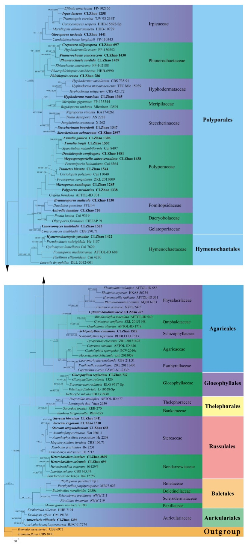
    Figure 1.  Maximum parsimony strict consensus tree illustrating the phylogeny of 52 species with related taxa in Agaricomycetes based on ITS sequences. Branches are labelled with maximum likelihood bootstrap equal to or greater than 70%, parsimony bootstrap equal to or greater than 50% and Bayesian posterior probabilities equal to or greater than 0.97, respectively. The taxa from the present study are indicated in black bold.

    Maximum parsimony analysis was applied to the ITS dataset sequences. Approaches to phylogenetic analysis followed Zhao & Wu (2017) and the tree construction procedure was performed in PAUP* version 4.0b10 (Swofford 2002). All characters were equally weighted and gaps were treated as missing data. Trees were inferred using the heuristic search option with TBR branch swapping and 1000 random sequence additions. Max-trees were set to 5000, branches of zero length were collapsed and all parsimonious trees were saved. Clade robustness was assessed using a bootstrap (BT) analysis with 1, 000 replicates (Felsenstein 1985). Descriptive tree statistics tree length (TL), consistency index (CI), retention index (RI), rescaled consistency index (RC), and homoplasy index (HI) were calculated for each Maximum Parsimonious Tree (MPT) generated. Sequences were also analyzed using Maximum Likelihood (ML) with RAxML-HPC2 through the Cipres Science Gateway (www.phylo.org; Miller et al. 2009). Branch support (BS) for ML analysis was determined by 1000 bootstrap replicates.

    MrModeltest 2.3 (Nylander 2004) was used to determine the best-fit evolution model for each data set for Bayesian inference (BI). Bayesian inference was calculated with MrBayes3.1.2 with a general time reversible (GTR+I+G) model of DNA substitution and a gamma distribution rate variation across sites (Ronquist & Huelsenbeck 2003). Four Markov chains were run for 2 runs from random starting trees for 1500 thousand generations (Fig. 1), and trees were sampled every 100 generations. The first one-fourth generations were discarded as burn-in. A majority rule consensus tree of all remaining trees was calculated. A majority rule consensus tree of all remaining trees was calculated. Branches were considered as significantly supported if they received maximum likelihood bootstrap (BS) > 70%, maximum parsimony bootstrap (BT) > 50%, or Bayesian posterior probabilities (BPP) > 0.95.

  • The ITS dataset (Fig. 1) included sequences from 102 fungal specimens representing 101 species. The dataset had an aligned length of 1389 characters, of which 308 characters were constant, 283 parsimony-uninformative, and 798 parsimony-informative. Maximum parsimony analysis yielded 1 equally parsimonious tree (TL = 8644, CI = 0.2678, HI = 0.7322, RI = 0.4535, RC = 0.1215). The best-fit model for ITS alignment estimated and applied in the Bayesian was GTR+I+G, lset nst = 6, rates = invgamma; prset statefreqpr = dirichlet (1, 1, 1, 1). Bayesian resulted in a similar topology with an average standard deviation of split frequencies = 0.029534 (BI), and the effective sample size (ESS) across the two runs is the double of the average ESS (avg ESS) = 354.

    The phylogeny (Fig. 1) inferred from ITS sequences demonstrated that fifty-two species nested in sixteen families, Auriculariaceae, Bondarzewiaceae, Dacryobolaceae, Fomitopsidaceae, Gelatoporiaceae, Gloeophyllaceae, Hymenochaetaceae, Hyphodermataceae, Irpicaceae, Phanerochaetaceae, Physalacriaceae, Polyporaceae, Schizophyllaceae, Schizoporaceae, Steccherinaceae and Stereaceae, belonging to six orders Agaricales, Auriculariales, Gloeophyllales, Hymenochaetales, Polyporales, Russulales in Agaricomycetes.

  • An alphabetical list (according to genus name) of wood-decaying fungi identified in these investigations is given below. The authors of scientific names are according to the second edition of Authors of Fungal Names (http://www.indexfungorum.org/AuthorsOfFungalNames.html). Substrate and collecting data are provided after the name of each species. The hosts are listed alphabetically, and within the same host tree, they are arranged by the order: living tree, dead standing tree, trunk, fallen branch and stump. The collectors and collection numbers are listed alphabetically, too (Dai 2011, 2012a).

    1. Antrodia tanakae (Murrill) Spirin & Miettinen, on the fallen branch of Acacia dealbata Link, CLZhao 720; on the stump of Acacia dealbata, CLZhao 1536

    2. Auricularia villosula Malysheva, on the fallen branch of Alnus nepalensis, CLZhao 1428; on the trunk of Juglans regia L., CLZhao 743; on the trunk of Quercus, CLZhao 1296; on the fallen branch of Quercus, CLZhao 1340

    3. Basidioradulum crustosum (Pers.) Zmitr., Malysheva & Spirin, on the fallen angiosperm branch, CLZhao 3028

    4. Bjerkandera adusta (Willd.) P. Karst, on the stump of Pinus yunnanensis, CLZhao 1555; on the dead tree of Quercus, CLZhao 1275

    5. Brunneoporus malicola (Berk. & M.A. Curtis) Audet, on the stump of Acacia dealbata, CLZhao 1524; on the trunk of Quercus acutissima Carr., CLZhao 1530

    6. Byssomerulius corium (Pers.) Parmasto, on the fallen angiosperm branch, CLZhao 1560; on the fallen branch of Pinus yunnanensis, CLZhao 734, CLZhao 781; on the fallen branch of Quercus, CLZhao 693, CLZhao 1266, CLZhao 1274, CLZhao 1313; on the stump of Quercus, CLZhao 1341

    7. Cinereomyces lindbladii (Berk.) Jülich, on the trunk of Pinus yunnanensis, CLZhao 1523

    8. Crepatura ellipsospora C.L. Zhao, on the trunk of Alnus, CLZhao 697; on the fallen branch of Quercus, CLZhao 868, CLZhao 1260, CLZhao 1265

    9. Cylindrobasidium laeve (Pers.) Chamuris, on the dead bamboo, CLZhao 756, CLZhao 767

    10. Daedaleopsis confragosa (Bolton) J. Schröt, on the trunk of Alnus nepalensis, CLZhao 1481

    11. Funalia gallica (Fr.) Bondartsev & Singer, on the trunk of Alnus nepalensis, CLZhao 1309; on the fallen branch of Alnus nepalensis, CLZhao 1306

    12. Funalia trogii Berk., on the trunk of Alnus nepalensis, CLZhao 1540; on the angiosperm trunk, CLZhao 741, CLZhao 3009, CLZhao 1557; on the fallen angiosperm branch CLZhao 1552

    13. Fuscoporia torulosa (Pers.) T. Wagner & M. Fisch, on the fallen branch of Quercus, CLZhao 1305

    14. Gloeophyllum sepiarium (Wulfen) P. Karst, on the trunk of Pinus yunnanensis, CLZhao 732, CLZhao 764; on the fallen branch of Pinus yunnanensis, CLZhao 774, CLZhao 904; on the stump of Pinus yunnanensis, CLZhao 731, CLZhao 784

    15. Gloeoporus dichrous (Fr.) Bres., on the fallen branch of Alnus nepalensis, CLZhao 1471

    16. Gloeoporus taxicola (Pers.) Gilb. & Ryvarden, on the trunk of Pinus armandii, CLZhao 1441

    17. Heterobasidion insulare (Murrill) Ryvarden, on the stump of Pinus yunnanensis, CLZhao 2899

    18. Heterobasidion orientale Tokuda, T. Hatt. & Y.C. Dai, trunk of Pinus yunnanensis, CLZhao 696

    19. Hymenochaete adusta (Lév.) Har. & Pat., J. Bot, on the trunk of Alnus, CLZhao 700

    20. Hymenochaete rheicolor (Mont.) Lév., on the trunk of Alnus, CLZhao 672, CLZhao 679; on the fallen branch of Quercus, CLZhao 666, CLZhao 671, CLZhao 682

    21. Hymenochaete villosa (Lév.) Bres., on the trunk of Quercus acutissima, CLZhao 1533

    22. Hymenochaetopsis corrugata (Fr.) S.H. He & Jiao Yang, on the fallen angiosperm branch, CLZhao 2893

    23. Hymenochaetopsis yasudae (Imazeki) S.H. He & Jiao Yang, on the fallen branch of Alnus nepalensis, CLZhao 1422, CLZhao 1445; on the living tree of Pinus armandii, CLZhao 1475; on the fallen branch of Pinus armandii, CLZhao 1486, CLZhao 1495, CLZhao 1549

    24. Hyphoderma transiens (Bres.) Parmasto, on the fallen angiosperm branch, CLZhao 1493; on the trunk of Populus yunnanensis Dode, CLZhao 1365

    25. Hyphodontia tropica Sheng H. Wu, on the fallen angiosperm branch, CLZhao 2898, CLZhao 2901

    26. Irpex lacteus (Fr.) Fr., on the trunk of Acacia dealbata, CLZhao 1258

    27. Lopharia cinerascens (Schwein.) G. Cunn., on the fallen branch of Alnus nepalensis, CLZhao 1499

    28. Megasporoporiella subcavernulosa (Y.C. Dai & Sheng H. Wu) B.K. Cui, on the fallen branch of Alnus nepalensis, CLZhao 1438, CLZhao 1466; on the stump of Alnus nepalensis, CLZhao 1412; on the fallen angiosperm branch, CLZhao 2966, CLZhao 2984, CLZhao 3016; on the stump of Cupressus funebris Endl., CLZhao 1444, CLZhao 1491

    29. Microporus xanthopus (Fr.) Kuntze, on the fallen branch of Alnus nepalensis, CLZhao 1503; on the fallen angiosperm branch, CLZhao 3012; on the trunk of Quercus, CLZhao 1253, CLZhao 1285; on the fallen branch of Quercus, CLZhao 1268, CLZhao 1304; on the stump of Quercus, CLZhao 1343

    30. Phanerochaete concrescens Spirin & Volobuev, on the fallen branch of Alnus nepalensis, CLZhao 1541, CLZhao 1545; on the fallen angiosperm branch, CLZhao 2929, CLZhao 2931, CLZhao 2939, CLZhao 2940, CLZhao 2945, CLZhao 2946, CLZhao 2949; on the trunk of Pinus yunnanensis, CLZhao 2916

    31. Phanerochaete sordida (P. Karst.) J. Erikss. & Ryvarden, on the trunk of Alnus, CLZhao 698; on the fallen branch of Alnus nepalensis, CLZhao 1459, CLZhao 1541, CLZhao 1545; on the angiosperm trunk, CLZhao 1461; on the fallen angiosperm branch, CLZhao 2929, CLZhao 2931, CLZhao 2939, CLZhao 2940, CLZhao 2945, CLZhao 2946, CLZhao 2949, CLZhao 4738, CLZhao 4746, CLZhao 4754; on the fallen branch of Pinus armandii, CLZhao 1515; on the trunk of Pinus yunnanensis, CLZhao 2916

    32. Phellinus gilvus (Schwein.) Pat., on the fallen branch of Alnus nepalensis, CLZhao 1334

    33. Phlebiopsis crassa (Lév.) Floudas & Hibbett, on the angiosperm trunk, CLZhao 786; on the fallen branch of Coriaria nepalensis Wall., CLZhao 1295, CLZhao 1308; on the trunk of Pinus yunnanensis, CLZhao 724; on the trunk of Quercus, CLZhao 1269, CLZhao 1314; on the fallen branch of Quercus acutissima, CLZhao 1532

    34. Polyporus arcularius (Batsch) Fr. on the stump of Coriaria nepalensis, CLZhao 1338; on the fallen branch of Quercus, CLZhao 1316

    35. Postia hibernica (Berk. Broome) Jülich, on the fallen angiosperm branch, CLZhao 2903; on the fallen branch of Pinus yunnanensis, CLZhao 2909

    36. Pulcherricium coeruleum (Lam.) Parmasto, on the fallen branch of Alnus nepalensis, CLZhao 1434; on the fallen angiosperm branch, CLZhao 3003, CLZhao 3020

    37. Schizophyllum commune Fr., on the trunk of Acacia dealbata, CLZhao 1527; on the fallen branch of Acacia dealbata, CLZhao 1528, CLZhao 1529; on the trunk of Alnus nepalensis, CLZhao 1537; on the stump of Alnus nepalensis, CLZhao 1562; on the stump of Eucalyptus robusta, CLZhao 1561

    38. Steccherinum bourdotii Saliba & A. David, on the fallen branch of Alnus nepalensis, CLZhao 1472; on the fallen branch of Pinus armandii, CLZhao 1347

    39. Steccherinum ochraceum (Pers. ex J.F. Gmel.) Gray, on the fallen angiosperm branch, CLZhao 2897, CLZhao 2968

    40. Stereum gausapatum (Fr.) Fr., on the trunk of Alder, 11 January 2017, CLZhao 668, CLZhao 669, CLZhao 673, CLZhao 677, CLZhao 683, CLZhao 694, CLZhao 707; on the dead tree of Quercus, 22 April 2017, CLZhao 1259, CLZhao 1270, CLZhao 1290, CLZhao 1300, CLZhao 1318, CLZhao 1320

    41. Stereum hirsutum (Willd.) Pers., on the fallen branch of Alnus nepalensis, CLZhao 1404, CLZhao 1405, CLZhao 1427, CLZhao 1455, CLZhao 1457, CLZhao 1465, CLZhao 1469, CLZhao 1470, CLZhao 1479, CLZhao 1482, CLZhao 1489, CLZhao 1498; on the fallen angiosperm branch, CLZhao 740; on fallen branch of Coriaria nepalensis, CLZhao 1559; on fallen branch of Pinus yunnanensis, CLZhao 2906; on the fallen branch of Quercus, CLZhao 1291

    42. Stereum rugosum Pers., on the trunk of Quercus, CLZhao 1310

    43. Stereum sanguinolentum (Alb. & Schwein.) Fr., on the trunk of Alnus, CLZhao 673; on the stump of Pinus yunnanensis, CLZhao 669

    44. Trametes hirsuta (Wulfen) Lloyd, on the fallen branch of Alnus nepalensis, CLZhao 1544; on the angiosperm trunk, CLZhao 1344; on the trunk of Pinus yunnanensis, CLZhao 739

    45. Trametes strumosa (Fr.) Zmitr., on the angiosperm trunk, CLZhao 718

    46. Trametes versicolor (L.) Lloyd, on the trunk of Alnus nepalensis, CLZhao 1539, CLZhao 1546; on the fallen branch of Alnus nepalensis, CLZhao 1431; on the stump of Alnus nepalensis, CLZhao 1510; on the angiosperm trunk, CLZhao 748, CLZhao 1293, CLZhao 1477; on the trunk of Cupressus funebris, CLZhao 1509; on the trunk of Quercus, CLZhao 1330; on the fallen branch of Quercus, CLZhao 686, CLZhao 714, CLZhao 1302, CLZhao 1307

    47. Trametopsis cervina (Schwein.) Tomšovský, on the trunk of Alnus nepalensis, CLZhao 1246; on the fallen branch of Quercus, CLZhao 1315

    48. Trichaptum abietinum (Dicks.) Ryvarden, on the angiosperm stump, CLZhao 719, CLZhao 723, CLZhao 736, CLZhao 777, CLZhao 3004; on the trunk of Pinus yunnanensis, CLZhao 730

    49. Tubulicrinis xantha C.L. Zhao, on the fallen branch of Pinus yunnanensis, CLZhao 2868, CLZhao 2869, CLZhao 2883

    50. Xylodon kunmingensis L. W. Zhou & C.L. Zhao, on the fallen angiosperm branch, CLZhao 3010, CLZhao 3019; on the stump of angiosperm, CLZhao 755; on the fallen branch of Pinus yunnanensis, CLZhao 752

    51. Xylodon nespori (Bres.) Hjortstam & Ryvarden, on the trunk of Cupressus funebris, CLZhao 1492

    52. Xylodon rimosissimus (Peck) Hjortstam & Ryvarden, on the fallen branch of Pinus armandii, CLZhao 1487

    • The researches were supported by the Yunnan Fundamental Research Project (Grant No. 202001AS070043) and the Science Foundation of Southwest Forestry University (Project No. 111715).
Figure (1)  Table (1) References (59)
  • About this article
    Cite this article
    X He, JZ Chen, CL Zhao. 2021. Diversity of wood – decaying fungi in Haikou Forestry Farm, Yunnan Province, P.R. China. Studies in Fungi 6(1):365−377 doi: 10.5943/sif/6/1/27
    X He, JZ Chen, CL Zhao. 2021. Diversity of wood – decaying fungi in Haikou Forestry Farm, Yunnan Province, P.R. China. Studies in Fungi 6(1):365−377 doi: 10.5943/sif/6/1/27
  • Catalog

    • About this article

    /

    DownLoad:  Full-Size Img  PowerPoint
    Return
    Return