SHORT COMMUNICATION   Open Access    

Distribution, regeneration status and threats to Senegalia venosa (Hochst. ex Benth.) Kyal. & Boatwr. in Hirmi Dryland areas of Northern Ethiopia

More Information
  • Received: 15 December 2023
    Revised: 17 April 2024
    Accepted: 18 April 2024
    Published online: 04 June 2024
    Tropical Plants  3 Article number: e018 (2024)  |  Cite this article
  • Based on the IUCN threat level, Senegalia venosa is one of the species that has endangered status and is restricted in Tigray and Gonder drylands. The purpose of this study was to identify the distribution of the Senegalia venosa species, assess its regeneration status, and map its location for potential future conservation efforts. Plant attributes and environmental data (including disturbance factors) were collected from plots (10 m × 10 m) where the species was found. Accordingly, Senegalia venosa was found in 11 sites in the study area. The number of mature, sapling, and seedling individuals in the sampled area was counted and density was computed. The mature plant of the species had the highest density (363.6 stems/ha) in terms of the species' regeneration status, followed by the sapling (254.5 stems/ha) and seedling (154.5 stems/ha). This implies Senegalia venosa has poor regeneration status, which could be associated with revealed disturbance factors including charcoal production, cutting and grazing.
  • Scale insects (Hemiptera: Coccomorpha) with hypogeic habits are considered of high phytosanitary relevance for coffee crops (Rubiaceae: Coffea spp.) in Colombia[1]. A total of 65 species of scale insects associated with coffee roots have been recorded in Colombia[24]. The most species-rich family is the Pseudococcidae with 28 species distributed in nine genera: Dysmicoccus Ferris, 1950 (13 spp.), followed by Pseudococcus Westwood, 1840 (four spp.), Phenacoccus Cockerell, 1893 (three spp.), Planococcus Ferris, 1950, and Spilococcus Ferris, 1950 (two spp. each), and Chorizococcus McKenzie, 1960, Distichlicoccus Ferris, 1950, Ferrisia Fullaway, 1923, and Paraputo Laing, 1929 (one sp. each). For the family Rhizoecidae, 19 species have been recorded in six genera, namely, Rhizoecus Kunckel d'Herculais, 1878 (13 spp.), Pseudorhizoecus Green, 1933 (two spp.), and Capitisetella Hambleton, 1977, Coccidella Hambleton, 1946, Geococcus Green, 1902, and Ripersiella Tinsley, 1899 (one sp. each). Other minor families include Coccidae and Ortheziidae (five spp. in each family), Xenococcidae (three spp.), and Putoidae and Diaspididae (two spp. in each family) and Margarodidae (one sp.). For this study all previous records were re-analysed with the purpose of providing an accurate list of species

    The taxonomic identification of scale insects by a morphological approach is particularly difficult, mainly for two reasons. First, they are small insects (usually < 5 mm) that require the preparation of slide-mounted specimens. Second, the taxonomic keys needed for morphological identifications are primarily designed for adult female specimens[5]. Differing from other insect orders (e.g., Coleoptera, Diptera and Hymenoptera), female scale insects lack well-defined tagmata, as well as sclerites, sutures, or discernible areas. Characters of taxonomic value in scale insects include cuticular processes, such as pores, ducts and, setae[5]. Recognizing these cuticular structures on such small bodies poses a difficult task for non-expert entomologists. To facilitate accessible identification, this manuscript offers an illustrated taxonomic key to scale insect species associated with coffee roots in Colombia and is aimed at users with basic knowledge of scale insect morphology.

    A careful revision of the specimens studied by Caballero et al.[2], preserved in the Scale Insect Collection at the Entomological Museum 'Universidad Nacional Agronomía Bogota' UNAB (Bogotá, Colombia), was carried out to exclude species that are doubtfully recorded from coffee roots in Colombia. This re-assessment allowed the compilation of an accurate list of species that could be included in the taxonomic key. Additional species and information from Caballero[3] and Caballero et al.[4] also were used for construction of the key. List of species recorded for Colombia and c ollection data of specimens analized are in Supplemmental Table S1 and S2 respectivately.

    The illustrated taxonomic key (Table 1) is based on the external morphology of the adult female with a dichotomous structure. Each couplet after the first one is numbered followed by the number of the preceding couplet in parenthesis, e.g. 12(7) means that couplet 12 is derived from couplet 7; the numbers at the end of the couplet indicate the next couplet in order to arrive at the species name that best matches the character states selected by the user. It is illustrated in most of the steps using microphotographs. Acquisition and analysis of images were done with a Lumenera 1-5C camera and the software Image Pro Insight 8.0. Designs were performed with Affinity Photo V 2.1 and Affinity Designer V 2.1 software. The taxonomic keys were structured with some adaptations of published taxonomic keys[615]. The general morphological terminology follows Kondo & Watson[5] with specific terminology for Coccidae[6,16], Margarodidae[17], Ortheziidae[7], Diaspididae[18], Pseudococcidae, Putoidae[8,9], and Rhizoecidae[10,11]. The abdominal segmentation is given as SabdI for abdominal segment 1 to SabdVIII for abdominal segment 8. All microphotographs are of adult female scale insects or their taxonomically important morphological structures.

    Table 1.  Illustrated taxonomic key.
    No.DetailsRef.
    1Abdominal spiracles present (Fig. 1a)2
    Abdominal spiracles absent (Fig. 1b)7
    Fig. 1 Abdominal spiracles (sp) on margin (a) present on Eurhizococcus colombianus, (b) absent on Distichlicoccus takumasai.
    2(1)Anal aperture without pores and setae (Fig. 2a); legs shorter than half of the transversal diameter of body (Fig. 2b); eyespots and mouthparts absentEurhizococcus colombianus

    Jakubski, 1965
    Anal aperture forming a well-developed anal ring with pores and setae (Fig. 2c); legs longer than transversal diameter of body; eyespots and mouthparts present (Fig. 2d)
    3
    Fig. 2 Eurhizococcus colombianus: (a) Anal aperture without pores and setae in the border, (b) section of mid body showing the length of hind leg (lel) and transversal body line (btl). Insignorthezia insignis: (c) Anal aperture with pores (po) and setae (st), (d) section of head with protruding eyespot (es) and labium (lb).
    3(2)Antennae each with eight segments (Fig. 3a)4
    Antennae each with fewer than five segments (Fig. 3b)5
    Fig. 3 (a) Eight-segmented antenna. (b) Four-segmented antenna.
    4(3)Transversal bands of spines absent in ventral region surrounded by an ovisac band (Fig. 4a); dorsal interantennal area without sclerosis (Fig. 4b)Insignorthezia insignis (Browne, 1887)
    Transversal bands of spine plates present in ventral region surrounded by an ovisac band (Fig. 4c); longitudinal sclerosis on dorsum in interantennal area (Fig. 4d)Praelongorthezia praelonga (Douglas, 1891)
    Fig. 4 Insignorthezia insignis: (a) Abdomen without transversal clusters of wax plates, (b) Dorsal interantennal area without sclerosis. Praelongorthezia praelonga: (c) Abdomen with transversal clusters of wax plates marked by dash lines, (d) dorsal interantennal area with a longitudinal sclerotic plate (ep).
    5(3)Antennae each with three segments (Fig. 5a)Newsteadia andreae Caballero, 2021
    Antennae each with four segments (Fig. 5b)6
    Fig. 5 (a) Three-segmented antenna of Newsteadia andreae. Note the presence of pseudosegmentation which gives the appearance of additional segments in the last antennal segment. (b) Four-segmented antenna of Mixorthezia minima.
    6(5)Dorsal area anterior to anal ring with simple pores on protuberances (Fig. 6a); ventral areas surrounding each coxa with a row of wax plate spines (Fig. 6b)Mixorthezia minima Koczné Benedicty & Kozár, 2004
    Dorsal area anterior to anal ring without simple pores or protuberances (Fig. 6c); ventral areas posterior to each coxa without wax plate spines (Fig. 6d)Mixorthezia neotropicalis (Silvestri, 1924)
    Fig. 6 Mixorthezia minima: (a) Dorsum of area anterior to anal ring with close-up of simple pores on protuberances (dash box); (b) ventral area posterior to each coxa with a row of wax plate spines (dash box). Mixorthezia neotropicalis: (c) Close-up of dorsum of area anterior to anal ring lacking simple pores on protuberances (dash box); (d) ventral area posterior to each coxa without associated wax plate spines.
    7(1)Anal plates present (Fig. 7a)8
    Anal plates absent (Fig. 7b)12
    Fig. 7 (a) Anal apparatus of Saissetia coffeae with anal plates (ap) covering the anal aperture (aa). (b) Anal apparatus of Pseudococcus sp. with anal aperture lacking anal plates.
    8(7)Antennae and legs with length similar to or shorter than spiracles (Fig. 8a)9
    Antennae and legs with length at least twice as long as spiracles (Fig. 8b)11
    Fig. 8 (a) Antenna (an) and foreleg (lg) (green lines), and anterior spiracle (sp) (yellow line) of Toumeyella coffeae showing their relative length. Note the similar size of the limbs and spiracle. (b) Antenna (an) and leg (lg) (green lines), and anterior spiracle (sp) (yellow line) of Coccus viridis showing their relative length. Note the relatively smaller size of the spiracle.
    9(8)Ventral tubular macroducts present (Fig. 9)Toumeyella coffeae
    Kondo, 2013
    Ventral tubular macroducts absent10
    Fig. 9 Ventral tubular macroducts (dash box) and close-up of macroducts (photo on right side).
    10(9)Orbicular pores (Fig. 10a) and cribriform platelets present (Fig. 10b); dorsal setae absent; opercular pores absentCryptostigma urichi (Cockerell, 1894)
    Orbicular pores and cribriform platelets absent; dorsal setae present (Fig. 10c); numerous opercular pores present throughout mid areas of dorsum (Fig. 10d)Akermes colombiensis Kondo & Williams, 2004
    Fig. 10 Cryptostigma urichi: (a) Orbicular pore and (b) close-up of a cribriform platelet. Akermes colombiensis: (c) Close-up of a dorsal body setae (dash box) and (d) close-up of opercular pores (arrows).
    11(8)Band of ventral tubular ducts in lateral and submarginal regions absent, ventral tubular ducts of one type; anal plates without discal setae (Fig. 11a); dorsal body setae capitate or clavate (Fig. 11b); perivulvar pores with seven or eight loculi, rarely with 10 loculi (Fig. 11c)Coccus viridis
    (Green, 1889)
    Band of ventral tubular ducts in lateral and submarginal regions present, submarginal region with two types of tubular ducts (Fig. 11d); anal plates with discal setae (Fig. 11e); dorsal body setae spine-like, apically pointed (Fig. 11f); perivulvar pores mostly with 10 loculi (Fig. 11g)Saissetia coffeae
    (Walker, 1852)
    Fig. 11 Coccus viridis: (a) Anal plates without discal setae; (b) dorsal body setae capitate (top) or clavate (below); (c) multilocular disc pores mostly with eight loculi. Saissetia coffeae: (d) Ventral submarginal region with two types of tubular ducts; (e) each anal plate with a discal seta; (f) dorsal body setae acute; (g) multilocular disc pores with mostly 10 loculi.
    12(7)Cerarii present on body margin, at least a pair on each anal lobe (Fig. 12a)13
    Cerarii absent on body margin (Fig. 12b)38
    Fig. 12 Abdominal body margin of (a) Pseudococcus sp. with three cerarii (dash box) and (b) Rhizoecus sp. (dash box) without cerarii.
    13(12)Enlarged oral collar tubular ducts composed of a sclerotized area surrounding the border and a set of flagellated setae (Ferrisia-type oral collar tubular ducts) (Fig. 13a)Ferrisia uzinuri
    Kaydan & Gullan, 2012
    Oral collar tubular ducts simple, not as above (Fig. 13b) or absent14
    Fig. 13 (a) Ferrisia-type oral collar tubular ducts with aperture of tubular duct (ad) surrounded by a sclerotized area (sa) and associated flagellate setae (fs). (b) Oral collar tubular ducts simple (arrows).
    14(12)Antenna with nine segments (Fig. 14a)15
    Antenna with eight segments (Fig. 14b) or fewer (Fig. 14c)19
    Fig. 14 Antenna with (a) nine segments, (b) eight segments and (c) seven segments.
    15(14)Cerarii with more than five conical setae (Fig. 15a); hind trochanter with six sensilla, three on each surface (Fig. 15b)16
    Cerarii with two lanceolate setae (Fig. 15c); hind trochanter with four sensilla, two on each surface (Fig. 15d)17
    Fig. 15 Puto barberi: (a) upper and lateral view of a cerarius, (b) close-up of the surface of trochanter with three sensilla (arrows). Phenacoccus sisalanus: (c) cerarius, (d) trochanter with two sensilla (arrows) on single surface.
    16(15)Cerarii with tubular ducts (Fig. 16a)Puto antioquensis
    (Murillo, 1931)
    Cerarii without tubular ducts (Fig. 16b)Puto barberi
    (Cockerell, 1895)
    Fig. 16 (a) Cerarius associated with tubular ducts (arrows). (b) Cerarius without tubular ducts.
    17(15)Oral collar tubular ducts absentPhenacoccus sisalanus Granara de Willink, 2007
    Oral collar tubular ducts present, at least on venter (Fig. 17)18
    Fig. 17 Ventral surface with oral collar tubular ducts (dash circles).
    18(17)Oral collar tubular ducts restricted to venterPhenacoccus solani
    Ferris, 1918
    Oral collar tubular ducts present on dorsum and venterPhenacoccus parvus Morrison, 1924
    19(14)Oral rim tubular ducts present (Fig. 18)20
    Oral rim tubular ducts absent26
    Fig. 18 Oral rim tubular ducts in upper view (dash circles) and close-up of lateral view.
    20(19)Oral rim tubular ducts present on venter onlyPseudococcus landoi (Balachowsky, 1959)
    Oral rim tubular ducts present on both dorsum and venter21
    21(20)Cerarii restricted to anal lobes (Fig. 19a)Chorizococcus caribaeus Williams & Granara de Willink, 1992
    Cerarii present, at least on the last five abdominal segments (Fig. 19b)22
    Fig. 19 Location of cerarii (dash boxes) on abdominal margin with close-up of cerarius (a) restricted to anal lobes (dash boxes) and (b) cerarii present on the last five abdominal segments.
    22(21)Circulus absent (Fig. 20a)23
    Circulus present (Fig. 20b)24
    Fig. 20 Ventral mid area of abdominal segments III and IV (dash box) of (a) Distichlicoccus takumasai without circulus and (b) Pseudococcus jackbeardsleyi with circulus.
    23(22)Multilocular disc pores present on venter of SabdIV and posterior segments (Fig. 21a); hind coxa with translucent pores and hind femur without translucent pores (Fig. 21b)Spilococcus pressus
    Ferris, 1950
    Multilocular disc pores absent, if some present, not more than three around vulvar opening (i.e. venter of SabdVII or SabdVIII); hind coxa without translucent pores (Fig. 21c) and hind femur with translucent pores (Fig. 21d)Distichlicoccus takumasai Caballero, 2021
    Fig. 21 Spilococcus pressus: (a) Ventral section of abdomen with multilocular disc pores (arrows); (b) hind leg with close-up of coxa with translucent pores (arrows). Distichlicoccus takumasai: (c) Hind coxa without translucent pores; (d) hind femur with translucent pores (arrows).
    24(22)Eyes without discoidal pores nor sclerotized surrounding area (Fig. 22a); circulus with transversal diameter 40 to
    60 µm (Fig. 22b)
    Pseudococcus luciae Caballero, 2021
    Eyes with discoidal pores and sclerotized surrounding area (Fig. 22c); circulus diameter 100 to 200 µm (Fig. 22d)26
    25(24)Oral rim tubular ducts on dorsal abdominal segments numbering three to eight; area between posterior ostiole and cerarius of SabdVII without oral rim tubular ducts (Fig. 23a)Pseudococcus elisae Borchsenius, 1947
    Oral rim tubular ducts on dorsal abdominal segments numbering 14 to 27; area between posterior ostiole and cerarius of SabdVII with an oral rim tubular duct (Fig. 23b)Pseudococcus jackbeardsleyi Gimpel & Miller, 1996
    Fig. 22 Pseudococcus luciae: (a) Eyespot without surrounding sclerotized area nor associated pores; (b) circulus ca. 58 µm wide. Pseudococcus jackbeardsleyi: (a) Eyespot with sclerotized area (sa) and associated pores (po); (d) circulus ca. 154 µm wide.
    Fig. 23 (a) Dorsal margin of abdominal segments VI to VIII, between cerarius of anal lobe (C1), cerarius of SabdVII (C2) and posterior ostiole (os) without oral rim tubular ducts. (b) Dorsal margin of abdominal segments VI to VIII, between cerarius of anal lobe (C1), cerarius of SabdVII (C2) and posterior ostiole (os) with an oral rim tubular duct and/or cerarius adjacent to SabdVII.
    26(19)Oral collar tubular ducts (Fig. 24) on both dorsum and venter27
    Oral collar tubular ducts restricted to venter28
    Fig. 24 Oral collar tubular duct in lateral view.
    27(26)Hind coxa with translucent pores (Fig. 25a); anal lobe with sclerotized bar, not on a sclerotized area (Fig. 25b); multilocular disc pores present posterior to fore coxaPlanococcus citri-minor complex
    Hind coxa without translucent pores (Fig. 25c); anal lobe without sclerotized bar, on a sclerotized area (Fig. 25d); multilocular disc pores absent posterior to fore coxaDysmicoccus quercicolus (Ferris, 1918)
    28(27)Oral collar tubular ducts absent on venter of both head and thorax.29
    Oral collar tubular ducts present on either head or thorax, but not on both areas (Fig. 26)30
    Fig. 25 Planococcus citri-minor complex: (a) Hind coxa with translucent pores (dash box) and (b) anal lobe with a sclerotization forming a bar (ab). Dysmicoccus quercicolus: (c) Hind coxa without translucent pores and (d) anal lobe with irregular broad sclerotized area (sa).
    Fig. 26 Marginal area of Dysmicoccus grassii, lateral to posterior spiracle (ps), with close-up of oral collar tubular ducts (oc) (left side).
    29(28)Translucent pores present on hind coxa, trochanter, femur and tibia (Fig. 27a); marginal clusters of oral collar tubular ducts on venter of SabdVI and SabdVIIDysmicoccus caribensis Granara de Willink, 2009
    Translucent pores restricted to hind femur and tibia (Fig. 27b); marginal clusters of oral collar tubular ducts present on venter of SabdIV to SabdVIIParaputo nasai
    Caballero, 2021
    Fig. 27 (a) Hind leg of Dysmicoccus caribensis with translucent pores on coxa (cx), trochanter (tr) and femur (fm), and tibia (tb). (b) Hind leg of Paraputo nasai with translucent pores restricted to femur (fm) and tibia (tb).
    30(28)Hind coxa with translucent pores (Fig. 28a)Dysmicoccus sylvarum
    Williams & Granara de Willink, 1992
    Hind coxa without translucent pores (Fig. 28b)31
    Fig. 28 (a) Translucent pores on hind coxa. (b) Translucent pores absent on hind coxa.
    31(30)Hind trochanter with translucent pores (Fig. 29a)Dysmicoccus varius
    Granara de Willink, 2009
    Hind trochanter without translucent pores (Fig. 29b)32
    Fig. 29 Translucent pores (a) on hind trochanter, (b) absent from hind trochanter.
    32(31)Oral collar tubular ducts present on margin of thorax (Fig. 30)33
    Oral collar tubular ducts absent from margin of thorax34
    Fig. 30 Prothorax margin of Dysmicoccus grassii with close-up of oral collar tubular ducts.
    33(32)Multilocular disc pores absent on SabdV; dorsal area immediately anterior to anal ring with tuft of flagellate setae; longest flagellate seta as long as diameter of anal ring (Fig. 31a), and discoidal pores larger than trilocular pores (Fig. 31b)Dysmicoccus radicis
    (Green, 1933)
    Multilocular disc pores present on SabdV; dorsal area immediately anterior to anal ring without a tuft of flagellate setae; flagellate setae much shorter than diameter of anal ring (Fig. 31c) and discoidal pores smaller than trilocular pores (Fig. 31d)Dysmicoccus grassii (Leonardi, 1913)
    34(32)Oral collar tubular ducts absent in interantennal area35
    Oral collar tubular ducts present in interantennal area (Fig. 32)36
    35(34)Translucent pores on hind leg restricted to tibia (Fig. 33a)Dysmicoccus perotensis
    Granara de Willink, 2009
    Translucent pores on hind leg present on tibia and femur (Fig. 33b)Dysmicoccus joannesiae-neobrevipes complex
    Fig. 31 Dysmicoccus radicis: (a) Area anterior to anal ring with a cluster of flagellate setae (fs) and anal ring (ar) showing the diameter of the different pores (dash box); (b) discoidal pores (dp) and trilocular pores (tp). Dysmicoccus grassii: (c) Area anterior to anal ring with scattered short flagellate setae (fs) contrasted with anal ring (ar) diameter (dash box); (d) discoidal pores (dp) and trilocular pores (tp) with similar diameter.
    Fig. 32 Interantennal area (dash box) of Dysmicoccus brevipes with close-up of oral collar tubular ducts.
    Fig. 33 (a) Hind leg of Dysmicoccus perotensis with close-up of femur and tibia with translucent pores on tibia only (arrows). (b) Hind leg of Dysmicoccus joannesiae-neobrevipes complex with close-up of femur and tibia with translucent pores (arrows).
    36(34)Hind coxa with translucent pores (see Fig. 28a)Dysmicoccus mackenziei
    Beardsleyi, 1965
    Hind coxa without translucent pores (see Fig. 28b)37
    37(36)Dorsal SabdVIII setae forming a tuft-like group, each seta conspicuously longer than remaining dorsal abdominal setae (Fig. 34a) and setal length similar to anal ring diameter (60–80 µm long)Dysmicoccus brevipes (Cockerell, 1893)
    Dorsal SabdVIII setae evenly distributed, each setae as long as remaining dorsal abdominal setae (Fig. 34b) and length less than half diameter of anal ringDysmicoccus texensis-neobrevipes complex
    38(12)Tritubular ducts absent39
    Tritubular ducts present (Fig. 35a-b)46
    Fig. 34 (a) Abdomen of Dysmicoccus brevipes with dorsal setae on SabdVIII (lfs) longer than setae on anterior segments (sfs). (b) Abdomen of Dysmicoccus texensis-neobrevipes complex with dorsal setae (ufs) along the abdominal segments of uniform length and scattered distribution.
    Fig. 35 (a) Tritubular duct in upper (left) and lateral view (right) with the border of the cuticular ring attached to tubules. (b) Tritubular duct with the border of the cuticular ring widely separated from tubules (arrows).
    39(38)Anal lobes strongly protruded, bulbiform (Fig. 36a) jutting out from margin for a distance equivalent to diameter of anal ring40
    Anal lobes shallow, if protruded, their length never more than half of diameter of anal ring (Fig. 36b)42
    Fig. 36 (a) Abdomen of Neochavesia caldasiae with anal lobes (al) protruding beyond the anal aperture (aa). (b) Abdomen of Ripersiella sp. with anal lobes (al) at the same level as the anal aperture (aa).
    40(39)Anal aperture located at the same level as the base of anal lobes (Fig. 37a); antennae located on ventral margin of headNeochavesia caldasiae (Balachowsky, 1957)
    Anal aperture located anterior to bases of anal lobes (Fig. 37b); antennae located on dorsum of head41
    Fig. 37 (a) Abdomen of Neochavesia caldasiae with anal aperture (aa) positioned between the anal lobes (al), at the same level as the bases of anal lobes (dash line). (b) Abdomen of Neochavesia eversi with anal aperture (aa) situated anterior to the bases of the anal lobes (al) (dash line).
    41(40)Antennae each with five segments, situated on a membranous base (Fig. 38a); length of hind claw less than length of hind tarsus (Fig. 38b)Neochavesia trinidadensis (Beardsley, 1970)
    Antennae each with four segments, situated on a sclerotized base (Fig. 38c); hind claw longer than hind tarsus (Fig. 38d)Neochavesia eversi (Beardsley, 1970)
    Fig. 38 (a) Antenna with four segments and a membranous base (mb). (b) Hind tarsus (green line) longer than the hind claw (red line). (c) Antenna with four segments and a sclerotized base (sb). (d) Hind tarsus (green line) shorter than hind claw (red line).
    42(39)Body setae capitate, at least on one surface (Fig. 39a)43
    Body setae never capitate (Fig. 39b)44
    Fig. 39 (a) Capitate setae. (b) Flagellate setae.
    43(42)Anal aperture without associated cells (Fig. 40a); three-segmented antennae (Fig. 40b); ventral setae in median
    and submedian regions capitate
    Capitisitella migrans
    (Green, 1933)
    Anal aperture surrounded by cells (Fig. 40c); six-segmented antennae (Fig. 40d); ventral setae in medial and submedial regions flagellateWilliamsrhizoecus coffeae
    Caballero & Ramos, 2018
    44(42)Three-segmented antennae (Fig. 41a); circulus present (Fig. 41b)Pseudorhizoecus bari
    Caballero & Ramos, 2018
    Five-segmented antennae (Fig. 41c); circulus absent45
    Fig. 40 Capitisitella migrans: (a) Anal aperture of surrounded only by setae; (b) antenna composed of three segments. Williamsrhizoecus coffeae: (c) Anal aperture of surrounded by setae and cells (flesh); (d) antenna composed of six segments.
    Fig. 41 Pseudorhizoecus bari: (a) Antenna composed of three segments and (b) circulus. (c) Antenna of Pseudorhizoecus proximus composed of five segments.
    45(44)Multilocular disc pores absent; anal aperture ornamented with small protuberances and two to five short setae, each seta never longer than 1/3 diameter of anal aperture, without cells (Fig. 42a)Pseudorhizoecus proximus
    Green, 1933
    Multilocular disc pores present (Fig. 42b); anal aperture not ornamented with small protruberances, ring with well-developed cells and six long setae, each seta as long as diameter of anal ring (Fig. 42c)Ripersiella andensis (Hambleton,
    1946)
    Fig. 42 (a) Anal aperture of Pseudorhizoecus proximus surrounded by protuberances (pr) and a few short setae (st). Ripersiella andensis: (b) Ventral section of abdomen with multilocular disc pores (mp); (c) anal aperture with a ring of cells and six long setae (se).
    46(38)Anal lobes strongly protruded, conical, each one with a stout spine at apex (Fig. 43a)Geococcus coffeae
    Green, 1933
    Anal lobes flat or barely protruded, without spines at apex (Fig. 43b)47
    47(46)Venter of abdomen with clusters of trilocular pores in medial region (Fig. 44a)Coccidella ecuadorina Konczné Benedicty & Foldi, 2004
    Venter of abdomen with trilocular pores evenly dispersed, never forming clusters in medial region (Fig. 44b)48
    Fig. 43 (a) Abdomen of Geococcus coffeae with protruding anal lobe (al) with a stout spine at the apex (sp). (b) Abdomen of Rhizoecus sp. with anal lobe (al) flat, with numerous flagellate setae (fs) at the apex.
    Fig. 44 (a) Ventral surface of Coccidella ecuadorina with clusters of trilocular pores (tc) (dash box) on medial region of abdomen. (b) Ventral surface of Rhizoecus sp. with trilocular pores (tr) scattered on venter of abdomen.
    48(47)Antennae with six well-developed segments (Fig. 45a)51
    Antennae with five well-developed segments (Fig. 45b), apical segment sometimes partially divided (Fig. 45c)49
    Fig. 45 (a) Six-segmented antenna. (b) Five-segmented antenna. (c) Five-segmented antenna with partially divided apical segment (pd). Note: antennal segments numbered in Roman numerals.
    49(48)Antennae length more than 140 µm (Fig. 46a); tritubular ducts of similar diameter to trilocular pores (± 2 µm variation) (Fig. 46b); tritubular ducts with space between ductules and edge as wide as the ductules (Fig. 46c); slender ductule, width/length ratio 1:6Rhizoecus coffeae
    Laing, 1925
    Antennae length less than 130 µm (Fig. 46d); tritubular ducts of diameter nearly twice diameter of trilocular pores (Fig. 46e); tritubular ducts with reduced space or without space between ductules and edge (Fig. 46f); stout ductule, width/length ratio 1:350
    50(49)Tubular ducts present (Fig. 47a); each anal lobe with around 28 dorsal setae of similar length, greater than 30 µm (Fig. 47b, al); and dorsal marginal clusters of setae on SabdVII 20–30 µm long (Fig. 47b, SabdVII)Rhizoecus setosus (Hambleton, 1946)
    Tubular ducts absent; each anal lobe with around 14 dorsal setae, with length less than 15 µm (Fig. 47c, al); dorsal marginal clusters of setae on SabdVII with length less 15 µm (Fig. 47c, SabdVII)Rhizoecus compotor
    Williams & Granara de Willink, 1992
    Fig. 46 (a) Antenna ca. 207 µm long. (b) Tritubular ducts (td) and trilocular pores (tp) with similar diameter. (c) Close-up of a tritubular duct indicating the space between the cuticular ring (mg) and the ductule (dt). (d) Antenna ca. 105 µm long. (e) Each tritubular duct (td) twice the diameter of a trilocular pore (tp). (f) Close-up of tritubular duct without a space between the cuticular ring (mg) and the ductule (dt).
    Fig. 47 Rhizoecus setosus: (a) Tubular ducts (td); (b) anal lobe (al) and abdominal segment (SabdVII) with marginal clusters of setae longer than 30 µm. (c) Abdomen of Rhizoecus compotor with marginal cluster of setae shorter than 20 µm on anal lobe (al) and abdominal segment (SabdVII).
    51(48)Fore tibia with at least one of two internal preapical setae spine-like (Fig. 48a-b)52
    Fore tibia with both internal preapical setae flagellate (Fig. 48c)56
    Fig. 48 Fore legs with preapical setae on tibia (ft): (a) one flagellate (fs) and one spine seta (ss), (b) with a pair of spine setae (ss), (c) with a pair of flagellate setae (fs).
    52(51)Fore tibia with one internal preapical spine-like setae and other seta flagellate (Fig. 48a); anal ring composed of spine-like setae (Fig. 49a); circulus absentRhizoecus spinipes (Hambleton, 1946)
    Fore tibia with both internal preapical setae spine-like (Fig. 48b); anal ring composed of flagellate-like setae (Fig. 49b); at least, one circulus present (Fig. 49c)53
    Fig. 49 (a) Anal ring (ar) of Rhizoecus spinipes with spine-like setae (ss). (b) Anal ring (ar) of Rhizoecus arabicus with flagellate setae (fs). (c) Circulus of Rhizoecus cacticans.
    53(52)Claw digitules setose and short, length less than half length of claw (Fig. 50a)54
    Claw digitules capitate and long, as long as claw (Fig. 50b)55
    Fig. 50 Claw with claw digitule: (a) setose (sd), (b) flagellate (fd).
    54(53)Anal ring with external row composed of 35 cells or more (Fig. 51a, ext); anal ring with external and internal rows separated by a space as wide as a cell of the external row (Fig. 51a, spc); anal ring cells without spicules (Fig. 51a, sp)Rhizoecus variabilis Hambleton, 1978
    Anal ring with external row composed of less than 30 cells (Fig. 51b, ext); anal ring with external and internal rows separated by a narrow space, as wide as half (or less) a cell of the external row (Fig. 51b, spc); anal ring cells with spicules (Fig. 51b, sp)Rhizoecus arabicus Hambleton, 1976
    Fig. 51 (a) Anal ring of Rhizoecus variabilis with external row (ext) of anal ring consisting of over 35 cells; external row separated from the internal row (int) by a similar width as the diameter of a cell (spc). (b) Anal ring of Rhizoecus arabicus with external row (ext) of anal ring with less than 30 cells; external row separated from the internal row (int) by a width less than half the diameter of a cell (spc); cells of the external row with spicules (sp).
    55(53)More than 80 tritubular ducts; circulus with basal diameter at least five times greater than apical diameter (Fig. 52a); stick-like genital chamber, parallel borders and all of similar width and structure, length across about two abdominal segments (169–175 µm long) (Fig. 52b)Rhizoecus atlanticus (Hambleton, 1946)
    Less than 50 tritubular ducts; circulus with basal diameter less than three times the apical diameter (Fig. 52c); genital chamber with basal third two times wider than anterior two-thirds, length across one abdominal segment (43–52 µm long) (Fig. 52d)Rhizoecus cacticans (Hambleton, 1946)
    Fig. 52 Rhizoecus atlanticus: (a) Circulus with diameter at base five times the apical diameter, (b) genital chamber tubular shape, length ca. 150 µm long. Rhizoecus cacticans: (c) Circulus with diameter at base about two times the apical diameter, (d) genital chamber with proximal section basiform and distal section tubular, with arms, length ca. 45 µm long.
    56(51)Multilocular disc pores absent on dorsumRhizoecus mayanus (Hambleton, 1946)
    Multilocular disc pores present on dorsum57
    57(56)Marginal prothoracic setae length greater than 50 µm (Fig. 53a); marginal SabdVII setae length greater than 45 µm (Fig 53b)Rhizoecus colombiensis Ramos-Portilla & Caballero, 2016
    Marginal prothoracic setae length less than 25 µm (Fig. 53c); marginal SabdVII setae length less than 30 µm (Fig. 53d)58
    Fig. 53 Rhizoecus colombiensis: (a) Body margin with a long seta (pts) (> 40 µm), longer than remaining setae in prothorax; (b) margin of abdominal segment VII (SabdVII) (st). with a long seta (pts) (> 40 µm), longer than remaining setae in abdomen. Rhizoecus americanus: (c) Margin of prothorax (pts) with setae of uniform length, shorter than 30 µm; (d) margin of abdominal segment VII (SabdVII) with setae (st) shorter than 30 µm.
    58(57)Tritubular ducts of two sizesRhizoecus caladii
    Green, 1933
    Tritubular ducts of three sizesRhizoecus americanus (Hambleton, 1946)
     | Show Table
    DownLoad: CSV

    The following illustrated taxonomic key (Table 1) is a tool for the identification of adult female scale insects (Hemiptera : Sternorrhyncha : Coccomorpha) associated with coffee roots in Colombia, which includes 59 species from seven families (see Supplemental Table S1).

    The taxonomic key includes 59 species associated with coffee roots. Hemiberlesia sp., Odonaspis sp., Rhizoecus stangei McKenzie, 1962, Spilococcus mamillariae (Bouche, 1844), Planococcus citri (Risso, 1813) and Planococcus minor (Maskell, 1897) were excluded from the key. In the case of the two armoured scale insects, the specimens were found in Berlese funnel samples associated with coffee roots[2], however, there is no evidence of these species feeding on the roots and there are no previous records of association of Hemiberlesia nor Odonaspis species with coffee roots.

    Previous records of single specimens of R. stangei and S. mamillariae by Caballero et al.[2] were determined as misidentifications of Rhizoecus caladii Green, 1933 and Spilococcus pressus Ferris, 1950, respectively. Spilococcus mamillariae is considered as an oligophagous species, but mainly associated with Cactaceae plants and feeding on the aerial parts of plants[19,20]. There are no records of S. mamillariae being found on any plant species of the family Rubiaceae, and hence we have removed this species from the list of species associated with coffee roots. The species R. stangei, which has been recorded only from Mexico and lacks information on its host plant[21] has apparently not been found since its original description[8].

    Planococcus citri and Pl. minor were listed also by Caballero et al.[2] as literature records. the morphological identification of P. citri and P. minor needs to be complemented with molecular and geographical analysis to be more accurate[22]. Therefore, the present key considers only identification to the Planococcus citri-minor complex.

    Furthermore, many specimens of Dysmicoccus collected from coffee roots in Colombia have morphological character states that overlap with Dysmicoccus neobrevipes Beardsley, 1959, Dysmicoccus joannesiae (Costa Lima, 1939) and Dysmicoccus texensis (Tinsley, 1900). The first case is a mix of character states of D. texensis and D. neobrevipes. The number of setae in the abdominal cerarii and the size of oral collar tubular ducts are the most important characters used to differentiate the adult females of Dysmicoccus species[8,23]. Adult females of D. texensis have a consistent pattern of only two setae in all thoracic and abdominal cerarii, along with a uniform size of oral collar tubular ducts (OC). On the other hand, D. neobrevipes varies in the number of setae in the cerarii, ranging from two to seven, accompanied by two distinct sizes of OC. These character states are generally constant among specimens found on the aerial parts of plants. However, among the specimens examined here, while the anal lobe cerarii consistently have two setae on the specimens of D. texensis found on the roots, the remaining cerarii display a variable number of setae, notably ranging from two to five, particularly within the abdominal cerarii. Furthermore, the OC of these specimens all are the same size. Regarding the differences in number of setae in the cerarii, Granara de Willink[23] underlined the need of more comprehensive studies to definitively separate these species.

    The second case involves D. joannesiae and D. neobrevipes. These species exhibit similarities in the number of setae on each cerarius (ranging from two to seven setae per cerarius) and differences in the number of clusters of OC along the abdominal margin; D. joannesiae has more than 25 clusters of OC and D. neobrevipes has fewer than 10 clusters of OC[8]. Granara de Willink also separated these two species by the presence of OC on the thorax and head[23] (present in D. neobrevipes and absent in D. joannesiae). Within the specimens of putative D. neobrevipes studied here, a few had clusters of OC numbering 15 to 20 along the abdominal margin and OC on the thorax and head. The primary challenge with addressing this dilemma lies in the fact that D. joannesiae has only been reported on Joannesia princeps Vell., 1798 (Euphorbiaceae) in Brazil and on Annona muricata (Annonaceae) intercepted in London from Saint Lucia[8,24]. Moreover, there has been no additional morphological variations recorded in the new records of D. joannesiae since its initial description in 1932 by Costa Lima. Therefore, the character states defining D. joannesiae are based on six type specimens. Based on these arguments, the following taxonomic key considers two species complex groups, namely the Dysmicoccus texensis-neobrevipes complex and the D. joannesiae-neobrevipes complex.

    Following article 31.1.2 of the International Commission of Nomenclature (ICZN), herein we make a change in nomenclature for Distichlicoccus takumasae Caballero, 2021. The ending -ae for takumasae is incorrect because the species was dedicated to Dr. Takumasa Kondo (a male coccidologist), and thus the correct ending is -i, hence the species epithet is herein amended to 'takumasai'. The corrected name is Distichlicoccus takumasai Caballero, 2021.

    After reviewing the species of scale insects associated with coffee roots in Colombia, we have compiled a list of 59 species (Supplemental Table S1). Although this study did not focus on the effect of habit (aerial vs underground) or host plant on the morphology of scale insects, we detected significant morphological variation within facultative hypogeal species. Until further studies allow an understanding of the overlap of character states between D. texensisD. neobrevipes and D. joannesiaeD. neobrevipes, we suggest considering these species as a morphological complex for hypogeal specimens. Further ecomorphological studies should be conducted to determine whether the morphology of a species may differ when feeding on the aerial parts compared when feeding on the underground parts of a host and to try to elucidate what factors trigger those changes, especially in species associated with coffee plants. As for the species complex, further collecting, morphological, and molecular studies should help elucidate these taxonomic problems.

    During the literature review performed for this study, we realized that most of the records of species are limited to mentioning the host but not the plant part on which collections were made, however, it is suspected that most species are normally collected from the aerial parts of the plant host. Although this taxonomic key is limited to root-associated species recorded in Colombia, this key could be useful for identifying scale insects associated with coffee in other tropical regions, extending also to species collected from the aerial parts of the hosts.

    The authors confirm contributions to the paper as follows: study conception and design: Caballero A, Kondo T; data collection: Caballero A, Kondo T; analysis and interpretation of results: Caballero A, Kondo T; draft manuscript preparation: Caballero A, Kondo T. Both authors reviewed the results and approved the final version of the manuscript.

    The data (microscopy slides of specimens) that support the findings of this study are available in the Scale insect repository of the entomological museum Universidad Nacional Agronomia Bogota – UNAB, Facultad de Ciencias Agrarias, Colombia. All data generated or analyzed during this study are included in this published article and its supplementary information files.

    The authors thank Dr. Andrea Ramos-Portilla for clarifying some aspects of the morphological variations of Rhizoecus species and Dr. Penny Gullan (Australian National University, Canberra, Australia) for reviewing an earlier version of the manuscript. Many thanks to Erika Valentina Vergara (AGROSAVIA) and Dr. Francisco Serna (Universidad Nacional de Colombia) for their help to access the Museum UNAB. Special thanks to Dr Giuseppina Pellizzari (University of Padova, Italy) for advice on scientific nomenclature. This study was financed by Colciencias (Programa Nacional de Ciencias Básicas [National Program on Basic Sciences]), code 110165843233, contract FP44842-004-2015), by the entomological museum UNAB (Facultad Ciencias Agrarias, Universidad Nacional de Colombia, sede Bogotá) and by Federación Nacional de Cafeteros.

  • The authors declare that they have no conflict of interest.

  • [1]

    Alemu S, Alemu S, Atnafu H, Awas T, Belay B, et al. 2021. Acacia venosa (errata version published in 2022). The IUCN Red List of Threatened Species

    [2]

    Khan ML, Rai JP, Tripathi RS. 1987. Population structure of some tree species in disturbed and protected subtropical forests of north-east India. Acta Oecologica 8(3):247−55

    Google Scholar

    [3]

    Senbeta F, Teketay D. 2001. Regeneration of indigenous woody species under the canopies of tree plantations in Central Ethiopia. Tropical Ecology 42(2):175−85

    Google Scholar

    [4]

    Girmay M, Bekele T, Lulekal E, Demissew S. 2022. Soil seed bank study of Hirmi woodland vegetation: Implications for restoration and conservation of natural vegetation, in Tigray, Northern Ethiopia. Trees, Forests and People 8:100249

    doi: 10.1016/j.tfp.2022.100249

    CrossRef   Google Scholar

    [5]

    Gebrehiwot K. 1997. Area enclosures as an approach in the management of dryland biodiversity: a case study in Tigray region, Northern Ethiopia. Workshop on Management of dryland biodiversity, Nairobi, Kenya.

    [6]

    Gebrelibanos T, Assen M. 2014. Effects of slope aspect and vegetation types on selected soil properties in a dryland Hirmi watershed and adjacent agro-ecosystem, northern highlands of Ethiopia. African Journal of Ecology 52(3):292−99

    doi: 10.1111/aje.12118

    CrossRef   Google Scholar

    [7]

    Gebru MG, Lulekal E, Bekele T, Demissew S. 2021. Use and management practices of medicinal plants in and around mixed woodland vegetation, Tigray Regional State, Northern Ethiopia. Ethnobotany Research and Applications 21:1−26

    doi: 10.32859/era.21.43.1-26

    CrossRef   Google Scholar

    [8]

    Tuxill J, Nabhan GP. 2001. People, plants and protected areas: A guide to in situ management. London and Sterling: Earthscan Publication Ltd. pp. 248. https://doi.org/10.4324/9781315071848

    [9]

    Vivero JL, Kelbessa E, Demissew S. 2006. Progress on the red list of plants of Ethiopia and Eritrea: Conservation and biogeography of endemic flowering taxa. In Taxonomy and ecology of African plants, their conservation and sustainable use, eds. Ghazanfar SA, Beentje HJ. UK: Kew Botanic Gardens. pp. 761−78.

    [10]

    Kelbessa E, Demissew S. 2014. Diversity of vascular plant taxa of the flora of Ethiopia and Eritrea. Ethiopian Journal of Biological Sciences 13:37−45

    Google Scholar

    [11]

    Girmay M, Bekele T, Demissew S, Lulekal E. 2020. Ecological and floristic study of Hirmi woodland vegetation in Tigray Region, Northern Ethiopia. Ecological Processes 9(1):53

    doi: 10.1186/s13717-020-00257-2

    CrossRef   Google Scholar

    [12]

    Rey PJ, Alcántara JM. 2000. Recruitment dynamics of a fleshy-fruited plant (Olea europaea): Connecting patterns of seed dispersal to seedling establishment. Journal of Ecology 88(4):622−33

    doi: 10.1046/j.1365-2745.2000.00472.x

    CrossRef   Google Scholar

    [13]

    Devi Khumbongmayum, Khan ML, Tripathi RS. 2006. Biodiversity conservation in sacred groves of Manipur, northeast India: Population structure and regeneration status of woody species. Biodiversity and Conservation 15:2439−56

    doi: 10.1007/s10531-004-6901-0

    CrossRef   Google Scholar

    [14]

    Lyaruu HV, Eliapenda S, Backéus I. 2000. Floristic, structural and seed bank diversity of a dry Afromontane forest at Mafai, central Tanzania. Biodiversity and Conservation 9(2):241−63

    doi: 10.1023/A:1008954526896

    CrossRef   Google Scholar

    [15]

    GIZ. 2014. Land use/land cover mapping of participatory forest management (PFM) project areas and adjacent SLMP (Sustainable Land Management Program) watersheds in three regions of Ethiopia. Mekelle University, Ethiopia

    [16]

    Hadera G. 2000. A study on the ecology and management of the Dess'a forest in the northeastern escarpment of Ethiopia. Thesis. Addis Ababa University, Ethiopia.

    [17]

    Wang J, Wang H, Cao Y, Bai Z, Qin Q. 2016. Effects of soil and topographic factors on vegetation restoration in opencast coal mine dumps located in a loess area. Scientific Reports 6:22058

    doi: 10.1038/srep22058

    CrossRef   Google Scholar

  • Cite this article

    Girmay M, Gebrehiwot K, Negussie A, Warkineh B, Alemu S, et al. 2024. Distribution, regeneration status and threats to Senegalia venosa (Hochst. ex Benth.) Kyal. & Boatwr. in Hirmi Dryland areas of Northern Ethiopia. Tropical Plants 3: e018 doi: 10.48130/tp-0024-0017
    Girmay M, Gebrehiwot K, Negussie A, Warkineh B, Alemu S, et al. 2024. Distribution, regeneration status and threats to Senegalia venosa (Hochst. ex Benth.) Kyal. & Boatwr. in Hirmi Dryland areas of Northern Ethiopia. Tropical Plants 3: e018 doi: 10.48130/tp-0024-0017

Figures(5)  /  Tables(1)

Article Metrics

Article views(2850) PDF downloads(508)

SHORT COMMUNICATION   Open Access    

Distribution, regeneration status and threats to Senegalia venosa (Hochst. ex Benth.) Kyal. & Boatwr. in Hirmi Dryland areas of Northern Ethiopia

Tropical Plants  3 Article number: e018  (2024)  |  Cite this article

Abstract: Based on the IUCN threat level, Senegalia venosa is one of the species that has endangered status and is restricted in Tigray and Gonder drylands. The purpose of this study was to identify the distribution of the Senegalia venosa species, assess its regeneration status, and map its location for potential future conservation efforts. Plant attributes and environmental data (including disturbance factors) were collected from plots (10 m × 10 m) where the species was found. Accordingly, Senegalia venosa was found in 11 sites in the study area. The number of mature, sapling, and seedling individuals in the sampled area was counted and density was computed. The mature plant of the species had the highest density (363.6 stems/ha) in terms of the species' regeneration status, followed by the sapling (254.5 stems/ha) and seedling (154.5 stems/ha). This implies Senegalia venosa has poor regeneration status, which could be associated with revealed disturbance factors including charcoal production, cutting and grazing.

    • Ethiopia is a country with a rich and diverse flora, estimated to have about 6,000 species of higher plants, of which 10% are considered to be endemic[1, 2]. The diverse physiographic features have contributed to the formation of different ecosystems characterized by variations in species diversity, vegetation types, soil types, and diverse climatic conditions. However, a significant number of species are threatened as a result of direct drivers such as habitat loss and change, over-harvesting, pollution, agricultural expansion, and climate change and indirect drivers such as population growth, changes in economic activities, socio-political factors, cultural factors and technological changes[3]. One of the threatened species found in Ethiopia is Senegalia venosa, also known as Acacia venosa, which belongs to the family Fabaceae[1]. The species is an endemic species to Ethiopia. The plant is only found in the Tigray and Gonder lowlands of Ethiopia, while it may exist in the escarpments of western Eritrea near to the Ethiopian border. It grows at altitudes ranging from 1200 to 2400 m. In the areas where the Senegalia venosa is located, it was found under poor regeneration status and regeneration potential. This is mostly because this plant species as well as other Acacia species are vital in sustaining the livelihood of the local population in firewood, charcoal manufacture, and fencing. The country's western escarpment and lowlands have a high ecological affinity for Gash-Barka (west Eritrean lowland)[1,4].

      Senegalia venosa is listed as endangered (EN) according to the International Union for Conservation of Nature (IUCN) criteria of threat level[1]. According to certain documented studies on the species, it has been utilized locally for fuel and medicinal purposes. Consequently, human disturbance, and grazing currently pose a high threat pressure on the species locally[4]. Therefore, the presence of Senegalia venosa in the IUCN red data list of Ethiopia highlights the need for conservation efforts to protect this species from further decline and extinction[3].

      This plant has an important role in the Ethiopian ecosystem and economy. Therefore, conservation efforts to protect the plant are not only important for the survival of this species but also for the preservation of the Ethiopian flora and the livelihoods of local communities. The objectives of the study were: (1) to assess the regeneration status of S. venosa; (2) to record the occurrence points of the species; and (3) to document the major threats to the species.

    • The study was conducted in Hirmi woodland vegetation and its environs. Hirmi woodland vegetation is stretched in the Northwest Zone of Tigray National Regional Administration, Ethiopia, over 30,900 hectares of area coverage (Fig. 1). The vegetation ecosystem is the home of several wildlife species, especially birds, reptiles and invertebrates, as well as being critical for the production of natural gum and incense[4].
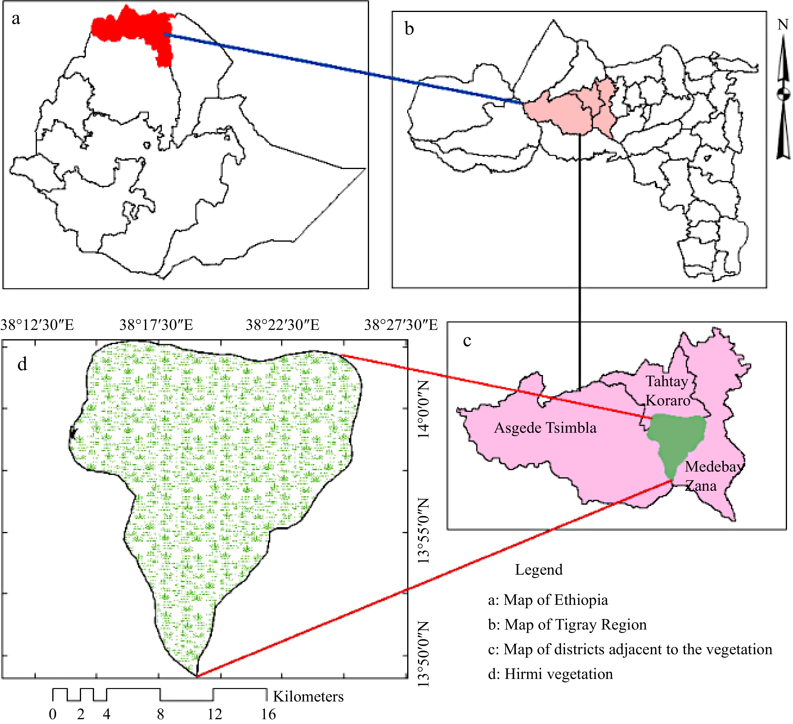
      Figure 1. 

      Map of Hirmi woodland (study area).

      Hirmi's vegetation is classified as Acacia-Comiphora, Combretum-Terminalia, and Dry-evergreen Afromontane forest type[4, 5]. Species including Senegalia venosa, Albizia malacophylla, Aloe elegans, Bidens macroptera, Lippia adoensis, Pennisetum glaucifolium, Phragmanthera macrosolen and Urtica simensis were among the dominant endemics species. Species such as Albizia malacophylla, Combretum hartmannianum and Combretum rochetianum as well as Senegalia venosa among the species that have scant populations and are found sparsely in Hirmi dryland ecosystem[6,7]. Currently, the study area is dominated by native remains and a few cultivated species. This is due to human disturbance and livestock interference in the forest.

    • To collect the required data for this study, a thorough survey which helps to identify the location of the species was conducted in the 30,900 hectares of the study area. At every site where the species is located, GPS points were tracked. Accordingly, a total of 11 plots with a size of 10 m × 10 m were laid in the site where the species is found following a purposive sampling approach. At every sampling plot, data related to the species abundance and growth habit were computed. The species growth habit with a height of ≥ 2 m and DBH ≥ 2 cm was considered as mature Senegalia venosa whereas those with a height of 1−2 m and a height ≤ 1 m were considered as saplings and seedlings, respectively[3, 8]. Environmental or ecological information, including disturbance signs, geographical coordinates (longitude and latitude) and altitude were recorded in each sample plot.

    • The collected data were analyzed in different approaches. ArcGIS version 10.5 was used to track the geographical distribution and map the sites where the species was located. Following that, a map was plotted that shows the locations of the 11 spatial coordinate points of the sample plots. Vis-à-vis the disturbances factors, it was measured and scaled from 0 to 4 based on visible signs of vegetation disturbance parameters including cutting, debarking, grazing, fire and charcoal production signs following[3]. Accordingly, values were coded: 0 − for plots with no any disturbance, 1 − for sample plots with one disturbance factor, 2 − for sample plots with two disturbance factors, 3 − for sample plots with three disturbance factors, and 4 − for sample plots that have more than three disturbance factors.

      The regeneration status of Senegalia venosa, was assessed by employing the total count of all seedlings, saplings and mature plant in each plot where the species were found following the techniques in[2]. Accordingly, the regeneration status was analyzed by calculating the density of seedlings, saplings and mature plants per the sample area as follows:

      Densityofmature/seedling/sapling=Thenumberofmature/seedling/saplingAreaofthesampleinahectare
    • The research findings indicate that the distribution of Senegalia venosa was restricted to specific locations within the Hirmi forest vegetation ecosystem. It was located in just 11 sites (Fig. 2). Even in the sample plots where the species was found, it was sparsely distributed between altitudes of 1,110−1,900 meters above sea level. This was associated typically with the existing topography and anthropic impacts. The species appeared (Fig. 3) in most of the plots at elevations between 1,700 and 1,900. Out of the 0.11 hectare sample plot, 40 mature, 19 saplings and 14 seedlings of Senegalia venosa were recorded. Accordingly, the mature Senegalia venosa has the highest density (363.6 species/ha) followed by saplings (172.7 species/ha) and seedlings (127.3 species/ha).
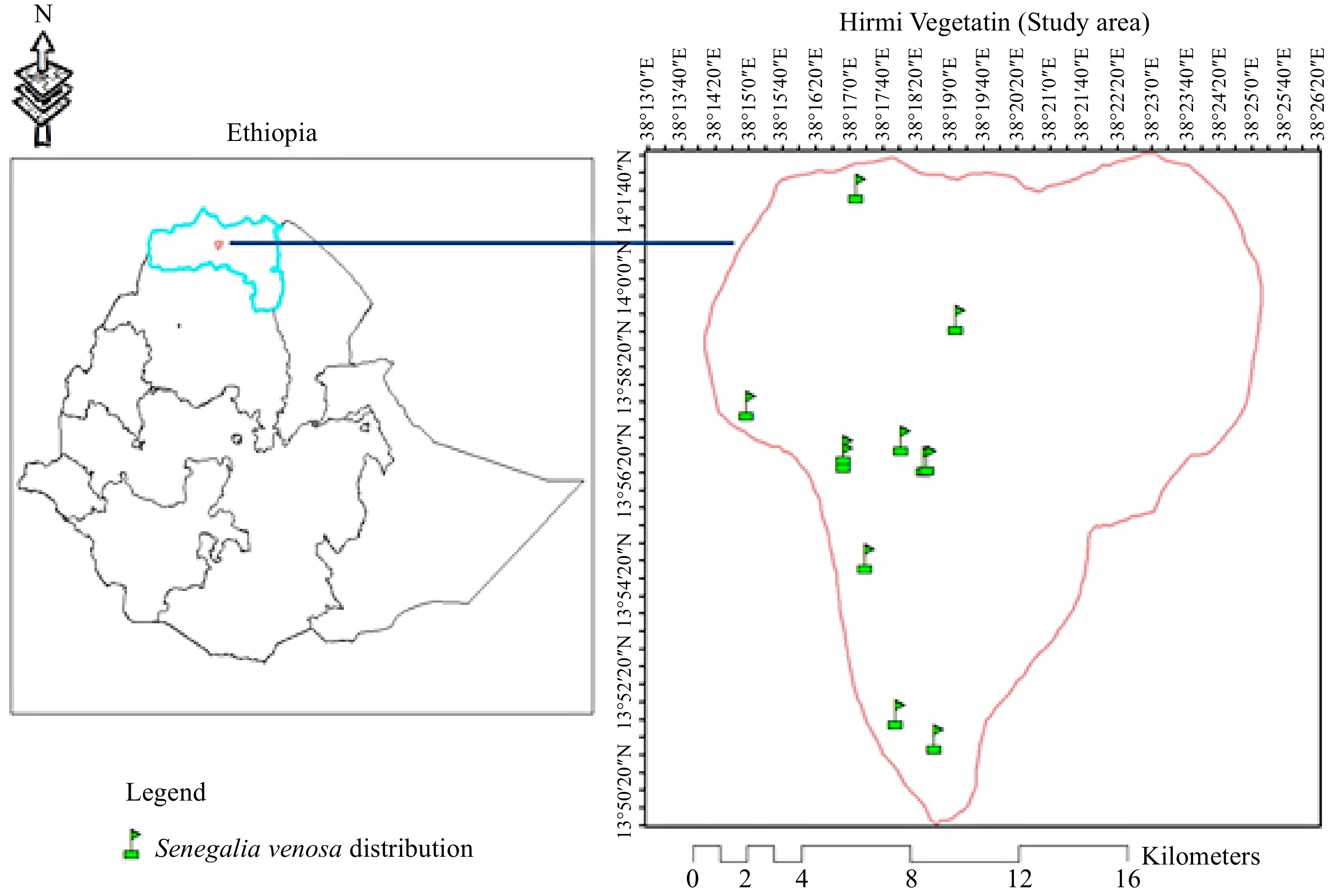
      Figure 2. 

      The map reveals the distribution of Senegalia venosa in Hirmi woodland vegetation.
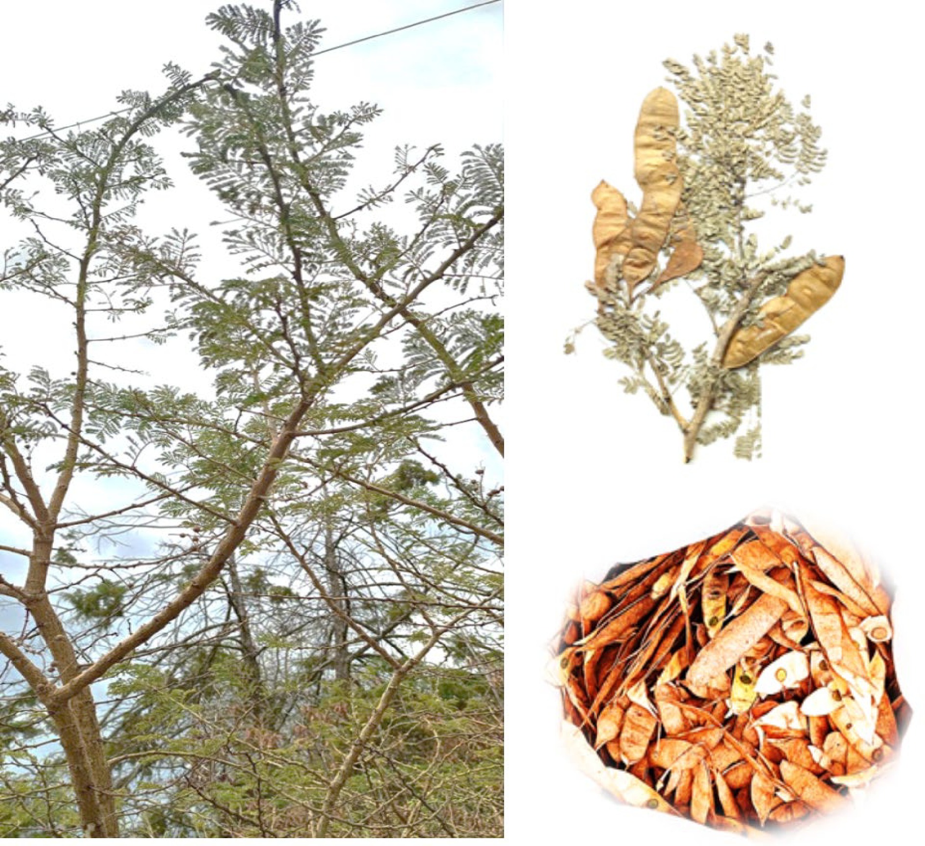
      Figure 3. 

      Images of different parts of Senegalia venosa.

    • The regeneration status of Senegalia venosa was characterized by computing the density of seedlings, saplings and maturity of the plant. A total of 40 mature Senegalia venosa as well as 28 saplings and 17 seedlings, were recorded in all sampled plots. In other words, the mature shrub of Senegalia venosa has the highest density followed by saplings and seedlings of the species (Fig. 4).
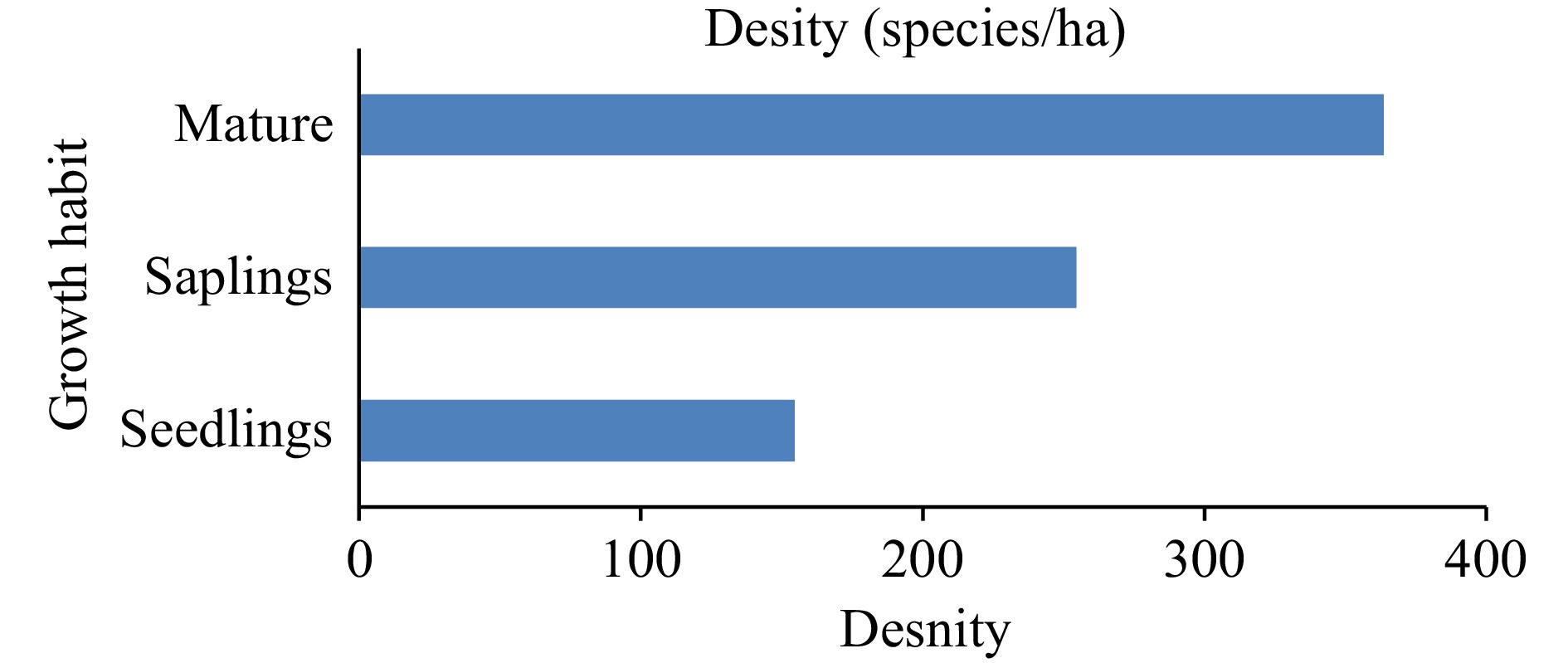
      Figure 4. 

      Regeneration status of Senegalia venosa.

    • According to the interviews made with the local communities in the study area as well as the researchers' field observation, anthropogenic disturbance signs such as charcoal production and cutting were frequently revealed in around the sampled area. Besides, livestock grazing and trampling were additional threats to the study species seedlings and saplings. The second plot has no visible disturbance whereas plot 5 has the highest rate of disturbance (Fig. 5).
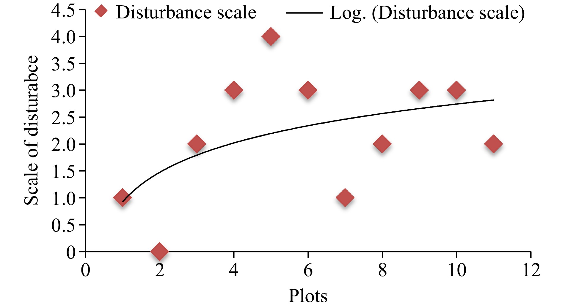
      Figure 5. 

      Scale of disturbance in the sample plots.

      Regarding the types of disturbances (Table 1), there was a high intensity of livestock disturbances such as grazing and trampling in plots 3, 4, 7, 9, and 11. Anthropogenic disturbances such as cutting and charcoal production were revealed in the 1st, 4th, 5th, 6th, and 10th plots.

      Table 1.  Locations and types of disturbance in sampled plots.

      PlotLatitudeLongitudeTypes of disturbance
      113.8550438.31161Cutting
      213.8626538.29898No
      313.9118638.28886Grazing
      413.9437238.2816Cutting and grazing
      513.9424138.30817Charcoal production & cutting
      613.9428738.30928Charcoal production & cutting
      713.9488938.30065Trampling
      813.9872538.31893Cutting
      913.98147238.26925Grazing & trampling
      1013.99635038.326788Cutting and cutting
      1114.0283838.2858Grazing
    • The vegetation of the Hirmi woodland is dominated by species of Acacia, Terminalia, Combretum, Albiza, Ficus, and Oxythenanthera abyssinica. It is one of the few habitats in the country where the plant Senegalia venosa is found[4]. The potential vegetation of Ethiopia studies by Vivero et al. and Kelbessa & Demissew[9, 10] also shows that ecosystems such as the Hirmi dryland can harbour Senegalia venosa due to favourable geographic conditions. However, the result of the study shows that the distribution of the species is restricted in a few sites (11 sites) out of the 30,900 hectares of Hirmi woodland vegetation. This indicates that the species are under critically endangered status either due to the existing anthropogenic threats[4, 11] or climate changes[3]. In line with this study, the poor regeneration status of species is mainly associated with climate change, habitat suitability and anthropogenic disturbances[12].

      Information related to the density ratio of seedlings, saplings and mature plants is used to determine the status of the regeneration of a given species and take pertinent conservation measures accordingly[13,14]. In this study density ratio density of Senegalia venosa was decreased from the mature of the plant to sapling and seedlings respectively. Based on the assessment criteria established by Khan et al.[2] & Khumbongmayum et al.[13], the vegetation's regeneration status is thus classified as poor. This outcome might be a result of the existing various disturbance factors, the demand of the species for medicinal value and the land use land cover dynamics due to agricultural expansions[4,15,16]. Additionally, because of the steep terrain where the study plant species is located, the soil is shallow, which could be difficult for seedling germinations. These findings were in line with a study by Wang et al.[17].

      Different disturbances were recorded in the majority of the sampled plots. However, the plots found near the center of the study area were relatively less disturbed since they are less accessible by humans and animals relative to these plots found on the edge of the vegetation area. Moreover, it was shown that heavy livestock grazing and trampling had a detrimental effect on Senegalia venosa seedling and sapling distribution. A similar result was reported in the study area by Girmay et al.[4,11].

    • The Senegalia venosa's poor regeneration status and limited ecological distribution as a result of various factors. This implies the requirement of further investigation on its propagation, and creating favourable niche and microclimate for better establishment of the species. Besides, avoiding unwanted anthropogenic disturbances, livestock pressures and other potential threats are some of the pertinent techniques to overcome the existing challenges. Establishing nursery sites to grow the seedlings and replanting in the study area to improve the poor population structure and recruitment succession of the species is prioritized. To do this, thorough research and surveys should be undertaken to determine the species' seed-producing and ripening phase so that it can be harvested appropriately for both in-situ and ex-situ conservation through nursery establishment. Additionally, this study underscores the necessity of raising the local community's understanding of how to manage and utilize plant species including Senegalia venosa in the study region sustainably.

    • The authors confirm their contribution to the paper as follows: study conceptualization, data curation, formal analysis and Writing of original draft: Girmay M, Gebrehiwot K; supervision, review & editing: Negussie A, Warkineh B; resources: Alemu S, Gidey T; methodology and software: Wondimu D, Kahsay G. All authors have reviewed the results and approved the final version of the manuscript.

    • The data that support the findings of this study are available from the corresponding author upon reasonable request.

      • We gratefully acknowledge the financial support from Mohamed bin Zayed Species Conservation to conduct this study.

      • The authors declare that they have no conflict of interest.

      • Received 15 December 2023; Accepted 18 April 2024; Published online 4 June 2024

      • Copyright: © 2024 by the author(s). Published by Maximum Academic Press on behalf of Hainan University. This article is an open access article distributed under Creative Commons Attribution License (CC BY 4.0), visit https://creativecommons.org/licenses/by/4.0/.
    Figure (5)  Table (1) References (17)
  • About this article
    Cite this article
    Girmay M, Gebrehiwot K, Negussie A, Warkineh B, Alemu S, et al. 2024. Distribution, regeneration status and threats to Senegalia venosa (Hochst. ex Benth.) Kyal. & Boatwr. in Hirmi Dryland areas of Northern Ethiopia. Tropical Plants 3: e018 doi: 10.48130/tp-0024-0017
    Girmay M, Gebrehiwot K, Negussie A, Warkineh B, Alemu S, et al. 2024. Distribution, regeneration status and threats to Senegalia venosa (Hochst. ex Benth.) Kyal. & Boatwr. in Hirmi Dryland areas of Northern Ethiopia. Tropical Plants 3: e018 doi: 10.48130/tp-0024-0017

Catalog

  • About this article

/

DownLoad:  Full-Size Img  PowerPoint
Return
Return