ARTICLE   Open Access    

Occurrence and interactions of arbuscular mycorrhizal fungi (Rhizophagus fasciculatus) and rhizospheric fungi in Saccharum officinarum L.

More Information
  • Received: 28 May 2024
    Revised: 09 August 2024
    Accepted: 20 August 2024
    Published online: 11 October 2024
    Tropical Plants  3 Article number: e033 (2024)  |  Cite this article
  • Different types of fungal species were obtained in the rhizosphere soil of AM fungus-infected and non-infected sugarcane plants.

    Outcome of our isolation process, abundance of rhizosphere fungi isolates was lower in plants infected with AM fungi than in plants without mycorrhizal infection.

    The occurrence and distribution of different rhizosphere fungi varied from plants infected with AM fungi compared to uninfected plants.

    We observe that parasitic fungi are virtually eliminated from the rhizosphere soil of plants infected with AM fungi.

  • Arbuscular Mycorrhizal fungi (AM fungi) promote plant growth and enhance nutrient uptake under unfavorable conditions. The present study investigates the interactions between AM fungi and rhizospheric fungi in sugarcane soil samples from sugarcane Pusa, Samastipur, districts, India. Sugarcane is the major crop mainly cultivated in these regions. Mycorrhizae and rhizosphere fungi were examined by isolating fungi from the soil of AM fungal infected and uninfected sugarcane crop field, 14 fungi belonging to nine genera isolates were obtained from the soil sample of AM fungi infected plant, on the other hand uninfected sugarcane yielded, 22 fungi belonging to 16 genera. In all, 10 fungi were common in both the rhizospheric soil of AM fungi-infected and uninfected sugarcane plants. The frequency and abundance of rhizospheric fungi were lower in plants infected by AM fungi than in plants without mycorrhizal infections. Furthermore, the presence of wilt-causing organisms and parasitic fungi was significantly lower in the soil of AM fungi-infected plants, while saprophytic organisms were more abundant. The frequency of different rhizosphere fungi during different months in a year fluctuated from 175 to 794 in AM fungi-infected plants, while the occurrence of different soil fungal isolates in the soil of non-infected plants fluctuated from 199 to 1,041. Parasitic fungi were almost eliminated from the soil of AM fungi-mediated plants. The results showed a significant interaction between AM fungi intensity and rooting time colonization compared to the control sample. Therefore, the present study data provides detailed knowledge on AM fungal inoculum and its effect on plant growth in a specific area.
    Graphical Abstract
  • One of the traditional techniques for increasing value and reducing agricultural produce spoilage is drying. Where more expensive alternative storage methods are used, this is especially crucial[1]. Through the addition of one or more energy sources, moisture from a product is removed throughout the drying process[2,3]. The physicochemical characteristics of the fruit are changed by drying, which can improve the flavor and texture of specific foods like raisins and dates[2]. It lowers the product's water activity (aw), and when the aw value drops to less than 0.6, it inhibits the growth and spread of spoiling bacteria[4]. Drying also reduces product weight, which reduces packing, storage, and shipping costs and ensures off-seasonal production[5,6]. The demand for dried fruit is rising globally as people become more health conscious[7].

    Worldwide pomegranate production is steadily rising, although large post-harvest losses are also common, according to reports[8]. When fruit is unsuitable for standard processing methods due to fruit cracking or sunburn, drying is a great way to reduce post-harvest losses because it extends shelf life and can be utilized to reduce food waste[9]. Numerous products, such as pharmaceuticals, snacks, cereals, quick drinks, and other confectionary items, employ dried pomegranate arils[10]. Dried pomegranate arils, also known as anardana, are utilized both medicinally and culinarily in several regions of the world, including India[11]. Dried arils can therefore be quite useful as value-added items that generate revenue. According to research conducted in the Indian Ramban area, anardana trade accounts for at least 41% of all annual household income[11].

    There are many different drying techniques. The most popular are freeze-drying, hot air drying, vacuum drying, and solar drying. Each technique has pros and cons in terms of the final product's quality and how efficiently it uses energy. Pre-drying procedures are frequently used in conjunction with drying. Pretreatment enhances the drying rate, product quality, and energy efficiency of the drying process. Enzymes that cause enzymatic browning, which lowers the product quality, are rendered inactive by pretreatments[1214]. Sensory qualities like color, texture, taste, scent, microbiological activity, and general acceptability are among the criteria that determine the quality of dried pomegranate arils[15]. These elements are crucial because they can have a big impact on customer preferences and, if not taken into full account, can lead to financial losses[16,17].

    Several studies[12,18,19] have looked at the impact of drying and pretreatment techniques on the general quality of the completed product. However, the combined impact of pretreatment and drying on quality was not sufficiently investigated. Reviews on pomegranate fruit often discuss the fruit's chemistry, nutritive value, and pharmacology. Therefore, by evaluating, highlighting, and reflecting on recent studies on pomegranate aril drying, this review seeks to close these gaps. This review paper compared and contrasted several pretreatment and drying setups with an emphasis on product quality.

    Heat, mass, and momentum exchanges all occur simultaneously during the drying of fruit materials in a sophisticated cellular architecture of biological tissue[20,21]. The characteristics of the material that affect the drying process are intricately dependent on size, shape, porosity, moisture content, and time[22]. For instance, the initial moisture level and the bioactive chemicals in pomegranate arils can vary depending on the cultivar and fruit ripeness. The understanding, engineering, and management of the drying process are further complicated by the intrinsic diversity of biological materials[23,24]. The mass, heat, and momentum transfer events that happen during a typical drying process are shown in Fig. 1[25]. Conduction and convection are the most common heat transfer methods, but radiation is typically only employed for high-end items due to its expensive cost[1,26]. Diffusion, capillary action, and bulk flow are only a few of the processes that might transfer mass. These mass transfer mechanisms must adapt to the ongoing physical changes in the material that take place as it dries out[22,27].

    Figure 1.  A visual representation of the drying processes of solid materials.

    Although it is native to Iran, the pomegranate (Punica granatum L.) is widely distributed worldwide[28]. It belongs to the family Lythraceae and is a deciduous shrub. It is a versatile plant that can be found growing in both semi-arid and subtropical climates. Pomegranates, however, need hot summer temperatures to ripen[29]. Pomegranate fruit has a non-uniform round shape and a range of hues depending on the cultivar and fruit development stage, including yellow, green, pink, deep red, deep purple, and black[30,31]. An outsized calyx crowns the fruit. The leading producers worldwide are Peru, Australia, South Africa, and Chile in the southern hemisphere, and India, China, and Iran in the northern hemisphere[32].

    Pomegranate is a one-of-a-kind fruit with distinct edible seeds (arils) that must be extracted by hand (Fig. 2)[33]. An aril is made up of a seed and fleshy, moist tissue surrounding the seed. Color, sweetness, juice content, and hardness of arils vary depending on cultivar and fruit maturity[30,31]. While the arils can be eaten fresh, they can also be made into jams, jellies, coloring agents, juices, vitamins, and anardana (dried arils). They can also be mixed into yoghurts, biscuits, and cereals[34]. Fresh pomegranate arils can be kept at 7 °C for up to 14 d without losing much quality. Dried pomegranate aril has an extremely low perishability, with a potential shelf life of more than 14 weeks in ambient air[35].

    Figure 2.  A typical breakdown of the material balance throughout the drying process for pomegranate aril.

    As demonstrated in Table 1, pomegranate is high in ellagitannins, gallic acids, ferulic acids, anthocyanins, flavonoids, fiber, and minerals like vitamin C, calcium, and phosphorus. Pomegranates' phenolic components and high vitamin C content (Table 1) have attracted the interest of both researchers and consumers due to their health advantages[3638]. As a result of its strong antioxidant activity and nutritional benefits, pomegranate is considered a superfruit. Pomegranate can also be employed in cosmetics and pharmacology due to its phytochemical and antioxidant qualities[39]. Pomegranate fruit extract (PFE) has bioactive elements that have been found to inhibit or prevent various types and levels of cancer[40]. Punicalagic acid, ellagic acid, urolithin, and luteolin are the most important pomegranate components known to have anticarcinogenic characteristics[40,41]. Pomegranate fruit has also been linked to the prevention of diseases such as Alzheimer's, hypertension, and diabetes[42,43]. Pomegranate supplements may also help during or after exercise because they have the potential to speed up hard exercise recovery[44].

    Table 1.  The nutritional composition of 100 g of pomegranate arils.
    NutrientValueUnit
    Water77.9g
    Energy346kJ
    Protein1.67g
    Total lipid fat1.17g
    Ash0.53g
    Total dietary fiber4g
    Total sugar13.7g
    Calcium10mg
    Phosphorus36mg
    Magnesium12mg
    Iron0.3mg
    Potassium236mg
    Sodium3mg
    Zinc0.35mg
    Vitamin C10.2mg
    Vitamin K16.4µg
    Vitamin E0.6mg
    Vitamin B-60.075mg
    Total choline7.6mg
    Folate38µg
    Adapted from United States Department of Agriculture (Agricultural Research Service), FoodData Central[52].
     | Show Table
    DownLoad: CSV

    Pretreatment procedures are utilized to improve the drying process's effect on product quality characteristics such as color, flavor, appearance, and some physicochemical aspects[45,46]. Figure 3 depicts the most often used pretreatment procedures[46]. A product is immersed in a chemical solution prior to drying in chemical pretreatment. Physical pretreatments, on the other hand, necessitate a physical alteration of the product. When drying with heat, Maillard reactions might occur, resulting in an unpleasant color change[47]. As a result, pretreatment techniques are critical in many drying applications, including the drying of pomegranate arils[45,48,49]. There is evidence that pretreatment reduces the product's exposure to heat by reducing drying time[50,51].

    Figure 3.  A classification of the numerous pretreatment techniques utilized in the drying process for pomegranate aril.

    Soaking in acidic solutions involves immersing the product to be dried in a hot acidic solution for many minutes before drying. Pretreatment with an acidic solution keeps the product's color and speeds up the drying process. The acidic solution suppresses polyphenol oxidase enzyme activity, slowing the rate of enzymatic browning (Fig. 3). Furthermore, numerous investigations[5355] have documented the retention of nutrients such as vitamin C in acidic solution pretreatment samples. Some acid-sensitive components, on the other hand, can be destroyed or leached away. As a result, while employing this strategy, this effect must be considered. In a pomegranate aril drying research, arils prepared with 3% citric acid had the highest sensory acceptance[56]. Vardin & Yilmaz[57] conducted research on the combined effect of acid blanching and subsequent drying temperature. The authors blanched the arils in 0.1% citric solution for 2 min at 80 ± 2 °C followed by drying at 55, 65, or 75 °C and discovered that drying at 55 °C had the maximum antioxidant capacity[46]. Understanding the connection between soaking in acid (balancing in acid) and the subsequent drying temperature is required to carry out the operation correctly.

    This entails immersing products in an alkaline solution. Alkaline solutions act by dissolving the wax covering on the fruit's surface, removing resistance to moisture transfer and increasing drying rate[58]. As a result, this pretreatment accelerates the drying process. However, the usage of alkaline solutions raises food safety concerns because the residue might be harmful to one's health[46,59]. In addition, although acidic solutions retain vitamin C, alkaline solutions leach it out and destroy it[46]. Samples dipped in ethyl oleate for roughly one minute revealed a considerably reduced drying rate: a 26.9, 28.5, and 27.2% decrease in drying time at drying air temperatures of 55, 65, and 75 °C, respectively, than the control[60].

    The fruit is dipped into a hypertonic solution, such as salt or sugar solutions, in this approach (Fig. 3). Because of the osmotic pressure differential, the hypertonic solution causes water to diffuse out of the fruit tissue[61]. When compared to other drying processes, osmotically pretreated dried products have great rehydration capability and little losses in quality parameters such as color, appearance, and nutrients[62]. Madhushree et al.[63] discovered that osmotic pretreatment (in 50oBrix sugar syrup concentrations) dried arils had high color retention. This could be owing to the samples' reduced exposure to oxygen when immersed in the sucrose solution. A separate investigation on the osmotic pretreatment of pomegranate arils with a 65°Brix sucrose solution revealed a decrease in drying rate in hot air drying at 70 °C compared to untreated control samples[64]. The scientists attributed the longer drying time (lower drying rate) to the creation of a dense sucrose layer beneath the fruit's surface, which created an additional barrier to moisture transfer. To that aim, the osmotic solution concentration must be assessed because it can result in prolonged drying times.

    Gaseous or liquid sulphur solutions have been used as a food preservation method and as a pretreatment in food drying procedures. Typically, sulphur solutions are utilized for their browning properties, both enzymatic and non-enzymatic[65]. In addition, sulfur pretreatment is associated with high vitamin C and A retention after drying, as well as inhibition of spoilage-related microbial proliferation[66].

    More et al.[23] compared physical pretreatments to chemical pretreatments with 1% potassium metabisulphide on arils. It was discovered that arils prepared with potassium metabisulphide had superior nutritional quality as well as improved color, flavor, taste, and overall acceptability (Table 2)[23]. As a result, processing of pomegranate arils with sulfur solutions can result in high-quality dried goods. Despite their anti-browning, antibacterial, antifungal, and nutrient retention qualities, sulphites might be harmful to one's health if the recommended dosage or daily intake is exceeded[67,68]. Furthermore, while the sulfur solutions maintain vitamins A and C, they deplete vitamin B1[65].

    Table 2.  Key findings in pomegranate aril pretreatment and drying studies.
    Pretreatment methodPretreatmentDrying techniqueKey findingsReference
    BlanchingWater blanching at 90 and 100 °CHot air oven dryingBlanched samples had a shorter drying time.[24]
    Water blanching at 80 °CHot air oven dryerBlanched samples had higher phytonutrient retention than unblanched samples.[69]
    Blanching using 0.1% citric solution
    at 80 ± 2 °C
    Cabinet tray dryerDrying process was shorter for blanched samples and there was a higher rate of bioactive compounds.[57]
    Sulphuring1% potassium metabisulphideSolar drying
    Cabinet tray dryer
    Freeze dryer
    Fruit of cv. Ganesh 1% potassium metabisulphide was of the highest quality and the highest acceptance.[23]
    Blanching and SulphuringHot water blanching 85 °C and 0.2% potassium metabisulphateMechanical dryer
    Solar dryer
    Keeping quality of mechanically dried arils was higher than the solar-dried arils.[70]
    Steam blanching, potassium metabisulphide and 0.3% Sulphur fumigationCabinet tray dryerThe highest dried aril quality was obtained from the combination of steam blanching and 0.3% Sulphur fumigation.[71]
    Steam blanching, sulphuring at 0.3%Vacuum dryer
    Hot oven dryer
    Sun drying
    Poly-tent house drying
    Room drying
    Sun drying had the highest moisture content reduction and the highest overall acceptance.[72]
    Hot water blanching 85 °C,
    potassium metabisulphite varying
    from 0.25% to 1%
    Hot air oven dryerThe best treatment was blanching in hot water at 85 °C for 1 min and then dipping the arils in 0.25% potassium metabisulphite.[70]
    Steam blanching, sulphuringSun drying
    Cabinet dryer
    Blanching reduced drying time. Cabinet drying of blanched samples without sulphuring was considered optimum for anthocyanins.[73]
    Acidic solution2%, 3% and 4% citric acidCabinet tray dryer

    3% acidic treatment was found to be the most acceptable.[56]
    Microwave100 and 200 W.Hot air oven dryer200 W pretreatment resulted in minimum energy utilization and drying time.[74]
    100 and 200 WHot air oven dryer200 W had the highest drying rate.[75]
    Osmotic treatmentSugar syrup, freezing at minus 18 °COpen sun drying,
    Solar tunnel dryer,
    Cabinet tray dryer
    Osmotic treatment and cabinet tray dryer produced dried arils with better physicochemical and sensory qualities.[63]
    • 100% pomegranate juice
    • 50% pomegranate and 50% chokeberry juice
    • 50% pomegranate and 50% apple
    • 50% apple and 50% chokeberry
    • 75% apple and 25% chokeberry
    Freeze drying
    Convective pre-drying vacuum microwave finish drying
    Vacuum-drying and freeze drying
    Pomegranate and chokeberry concentrated juice improved the quality of the dried arils.[12]
    Sucrose solutionHot air oven dryingPretreatment increased the drying time of the samples.[64]
     | Show Table
    DownLoad: CSV

    The dipping or soaking of a product in an alcoholic solution, usually ethanol, is known as alcoholic pretreatment. Ethanol dissolves the cell wall components, which increases moisture loss and thus the drying rate[76]. Several fruits, including melon (Cucumis melo L.) and apples (Malus domestica), have been pretreated in alcoholic solutions before drying[77,78]. However, no investigations on the pretreatment of pomegranate arils with alcohol were reported. This could be owing to the aril's waxy layer, which could impede permeability and hence the efficacy of the alcohol pretreatment[79].

    In this method, the fruit is dipped, submerged, or sprayed with a liquid solution that forms a thin coating layer on the product's surface and then dried (Fig. 3). According to studies, the use of edible coatings can help to retain the color, texture, and nutrient retention of dried items[80]. It is critical to note that the drying pace and dried product quality are affected by the coating thickness, drying method, and coating solution. Most of the research on edible coverings for pomegranate arils has focused on cold preservation and packing.

    Acoustic cavitation is utilized to rupture cell walls using ultrasound pretreatment[81]. According to one investigation on the effect of sonification on osmotic dehydration and subsequent air drying of pomegranate arils, ultrasonography caused a 2-fold and 2.7-fold increase in water loss[82]. The authors hypothesized that ultrasonic promoted cell wall disintegration and enhanced permeability. While using ultrasonic improved color quality, it also reduced anthocyanin content when compared to osmotically dehydrated samples[82].

    Blanching is a pretreatment technique that involves rapidly heating and then cooling a product that will be dried[45]. Blanching can be used to inactivate enzymes that potentially degrade product quality, such as polyphenol oxidase, peroxidase, and polygalacturonase[4]. Unwanted sensory traits in color, flavor, texture, and nutritional aspects are examples[4,83]. Blanching also improves cell membrane permeability, resulting in a faster drying rate[45]. Furthermore, blanching kills bacteria that might cause product spoiling[45,83]. As indicated in Fig. 3, there are several blanching processes, including hot water blanching and modern technologies such as microwave blanching and infrared blanching.

    Adetoro et al.[18] discovered that blanching pomegranate arils in hot water accelerated drying rates compared to unblanched samples. In a separate investigation, the authors found that blanching arils at 90 °C for 30 s and drying at 60 °C had higher total anthocyanin content and radical scavenging activity than blanching at 100 °C for 60 s and drying at 60 °C. In another work, Karaaslan et al.,[69] blanched arils in water at 80 °C for 2 min to investigate the effects of temperature and pretreatment on the arils. The authors discovered that while 75 °C had the fastest drying time, 55 °C had the maximum anthocyanin concentration, phenolic content, and antioxidant capacity. In other studies, pretreatment procedures are combined to produce high-quality dried fruit products.

    Singh et al.[73] conducted a study to evaluate the drying of pomegranate seeds under various drying conditions. They discovered that blanched samples dried faster and had higher acidity than sulphured samples. Furthermore, the anthocyanin concentration of blanched samples was higher than that of blanched and sulphured samples using the mechanical dryer. The authors hypothesized that the reduced anthocyanin concentration seen after sun drying was caused by the long drying hours in the sun. They also suggested that pomegranate arils be dried using blanching rather than sulphuring to achieve the maximum nutritional quality. Sharma et al.[70] investigated ideal methods for drying pomegranate arils by blanching them in hot water at 85 °C for one minute and then immersing them in a solution of potassium metabisulphite with concentrations ranging from 0.25% to 1% for two minutes (Table 2). The highest potassium metabisulphite content resulted in the lowest acidity.

    Thakur et al.[71] used steam blanching for 30 s and 0.3% sulphur fumigation for one hour to standardize pretreatments for dried arils from wild pomegranate. The authors discovered that cabinet drying outperforms solar drying and open sun drying. The solar dryer ranked second in terms of sensory characteristics like texture, taste, and general acceptability. Sun-dried pomegranate arils, on the other hand, exhibited the highest reduction in moisture content and overall acceptance when compared to vacuum drying, hot air oven drying, polytent drying, and room temperature drying, according to Bakshi et al.[72].

    The effect of pulsed electric field treatment on the behavior of microwave-assisted hot air drying of pomegranate arils was examined by Amiali et al.[84]. When compared to drying at 70 °C, they found that pulsed electric field treatment was only advantageous when the subsequent drying process was done at the lowest temperature (50 °C). The lower the temperature, the higher the overall phenolic concentration. Arils treated with a pulsed electric field had a 21.02% higher total phenolic content than untreated arils.

    Various pretreatment procedures on pomegranate arils have been utilized with the goal of conserving physicochemical, physical, and chemical properties or enhancing drying rates. However, pretreatment standardization is currently restricted and underexplored. Table 2 lists some of the methods for preparing dried pomegranate arils.

    Despite the fact that pomegranate is a fruit with numerous health and nutritional benefits, it is now a modest crop with limited marketability. The difficulty in collecting the interior edible seeds (arils) is the greatest impediment to realizing the full potential of this unusual fruit[29,31]. Only manual extraction of pomegranate arils for laboratory scale testing is described in the literature.

    Pomegranates are first cleaned and sorted for uniformity in color, size, shape, and weight before aril extraction[18,85]. All pomegranates should be washed. To avoid introducing bacteria into the arils when the fruit is sliced open, excess water from the fruit surface is dried before cutting[86]. Following that, the fruit is cut along the ridges and the segments are gently pulled apart to form a flower-like structure. The deconstructed pomegranate is then flipped over a bowl of water and gently tapped with a wooden spoon on the skin side. As a result, the arils will begin to come out without being broken. Once all arils have dropped out, the white membranes are skimmed off the surface of the water as it floats, the water is drained, the arils are separated, and the surplus water is gently patted off with a towel. This method involves cutting the pomegranate with a knife, which results in a loss of more than 30% of the arils owing to mechanical damage[86]. As a result, a better approach was required, such as the machinery developed by Schmilovitch et al.[87], which allows opening the fruit without cutting, extracting the arils with minimal damage, separating the arils from extraneous materials, and delivering clean arils to a packaging machine. This technology could be used to produce dried pomegranate on a greater scale.

    The drying time is determined by the pretreatment process, kind, and technique of dehydration used. The dehydration methods that will be investigated in this study are low-temperature drying and high-temperature drying.

    Low-temperature drying methods employ temperatures ranging from subzero to 50 °C[49,88]. These drying processes are time-consuming and are usually utilized for temperature-sensitive goods like herbs. Furthermore, low-temperature drying procedures reduce the risk of scorching the fruit, protecting heat-sensitive components such as vitamin C[89]. Freeze drying, vacuum drying, and sun drying are all low-temperature drying procedures[90]. Pomegranate arils dried using low-temperature procedures such as freeze drying and vacuum drying have a more acceptable look and nutrient composition than most high-temperature drying methods[12,91].

    Freeze-drying (FD) is a low-temperature technology frequently used for drying food samples for high-quality or heat-sensitive products[92]. It is also recognized as one of the most expensive, time-consuming, and energy-intensive procedures in the food industry[93]. It entails removing moisture from food ingredients under low temperature and high vacuum via ice sublimation[48]. Because the product is frequently frozen, it is also known as sublimation drying.

    Adetoro et al.[94] freeze-dried fresh pomegranate arils at a freezer temperature of −80 °C for 96 h to examine the effect of drying procedures on pomegranate arils. The researchers discovered that color, total phenolic compounds (TPC), total anthocyanin content (TAC), and radical-scavenging activity stability differed significantly from the hot air-drying procedure (Table 3). More et al.[23] investigated the effect of drying procedures on the quality of dried pomegranate arils from three varieties. The authors discovered that FD produced the greatest results in terms of color, flavor, taste, and nutritional factors across all cultivars. However, FD stood out due to its prolonged drying duration, 24−48 h, when compared to solar drying (17 h) and hot air drying (10 h). A study on the influence of freeze-drying on the color attributes of 'Assiuty' pomegranate arils revealed that FD had the best color attributes (L* value of 46.50 ± 4.4 and a* value of 13.97 ± 1.23)[95]. Gölükcü[91] discovered that the FD had the maximum phenolic matter content (5580 mg/kg), followed by vacuum, convective, and sun-dried pomegranate arils (Table 3). Caln-Sánchez et al.[96] investigated the chemical composition, antioxidant capability, and sensory quality of pomegranate arils and rind after exposure to FD. The investigators found that FD pomegranate arils retained the most sensory characteristics and punicalagin content. Similarly, Cano-Lamadrid et al.[12] discovered the best sensory profile and sweetness in FD pomegranate arils at 65 Pa for 24 h at −60 °C. The drying kinetics, total bioactive content, in-vitro bio accessibility of bioactive compounds, and color and microstructural features of pomegranate arils were also studied[97]. When compared to alternative drying methods, the FD was shown to be the best approach in terms of final product quality and has been highly recommended by multiple reviewers[48]. FD arils have been demonstrated to have a higher bioactive chemical content, less shrinking, and excellent color quality. The FD for pomegranate arils has a disadvantage in terms of bioactive chemical recovery when compared to other methods, as well as extensive drying times[97]. Furthermore, FD is costly due to high energy consumption and initial investment expenses[98].

    Table 3.  A summary of the many techniques for drying pomegranate arils.
    CultivarDrying methodDrying
    time (h)
    Drying conditionInitial moisture
    content (%)
    Final moisture
    content (%)
    Key findingsReference
    WonderfulFreeze dryer96−80 °C
    5,999.1 Pa
    74.7 (w.b.)FD showed a higher color shift (19.6% ± 2,77%) at week 4 compared to hot air drying at week 0.[94]
    Ganesh, Bhagwa and AraktaFreeze dryer24−48−45 °C79.9 (w.b.)
    80.5 (w.b.)
    78.9 (w.b.)
    9.65−9.9 (w.b.)
    9.8−10.2 (w.b.)
    9.8−9.9 (w.b.)
    Arakta pre-treated with 1% potassium metabisulphide had the highest ascorbic acid concentration (6.81 ± 0.07 mg 100 g−1).[23]
    AssiutyFreeze dryer36−70 °CTA (18.80, 2.80 mg 100 g−1), TP (608.09a, 41.26 mg 100 g−1), DPPH (68.91, 0.72%), and ABTS (2,956.59c, 120 mol trolox
    100 g−1) were all higher in frozen pomegranate arils than in freeze-dried arils.
    [95]
    Mollar de Elche24−60 °C, 65 Pa,81.5 (w.b.)FD showed higher anthocyanin content (646 mg kg−1) compared to osmotic drying and conventional drying.[12]
    Hicaznar36−20 °C, 100 Pa for
    12 h
    −70 °C, 0.26 Pa
    76.96 (w.b.)10 (w.b.)Arils dried using FD had the highest magnesium content (96.17 ± 6.95 mg kg−1), manganese content (0.96 ± 0.05 mg kg−1) and zinc content (3.93 ± 0.07 mg kg−1) compared to sun drying, hot air drying and vacuum drying.[91]
    KebenFreeze dryer57−55 °C77.6 ± 1 (d.b.)20 ± 1 (d.b.)FD had lower bioactive recovery during in-vitro gastrointestinal digestion (TPC of 2.92%, ABTS of 6.12% and CUPRAC of 38.85%) compared to vacuum drying, hot air drying and ultrasound-assisted vacuum drying.[97]
    KebenVacuum dryer10.855 °C77.6 ± 1 (d.b.)20 ± 1 (d.b.)Vacuum drying had the highest bio accessibility recovery of bioactive compounds at 10.32% compared to HAD, FD and ultrasound-assisted vacuum drying.[97]
    HicaznarVacuum dryer3.7
    4.6
    7.8
    75 °C
    65 °C
    55 °C
    85,000 Pa
    78.1 ± 0.2 (w.b.)16 (w.b.)Drying temperatures of 75°C resulted in higher degradation of anthocyanins, phenolic compounds and antioxidant capacity (20.0%, 51.0%, 29.7% ± 0.28% respectively).[99]
    HicaznarVacuum dryer2455 °C
    3 500 Pa
    76.96 (w.b.)10 (w.b.)FD had the highest quality attributes such as TAC-1288.73 mg/kg) compared to VD and HAD however, due to physical changes that were undesirable physical changes, VD and HAD are recommended for pomegranate aril drying.[91]
    Wild pomegranates (cultivar- unspecified)Vacuum dryer1342 ± 2 °CVD had a higher sensorial overall acceptance (17.1) compared to sun drying (16.2) and room temperature (12.6).[72]
    BasseinSun drying1715.73
    (unspecified)
    Drying rate of pomegranate arils is affected by tray load and the recommended tray load is 1.25 kg m−2[73]
    Wild pomegranates (cv. Unspecified)Solar poly-tunnel14014.9−28.4 °C
    48.5%−74% -RH
    0.639−0.944 -wind speed
    Arils dried in the solar poly-tunnel had higher ascorbic acid, anthocyanins, and phenol content, 12.7 mg 100 g−2, 28.12 mg 100 g−2, and 108.60 mg 100 g−2 respectively than open sun drying.[100]
    SefriIndirect solar dryer75
    10
    6
    4
    40 °C
    50 °C
    60 °C
    75 °C
    78 ± 0.1(w.b.)The optimal water activity for drying and storing arils is 0.3684 ± 0.03.[101]
    Wild pomegranates
    (cv. Unspecified)
    Solar tunnel30−45 °CArils from the Karsog location had the highest TSS, sugars, anthocyanin, total phenols and antioxidant activity[102]
    Microwave2.3
    1.3
    0.7
    270 W
    450 W
    630 W
    70.25 ± 0.5(w.b.)10(d.b.)Color changes increased from 6.77−13.11 with an increase in microwave power from 270−630 W and were lower compared to arils dried using HAD.[85]
    HicazMicrowave1.2
    0.6
    0.4
    210 W
    350 W
    490 W
    23.93 ± 1.4 (unspecified)22.2
    (unspecified)
    Based on quality parameters, a microwave drying power of 350 W was recommended for drying pomegranate arils.
    Sweet acid
    (cv. Unspecified)
    Infrared4.3
    2.2
    1.6
    50 °C
    60 °C
    70 °C
    78 ± 0.2 (w.b.)9 ± 0.2 (d.b.)
    22.2
    (unspecified)
    Drying time for infrared drying was less than for HAD.[103]
    HicazHAD2450, 60, 70 °C at
    1.0 m/s air velocity
    To obtain better dried aril quality, 60 °C was recommended for drying pomegranate arils.[104]
    Wild pomegranates
    (cv. Unspecified)
    HAD16.542 ± 2 °CSun drying resulted in a maximum loss of moisture compared to VD, HAD, poly-tent house drying and room drying.[72]
    Wild pomegranates
    (cv. Unspecified)
    HAD1062 ± 2 °CHAD achieved the highest total soluble solids (39.6°Brix) and drying rate compared to solar drying and open sun drying.[105]
    KebenHAD555 °C; with 1.3 m s−1 constant air velocity77.6 ± 1 (d.b.)20 ± 1 (d.b.)In comparison to hot air oven drying, ultrasound-assisted vacuum drying and freeze-drying have higher quality characteristics.[97]
    Bassein seedlessHAD5−660 ± 5 °C. Airflow in
    the dryer was 1.2−1.8 m s−1.
    8.98 ± 0.091For the finest preparation of anardana, blanched samples (with sulphur) should be dried in a cabinet.[73]
    Mollar de ElcheVacuum-
    microwave
    240, 360, 480 W and pressure ranging
    from 4,000−6,000 Pa
    80.4 (w.b.)Arils dried using vacuum microwave drying at 240 W had the highest sensorial scores for odour and aroma at 3.1 and 5.6 respectively.[106]
     | Show Table
    DownLoad: CSV

    Vacuum drying (VD) is the process of subjecting items to low pressure in a vacuum. Because of the low pressure, water has a lower boiling point, allowing samples to be dried at low temperatures. As a result, VD is appropriate for heat and/or oxygen-sensitive items. During VD, heat transmission can occur by conduction, radiation, or microwave energy. VD is distinguished by faster drying times when compared to FD, and the products are not initially frozen as necessary for FD[107]. This low-temperature operation, combined with the elimination of oxygen during vacuum drying, allows nutrients and bioactive components such as phenolic compounds and vitamins to be retained[108,109].

    Ozay-Arancioglu et al.[97] investigated the influence of drying methods on dried pomegranate arils by comparing four distinct drying techniques: FD, VD, ultra-assisted vacuum drying, and hot air drying. They discovered that VD had better antioxidant capacity values than the samples tested for ABTS following FD. According to Gölükcü[91], VD is second only to FD as the finest choice for producing dried pomegranate (Hicazar) arils. Another study found that arils dried at 55 °C had higher phytonutrient levels than those dried at 65 and 75 °C under vacuum conditions[69]. When compared to other drying procedures, vacuum drying produces products with higher levels of phytochemical components. However, drying times range from 7.8 to 24 h at 55 °C, contributing to high costs, and products can only be dried in batches[91,99,110].

    Sun drying is one of the oldest and most used methods of drying. Sun drying is a low-cost, renewable energy-based drying process. In a nutshell, products are laid out on a flat area where they can be fully exposed to the sun for as long as possible. Because the drying process is dependent on solar radiation, the temperature is low and the drying process can be lengthy, taking approximately 15 d for pomegranate arils[60]. Furthermore, exposure to light and oxygen can lead to decreased preservation of substances like vitamin C. Furthermore, solar drying is an uncontrolled process with substantial risks of pest contamination, dust exposure, and product remoistening at night. To increase safety, solar dryers and solar tunnels are proposed to reduce pest and dust contamination[111]. Solar dryers use a contained environment comprised of a transparent or opaque cover, resulting in either direct or indirect drying[112]. The indirect drier system captures solar heat and transfers it to the product drying chamber via a second solar collector. A solar tunnel dryer is typically large in size and has a clear cover (Fig. 4). To regulate drying conditions such as temperature and relative humidity within the tunnel, solar tunnels often require a forced convection facility. A solar tunnel dryer may also include a solar air heater[111]. Solar dryers have the potential to boost drying temperatures, resulting in a quicker drying time[113].

    Figure 4.  This image depicts a sample of wild pomegranate arils being dried in a solar tunnel drier. Reprinted from Thakur et al.[102].

    In a comparison of hot air drying (60 °C), solar tunnel drying, and sun drying by Madushree et al.[63], hot air-dried arils were shown to have the highest quality. However, of all the drying techniques, solar drying had the greatest L* values (lightness), a desired quality. This was due to the comparatively low temperature of sun drying. Additionally, Bakshi et al.[72] discovered that when compared to vacuum drying, oven drying (42 ± 2 °C), and room drying (23 ± 2 °C), sun dried arils had the highest sensory overall acceptance and the lowest moisture content. In their comparative analysis of drying techniques, Singh et al.[73] discovered that hot air-dried pomegranate arils had the greatest anthocyanin and acidity contents. But in hot-air oven-dried samples, undesirable non-enzymatic browning was most pronounced. Sharma & Thakur[100] demonstrated that the quality of arils dried in solar polytunnels was superior to that of arils dried in the open sun (Fig. 4). The ascorbic acid, anthocyanins, and phenols were found to be significantly greater in the sun polytunnel dried arils, according to the authors. They also received superior sensory ratings for color, texture, taste, and acceptability.

    Temperatures above 50 °C are used in high-temperature drying processes[88]. These drying processes are energy intensive, have large operating expenses, and so are costly. These technologies rely on fossil fuels, which pollute the environment where they are generated and utilized, and their continued usage is seriously harming our environment[114]. The drying mechanism is designed such that there is a controlled direct or indirect heat transmission to the product, leading in moisture elimination. These drying procedures may not be suited for some foods because they may induce nutritional breakdown[115]. Furthermore, high temperatures might cause product shrinkage and distortion. Hot-air drying ovens, steam drying, heat pump drying, and spray drying are all examples of high-temperature drying processes.

    Using forced convection, hot air oven drying (HAD) eliminates moisture from materials. Objects dry out through evaporation when hot air is forced through and around the substance. As a result, the dried product's flavor, color, nutrients, and ability to rehydrate may alter unintentionally[97,104,106]. The HAD techniques were shown to have a comparatively high total color change by Ozay-Arancioglu et al.[97]. As shown in Fig. 5, hot air-dried arils were darker than freeze-dried and vacuum-dried arils.

    Figure 5.  Illustration of dried pomegranate arils that have been dried using different methods. (a) Freeze drying, (b) vacuum drying, and (c) hot air drying. Adapted from Ozay-Arancioglu et al.[97].

    Başlar et al.[99] prepared dried aril samples using the hot air-drying process and subjected them to various quality assessments. Fresh aril samples were dried at three different temperatures (55, 65, and 75 °C). According to the authors' findings, high temperatures and short drying times are optimal for retaining valuable food biocomponents. However, whereas bioactive chemical losses increased over time, they degraded faster at higher temperatures. The antioxidant activity, on the other hand, decreased with drying time and was unaffected by drying temperatures. Horuz & Maskan[104] investigated the effect of hot air drying on pomegranate aril cv. Hicaz at three different drying temperatures and compared quality metrics such color, shrinkage, rehydration capacity, and drying time (Table 3). The authors suggested 60 °C for pomegranate aril HAD. The authors also discovered that shrinkage was greater in HAD than in microwave drying. In a second study, researchers discovered that the optimal drying temperature for retaining bioactive chemicals when drying pomegranate arils (cv. Hicaznar) in a hot air dryer was 65 °C[116]. When compared to the sun drying method for anardana made from wild pomegranate, Bhat et al.[105] discovered HAD dried arils with maximum acidity of 13.72%, phenols of 110.7 mg per 100 g, total sugars (24.2%), and reducing sugars (21.2%).

    However, Bakshi et al.[72] carried out a study with lower temperatures in which they studied the influence of different drying processes on the moisture content of dried pomegranate aril (cv. Wild). Lower temperatures were employed to gain insight into the quality of the dried product when compared to the low temperature drying methods used in the study, such as sun drying, poly tent house drying, room drying, and VD. When compared to alternative drying methods, the authors discovered that HAD (42 ± 2 °C ) for 16.5 h and drying in room at normal air (23 ± 2 °C ) for 10−12 d produced in the greatest loss of moisture from fresh arils of wild pomegranate (75.12%).

    Singh et al.[73] evaluated the influence of different drying conditions on the quality of dried pomegranate arils (Bassien Seedless) samples (Table 3). The scientists discovered that sun-drying preserved more MC while drying was faster with a HAD dryer and generally recommended it as a better strategy for preparing dried pomegranate arils.

    HAD drying of pomegranate arils is a standard drying procedure that can be utilized in commercial settings. Although HAD does not generate high-quality goods like FD, it does provide better TSS, TA, and antioxidant capacity stability. Furthermore, although having a higher rate of bioactive component degradation, higher temperatures may result in higher retention compared to approaches such as solar drying due to the short drying times.

    Electrical current is passed through the pomegranate aril during electric drying techniques including ohmic heating. The intrinsic resistance of the aril induces internal heating as the electrical current flows through it[117,118]. Ohmic heating is typically employed for liquid, viscous, and particle-containing foods[119]. Regardless of the meal's densities, food products prepared using this approach are heated quickly and uniformly[120].

    Dielectric techniques, on the other hand, use electromagnetic waves to directly produce heat inside the product, such as microwave, radio frequency drying, and infrared radiation[121]. Dielectric heating results from the conversion of electromagnetic energy to kinetic energy by dipolar molecules oscillating in accordance with the rapidly oscillating electric field[122,123]. Compared to traditional methods like hot air drying, dielectric technologies dry materials more quickly[124]. Additionally, the items are of a higher caliber than those produced by traditional drying techniques.

    Microwave drying (MD) is one of the emerging drying technologies. Unlike other techniques, MD utilizes volumetric heating to rapidly dehydrate the sample material[104]. Some studies[85,104] have indicated that arils desiccated at 150 W microwave power and 58 bar (abs) pressure produced the highest quality arils. In another study, microwave power of 80 W and vacuum pressure of 60 mm Hg provided the highest drying efficiency and qualitative attributes, including color and texture[125]. Horuz & Maskan[104] observed that microwave-dried pomegranate samples had lower levels of shrinkage and bulk density than hot air-dried samples. The authors also noted that microwave drying caused a greater loss of color in terms of total color difference (E) compared to air drying. It was observed that microwave-dried samples had a brownish hue.

    Drying with MD reduces drying time, but essential quality parameters, such as color, are sacrificed. Since the product is directly heated, the lack of heating uniformity during MD, which is difficult to control and could contribute to product burning has been cited as a disadvantage[126].

    Infrared drying is an effective technique of drying in which the product is heated directly without the use of air as the drying medium. In a comparative study of drying methods (hot air drying and infrared drying) to dry pomegranate arils, the authors discovered that infrared drying effectively dried pomegranate arils and that the polyphenol content in arils dried using infrared drying was higher at 50 and 60 °C than in arils dried using hot air drying at similar temperatures[103]. Therefore, pomegranate arils can be infrared-dried at 50 °C for optimal nutrient retention[103]. In contrast, a distinct study dried pomegranate arils under near-infrared vacuum conditions and found that drying at 60 °C and 20 kPa air pressure resulted in optimal colour retention and shrinkage[127].

    Although infrared drying is a rapid drying method, it is challenging to control due to parameters such as infrared intensity and radiation distance, and its energy consumption is unpredictable[26].

    Drying kinetics is the study of how factors that influence the removal of moisture from products during a drying process interact[110]. The drying kinetics of a substance is dependent on its thermal and mass transport properties. Understanding drying kinetics relates to process variables, and therefore aids in identifying suitable drying methods and controlling drying processes[57,96,116]. For optimal operating conditions, drying kinetics can be used to estimate drying time, energy requirements, and drying efficiency[2]. Due to the complexity of the drying phenomenon, however, mathematical models describing the drying kinetic of biological tissues are devised based on the time history of the moisture ratio from a controlled drying experiment[128]. There are numerous mathematical models, but Table 4 provides a summary of the most used models.

    Table 4.  A summary of the mathematical equations that are most commonly used to model the drying kinetics of pomegranate arils is shown here.
    Model nameModel expressionReference
    PageMR = exp(-ktn)[129]
    NewtonMR = exp(-kt)[130]
    Henderson and PabisMR = aexp(-kt)[131]
    Midili et alMR = aexp(-ktn) + bt[132]
    Wang and SinghMR = 1 + at + btn[133]
    Two TermMR = (aexp(-k0 t) + bexp(-k1 t))[134]
    LogarithmicMR = aexp(-kt) + c[135]
     | Show Table
    DownLoad: CSV

    The most common models that best describe the hot air drying of pomegranate arils include the Logarithmic, Midili and Page models[24,99]. Baslar et al.[99] found the Logarithmic model as the best in describing hot air drying of arils at 55, 65 and 75 °C. In another study, it was found the Sigmoid model describing the kinetics of hot air drying of pomegranate arils at similar drying temperatures[116].

    In their study on the infrared drying of pomegranate arils, Briki et al.[103] discovered that the Midili model was the most accurate representation of the drying kinetics. In a similar manner, it was discovered that the Midili model provided the best fit to the experimental data for drying using a combination of infrared and hot air[136]. Another investigation indicated that the Aghbashlo model provided the greatest fit for the data obtained from near-infrared vacuum drying at a temperature of 60 °C[127].

    Based on measurement data from three pomegranate cultivars (cvs. 'Acco', 'Herkaswitz', and 'Wonderful') at 60 °C, Adetoro et al. demonstrated cultivar as another influencing factor in selecting an optimal model. While the drying data of the blanched samples of all the cultivars in this investigation were best fit by the Logarithmic model, the unblanched samples of 'Acco' and 'Herkaswitz' and 'Wonderful' were best fitted by the Page and Midili models, respectively[24].

    The bioactive chemicals retained and drying periods may be affected by the pretreatment process, although frequently the drying kinetics of both untreated and pretreated samples may be described by the same model. The Page and Modified Page were determined to be the best models to fit the drying data of both blanched and unblanched pomegranate arils under vacuum air drying by Karaslaan et al.[69]. In a different investigation, the Page and Modified Page were shown to be the model that best suited the drying data of pomegranate arils that had been bathed in citric acid and dried by hot air[57]. Although the drying rate of pre-treated samples is higher than that of untreated samples, the scientists noted that the same models were found to best reflect the drying kinetics. The Page, Logarithmic, and Midili drying models are the most popular and effective drying models for pomegranate arils employing HAD, MD, and VD. The mathematical models that were utilized to explain the drying kinetics of pomegranate arils are summarized in Table 5.

    Table 5.  A synopsis of the results of mathematical modeling of the kinetics of drying pomegranate arils.
    CultivarDrying methodDrying parametersPretreatmentSuitable drying modelReference
    cv. HicazHAD55, 65, 75 °C0.1% citric acidPage and Modified Page[57]
    cv. HicaznarHAD55, 65, 75 °CSigmoid[116]
    HAD55, 55, 60 °CPage[137]
    HAD50, 60, 70 °CPage[85]
    MD270, 450, and 630 WPage[85]
    cv. HicaznarVD55, 65, 75 °CHot water blanchingPage and Modified page[69]
    HAD45, 50, 55, 60, 65, and 70 °CMicrowaveMidili[75]
    cvs. Acco, Herskawitz and WonderfulHAD60 °CHot water blanching (at 90 and 100 °C, each for 30 s
    and 60 s)
    Logarithmic, Page,
    Midili for unblanched arils
    Midili and Page for blanched
    [24]
     | Show Table
    DownLoad: CSV

    The drying procedure aids in reducing bacterial growth, which can result in reducing spoilage. However, while food is being dried, other changes may occur that degrade its quality. To determine the product's expiration life, chemical, physical, physicochemical, and microbiological changes are monitored[138,139]. These modifications are affected by stowage, environment, and packaging methods. The most significant factor affecting the integrity of stored food products is temperature[140]. Consequently, most shelf-life experiments are designed to evaluate the temperature-time history in relation to changes in product quality[138,141]. To accurately evaluate quality changes and safety, shelf-life evaluations should ideally simulate actual storage conditions[142]. In the case of desiccated goods, the actual storage period is lengthy, and the evaluation of shelf-life can become time-consuming and expensive. When the actual storage time is lengthy for practical reasons, an accelerated shelf-life test or analysis of the worst-case scenario is employed[142]. Ordinarily, the end of shelf life is determined by relevant food legislation, guidelines issued by enforcement authorities or agencies, guidelines issued by independent professional organizations, current industrial best practices, self-imposed end-point assessment, and market data[142].

    Appropriate packaging material can help to reduce quality losses. The packaging and shelf-life tests on dried pomegranate arils are summarized in Table 6. Sharma et al.[70] examined the packaging of dried pomegranate arils with high-density polyethylene (HDPE), low-density polyethylene (LDPE), and polypropylene (PP) films. For 12 months, samples were held at 7 °C and ambient (14−39 °C). They found that HDPE retained the most color, total tannins, and acidity while gaining the least moisture. The authors proposed a safe storage time of 6 and 9 months for HDPE packed dried pomegranate arils under ambient and refrigerated conditions, respectively.

    Table 6.  A list of the several types of containers used to store dried pomegranate arils.
    Drying methodDrying temperaturePackaging materialStorage period (months)Shelf-life performanceReference
    MC drier
    Solar cabinet drier
    Open sun
    62−64 °C
    50−55 °C
    18−24 °C
    Aluminum laminated polyethylene pouch, polyethylene pouches and thermoform trays.6Aluminum laminated polyethylene pouches were best for packaging.[105]
    Microwave-vacuum drying38 °CHigh-density polypropylene (HDPP) and aluminum laminated polyethylene (ALP).3−6Pomegranate arils stored showed that ALP is more protective than HDPP.[35]
    Solar tunnel30−45 °CGunny bags, aluminum laminated polyethylene pouches (ALP) and vacuum-sealed aluminum laminated polyethylene pouches (ALPV).12Both refrigerated and ambient storage can securely preserve dried pomegranate samples for 12 months. Best performance was ALP with vacuum and cold storage.[144]
    Hot air dryer60 °CAluminum laminated polyethylene pouch (ALP), polyethylene pouch (PEP), and thermofoam tray (TT) covered in shrinkable polypropylene transparent sheet.6Moisture absorbers aid in the preservation of samples.[100]
     | Show Table
    DownLoad: CSV

    Bhat et al.[105] compared aluminum-laminated polyethylene (ALP, 99.8 g m−2) and polyethylene pouches (93.9 g m−2) each storing 100 g dried pomegranate arils and stored at ambient (15−25 °C). The authors discovered that aluminum laminated polyethylene pouches performed best after a 6-month storage period. Dak et al. [35] compared HDPP and ALP under accelerated shelf-life conditions (38 ± 1 °C and 90% ± 1% relative humidity) and evaluated the correlation between packaging material, storage period, and anthocyanin, phenolics, TSS, TA, and microbial count. The authors estimated that HDPP and ALP have shelf lives of 96 and 187 d, respectively. The above two research confirmed that ALP had the best performance features. This could be owing to the pouches' thickness and the opaque barrier of the aluminum lamination. The use of opaque packaging material may extend the shelf life of pomegranate arils by minimizing photodegradation of components such as carotenoids, flavonoids, and lipids, which can alter qualitative qualities such as aroma, texture, and color[143]. Based on safe consumption criteria, Mokapane et al.[19] proposed a shelf life of 5 months for citric acid pretreatment and dried arils wrapped in kraft paper pouches.

    In a second investigation, Thakur et al.[144] examined the effectiveness of gunny bags, ALP, and ALP combined with vacuum for storing dried pomegranate arils for a period of one year. According to the authors' findings, ALP performed best when performed under vacuum. The various types of containers that are used to store dried pomegranate arils are broken down into categories and shown in Fig. 6.

    Figure 6.  Different types of storage bags for dried pomegranate arils. (a) Aluminum-laminated polyethylene pouches, (b) vacuum-sealed aluminum-laminated polyethylene pouches, (c) gunny bags and (d) transparent polyethylene pouch. From Thakur et al.[144], modified.

    Sharma & Thakur[100] investigated the effect of active packaging on the quality features of dried wild pomegranate arils over a 6-month storage period. Salt or sugar sachets were inserted in ALP pouches or Thermofoam trays that had been wrapped in shrinkable polypropylene transparent film or polyethylene pouches. The researchers reported that for arils dried in a mechanical drier, ALP pouches had the best quality retention of criteria such as ascorbic acid, anthocyanins, total phenols, color, and texture. Furthermore, the inclusion of salt or sugar in active packing aids in the production of high-quality dried arils. However, salt-based active packaging had somewhat higher TA, ascorbic acid content, total sugars, anthocyanin content, and total phenols than sugar-based active packaging.

    The shelf life of dried pomegranate aril can range anywhere from three months to a year depending on the pretreatment, drying procedures and packaging that were used. There is, however, no definitive guideline about the influence that pretreatment and drying procedures have on the quality characteristics of the product when it is being stored. Many studies on the shelf life of dried pomegranate arils focus on examining the influence that different types of packaging material have on the storage life. These studies pay less attention to the physical and microbiological changes that accompany quality alterations. Additionally, a specialized shelf-life testing process and quality standard for dried pomegranate arils can be difficult to locate in the literature.

    While high temperatures have a positive influence on the drying rate, it has a negative effect on the product's texture, color, and nutritional content. Because of this, lower temperatures are often ideal for retaining the pomegranate arils' nutritional value and maintaining their consistency. Freeze dryers offer the best result in this regard. Freeze-drying, on the other hand, is a time- and money-consuming process. When paired with other types of pretreatment, inexpensive procedures such as sun dryers can be adjusted to produce high retention on chemicals that are virtually identical to those obtained in freeze dryers. However, before implementation, it is necessary to examine the intended outcome of the pretreated arils. This is because pretreatments affect both the drying rate and the retention of nutrients. Therefore, in order to completely optimize the process, it is necessary to have an awareness of the interaction that occurs between the pretreatments and the subsequent drying procedure. It is proposed that recommendations be formulated to assist in the manufacture and marketing of dried pomegranate arils that are consistent, nutritious, and hygienically safe.

    The authors confirm contribution to the paper as follows: study conception and design: Ambaw A, Opara UL; project administration and supervision: Ambaw A, Opara UL; draft manuscript preparation: Maphosa B; manuscript review and editing: Ambaw A. all authors reviewed the results and approved the final version of the manuscript.

    Data sharing not applicable to this article as no datasets were generated or analyzed during the current study.

    This work is based on the research supported wholly/in part by the National Research Foundation of South Africa (Grant No. 64813). The opinions, findings and conclusions or recommendations expressed are those of the author(s) alone, and the NRF accepts no liability whatsoever in this regard. Research reported in this publication was supported in part by the Foundation for Food and Agriculture Research under award number 434—grant ID: DFs-18- 0000000008.

  • The authors declare that they have no conflict of interest.

  • [1]

    Kastner T, Chaudhary A, Gingrich S, Marques A, Persson UM, et al. 2021. Global agricultural trade and land system sustainability: Implications for ecosystem carbon storage, biodiversity, and human nutrition. One Earth 4:1425−43

    doi: 10.1016/j.oneear.2021.09.006

    CrossRef   Google Scholar

    [2]

    Byerlee D, Falcon WP, Naylor R. 2017. The tropical oil crop revolution: food, feed, fuel, and forests. Oxford, UK: Oxford University Press

    [3]

    Finucane ML, Acosta J, Wicker A, Whipkey K. 2020. Short-term solutions to a long-term challenge: rethinking disaster recovery planning to reduce vulnerabilities and inequities. International Journal of Environmental Research and Public Health 17:482

    doi: 10.3390/ijerph17020482

    CrossRef   Google Scholar

    [4]

    Ghosh A, Khanra S, Haldar G, Bhowmick TK, Gayen K. 2019. Diverse cyanobacteria resource from north east region of India for valuable biomolecules: phycobiliprotein, carotenoid, carbohydrate and lipid. Current Biochemical Engineering 5:21−33

    doi: 10.2174/2212711905666180817105828

    CrossRef   Google Scholar

    [5]

    Prasad K. 2023. Role of microbial technology in agriculture by improving soil health, plant broad-mindedness, crop quality and productivity for sustaining rapid population. Life Science 1(1):87−105

    Google Scholar

    [6]

    Prasad K. 2023. Symbiotic endophytes of glomalin AM fungi, rhizobium, and PGPR potential bio stimulants to intensive global food production for sustainable agriculture system. Journal of Microbes and Research 2(2):1−23

    doi: 10.58489/2836-2187/012

    CrossRef   Google Scholar

    [7]

    Wahab A, Muhammad M, Munir A, Abdi G, Zaman W, et al. 2023. Role of Arbuscular mycorrhizal fungi in regulating growth, enhancing productivity, and potentially influencing ecosystems under abiotic and biotic stresses. Plants 12:3102

    doi: 10.3390/plants12173102

    CrossRef   Google Scholar

    [8]

    Sahoo G, Wani AM, Swamy SL, Roul PK, Dash AC, et al. 2022. Livelihood strategy and sustainability aspects in industrialization as a source of employment in rural areas. In Social Morphology, Human Welfare, and Sustainability, eds. Hassan MI, Sen Roy S, Chatterjee U, Chakraborty S, Singh U. Cham: Springer. pp. 643−70. doi: 10.1007/978-3-030-96760-4_26

    [9]

    Brevik EC, Slaughter L, Singh BR, Steffan JJ, Collier D, et al. 2020. Soil and human health: current status and future needs. Air Soil and Water Research 13:178622120934441

    doi: 10.1177/1178622120934441

    CrossRef   Google Scholar

    [10]

    Smith SE, Smith FA. 2011. Roles of arbuscular mycorrhizas in plant nutrition and growth: new paradigms from cellular to ecosystem scales. Annual Review of Plant Biology 62:227−50

    doi: 10.1146/annurev-arplant-042110-103846

    CrossRef   Google Scholar

    [11]

    Scheublin TR, Sanders IR, Keel C, van der Meer JR. 2010. Characterisation of microbial communities colonising the hyphal surfaces of arbuscular mycorrhizal fungi. ISME Journal 4:752−63

    doi: 10.1038/ismej.2010.5

    CrossRef   Google Scholar

    [12]

    Read DJ, Duckett JG, Francis R, Ligrone R, Russell A. 2000. Symbiotic fungal associations in ‘lower’ land plants. Philosophical Transactions of the Royal Society of London Series B–Biological Sciences 355:815−30

    doi: 10.1098/rstb.2000.0617

    CrossRef   Google Scholar

    [13]

    Prasad K. 2022. Influence of PGPR, AM fungi and conventional chemical fertilizers armament on growth, yield quality, nutrient’s translocations, and quercetin content in onion crop cultivated in semi-arid region of India. Journal of Microbiology & Biotechnology 7(1):000214

    doi: 10.23880/oajmb-16000214

    CrossRef   Google Scholar

    [14]

    Prasad K. 2022. Potential impression of arbuscular mycorrhizal fungi on agricultural growth, productivity, and environment toward global sustainable development for green technology. In Applied Mycology, Fungal Biology, ed. Shukla AC. Switzerland: Springer. pp. 111−36. doi: 10.1007/978-3-030-90649-8_5

    [15]

    Bagyaraj DJ, Menge JA. 1978. Interaction between a VA mycorrhiza and Azotobacter and their effects on rhizosphere microflora and plant growth. New Phytologist 80:567−673

    doi: 10.1111/j.1469-8137.1978.tb01588.x

    CrossRef   Google Scholar

    [16]

    Caron M, Fortin JA, Richard C. 1986. Effect of Glomus intraradices on infection by Fusarium oxysporum f. sp. radicis-lycopersici on tomato over a 12-week period. Canadian Journal of Botany 64:552−56

    doi: 10.1139/b86-070

    CrossRef   Google Scholar

    [17]

    Prasad K. 1998. Biological control of rhizospheric microflora of Saccharum officinarum L. plants through vesicular arbuscular mycorrhizal (Glomus fasciculatum) fungi. Biome 8 (1−2):131−36

    [18]

    Prasad K, Warke RV, Khadke K. 2019. Management of soilborne pathogens to improve production of pulses using organic Technologies for sustainable agriculture. International Journal of Research and Analytical Reviews 6(2):82−101

    Google Scholar

    [19]

    Scchoenback F, Dehne HW. 1979. Investigations on the influence of endotropic mycorrhiza on plant disease of fungal parasites on shoots, Olpidium brassicae. TMV Zeitschrift fur Pflanzen krank heilen und Pflanschutz 86:103−12

    Google Scholar

    [20]

    Scchoenback F. 1979. Endomycorrhiza in relation to plant disease in soil born plant pathogens. In Soilborne Plant Pathogens: Concepts and Connections, eds. Schippers B, Games W. London: Academic Press. pp. 271−80

    [21]

    Daniels AB, Skipper HD. 1982. Methods for the recovery and quantitative estimation of propagules from soil. In Methods and Principles of Mycorrhizal Research, ed. Schenck NC. St. Paul, Minnesota, USA: APS Press. pp. 29−35

    [22]

    Schenck NC, Perez Y 1990. Manual for the identification of VA-mycorrhizal fungi. 3rd Edition. Gainesville, Fla, USA: Synergistic Publications.

    [23]

    Datta P, Kulkarni M. 2012. Arbuscular mycorrhizal fungal diversity in sugarcane rhizosphere in relation with soil properties. Notulae Scientia Biologicae 4(1):66−74

    doi: 10.15835/nsb416567

    CrossRef   Google Scholar

    [24]

    dos Santos Lucas L, Neto AR, de Moura JB, de Souza RF, Santos MEF, et al. 2022. Mycorrhizal fungi arbuscular in forage grasses cultivated in Cerrado soil. Scientific Reports 12:3103

    doi: 10.1038/s41598-022-07088-5

    CrossRef   Google Scholar

    [25]

    Hartoyo B, Trisilawati O. 2021. Diversity of Arbuscular Mycorrhiza Fungi (AMF) in the rhizosphere of sugarcane. IOP Conference: Series Earth and Environmental Science 653:012066

    doi: 10.1088/1755-1315/653/1/012066

    CrossRef   Google Scholar

    [26]

    Hijri M, Redecker D, Petetot JA, Voigt K, Wöstemeyer J, et al. 2002. Identification and Isolation of Two Ascomycete Fungi from Spores of the Arbuscular Mycorrhizal Fungus Scutellospora castanea. Applied and Environmental Microbiology 68:4567−73

    doi: 10.1128/AEM.68.9.4567-4573.2002

    CrossRef   Google Scholar

    [27]

    Menge JA, Timmer LW. 1982. Procedures for inoculation of plants with vesicular arbuscular mycorrhiza in the laboratory green house. In Methods and Principles of Mycorrhizal Research, ed. Schenck NC. St. Paul, Minnesota, USA: APS Press. pp. 59-68

    [28]

    Aguilera P, Becerra N, Alvear M, Ortiz N, Turrini A, et al. 2022. Arbuscular mycorrhizal fungi from acidic soils favors production of tomatoes and lycopene concentration. Journal of the Science of Food and Agriculture 102(6):2352−58

    doi: 10.1002/jsfa.11573

    CrossRef   Google Scholar

    [29]

    Prasad SS, Bilgrami RS. 1969. Investigation on disease of 'Litchi' 1. Phyllosphere mycoflora of Litchi chinensis in relation to fruit rot. Indian Phytopathology 22:507−10

    Google Scholar

    [30]

    Agarwal GP. 1991. Biological Plant Protection: Recent Developments. Presidential address. Botany Section. Proc. Indian Science Congress Association, 78 session. pp. 1−20

    [31]

    Kerhi HK, Chandra S. 1989. Mycorrhizal infection and its relation to rhizospheric microflora in urad under stress conditions. In Mycorrhizae for Green Asia, eds. Mahadevan N, Raman N, Natrajan K. Madras, India: Alamu Publication. pp. 219−21

    [32]

    de Oliveira TC, Uehara HM, da Silva LD, Tavares GG, Santana LR, et al. 2019. Produtividade da soja em associação ao fungo micorrízico arbuscular Rhizophagus clarus cultivada em condições de campo. Revista De Ciências Agroveterinárias 18:530−35

    doi: 10.5965/223811711832019530

    CrossRef   Google Scholar

    [33]

    Shrivastava AK, Srivastava AK, Solomon S. 2011. Sustaining sugarcane productivity under depleting water resource. Current Science 101:748−54

    Google Scholar

    [34]

    Kumar T, Ghose M. 2001. Status of arbuscular mycorrhizal fungi (AMF) in the Sundarbans of India in relation to tidal inundation and chemical properties of soil. Wetlands Ecology and Management 16:471−83

    Google Scholar

    [35]

    Wu T, Hao W, Lin X, Shi Y. 2002. Screening of arbuscular mycorrhizal fungi for the revegetation of eroded red soils in subtropical China. Plant and Soil 239:225−35

    doi: 10.1023/A:1015078207757

    CrossRef   Google Scholar

    [36]

    Zambolin L, Schenck NC. 1983. Reduction of the effect of pathogenic root infecting fungi on soybean by the mycorrhizal fungus, Glomus mosseae. Phytopathology 73:1402−5

    doi: 10.1094/Phyto-73-1402

    CrossRef   Google Scholar

    [37]

    Caron M, Fortin JA, Richard C. 1986. Effect of inoculation sequence on the interaction between Glomus intraradices and Fusarium oxysporum f. sp. Radicis-lycopersiciin tomatoes. Canadian Journal of Plant Pathology 8:12−16

    doi: 10.1080/07060668609501835

    CrossRef   Google Scholar

    [38]

    Sparling GP, Tinker PB. 1978. Mycorrhizal infection in Pennine grassland II. Effect of mycorrhizal infection on the growth of some upland grasses on irradiated soils. Journal of Applied Ecology 15:951−58

    doi: 10.2307/2402790

    CrossRef   Google Scholar

    [39]

    Dehne HW. 1982. Interaction between vesicular arbuscular mycorrhizal fungi and plant pathogens. Phytopathology 72:1115−19

    Google Scholar

    [40]

    Jalali BL, Chand H. 1989. Role of vesicular arbuscular mycorrhizae in biological control of plant disease. In Mycorrhizae for Green Asia, eds. Mahadevan N, Raman N, Natrajan K. Madras, India: Alamu Publication. pp. 209−11

  • Cite this article

    Prasad K. 2024. Occurrence and interactions of arbuscular mycorrhizal fungi (Rhizophagus fasciculatus) and rhizospheric fungi in Saccharum officinarum L. Tropical Plants 3: e033 doi: 10.48130/tp-0024-0035
    Prasad K. 2024. Occurrence and interactions of arbuscular mycorrhizal fungi (Rhizophagus fasciculatus) and rhizospheric fungi in Saccharum officinarum L. Tropical Plants 3: e033 doi: 10.48130/tp-0024-0035

Figures(0)  /  Tables(5)

Article Metrics

Article views(1416) PDF downloads(260)

Other Articles By Authors

ARTICLE   Open Access    

Occurrence and interactions of arbuscular mycorrhizal fungi (Rhizophagus fasciculatus) and rhizospheric fungi in Saccharum officinarum L.

Tropical Plants  3 Article number: e033  (2024)  |  Cite this article

Abstract: Arbuscular Mycorrhizal fungi (AM fungi) promote plant growth and enhance nutrient uptake under unfavorable conditions. The present study investigates the interactions between AM fungi and rhizospheric fungi in sugarcane soil samples from sugarcane Pusa, Samastipur, districts, India. Sugarcane is the major crop mainly cultivated in these regions. Mycorrhizae and rhizosphere fungi were examined by isolating fungi from the soil of AM fungal infected and uninfected sugarcane crop field, 14 fungi belonging to nine genera isolates were obtained from the soil sample of AM fungi infected plant, on the other hand uninfected sugarcane yielded, 22 fungi belonging to 16 genera. In all, 10 fungi were common in both the rhizospheric soil of AM fungi-infected and uninfected sugarcane plants. The frequency and abundance of rhizospheric fungi were lower in plants infected by AM fungi than in plants without mycorrhizal infections. Furthermore, the presence of wilt-causing organisms and parasitic fungi was significantly lower in the soil of AM fungi-infected plants, while saprophytic organisms were more abundant. The frequency of different rhizosphere fungi during different months in a year fluctuated from 175 to 794 in AM fungi-infected plants, while the occurrence of different soil fungal isolates in the soil of non-infected plants fluctuated from 199 to 1,041. Parasitic fungi were almost eliminated from the soil of AM fungi-mediated plants. The results showed a significant interaction between AM fungi intensity and rooting time colonization compared to the control sample. Therefore, the present study data provides detailed knowledge on AM fungal inoculum and its effect on plant growth in a specific area.

    • As world population growth, urbanization, and industrialization shrink arable land, food production capacity is rapidly increasing the demand. Additionally, climate change's major threats to sustaining agriculture, including varying temperatures, droughts, and floods, affect agricultural practices[1]. Most importantly, exportation has a high fiscal assessment. However, sugarcane fields provide an alternative to traditional fossil fuels as an energy source. In addition, these plants are often very tolerant of changing climatic conditions, developing a sustainable option in specific regions[2]. These impacts have had a long-term effect on the planet and climate change manifests itself more and more every day[3]. To achieve this balance, agro-farming systems are important by using ecological resources for food production to minimize the negative impact of the manufacturing process on the natural environment[46]. Therefore, this information clearly shows that soil resources play an important role in agriculture and very careful maintenance of essential resources are needed to ensure the extended duration and sustainable agricultural systems[69].

      Soil microbial communities perform a vital function in influencing ecosystem functioning nutrients driving plant health and soil structure. The symbiosis of AM fungal species with plant roots is crucial in these processes. Some research has determined that mycorrhization can substantially change the composition and function of association with soil microbial communities[1012]. AMF fungi significantly shift in the soil microbiome, enhancing nutrient availability and plant growth. Similarly, ectomycorrhizal fungi (EMF) also promote the entophytic related bacteria and suppressed pathogenic microbes in forest ecosystems[10].

      AM fungi are often associated with soil microbes and are incorporated into plant roots to change morphology and physiology[10]. Among other physiological changes, root quality and quantity of exudation are altered, and thus the microbial composition rhizosphere modifications. Several research studies have revealed that AM fungi and its inter relationship with the plant roots, which provides nutrients and signals to the other part of plants[6,11]. Once mycorrhizae are available, the number and imperativeness of these nitrogen fixers improve. Mycorrhizae too help the plant to withstand disease from other parasites and indeed microbes. This may be because the plant, being superiorly fed, is more advantageous and has superior resistance to the trespasser.

      Only limited studies have been carried out on the interaction of root-associated fungi with other microorganisms in rhizospheric soils. AM fungi attached roots had low vulnerability to some cultivar’s pathogens. Therefore, in recent years, researchers have expanded their knowledge of the control of AM fungi in the rhizosphere of plants harboring soil-borne pathogens. Much research has reported that the colonization of roots by AM fungi develop immunity resistance against plant pathogens[1215]. However, some studies revealed that AM fungi make the roots of the host plant susceptible to root pathogens which increase the chances of infection rather than prevent infection[16,17]. It has also been reported[12] that certain rhizospheric microorganisms trigger the association of roots with AM fungi. However, it is still unclear which of the rhizospheric organisms help in the successful colonization of AM fungi into the plant host. It is believed that rhizospheric microbes bring about a measurable shift in the absorptivity of root cells by damaging root tissue, altering root metabolism, utilizing certain root exudates, or secreting toxins.

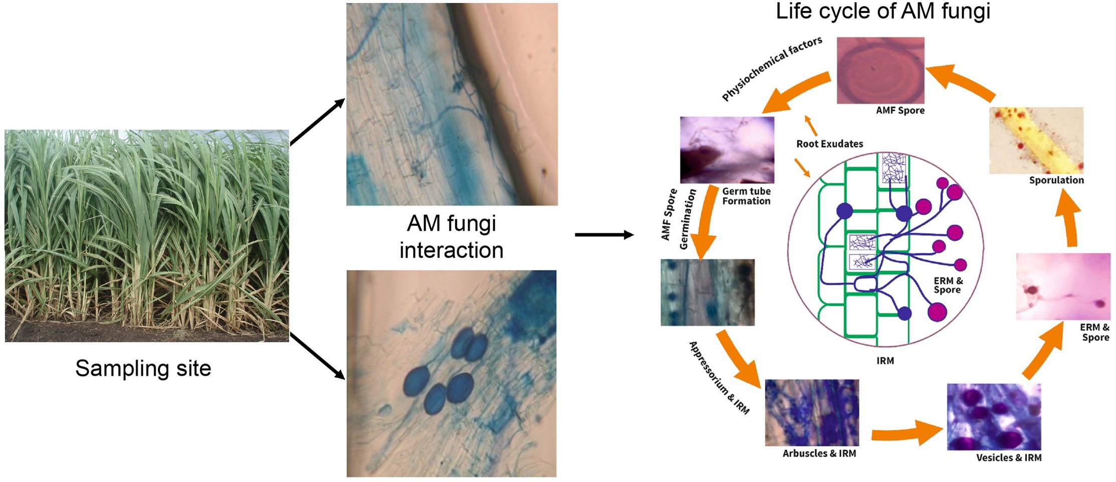
      The present investigation deals with the fungi isolated from the rhizosphere of AM fungi mediated and uninfected (AM fungi free) sugarcane plants, monthly and seasonal fluctuations and abundance of individual organisms, comparative monthly and seasonal fluctuations in AM fungi populations in soil, the percentage of AM fungi interaction in roots, and occurrence of total rhizosphere fungi of AM fungi infected and uninfected plants of Saccharum officinarum.

    • In Bihar (India), sugarcane crops are grown mainly in the western region namely Pusa, Samastipur District, the districts are known as main burgeoning areas. Sugarcane is also cultivated in other parts of the region. Soil samples were collected in different months from January 2019 to December 2020 at 10 different taluks and locations. Temperature 30−40 °C, environmental and soil variations (clay and red soil). These samples were collected at the end of each month in different locations and stored in polyethylene bags at 4 °C for further analysis.

    • AM fungal species were subjected and isolated from sugarcane field soil samples. Initially, the place for the collection of rhizospheric soil samples from various places of the sugarcane field were selected, then the collected samples were maintained at 4 °C. AM fungi single spores were collected by using slightly modified wet sieving and decanting methods[18,19]. Surface sterilization of spores was achieved by applying 2% (w/v) chloramine T and streptomycin sulfate (200 μg ml−1) for 20 min and then cleaning several times using sterile distilled water. Single AM fungal spore cultures were raised by following the standard funnel technique[20] with Zea mays as a host plant. Three months of soil samples were collected, and spores were harvested. The samples were first subjected to a morphogenetic and micrometric study where individual AM fungal spore morphology, size, shape, color, surface, and structure of hyphae using Melzer’s reagent were determined. The present AM fungal genera morphological identification study results support previous findings where Glomus spp. was found in the sugarcane soil samples (Table 1)[2124].

      Table 1.  AM fungi infection in roots and spores' population spores in rhizosphere in soil for different sugarcane fields.

      S. No. Sugarcane
      cultivated field
      Hyphal type Arbuscles Vesicles AMF infection
      level
      AMF spore of
      10 gm soil
      AMF spp.
      Broad Thin
      1 Field 1 +++ ++ +++ 56% 12 Rf, Ga
      2 Field 2 +++ ++ +++ 71% 13 Rf, Ga, Gi, Gc
      3 Field 3 + + + 34% 8 Rf, At
      4 Field 4 9 Rf, Gal, Gac
      5 Field 5 ++ + 48% 11 Rf, Ri, Gc, Gg
      6 Field 6 ++ + ++ 52% 12 Rf, Ga, AL
      7 Field 7 ++ + +++ 39% 14 Rf, G, Sc
      8 Field 9 10 Rf, Ri, Gm
      9 Field 9 ++ ++ +++ 53% 12 Rf, Ga, Fm
      10 Field 10 ++ + ++ 56% 10 Rf, Ga, Fm
      +, Poor; ++, Moderate; +++, Abundant; −, Absent. Rf, R. fasciculatus; Ga, G. aggregatum; Ri, R. intraradices; Gm, F. mosseae; Gc, G. constrictum; Gac, G. macrocarpum; Gal, Gigaspora albida; At, Acaulospora tuberculate; AL, A. laevis; Sc, Sclerocystis spp.
    • Fresh samples containing 2−3 g of roots were used to assess the colonization percentage by staining. Fixed roots were cleaned with tap water, applied in 10% KOH, acidified with 1N HCL, and stained with 0.05% trypan blue. Quantification of root colonization of AM fungus was conducted by using the gridline cross-section technique[25] and 100 root sections of each sample were observed under a light microscope[26,27]. AM fungi colonization in plant root systems such as vesicles, arbuscules, and hyphae at fixed points were observed and the percentage of AM fungal colonization in the sugarcane root systems were calculated.

    • Experimental pots (60 cm × 50 cm × 50 cm) were filled with 50 kg of sieved and sterilized soil. The soil was crushed through a 4 mm sieve. The soil used was phosphorus deficient (Olsen P. 6.87 μg−1 soil) with a pH of 8.4 (1:2, Soil : water suspension). The inoculum of Rhizophagus fasciculatus was applied through a layering technique[28]. Roots of non mycorrhizal plants served as the control.

    • Seeds of sugarcane were obtained from the Sugarcane Research Institute, Pusa, Samastipur, Bihar, India.

    • Surface sterilized seeds (0.1% aqueous HgCl2 for 30 min) of the B.O.109 variety of sugarcane were used. Two eye seeds were used in each pot. The seedlings were thinned out into one in each pot after 25 d of sowing. The potted plants receiving the above treatments were left on greenhouse benches with the temperature ranging between 25 to 35 °C in a randomized complete block design to minimize any positional effects with six replications per treatment. After 30 d, all plants were given 60 ml of half-strength Hoagland's nutrient solution without phosphate once a week. Water was added as needed to maintain the growth medium at 60% of its limit water holding capacity.

    • To isolate fungi from the rhizosphere of AM fungi, plants were dug up with an intact root system using a specially designed sharp and long spade. Roots of both plants were carefully collected at intervals of 30 d. Excess soil adhering to the roots was removed by gentle shaking, and root tips (about 2.3 cm long) were cut with sterilized scissors and transferred to 100 ml of sterilized water (in a 250 ml Erlenmeyer flask). The flask was then subjected to vigorous shaking to obtain a homogeneous suspension of rhizosphere soil. From this suspension, a 1:10,000 dilution was prepared. About 20 ml of sterile Martin Rose Bengal agar medium (10.0 g glucose, 5.0 g peptone, 1.0 g KH2PO4, 0.5 g MgSO4 7H2O, 0.020 g rose bengal, 0.03 g agar-agar and 1,000 ml distilled water) was added. The Petri plates were rotated in different directions so that the suspension was completely mixed with the culture medium, and the inoculum was uniformly distributed on the plate. Five replicates were used for each set. The sealed petri plates were incubated at 28 ± 2 °C for 1 week.

    • After the necessary incubation period, individual fungal colonies developing on agar plates were aseptically transferred to sterilized potato-dextrose agar slants. The pure culture of each isolate was obtained by successive sub-culturing. The morphological characters of different isolates were studied microscopically (450×) and identified by comparing the characters of known species mentioned in the relevant literature. Some of the cultures whose identities were doubtful were sent to C.A.B. Mycological Institute, Kew (UK), for identification and confirmation. The various fungal species isolated from rhizospheres of AM fungi treated and untreated plants gave the qualitative number of fungi associated with the rhizospheres of aforesaid plants.

    • Quantitative analysis of rhizosphere fungi was performed by calculating the percentage frequency and abundance of different isolated fungi in different months. The formulas used to determine percentage frequency and percentage abundance were similar to those followed by Prasad & Bilgrami[29].

      Percentagefrequency=No. ofobservationsinwhichspeciesappearedTotalno.ofobservations×100
      Percentageabundance=Totalno.ofcoloniesofaspeciesinallobservationsTotalno.ofcolonies×100
    • Statistical analysis was performed using Statistical Package for the Social Sciences (SPSS) to evaluate the colonization and spore density of AM fungi in root tissues. Obtained results were analyzed using one-way variance, and significant differences were expressed at the significance level (p < 0.05) by Duncan's multiple range test.

    • In this experiment, an assessment of AM fungal species association into the root part of sugarcane crop plants, as well as fungal spore density in soil samples of different fields is presented in Table 1. The results of the occurrence of species, fields out of a total of 10 fields were found to have the ability to host mycorrhizal root infection because of this inquiry. Based on the results the length of the roots that were analyzed, the level of mycorrhizal fungal association of root tissues varied from 34−71. Field 2 plants had root length infection, whereas Field 3 had the least amount of root colonization (Table 1). The values for the infection spectrum of examined fields were determined to be 56%, 71%, 34%, 48%, 52%, 39%, 53%, and 56% accordingly for Fields 1−10 respectively (except Field 4 and Field 8 as they didn't yield any occurance). The fungal infection that caused AM fungi was made up of hyphae, vesicles, and arbuscules of the fungus. There was a significant amount of variation in the infection rate amongst the different field plants. The gathered soil samples to determine the AM fungi density were variations of sorts and exhibited a broad assortment of soils. In this soil range, the soil had a high population of mycorrhizal spores, and Glomus sp. were most abundant.

      The AM fungal concentration in the soil varied from 8−14 in 10 g−1 soil. Field 7 plants were found to have the highest spore density, whereas Field 3 had the lowest. A wide range of spores, the major species of which belonged to the genus Glomus, were extracted from the soil and the root washings. However, a zygospore of Acaulospora and Gigaspora as well as sporocarps of Sclerocystis were also found, although very rare.

      The determination of the various sugarcane plants, soil samples taken from the rhizospheres of all the sugarcane fields were treated to isolate the various fungal propagules. Following are some of the fungi that have been identified based on the characteristics of their spores: six species of Glomus, namely Rhizophagus fasciculatus, Glomus aggregatum, Rhizophagus intraradices, Funneliformis mosseae, and Glomus constrictum and Glomus macrocarpum; two species of Gigaspora, namely Claroideoglomus claroideum and G. gigantea and two species of Acaulospora namely Acaulospora tuberculate and A. laevis and one species of Sclerocystis spp. The results showed that AM fungal species were predominant, and mostly belong to these species of the current soil samples at different sugarcane fields followed by R. fasciculatum (10 sugarcane fields), G. aggregatum (five fields), R. intraradices (three fields), F. mosseae (two fields), G. constrictum (two fields), and G. macrocarpum (one field). Additionally, the species of Gigaspora (Gigaspora albida, and G. gigantea) were observed in the sampling soil from one field. The presence of spores and mycorrhizal root colonization was not found to have any definitive relationship with one another. As a result of the fact that the proliferation of an endomycorrhiza relies on its contact with plant roots, the quantity of its spores in soils is likely to change, as was shown in the current experiment. AM fungus has one of the major roles in symbiotic relationships with plant roots and enhances the growth development in humid soil nature, particularly in waterless zones. Soil conditions have a significant effect on the extent to which mycorrhizal fungal populations are active, as measured by the number of spores produced and the extent to which roots are infected[28].

    • Many varieties of soil fungal species were isolated from AM fungal-treated plants when compared to non-treated plants that showed a huge number of isolates. Statistical analysis revealed significantly higher root colonization compared to non-inoculated plots (control). Altogether, 14 fungi belonging to nine different genera (Tables 2 & 3) were isolated from AM fungi-infected plants. Out of the total isolates, two species belonged to Phycomycetes, one Ascomycete species, and the remaining 11 species to Deuteromycetes. The rhizosphere soil of Control (AM fungi-free) sugarcane plants (Tables 2 & 3) however, yielded 22 fungi belonging to 16 different genera. Among the isolated organisms, three species belonged to Phycomycetes, two species to Ascomycetes, and the remaining 17 species to Deuteromycetes. In all, 10 fungi viz., M. recemosus, Rhizopus oryzae, Chaetomium globosum, Aspergillus candidus, A. flavus, A. niger, A. terreus, Cladosporium herbarum, Pestalotia glandicola, and Macrophomina phaeolina were found to be common in both the rhizosphere of AM fungi inoculated and uninoculated sugarcane plants. Besides the aforementioned fungi, the rhizosphere of uninoculated plants also yielded 12 more fungi viz. Rhizopus varians, Neocosmospora vasifecta, Alternaria alternata, Curvularia lunata, Helminthosporium halodes, C. sacchari, F. moniliformae, F. solani, F. semitectum, Rhizoctonia solani, Myrothecium roridum, and Verticillium albo-atrum. The rhizosphere of AM fungi inoculated plants, however, yielded only four fungi that were not found in the rhizospheric soil of uninoculated plants viz., Aspergillus sydowi, P. chrysogenum, P. lilacinum, and T. harzianum.

      Table 2.  Presence (+) and absence (−) of genera of arbuscular mycorrhizal fungi in infected and uninfected (AM fungi free) plants of Saccharum officinarum L.

      Fungi AM fungi infected Control
      (AM fungi free)
      Phycomycetes
      Mucor racemosus Fres + +
      Rhizopus oryzae Went & Gerling + +
      Rhizopus varians Povah +
      Ascomycetes
      Chaetomium globosum Kunze & Shorr + +
      Neocosmospora vasinfecta Smith +
      Deute romyetes
      Alternaria alternata (Fr), Keisler +
      Aspergillus candidus Link. + +
      Aspergillus flavus Link. + +
      Aspergillus niger Van Tiegh. + +
      Aspergillus sydowi (Bainer & Sartory) +
      Aspergillus terreus Thom + +
      Cladosporium herbarum (Pors). Link + +
      Cephalosporium sacchari Butler +
      Curvularia lunata (Wakker) Boedijn +
      Fusarium moniliforme Sheldon +
      Fusarium solani (Mart.) Sace. +
      Fusarium semifectum Berk & Rev. +
      Helminthosporium halodes Drechs. +
      Macrophomina phasealina (Tassi) Goid + +
      Mycothecium roridum Tode exfr +
      Penicillium chrysogenum Thon +
      Penicillium lilacinum Thon +
      Pestalotia glandicola (Cast) Stey + +
      Rhizoctonia solani Kuhn. +
      Trichoderma harzianum Rafai +
      Verticillium albo-atrum Reink & Berthold. +
      Total number of fungi 14 12

      Table 3.  Monthly fluctuations in the percentage frequency of rhizosphere mycoflora of AM fungi infected and uninfected plants of Saccharum officinarum L.

      Fungi Jan Feb. March April May June July Aug. Sep. Oct. Nov. Dec.
      AI AU AI AU AI AU AI AU AI AU AI AU AI AU AI AU AI AU AI AU AI AU AI AU
      F F F F F F F F F F F F F F F F F F F F F F F F
      Mucor racemosus 60 65 58 64 57 60 30 35 30 38 35 45 48 54 55 58 56
      Rhizopus oryzae 85 88 70 85 65 84 60 72 35 70 30 65 70 40 74 35 79 75 80 80 84 86 86 86
      Rhizopus varians 64 80 68 66 58 60 60 66 72
      Chaetomium globosum 34 52 40 26 45 32 42 34 50 36
      Neocosmospora vainfecta 17 10 15 14 10 18
      Alternaria alternata 28 35 32 35 30 28
      Aspergillus candidus 66 70 62 36 40 60 65 62 70 52 60 56 66 60 76
      Aspergillus flavus 74 82 52 65 60 62 70 80 71 75 75 80 76 85 82 82
      Aspergillus niger 80 86 78 86 70 78 65 65 52 67 35 65 40 45 50 64 64 70 75 72 76 75 80 86
      Aspergillus sydowi 52 34 52 60 56 54
      Aspergillus terreus 70 85 70 55 50 62 56 64 68 68 64 70 75 78 76 72 66 80
      Cladosporium herbarum 29 35 40 50 48 48 45 32 38 34 35
      Cephalasporium sacchari 40 33 50 48 42 34 30
      Curvularia lunata 50 45 43 45 50 52 55
      Fusarium moniliforme 40 36 45 50 45 30 32 28
      Fusarium solani 45 50 44 36 26 30 35 55 52 48
      Fusarium hemitectum 42 50 45 36 20 46 48 56 55 54
      Helminthosporium halodes 35 32 40 36 45
      Macrophomina phaseolina 35 40 35 22 30 40 35 36 45 46 50
      Myrothecium roridum 40 35 25 30 22 35 20 30
      Penicillum chrysogenum 42 52 30 32 28 33 47 52 60
      Penicillum lilacinum 50 60 64 70 55 54 60
      Pestalotia glandicola 55 52 52 46 54 55 64 65 58 62 60 55
      Rhizoctonia solani 30 35 32 35 40 25
      Trichoderma harzianum 78 56 60 68 64 76 70 76 80
      Verticillium albo-atrum 30 25 35 26 30 45 35 22 30 50
      AI, AMF infected ; AU, AMF Uninfected; F, Percentage frequency; −, Percentage frequency NIL.

      It is noteworthy that C. sacchari, and F. moniliforme, which cause sugarcane wilt in this region, were not obtained from the rhizosphere soil of AM fungal inoculated plants. Although these two fungi were isolated from rhizosphere soil, they were not inoculated into plants.

    • The data presented in Tables 4 & 5 show that the percentage frequency of AM fungi and the percentage of various rhizospheric fungi were higher in non-inoculated sugarcane plants and different months. Generally, the percentage of different fungi is highest in August, September, October and November. The percentage of AM fungal frequency and abundance of these fungi decreased during April, May, and June. The rhizosphere of AM fungi was compared with the fungi extracted from the soil, and the level of frequency and abundance of AM fungi were found to be different. The percentage frequency and percentage abundance of different species of Mucor, Rhizopus, Aspergillus, Penicillium, and Trichoderma were significantly higher in the rhizosphere soil of AM fungi and non-inoculated plants.

      Table 4.  Monthly fluctuations in the percentage abundance of rhizosphere mycoflora of AM fungi infected and AM fungi free plants of Saccharum officinarum L.

      Fungi Jan. Feb. March April May June July Aug. Sep. Oct. Nov. Dec.
      AI AU AI AU AI AU AI AU AI AU AI AU AI AU AI AU AI AU AI AU AI AU AI AU
      F F F F F F F F F F F F F F F F F F F F F F F F
      Mucor racemosus 7.70 7.85 7.60 8.75 7.56 7.70 5.45 6.15 6.45 6.80 6.15 6.95 7.80 7.35 7.90 7.62 8.50
      Rhizopus oryzae 14.42 14.92 13.72 14.45 13.10 14.40 13.05 13.76 8.25 13.70 8.80 13.10 10.70 11.25 14.20 14.85 12.43 14.25 14.43 15.25 14.40 14.65 14.62 14.70
      Rhizopus varians 12.25 14.75 12.82 12.05 11.96 11.95 12.95 11.95 12.08
      Chaetomium globosum 6.15 8.10 6.80 4.45 7.15 4.78 7.25 4.95 7.70 5.05
      Neocosmospora vainfecta 1.95 1.88 1.95 1.75 1.70 2.02
      Alternaria alternata 5.45 5.90 5.85 5.70 5.68 5.55
      Aspergillus candidus 8.72 13.10 12.85 3.40 10.25 8.60 13.10 9.35 14.20 8.95 12.45 11.85 10.96 14.25
      Aspergillus flavus 12.76 14.25 12.80 12.95 12.65 12.75 13.12 13.10 13.10 13.95 13.45 14.10 13.48 14.95 14.56 14.56
      Aspergillus niger 13.60 15.45 13.20 15.45 12.10 14.85 11.45 11.40 5.12 14.05 8.95 13.85 9.85 9.50 10.26 13.75 11.40 14.15 12.75 14.20 12.80 14.60 13.65 15.45
      Aspergillus sydowi 9.05 8.70 8.60 9.35 8.95 10.96
      Aspergillus terreus 11.76 14.78 13.10 12.32 11.70 12.80 12.35 14.05 13.15 13.65 11.48 13.10 12.75 13.75 13.76 13.80 13.10 14.45
      Cladosporium herbarum 5.25 5.45 6.48 7.55 7.48 7.45 7.35 5.45 6.65 5.52 5.65
      Cephalasporium sacchari 6.05 5.45 7.52 7.48 6.46 5.52 5.52
      Curvularia lunata 6.70 6.40 6.30 6.40 6.75 6.75 6.85
      Fusarium moniliforme 5.10 4.95 6.15 8.16 7.15 4.60 4.75 4.35
      Fusarium solani 6.15 7.06 6.05 4.95 4.32 4.60 5.09 5.75 5.65 5.45
      Fusarium semitectum 5.90 7.15 6.78 5.16 5.05 6.25 6.35 6.85 6.80 6.75
      Penicillum chrysogenum 5.80 10.20 5.15 5.20 4.90 5.15 6.35 7.96 10.56
      Helminthosporium halodes 6.10 5.35 6.15 6.15 6.60
      Macrophomina phaseolina 8.95 9.15 8.25 3.30 4.10 3.15 5.15 3.10 6.10 7.06 8.32
      Myrothecium roridum 4.75 3.65 3.15 3.64 3.10 3.46 2.60 3.95
      Penicillum lilacinum 9.15 11.10 11.15 12.30 10.50 12.10 10.70
      Pestalotia glandicola 10.15 10.05 10.10 9.86 10.20 10.25 11.35 11.40 10.60 11.15 10.85 10.15
      Rhizoctonia solani 3.42 4.15 4.08 4.26 4.96 2.15
      Trichoderma harzianum 13.20 12.30 12.40 12.70 10.35 13.10 12.76 13.10 13.40
      Verticillium albo-atrum 2.01 2.05 3.09 2.15 2.96 4.98 3.26 1.95 2.35
      AI, AMF infected ; AU, AMF Uninfected. F, Percentage frequency; −, Percentage frequency NIL.

      Table 5.  Monthly fluctuations in the percentage abundance of rhizosphere mycoflora of AM fungi infected and AM fungi free plants of Saccharum officinarum L.

      Fungi Summer Monsoon Winter Annual
      March AMF
      infected
      June AMF free July AMF
      infected
      October AMF free November AMF
      infected
      February AMF free March AMF
      infected
      February AMF free
      F A F A F A F A F A F A F A F A
      Mucor racemosus 14 1.89 15 1.92 37 6.33 28 5.1 57 7.56 60 8.25 36 5.26 34 5.09
      Rhizopus oryzae 47 10.80 73 13.74 76 12.94 57 13.90 81 14.29 86 14.68 68 12.67 72 14.10
      Rhizopus varians 17 3.20 61 12.22 65 12.74 49 9.39
      Chaetomium globosum 10 1.70 14 2.30 22 3.60 26 4.03 26 4.12 13 2.11 19 3.08
      Neocosmospora vainfecta 3 0.47 10 1.35 9 0.99 7 0.16
      Alternaria alternata 8 1.46 23 4.23 16 2.83 13 2.38
      Aspergillus candidus 9 0.85 10 2.56 43 6.72 49 9.93 45 4.92 68 13.01 33 4.16 42 8.50
      Aspergillus flavus 28 6 32 6.47 54 12.43 59 13.63 50 9.65 62 10.75 47 9.36 51 10.29
      Aspergillus niger 55 9.40 68 13.53 57 11.06 62 12.90 79 13.31 83 15.23 67 11.26 71 13.88
      Aspergillus sydowi 8 2.17 42 6.72 56 5.00 26 4.63
      Aspergillus terreus 22 6.00 50 6.47 66 12.43 70 13.63 53 9.65 59 10.75 48 9.36 53 10.29
      Cladosporium herbarum 7 1.31 9 1.36 30 4.85 42 7.25 9 1.38 9 1.41 15 2.51 21 3.34
      Cephalasporium sacchari 8 1.36 35 5.36 27 4.21 23 3.64
      Curvularia lunata 47 6.55 38 4.98 28 3.86
      Fusarium moniliforme 9 1.23 43 6.51 25 3.55 26 3.76
      Fusarium solani 20 2.75 37 4.94 49 6.07 35 4.58
      Fusarium semitectum 20 2.98 46 6.12 50 6.65 38 6.25
      Helminthosporium halodes 8 1.34 30 4.73 9 1.52 16 2.53
      Macrophomina phaseolina 9 2.23 19 4.35 25 2.38 28 3.83 24 3.84 11 1.54 23 4.01
      Myrothecium roridum 9 0.91 28 3.33 23 2.82 20 3.36
      Penicillum chrysogenum 8 1.28 35 5.40 52 8.63 31 5.11
      Penicillum lilacinum 47 8.48 57 10.28 35 6.26
      Pestalotia glandicola 11 2.46 30 5.38 30 5.41 43 7.87 59 10.38 28 5.24 30 5.27
      Rhizoctonia solani 27 3.32 27 2.43 16 1.92
      Trichoderma harzianum 29 6.17 70 12.22 59 9.92 52 9.44
      Verticillium albo-atrum 15 1.31 33 3.28 34 3.09 27 2.56
      F, percentage frequency; A, percentage abundance; −, percentage frequency NIL.

      Whereas the proportion of frequency and abundance of M. phaseolina in the rhizosphere soil of AM fungi is less. Additionally, the remaining species were moderate in the soil samples. It was generally observed that the frequency of representation of saprophytic fungi like Rhizopus, Aspergillus, Penicillium, and Trichoderma was high, while root rot-causing fungi like Fusarium, Cephalosporium, and Rhizoctonia were almost nil in AM fungi inoculated plants. The percentage frequency and abundance of fungi run parallel to each other during different months throughout the year.

      A marked variation in the percentage frequency and abundance (Table 5) of different fungi occurs during different seasons in both AM fungi inoculated and uninoculated in the rhizospheric soil of sugarcane plants. A minimum number of fungi and minimum percentage of frequency of different fungi was recorded during the summer. C. globosum, and P. lilacinum were found to be absent during the summer in the rhizosphere soil of AM fungi-infected plants and C. lunata and P. glandicola were found to be absent during the summer season of uninoculated plants. Winter proved to be the most favorable season for most of the fungi. The occurrence and distribution of several fungi were shown to be higher in the rhizosphere soil of inoculated and non-inoculated plants than AM fungi during this season. In general, the late monsoon and early winter period was found to be the best for growth and development of these fungi. Among the various organisms isolated from the rhizosphere soil, R. oryzae and A. niger proved to be most dominant and tolerant of environmental conditions as they were found throughout the year in the rhizosphere soil of both AM fungi inoculated and uninoculated sugarcane plants. A. sydowi, C. globosum and P. chrysogenum in AM fungi inoculated and Neocosmospora vasinfects, A. alternata, A. candidus, C. lunata, T. harzianum, F. semifectum, M. phaseolina, R. solani, M. roridum and V. albo-atrum in uninoculated plants, however, were found to be sensitive to environmental changes as they appeared only for a few months.

    • The recovered number of fungal populations in the rhizosphere soil of AM fungi inoculated and uninoculated sugarcane plants fluctuated during different months (Tables 35). The sampling results indicated that total fungal frequency was highest in the rhizosphere soil of AM fungi inoculated and non-inoculated plants during the different seasons of winter (November–February), monsoon (July–October), and summer (March–June). The AM fungal population of spores in the rhizosphere soil and the concentration of mycorrhizal attachment in the roots of sugarcane plants also fluctuated in different months. The maximum population of AM fungal spores and severity of root infection was observed in July and the minimum population of AM fungal spores occurred in February, while the maximum fungal population (i.e., total frequency and abundance of fungi) occurred in October and the minimum frequency, and an abundance of fungi occurred in May. The fluctuations in spore population and root infection were caused by environmental factors and seasonal variation. The maximum concentration of AM fungi spores and root colonization were recorded from April to July and the minimum concentration from November to March. In winter, an inverse relationship was observed between rhizosphere fungi and the AM fungi. The concentration of different rhizosphere fungi was higher in winter but the number of AM fungi and their attachment in roots were lower. Control soils contain opportunistic fungi that are naturally present in the soil.

      Statistical analysis revealed a significant treatment effect over different periods, with plots that received AM fungi inoculation displaying significantly higher root colonization compared to non-inoculated plots (control). These fungi are part of the soil's intrinsic microbial community and sustain themselves over time through natural processes.

    • The rhizosphere incidence of AM fungi varied with the number of fungal populations in treated and non-treated plants. The occurrence and distribution of many fungal species were reduced or totally suppressed in the rhizospheric soil of AM fungi-inoculated plants. The beneficial effect of mycorrhizae is not only attributed to nutritional factors but also to the protection of roots against soil pathogens. Agarwal[30] reported that mycorrhizal fungi utilize the surplus of nutrients from soil which becomes a limiting factor for other soil fungi. He also reported that mycorrhizal fungi may trigger the production of antifungal substances which may reduce the chances of infection by soil pathogens. In the present experiments also, the parasitic fungi were almost eliminated from the rhizosphere soil of AM fungi-mediated plants.

      It was observed the AM fungal species diversity and denticity in sugarcane field soil samples of 10 different locations from Pusa, Samastipur, Bihar, India (Table 1). Rhizosphere fungal populations depend on the micro-environment and nutrient status, which depends on the physiological state of the plant[31] and the pattern of its root exudation[32]. Since the physiological condition of plants and the pattern of their root exudation are expected to vary in different seasons, and stages of plant growth, the fungal population in the rhizosphere varies accordingly (Table 2). Although the occurrence of fungal species was significantly different compared to the control, the present results are in conformity with the observations made by Bagyaraj & Menge[15]. The harshness of mycorrhizal root attachment and the population of AMF spores in rhizospheric soil vary from season to season, due to plant age and plant physiology. However, these findings are similarly presented in previous research work, where root exudation of rhizospheric fungi and their fluctuations in environmental conditions[1215].

      AM fungal species were commonly identified in rhizosphere soils of all studied sugarcane fields. Previous research[3335] have reported that plants previously inoculated with AMF exhibited resistance toward soil-borne diseases like wilt and root. Zambolin & Schenck[36] observed the interaction of G. mosseae and M. phaseolina species that the colonization in the host root of soybeans and inhibits pathogens, and enhances the growth in the host. Similarly, Caron et al.[37] studied that root colonization by Glomus species was not affected by the presence of Fusarium. The number of campaigns of Fusarium sp. plants were consistently absent when inoculated with a mycorrhizal endophyte. In this study, the occurrence and diversity of G. fasciculatum totally inhibited the disease-causing pathogens viz., F. moniliforme and C. sacchari in the rhizosphere soil of AM fungi-mediated sugarcane plants (Table 2). Thus, the findings of the diversity results of maximum fungal species found in the winter (November–February), monsoon (July–October), and summer (March–June). The AM fungal population of spores in the rhizosphere soil and the concentration of mycorrhizal attachment in the roots of sugarcane plants also fluctuated in different months.

      Mycorrhizal colonization rate showed a significant difference between treated and untreated plants (Table 2). These results are correlated with the study conducted on maize sorghum, where AMF inoculation considerably increased soil nutrient availability[12]. A few previous researchers have revealed that the variation of AM fungal species intensity, their host plants' age, phonology, soil nature, root morphology, and environmental factors also may be affected. Previous studies indicated that the intraradical development of AM fungi is highly influenced by plant species, soil pH, and phosphorus content[1214,36,3840].

      Different diversity of soil fungal communities was observed in the sugarcane soil samples at different locations of the sampling sites. Among these identified AM fungal species, R. fasciculatus, G. aggregatum, R. intraradices, F. mosseae, and G. constrictum and G. macrocarpum; two species of Gigaspora, namely Claroideoglomus claroideum and G. gigantean and two species of Acaulospora, namely Acaulospora tuberculate and A. laevis and one species of Sclerocystis spp. However, AM fungal community variations and frequency being higher especially Glomus species compared to the control sample (Tables 15). The results of the AM fungal diversity study are very similar to previous reports where there is a correlation between diversity and abundance[1113,37]. Thus, this study suggests that sugarcane root serves as a suitable host for these AM species to form a mutualistic symbiosis.

      Hence, the study presents clear and significant results, demonstrating the significant impact of AMF on sugarcane growth and yield, along with a noticeable differences between the seasonal abundance of fungal species. However, the findings were limited to the specific conditions and cultivars used in this study, and their applicability to other regions or sugarcane varieties could be addressed.

    • The author confirms sole responsibility for all aspects of this article.

    • The data that support the findings of this study are available on request from the corresponding author.

    • Author is thankful to the Director, Sugarcane Research Institute, Pusa, Samastipur for Sugarcane seeds and Prof. J.J. Deploey, Pennsylvania University, Pennsylvania, USA for his encouragement and his valuable guidance and comments on the manuscript.

      • The author declares that there is no conflict of interest.

      • Received 28 May 2024; Accepted 20 August 2024; Published online 11 October 2024

      • Different types of fungal species were obtained in the rhizosphere soil of AM fungus-infected and non-infected sugarcane plants.

        Outcome of our isolation process, abundance of rhizosphere fungi isolates was lower in plants infected with AM fungi than in plants without mycorrhizal infection.

        The occurrence and distribution of different rhizosphere fungi varied from plants infected with AM fungi compared to uninfected plants.

        We observe that parasitic fungi are virtually eliminated from the rhizosphere soil of plants infected with AM fungi.

      • Copyright: © 2024 by the author(s). Published by Maximum Academic Press on behalf of Hainan University. This article is an open access article distributed under Creative Commons Attribution License (CC BY 4.0), visit https://creativecommons.org/licenses/by/4.0/.
    Figure (0)  Table (5) References (40)
  • About this article
    Cite this article
    Prasad K. 2024. Occurrence and interactions of arbuscular mycorrhizal fungi (Rhizophagus fasciculatus) and rhizospheric fungi in Saccharum officinarum L. Tropical Plants 3: e033 doi: 10.48130/tp-0024-0035
    Prasad K. 2024. Occurrence and interactions of arbuscular mycorrhizal fungi (Rhizophagus fasciculatus) and rhizospheric fungi in Saccharum officinarum L. Tropical Plants 3: e033 doi: 10.48130/tp-0024-0035

Catalog

  • About this article

/

DownLoad:  Full-Size Img  PowerPoint
Return
Return