-
Overpopulation in Egypt has led to an increases in the demand for food supply, which has focused the attention to raising crop production. This has led to an increase in the application of chemical fertilizers, especially in the cultivation of vegetable crops, where large amounts of chemical fertilizers are used to obtain the highest productivity[1]. The rates of chemical fertilizers applied in the cultivation of vegetable crops are considerably higher compared to other crops because of intensive cultivation and vegetative organs (leaves) as final products[2−4]. This in turn can lead to negative side effects on the environment and human health issues[5,6]. The addition of mineral fertilizers cannot replace the soil organic matter (SOM) that may be lost due to intensive crop production[7,8]. Maintaining appropriate levels of organic matter in soils is important for the sustainable management of light soils[9]. Instead, vermicompost (VC) and compost (CT) applications may improve the content of SOM. Compost and vermicompost could be alternatives to chemical fertilizers[9,10]. Vermicompost is the product of organic matter degradation through the interaction between earthworms and microorganisms[11,12]. Vermicompost is considering a biofertilizer with a varied microbial community[13−15]. Vermicompost contains nutrients such as P, K, Ca, and Mg[16,17]. Vermicomposting and composting are two distinct processes, and it is crucial not to confuse the two[18]. The vermicomposting process produces a high diversity and number of microorganisms because the temperature during VC production is suitable for worms[19]. A decrease in the C/N ratio of the vermicompost was testified than the compost[20].
Zucchini (Cucurbita pepo L.) is one of the most important vegetable crops grown in Egypt. Zucchini plants are very diverse and popular for human consumption throughout the world[21]. However, zucchini fruit contains many nutrients, bioactive compounds (antioxidants, flavonoids, vitamins) and have a high amount of dietary fiber, which is very low in calories[21]. Intensive zucchini production in Egypt requires higher rates of chemical fertilizers.
Soil degradation and its expansion are considered the greatest problem in Egypt, directly affecting food security and crop production[22,23]. Soil nutrients are necessary for crop growth and development and are critical factors for soil fertility along with adequate soil moisture, which is considered a key factor for crop growth and yield[24]. Therefore, manipulating nutrient release is an advanced and effective way to maintain sustainable crop production including zucchini[25,26]. Farmers assume that the extensive use of CF leads to better yields of various crops without considering the hazardous effects on the environment. The continuous use of chemical fertilizers only has a negative impact on soil fertility[27,28].
To ensure a healthy diet and minimize environmental risks of chemical fertilizer, and in line with sustainable development programs that call for a return to nature, this study was conducted to assess replacing chemical fertilizers with the vermicompost and compost on soil fertility, growth, and yield of zucchini cultivation under field conditions. It was hypothesized that the compost and vermicompost application would help produce zucchini fruit free from the residual effects of the chemical fertilizer and improve sandy soil properties under the arid conditions of Egypt.
-
This field experiment was carried out in 2021 and 2022 seasons at a commercial vegetable farm in Assiut City, Egypt (27°12'16.67" N; 31°09'36.86" E). Soil samples (0−20 cm) were collected, air-dried and ground to pass through a 2-mm sieve. Particle size distribution was determined according to the pipette method[29]. Soil pH and EC were determined in a 1:2.5 suspension using pH and EC-meter, respectively. The soil organic matter (OM) was determined by using the method of Jackson[29], while total calcium carbonate in the soil was determined by Collin's calcimeter method[29]. Available soil P was determined[30]. While available soil N and available K were determined according to Kompała-Bąba et al.[31]. The soil characterization before cultivation is presented in Table 1. Compost was produced from plant residues, obtained from the Nile Company, Egypt, while vermicompost was brought from the Agricultural Research Center (ARC), Egypt. Some chemical characteristics of the compost and vermicompost are presented in Table 2.
Table 1. Some physical and chemical properties of the soil before experiment.
Property Unit Value Sand g·kg−1 457 ± 10.5 Silt g·kg−1 327 ± 6.90 Clay g·kg−1 216 ± 5.40 Texture − Silty loam CaCO3 g·kg−1 32.7 ± 2.57 EC (1:2.5) dS·m−1 0.63 ± 0.10 pH (1:2.5) − 7.90 ± 0.05 OM g·kg−1 11.3 ± 1.39 Available N mg·kg−1 60.4 ± 5.40 Available P mg·kg−1 14.7 ± 1.30 Available K mg·kg−1 397 ± 5.30 Table 2. Characteristics of the compost and vermicompost.
Property Unit Compost Vermicompost EC (1:5) dS·m−1 4.39 ± 0.07 3.89 ± 0.82 pH (1:5) − 7.77 ± 0.07 7.88 ± 0.05 (OC) g·kg−1 203 ± 3.12 237.6 ± 2.89 Total N g·kg−1 16.4 ± 2.31 16.9 ± 6.76 Total P g·kg−1 6.80 ± 1.43 12.9 ± 0.84 Total K g·kg−1 18.7 ± 0.80 10.9 ± 0.90 C/N ratio − 12.4 ± 0.70 14.1 ± 1.20 Each value represents a mean ± standard error (SE) of five replicates. Field experiment
-
The experimental plot dimension was 4.5 m × 3.0 m with three terracing at a distance of 90 cm with ridge spacing of 40 cm in a row (18 plants·plot−1 equal to 13,330 plants·ha−1). The treatments were: CO = control, CF = chemical fertilizer, CT = compost, and VC = vermicompost, and were arranged in a complete randomized design with five replicates. For the CF treatment, the recommended NPK fertilizers were applied at the rate of 178.5 kg N·ha−1, 71.4 kg P2O5·ha−1, and 119 kg K2O·ha−1. Compost and vermicompost were applied as full dose before planting, nitrogen fertilizer as urea (46% N) was added in three doses: 20% as basal application, 40% 15 d after sowing (DAS), and 40% 30 DAS. Potassium fertilizer as potassium sulfate was applied in three splits: 40% at 30 DAS, 30% 30 d from the first addition, and 30% 30 d after the second addition; while the phosphorus fertilizer (superphosphate) was applied in one split as a basal application before sowing. The application rates of compost and vermicompost were 7.15 t·ha−1 according to the Egyptian Ministry of Agriculture and were applied as full dose before sawing. The compost and vermicompost and chemical fertilizer was mixed well with soil by raking to a depth of 10 cm. Seeds of zucchini (Cucurbita pepo L.) were planted directly in soil on Feb 15th 2021 and 2022 growing seasons.
Data collection
-
The zucchini plant samples were collected 50 DAS and the fresh and dry weights were determined using an electronic balance. In addition, the total chlorophyll index (SPAD) of the leaves was measured using (502-m Konica Minolta, Inc., Tokyo, Japan) taking four measurements per leaf. The zucchini fruit were harvested 90 DAS and yield parameters such as length of fruit, fruit diameter, fruit number, fruit weight, and fruit yield were recorded.
Plant analysis
-
The zucchini fruit samples were cleaned, and washed with distilled water, oven-dried (70 °C) for 48 h and then the digested by methods adopted by Page et al.[32] and finally the total concentrations of N, P, and K were measured according to previously described methods described[32,33].
Soil analysis at harvesting
-
After harvesting, soil samples were taken from each pot, air-dried, crushed, passed through a 2 mm sieve and then available, N, P, and K were determined according to methods described by Page et al.[32].
Nutrient use efficiency (NUE) is defined as the fruit yield obtained per amount of applied fertilizer and was calculated as:
where, Yt = fruit yield of treatment (kg); Y0 = fruit yield of control (kg) and N is the amount of applied fertilizers (kg).
Plant uptake = Dry weight × Nutrient concentration.
Statistical analysis
-
Statistical analyses were run using analysis of variance technique by using statistics 8.10 software packages. Means of treatments were compared using the Duncan tests (p < 0.05). Principal component analysis (PCA) were run by Past software, version 4.06 and also the correlations among the soil properties and plant traits were calculated.
-
Application of CT, VC, and CF treatments significantly (p < 0.05) increased the fresh and dry weights of zucchini plants compared with CO treatment in the 2021 and 2022 seasons (Fig. 1). The fresh weight of the treatments could be arranged in a descending order: CT > VC > CF > CO, while in the dry weight can be arranged in a descending order: CT > CF > VC > CO. The highest leaf SPAD value was recorded in CT treatment. Significant differences were found in the fruit number, fruit length, fruit diameter, and zucchini yield among treatments (Fig. 2). Compared to the CO treatment, the fruit number per plant increased in the CF, VC, and CT by about 21%, 10%, and 37%, respectively. Fruit weight increased by about 1%, 5%, and 8%, respectively, in the CF, VC, and CT treatments, while fruit length increased by about 24%, 10%, and 13%, respectively. Compared to the CO treatment, the fruit diameter increased by about 21%, 6%, and 10%, respectively, while CF, VC, and CT treatments increased fruit dry weight by about 13%, 9%, and 5%, respectively. The highest yield of zucchini fruit was recorded in CT treatment in the two seasons. Significant difference was found in the total soluble solids and total concentration of N, P, and K in zucchini fruit (Fig. 3). The CF treatment, followed by VC recorded the highest total soluble solids and total NPK contents.
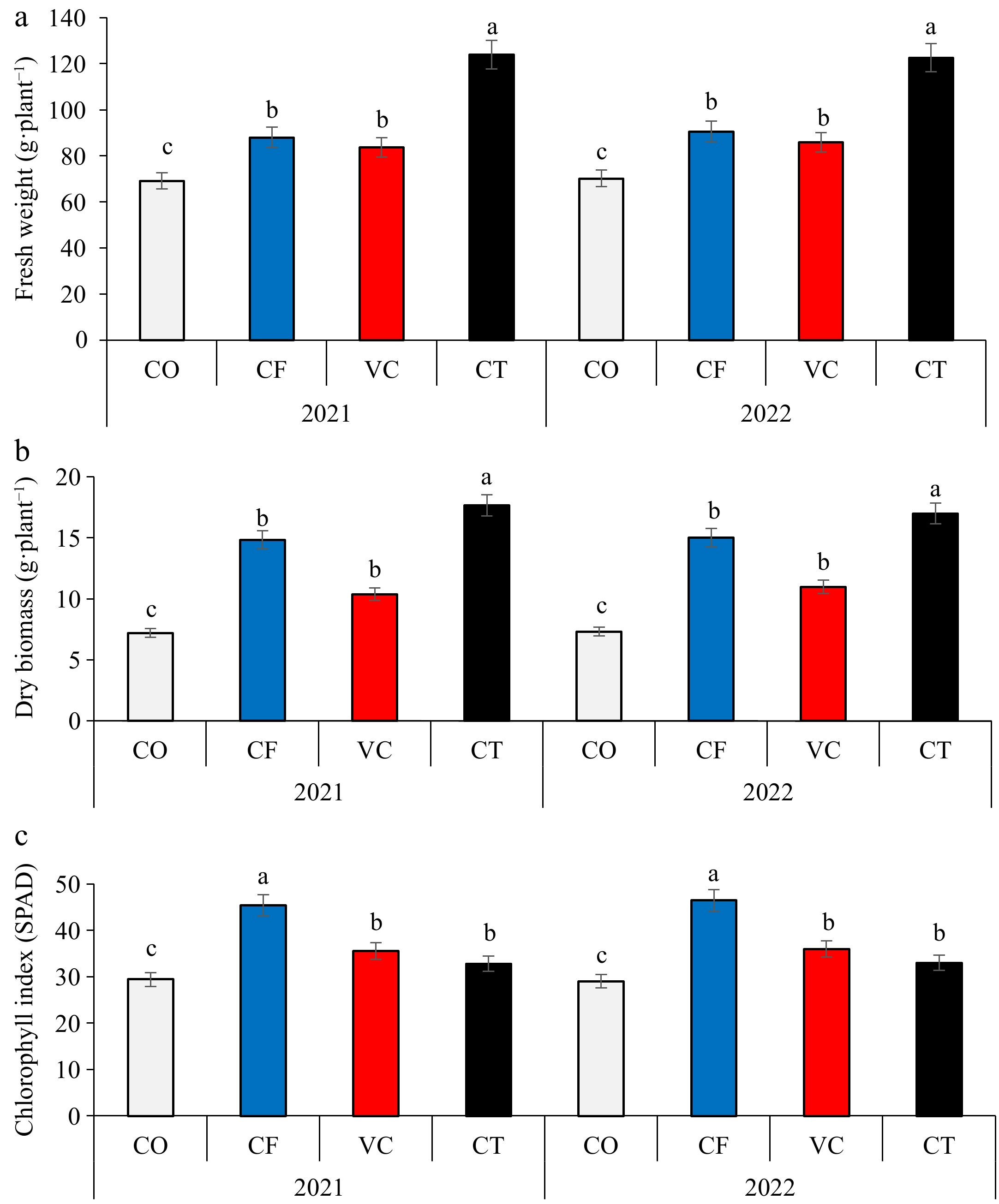
Figure 1.
Effect of compost and vermicompost application on (a) fruit fresh biomass, (b) fruit dry biomass, and (c) chlorophyll index of zucchini plants.
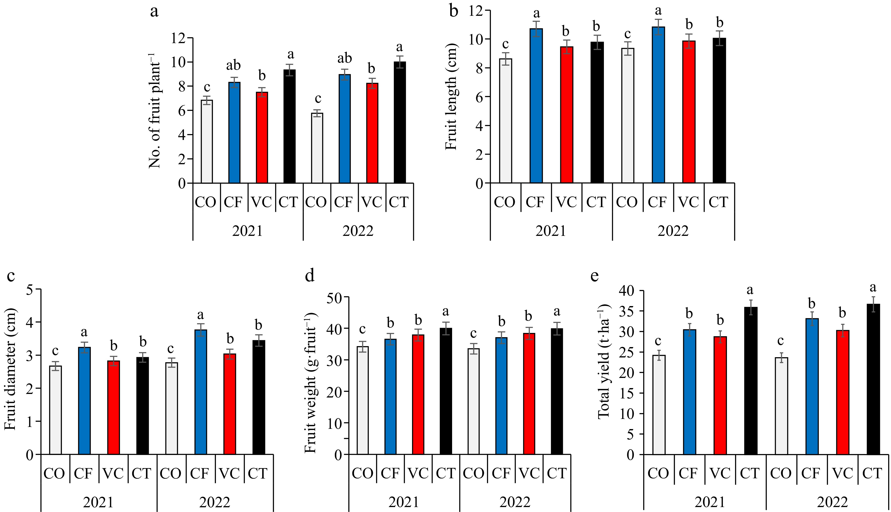
Figure 2.
Impact of different fertilizers treatment on (a) fruit number, (b) fruit length, (c) fruit diameter, (d) fruit weight, and (e) yield.
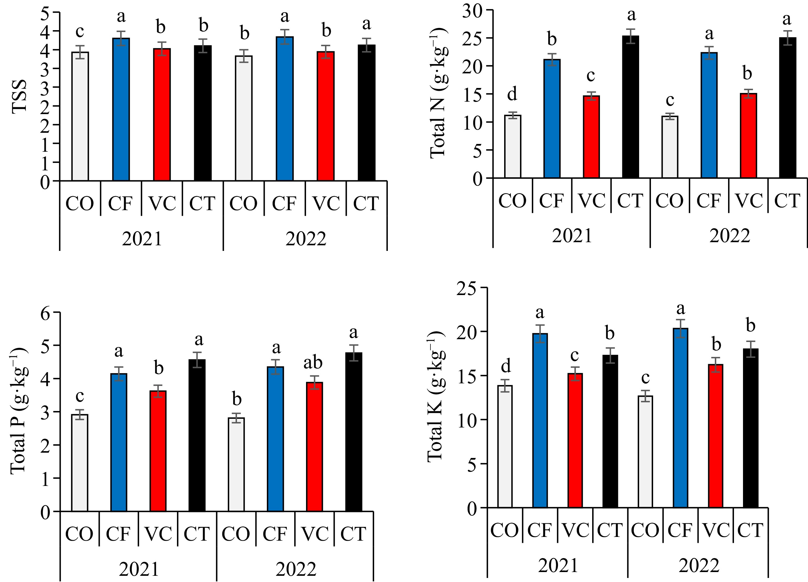
Zucchini uptake and nutrient use efficiencies
-
A significant difference was found in the uptake of NPK by zucchini fruit between the treatments (Table 3). The CT treatments significantly increased the uptake of NPK. Consequently, the uptake of NPK could be arranged in the descending order: CT > CF> VC > CO treatments in the 2021 and 2022 seasons. Significant differences were found in the agronomic NUE, PUE, and PUE by zucchini fruit among the treatments. In general, the CT treatment significantly increased the agronomic NUE, PUE, and PUE by zucchini fruit (Table 3).
Table 3. Effect of different fertilizer treatments on total NPK uptake and nutrients use efficiency.
Year Treatments N uptake (kg·ha−1) P uptake (kg·ha−1) K uptake (kg·ha−1) NUE (kg·kg−1) PUE (kg·kg−1) KUE (kg·kg−1) 2021 CO 27.83 ± 0.36d 7.25 ± 0.75d 34.46 ± 1.32c − − − CF 108.25 ± 1.23b 21.22 ± 1.21b 101.24 ± 3.12a 34.9 ± 0.87b 87.2 ± 2.12b 52.3 ± 1.45b VC 52.46 ± 1.02c 12.98 ± 1.54c 54.55 ± 2.54b 25.2 ± 0.56c 63.1 ± 1.88c 37.8 ± 0.98c CT 154.24 ± 2.33a 27.85 ± 0.97a 105.43 ± 2.64a 65.3 ± 1.34a 163.3 ± 3.21a 98.0 ± 1.98a 2022 CO 27.89 ± 0.54d 5.93 ± 0.54d 32.10 ± 0.43c − − − CF 115.99 ± 2.43b 22.60 ± 1.03b 105.60 ± 1.55a 53.2 ± 0.88b 133.0 ± 3.65b 79.8 ± 1.56b VC 57.16 ± 1.54c 14.74 ± 0.87c 61.61 ± 0.98b 37.2 ± 0.98c 92.9 ± 0.78c 55.8 ± 0.78c CT 146.99 ± 2.32a 28.06 ± 1.32a 105.82 ± 1.01a 72.8 ± 1.23a 182.1 ± 3.11a 109.2 ± 0.54a Means within a column followed by the same letter do not differ significantly (p < 0.05) according to DMRT. Soil chemical properties
-
After harvest, significant differences were recorded in pH, EC, OM, and availability of soil NPK contents among the different fertilizers (Table 4). At harvest, the CT and VC treatments slightly reduced the soil pH, while, the pH value in the CF treatment increased to 7.83 and 7.85 in 2021 and 2022, respectively. CF treatment recorded the highest pH value, while CT treatment recorded the lowest in the 2021 and 2022 growing seasons. Compared to the CO treatment, the CF, VC, and CT significantly (p < 0.05) increased the EC value by 39%, 17%, and 53%, respectively. A significant difference was observed in soil available N and K between the treatments. Soil available N and soil available K was significantly enhanced by the CT and VC treatments. Consequently, soil available P and soil available K could be arranged in descending order: CT > CF > VC > CO treatments.
Table 4. Effects of different fertilizer treatments on some selected soil properties.
Year Treatments pH EC (dS·m−1) OM (g·Kg−1) Available N (mg·kg−1) Available P (mg·kg−1) Available K (mg·kg−1) 2021 CO 7.80 ± 0.02a 0.38 ± 0.01c 10.9 ± 0.21c 50.4 ± 1.12c 8.71 ± 1.43c 221.7 ± 1.76c CF 7.83 ± 0.01a 0.53 ± 0.01a 10.7 ± 0.02c 60.2 ± 1.23b 16.4 ± 0.92a 466.4 ± 4.12b VC 7.76 ± 0.02b 0.45 ± 0.02b 13.5 ± 0.10b 67.8 ± 1.23a 12.7 ± 1.32b 452.7 ± 4.98b CT 7.71 ± 0.01c 0.58 ± 0.02a 16.1 ± 0.12a 71.9 ± 1.04a 18.8 ± 0.23a 689.2 ± 3.23a 2022 CO 7.77 ± 0.05b 0.36 ± 0.02c 10.6 ± 0.88c 54.8 ± 2.02b 9.20 ± 1.34c 201.9 ± 2.54c CF 7.85 ± 0.03a 0.58 ± 0.02a 10.6 ± 0.76c 58.0 ± 1.98b 17.7 ± 1.43a 426.5 ± 1.76b VC 7.74 ± 0.01b 0.53 ± 0.01b 14.5 ± 0.78b 64.9 ± 2.32a 11.4 ± 0.98b 462.9 ± 1.34b CT 7.68 ± 0.03c 0.62 ± 0.03a 17.2 ± 0.98a 68.7 ± 1.32a 17.6 ± 1.23a 609.3 ± 4.44a Means within a column followed by the same letter do not differ significantly (p < 0.05) according to DMRT. Correlations between soil characteristics and zucchini traits
-
The first two principal components contributed 93.7% of the differences between soil properties and zucchini parameters (Table 5). PC1 contributed about 65.1% of the differences and was significantly and positively correlated with soil electrical conductivity (EC), organic matter (OM), available N (AN), available P (AP), available K (AK), average fruit number (AFN), fruit weight (FW), fresh biomass (Fb), dry biomass (Db), nitrogen uptake (NUp), and phosphorus uptake (Pup). While PC2 contributed about 20.8% of the differences and were correlated positively with fruit length (FL), fruit diameter (FD), fruit dry matter (DM), total chlorophyll (TCh), total soluble solids (TSS), potassium uptake (KUp), and total yield (TY), and negatively correlated with the soil pH. The addition of CT and CF treatments positively increased the nutrient availability and zucchini growth indicators.
Table 5. Correlations coefficient among soil properties and zucchini traits.
Variables pH EC OM AN AP AK AFN FW FL FD DM TCh Fb Db TSS NUp Pup KUp TY pH 1.0 EC −0.46 1.0 OM −0.98 0.59 1.0 AN −0.88 0.68 0.95 1.0 AP −0.58 0.99 0.70 0.74 1.0 AK −0.69 0.94 0.81 0.88 0.97 1.0 AFN −0.56 0.99 0.67 0.71 1.00 0.95 1.0 FW −0.93 0.67 0.98 0.99 0.75 0.88 0.73 1.0 FL 0.19 0.75 −0.01 0.21 0.64 0.57 0.65 0.14 1.0 FD 0.29 0.71 −0.12 0.08 0.59 0.48 0.60 0.02 0.99 1.0 DM 0.30 0.49 −0.11 0.19 0.36 0.39 0.35 0.07 0.89 0.84 1.0 TCh 0.53 0.44 −0.36 −0.10 0.29 0.23 0.30 −0.19 0.92 0.93 0.93 1.0 Fb −0.78 0.91 0.85 0.83 0.96 0.96 0.96 0.87 0.41 0.35 0.14 0.03 1.0 Db −0.45 1.00 0.58 0.65 0.99 0.93 0.99 0.65 0.75 0.71 0.46 0.43 0.91 1.0 TSS 0.31 0.69 −0.15 0.05 0.58 0.46 0.59 −0.01 0.98 1.00 0.82 0.93 0.33 0.70 1.0 NUp −0.45 0.99 0.57 0.63 0.99 0.92 0.99 0.64 0.72 0.69 0.42 0.40 0.91 1.00 0.68 1.0 Pup −0.47 1.00 0.61 0.71 0.98 0.96 0.98 0.70 0.76 0.70 0.53 0.45 0.90 0.99 0.69 0.98 1.0 KUp 0.18 0.79 −0.02 0.16 0.69 0.57 0.70 0.11 0.98 0.99 0.78 0.87 0.46 0.79 0.99 0.78 0.78 1.0 TY −0.35 0.63 0.51 0.75 0.59 0.73 0.56 0.65 0.63 0.50 0.77 0.50 0.52 0.59 0.47 0.54 0.69 0.50 1.0 Electrical conductivity (EC), organic matter, (OM), available N (AN), available P (AP), available K (AK), fruit number (AFN), fruit weight (FW), fresh biomass (Fb), dry biomass (Db), N uptake (NUp), P uptake (Pup), fruit length (FL), fruit diameter (FD), fruit dry matter (DM), total chlorophyll (TCh), total soluble solids (TSS), P uptake (KUp) and yield (TY). -
The current study showed that compost and vermicompost application significantly improved soil fertility through increases in OM content, soil availability of N, P, and K. Therefore, these changes in soil characteristics led to improvement in the growth, yield, fruit quality, and nutrient uptake of zucchini. At harvest, the compost and vermicompost treatments slightly reduced the soil pH probably due to the production and release of hydrogen ions, organic acids, and carbon dioxide gases as suggested by previous studies[34−36]. The increase in soil salinity that accompanies the application of composts is a major environmental concern[37,38]. However, the data obtained indicated that the compost and vermicompost application has increased the soil EC slightly. These results may be due to the higher nutrient uptake by zucchini. Significant difference was observed in the availability of N and P contents among treatments. These results could be attributed to the higher P and K contents in compost and vermicompost. The application of compost and vermicompost significantly increased soil OM content. Organic matter addition directly through the use of compost and vermicompost could enhance the zucchini growth, and yield. In addition, the residual impact of compost and vermicompost increased the zucchini yield[39−42]. Organic matter provides substrates for decomposing microbes, which led to improved soil structure and increased soil holding capacity[43−46]. The application of compost and vermicompost recorded the highest values of N, P, and K soil availability and uptake confirming their ability to increase the efficient use of N, P, and K applied fertilizers. The growth, quality, and yield of zucchini were improved by the application of compost and vermicompost. Positive effects of compost and vermicompost amendments on the growth and fruit yield of zucchini may be related to the supply of mineral nutrients during the mineralization of compost and vermicompost[47,48]. Increases in dry weight of the zucchini plant can be explained through increased nutrient release through direct application of compost and vermicompost and increase the soil microorganisms, which enhance sand soil fertility[49,50]. Soils under semi-arid conditions, in Egypt, are highly alkaline and consequently, reducing the soil pH by using compost and vermicompost is important to increase the soil fertility.
-
The research findings highlight the significant positive effects of compost and vermicompost application on growth, yield of Zucchini, fruit uptake, as well as some soil fertility properties. Notably compost and vermicompost applications significantly improved soil organic matter, availability of soil nitrogen, phosphorus, and potassium and could be applied partially with chemical fertilizers in zucchini cultivation under field conditions. The application of compost and vermicompost resulted in enhancements across various parameters, including fruit growth and fruit yield. Compost and vermicompost modified the soil properties which increased zucchini grain yield.
-
The authors confirm contribution to the paper as follows: study conceptualization, methodology, investigation, formal analysis, visualization, and writing the original draft: Rekaby SA, Ghoneim AM, Gebreel M, Ali WM, Yousef AF, Mahmoud E. All authors reviewed the results and approved the final version of the manuscript.
-
The datasets generated during and/or analyzed during the current study are available from the corresponding author on reasonable request.
-
The authors express their gratitude to the Faculty of Agriculture, Al-Azhar University (Assiut Branch), Egypt, for their assistance during this work. The authors would like to express their gratitude to the Field Crops Research Institute, Agricultural Research Center, Egypt.
-
The authors declare that they have no conflict of interest.
- Copyright: © 2024 by the author(s). Published by Maximum Academic Press, Fayetteville, GA. This article is an open access article distributed under Creative Commons Attribution License (CC BY 4.0), visit https://creativecommons.org/licenses/by/4.0/.
-
About this article
Cite this article
Rekaby SA, Ghoneim AM, Gebreel M, Ali WM, Yousef AF, et al. 2024. Impact of some organic fertilizers on nutrients uptake, yield of Zucchini (Cucurbita pepo L.) and soil fertility properties. Technology in Agronomy 4: e030 doi: 10.48130/tia-0024-0029
No. | Details | Ref. | |
1 | Abdominal spiracles present (Fig. 1a) | 2 | |
− | Abdominal spiracles absent (Fig. 1b) | 7 | |
![]() | |||
Fig. 1 Abdominal spiracles (sp) on margin (a) present on Eurhizococcus colombianus, (b) absent on Distichlicoccus takumasai. | |||
2(1) | Anal aperture without pores and setae (Fig. 2a); legs shorter than half of the transversal diameter of body (Fig. 2b); eyespots and mouthparts absent | Eurhizococcus colombianus Jakubski, 1965 | |
− | Anal aperture forming a well-developed anal ring with pores and setae (Fig. 2c); legs longer than transversal diameter of body; eyespots and mouthparts present (Fig. 2d) | 3 | |
![]() | |||
Fig. 2 Eurhizococcus colombianus: (a) Anal aperture without pores and setae in the border, (b) section of mid body showing the length of hind leg (lel) and transversal body line (btl). Insignorthezia insignis: (c) Anal aperture with pores (po) and setae (st), (d) section of head with protruding eyespot (es) and labium (lb). | |||
3(2) | Antennae each with eight segments (Fig. 3a) | 4 | |
− | Antennae each with fewer than five segments (Fig. 3b) | 5 | |
![]() | |||
Fig. 3 (a) Eight-segmented antenna. (b) Four-segmented antenna. | |||
4(3) | Transversal bands of spines absent in ventral region surrounded by an ovisac band (Fig. 4a); dorsal interantennal area without sclerosis (Fig. 4b) | Insignorthezia insignis (Browne, 1887) | |
− | Transversal bands of spine plates present in ventral region surrounded by an ovisac band (Fig. 4c); longitudinal sclerosis on dorsum in interantennal area (Fig. 4d) | Praelongorthezia praelonga (Douglas, 1891) | |
![]() | |||
Fig. 4 Insignorthezia insignis: (a) Abdomen without transversal clusters of wax plates, (b) Dorsal interantennal area without sclerosis. Praelongorthezia praelonga: (c) Abdomen with transversal clusters of wax plates marked by dash lines, (d) dorsal interantennal area with a longitudinal sclerotic plate (ep). | |||
5(3) | Antennae each with three segments (Fig. 5a) | Newsteadia andreae Caballero, 2021 | |
− | Antennae each with four segments (Fig. 5b) | 6 | |
![]() | |||
Fig. 5 (a) Three-segmented antenna of Newsteadia andreae. Note the presence of pseudosegmentation which gives the appearance of additional segments in the last antennal segment. (b) Four-segmented antenna of Mixorthezia minima. | |||
6(5) | Dorsal area anterior to anal ring with simple pores on protuberances (Fig. 6a); ventral areas surrounding each coxa with a row of wax plate spines (Fig. 6b) | Mixorthezia minima Koczné Benedicty & Kozár, 2004 | |
− | Dorsal area anterior to anal ring without simple pores or protuberances (Fig. 6c); ventral areas posterior to each coxa without wax plate spines (Fig. 6d) | Mixorthezia neotropicalis (Silvestri, 1924) | |
![]() | |||
Fig. 6 Mixorthezia minima: (a) Dorsum of area anterior to anal ring with close-up of simple pores on protuberances (dash box); (b) ventral area posterior to each coxa with a row of wax plate spines (dash box). Mixorthezia neotropicalis: (c) Close-up of dorsum of area anterior to anal ring lacking simple pores on protuberances (dash box); (d) ventral area posterior to each coxa without associated wax plate spines. | |||
7(1) | Anal plates present (Fig. 7a) | 8 | |
− | Anal plates absent (Fig. 7b) | 12 | |
![]() | |||
Fig. 7 (a) Anal apparatus of Saissetia coffeae with anal plates (ap) covering the anal aperture (aa). (b) Anal apparatus of Pseudococcus sp. with anal aperture lacking anal plates. | |||
8(7) | Antennae and legs with length similar to or shorter than spiracles (Fig. 8a) | 9 | |
− | Antennae and legs with length at least twice as long as spiracles (Fig. 8b) | 11 | |
![]() | |||
Fig. 8 (a) Antenna (an) and foreleg (lg) (green lines), and anterior spiracle (sp) (yellow line) of Toumeyella coffeae showing their relative length. Note the similar size of the limbs and spiracle. (b) Antenna (an) and leg (lg) (green lines), and anterior spiracle (sp) (yellow line) of Coccus viridis showing their relative length. Note the relatively smaller size of the spiracle. | |||
9(8) | Ventral tubular macroducts present (Fig. 9) | Toumeyella coffeae Kondo, 2013 | |
− | Ventral tubular macroducts absent | 10 | |
![]() | |||
Fig. 9 Ventral tubular macroducts (dash box) and close-up of macroducts (photo on right side). | |||
10(9) | Orbicular pores (Fig. 10a) and cribriform platelets present (Fig. 10b); dorsal setae absent; opercular pores absent | Cryptostigma urichi (Cockerell, 1894) | |
− | Orbicular pores and cribriform platelets absent; dorsal setae present (Fig. 10c); numerous opercular pores present throughout mid areas of dorsum (Fig. 10d) | Akermes colombiensis Kondo & Williams, 2004 | |
![]() | |||
Fig. 10 Cryptostigma urichi: (a) Orbicular pore and (b) close-up of a cribriform platelet. Akermes colombiensis: (c) Close-up of a dorsal body setae (dash box) and (d) close-up of opercular pores (arrows). | |||
11(8) | Band of ventral tubular ducts in lateral and submarginal regions absent, ventral tubular ducts of one type; anal plates without discal setae (Fig. 11a); dorsal body setae capitate or clavate (Fig. 11b); perivulvar pores with seven or eight loculi, rarely with 10 loculi (Fig. 11c) | Coccus viridis (Green, 1889) | |
− | Band of ventral tubular ducts in lateral and submarginal regions present, submarginal region with two types of tubular ducts (Fig. 11d); anal plates with discal setae (Fig. 11e); dorsal body setae spine-like, apically pointed (Fig. 11f); perivulvar pores mostly with 10 loculi (Fig. 11g) | Saissetia coffeae (Walker, 1852) | |
![]() | |||
Fig. 11 Coccus viridis: (a) Anal plates without discal setae; (b) dorsal body setae capitate (top) or clavate (below); (c) multilocular disc pores mostly with eight loculi. Saissetia coffeae: (d) Ventral submarginal region with two types of tubular ducts; (e) each anal plate with a discal seta; (f) dorsal body setae acute; (g) multilocular disc pores with mostly 10 loculi. | |||
12(7) | Cerarii present on body margin, at least a pair on each anal lobe (Fig. 12a) | 13 | |
− | Cerarii absent on body margin (Fig. 12b) | 38 | |
![]() | |||
Fig. 12 Abdominal body margin of (a) Pseudococcus sp. with three cerarii (dash box) and (b) Rhizoecus sp. (dash box) without cerarii. | |||
13(12) | Enlarged oral collar tubular ducts composed of a sclerotized area surrounding the border and a set of flagellated setae (Ferrisia-type oral collar tubular ducts) (Fig. 13a) | Ferrisia uzinuri Kaydan & Gullan, 2012 | |
− | Oral collar tubular ducts simple, not as above (Fig. 13b) or absent | 14 | |
![]() | |||
Fig. 13 (a) Ferrisia-type oral collar tubular ducts with aperture of tubular duct (ad) surrounded by a sclerotized area (sa) and associated flagellate setae (fs). (b) Oral collar tubular ducts simple (arrows). | |||
14(12) | Antenna with nine segments (Fig. 14a) | 15 | |
− | Antenna with eight segments (Fig. 14b) or fewer (Fig. 14c) | 19 | |
![]() | |||
Fig. 14 Antenna with (a) nine segments, (b) eight segments and (c) seven segments. | |||
15(14) | Cerarii with more than five conical setae (Fig. 15a); hind trochanter with six sensilla, three on each surface (Fig. 15b) | 16 | |
− | Cerarii with two lanceolate setae (Fig. 15c); hind trochanter with four sensilla, two on each surface (Fig. 15d) | 17 | |
![]() | |||
Fig. 15 Puto barberi: (a) upper and lateral view of a cerarius, (b) close-up of the surface of trochanter with three sensilla (arrows). Phenacoccus sisalanus: (c) cerarius, (d) trochanter with two sensilla (arrows) on single surface. | |||
16(15) | Cerarii with tubular ducts (Fig. 16a) | Puto antioquensis (Murillo, 1931) | |
− | Cerarii without tubular ducts (Fig. 16b) | Puto barberi (Cockerell, 1895) | |
![]() | |||
Fig. 16 (a) Cerarius associated with tubular ducts (arrows). (b) Cerarius without tubular ducts. | |||
17(15) | Oral collar tubular ducts absent | Phenacoccus sisalanus Granara de Willink, 2007 | |
− | Oral collar tubular ducts present, at least on venter (Fig. 17) | 18 | |
![]() | |||
Fig. 17 Ventral surface with oral collar tubular ducts (dash circles). | |||
18(17) | Oral collar tubular ducts restricted to venter | Phenacoccus solani Ferris, 1918 | |
− | Oral collar tubular ducts present on dorsum and venter | Phenacoccus parvus Morrison, 1924 | |
19(14) | Oral rim tubular ducts present (Fig. 18) | 20 | |
− | Oral rim tubular ducts absent | 26 | |
![]() | |||
Fig. 18 Oral rim tubular ducts in upper view (dash circles) and close-up of lateral view. | |||
20(19) | Oral rim tubular ducts present on venter only | Pseudococcus landoi (Balachowsky, 1959) | |
− | Oral rim tubular ducts present on both dorsum and venter | 21 | |
21(20) | Cerarii restricted to anal lobes (Fig. 19a) | Chorizococcus caribaeus Williams & Granara de Willink, 1992 | |
− | Cerarii present, at least on the last five abdominal segments (Fig. 19b) | 22 | |
![]() | |||
Fig. 19 Location of cerarii (dash boxes) on abdominal margin with close-up of cerarius (a) restricted to anal lobes (dash boxes) and (b) cerarii present on the last five abdominal segments. | |||
22(21) | Circulus absent (Fig. 20a) | 23 | |
− | Circulus present (Fig. 20b) | 24 | |
![]() | |||
Fig. 20 Ventral mid area of abdominal segments III and IV (dash box) of (a) Distichlicoccus takumasai without circulus and (b) Pseudococcus jackbeardsleyi with circulus. | |||
23(22) | Multilocular disc pores present on venter of SabdIV and posterior segments (Fig. 21a); hind coxa with translucent pores and hind femur without translucent pores (Fig. 21b) | Spilococcus pressus Ferris, 1950 | |
− | Multilocular disc pores absent, if some present, not more than three around vulvar opening (i.e. venter of SabdVII or SabdVIII); hind coxa without translucent pores (Fig. 21c) and hind femur with translucent pores (Fig. 21d) | Distichlicoccus takumasai Caballero, 2021 | |
![]() | |||
Fig. 21 Spilococcus pressus: (a) Ventral section of abdomen with multilocular disc pores (arrows); (b) hind leg with close-up of coxa with translucent pores (arrows). Distichlicoccus takumasai: (c) Hind coxa without translucent pores; (d) hind femur with translucent pores (arrows). | |||
24(22) | Eyes without discoidal pores nor sclerotized surrounding area (Fig. 22a); circulus with transversal diameter 40 to 60 µm (Fig. 22b) | Pseudococcus luciae Caballero, 2021 | |
− | Eyes with discoidal pores and sclerotized surrounding area (Fig. 22c); circulus diameter 100 to 200 µm (Fig. 22d) | 26 | |
25(24) | Oral rim tubular ducts on dorsal abdominal segments numbering three to eight; area between posterior ostiole and cerarius of SabdVII without oral rim tubular ducts (Fig. 23a) | Pseudococcus elisae Borchsenius, 1947 | |
− | Oral rim tubular ducts on dorsal abdominal segments numbering 14 to 27; area between posterior ostiole and cerarius of SabdVII with an oral rim tubular duct (Fig. 23b) | Pseudococcus jackbeardsleyi Gimpel & Miller, 1996 | |
![]() | |||
Fig. 22 Pseudococcus luciae: (a) Eyespot without surrounding sclerotized area nor associated pores; (b) circulus ca. 58 µm wide. Pseudococcus jackbeardsleyi: (a) Eyespot with sclerotized area (sa) and associated pores (po); (d) circulus ca. 154 µm wide. | |||
![]() | |||
Fig. 23 (a) Dorsal margin of abdominal segments VI to VIII, between cerarius of anal lobe (C1), cerarius of SabdVII (C2) and posterior ostiole (os) without oral rim tubular ducts. (b) Dorsal margin of abdominal segments VI to VIII, between cerarius of anal lobe (C1), cerarius of SabdVII (C2) and posterior ostiole (os) with an oral rim tubular duct and/or cerarius adjacent to SabdVII. | |||
26(19) | Oral collar tubular ducts (Fig. 24) on both dorsum and venter | 27 | |
− | Oral collar tubular ducts restricted to venter | 28 | |
![]() | |||
Fig. 24 Oral collar tubular duct in lateral view. | |||
27(26) | Hind coxa with translucent pores (Fig. 25a); anal lobe with sclerotized bar, not on a sclerotized area (Fig. 25b); multilocular disc pores present posterior to fore coxa | Planococcus citri-minor complex | |
− | Hind coxa without translucent pores (Fig. 25c); anal lobe without sclerotized bar, on a sclerotized area (Fig. 25d); multilocular disc pores absent posterior to fore coxa | Dysmicoccus quercicolus (Ferris, 1918) | |
28(27) | Oral collar tubular ducts absent on venter of both head and thorax. | 29 | |
− | Oral collar tubular ducts present on either head or thorax, but not on both areas (Fig. 26) | 30 | |
![]() | |||
Fig. 25 Planococcus citri-minor complex: (a) Hind coxa with translucent pores (dash box) and (b) anal lobe with a sclerotization forming a bar (ab). Dysmicoccus quercicolus: (c) Hind coxa without translucent pores and (d) anal lobe with irregular broad sclerotized area (sa). | |||
![]() | |||
Fig. 26 Marginal area of Dysmicoccus grassii, lateral to posterior spiracle (ps), with close-up of oral collar tubular ducts (oc) (left side). | |||
29(28) | Translucent pores present on hind coxa, trochanter, femur and tibia (Fig. 27a); marginal clusters of oral collar tubular ducts on venter of SabdVI and SabdVII | Dysmicoccus caribensis Granara de Willink, 2009 | |
− | Translucent pores restricted to hind femur and tibia (Fig. 27b); marginal clusters of oral collar tubular ducts present on venter of SabdIV to SabdVII | Paraputo nasai Caballero, 2021 | |
![]() | |||
Fig. 27 (a) Hind leg of Dysmicoccus caribensis with translucent pores on coxa (cx), trochanter (tr) and femur (fm), and tibia (tb). (b) Hind leg of Paraputo nasai with translucent pores restricted to femur (fm) and tibia (tb). | |||
30(28) | Hind coxa with translucent pores (Fig. 28a) | Dysmicoccus sylvarum Williams & Granara de Willink, 1992 | |
− | Hind coxa without translucent pores (Fig. 28b) | 31 | |
![]() | |||
Fig. 28 (a) Translucent pores on hind coxa. (b) Translucent pores absent on hind coxa. | |||
31(30) | Hind trochanter with translucent pores (Fig. 29a) | Dysmicoccus varius Granara de Willink, 2009 | |
− | Hind trochanter without translucent pores (Fig. 29b) | 32 | |
![]() | |||
Fig. 29 Translucent pores (a) on hind trochanter, (b) absent from hind trochanter. | |||
32(31) | Oral collar tubular ducts present on margin of thorax (Fig. 30) | 33 | |
− | Oral collar tubular ducts absent from margin of thorax | 34 | |
![]() | |||
Fig. 30 Prothorax margin of Dysmicoccus grassii with close-up of oral collar tubular ducts. | |||
33(32) | Multilocular disc pores absent on SabdV; dorsal area immediately anterior to anal ring with tuft of flagellate setae; longest flagellate seta as long as diameter of anal ring (Fig. 31a), and discoidal pores larger than trilocular pores (Fig. 31b) | Dysmicoccus radicis (Green, 1933) | |
− | Multilocular disc pores present on SabdV; dorsal area immediately anterior to anal ring without a tuft of flagellate setae; flagellate setae much shorter than diameter of anal ring (Fig. 31c) and discoidal pores smaller than trilocular pores (Fig. 31d) | Dysmicoccus grassii (Leonardi, 1913) | |
34(32) | Oral collar tubular ducts absent in interantennal area | 35 | |
− | Oral collar tubular ducts present in interantennal area (Fig. 32) | 36 | |
35(34) | Translucent pores on hind leg restricted to tibia (Fig. 33a) | Dysmicoccus perotensis Granara de Willink, 2009 | |
− | Translucent pores on hind leg present on tibia and femur (Fig. 33b) | Dysmicoccus joannesiae-neobrevipes complex | |
![]() | |||
Fig. 31 Dysmicoccus radicis: (a) Area anterior to anal ring with a cluster of flagellate setae (fs) and anal ring (ar) showing the diameter of the different pores (dash box); (b) discoidal pores (dp) and trilocular pores (tp). Dysmicoccus grassii: (c) Area anterior to anal ring with scattered short flagellate setae (fs) contrasted with anal ring (ar) diameter (dash box); (d) discoidal pores (dp) and trilocular pores (tp) with similar diameter. | |||
![]() | |||
Fig. 32 Interantennal area (dash box) of Dysmicoccus brevipes with close-up of oral collar tubular ducts. | |||
![]() | |||
Fig. 33 (a) Hind leg of Dysmicoccus perotensis with close-up of femur and tibia with translucent pores on tibia only (arrows). (b) Hind leg of Dysmicoccus joannesiae-neobrevipes complex with close-up of femur and tibia with translucent pores (arrows). | |||
36(34) | Hind coxa with translucent pores (see Fig. 28a) | Dysmicoccus mackenziei Beardsleyi, 1965 | |
− | Hind coxa without translucent pores (see Fig. 28b) | 37 | |
37(36) | Dorsal SabdVIII setae forming a tuft-like group, each seta conspicuously longer than remaining dorsal abdominal setae (Fig. 34a) and setal length similar to anal ring diameter (60–80 µm long) | Dysmicoccus brevipes (Cockerell, 1893) | |
− | Dorsal SabdVIII setae evenly distributed, each setae as long as remaining dorsal abdominal setae (Fig. 34b) and length less than half diameter of anal ring | Dysmicoccus texensis-neobrevipes complex | |
38(12) | Tritubular ducts absent | 39 | |
− | Tritubular ducts present (Fig. 35a-b) | 46 | |
![]() | |||
Fig. 34 (a) Abdomen of Dysmicoccus brevipes with dorsal setae on SabdVIII (lfs) longer than setae on anterior segments (sfs). (b) Abdomen of Dysmicoccus texensis-neobrevipes complex with dorsal setae (ufs) along the abdominal segments of uniform length and scattered distribution. | |||
![]() | |||
Fig. 35 (a) Tritubular duct in upper (left) and lateral view (right) with the border of the cuticular ring attached to tubules. (b) Tritubular duct with the border of the cuticular ring widely separated from tubules (arrows). | |||
39(38) | Anal lobes strongly protruded, bulbiform (Fig. 36a) jutting out from margin for a distance equivalent to diameter of anal ring | 40 | |
− | Anal lobes shallow, if protruded, their length never more than half of diameter of anal ring (Fig. 36b) | 42 | |
![]() | |||
Fig. 36 (a) Abdomen of Neochavesia caldasiae with anal lobes (al) protruding beyond the anal aperture (aa). (b) Abdomen of Ripersiella sp. with anal lobes (al) at the same level as the anal aperture (aa). | |||
40(39) | Anal aperture located at the same level as the base of anal lobes (Fig. 37a); antennae located on ventral margin of head | Neochavesia caldasiae (Balachowsky, 1957) | |
− | Anal aperture located anterior to bases of anal lobes (Fig. 37b); antennae located on dorsum of head | 41 | |
![]() | |||
Fig. 37 (a) Abdomen of Neochavesia caldasiae with anal aperture (aa) positioned between the anal lobes (al), at the same level as the bases of anal lobes (dash line). (b) Abdomen of Neochavesia eversi with anal aperture (aa) situated anterior to the bases of the anal lobes (al) (dash line). | |||
41(40) | Antennae each with five segments, situated on a membranous base (Fig. 38a); length of hind claw less than length of hind tarsus (Fig. 38b) | Neochavesia trinidadensis (Beardsley, 1970) | |
− | Antennae each with four segments, situated on a sclerotized base (Fig. 38c); hind claw longer than hind tarsus (Fig. 38d) | Neochavesia eversi (Beardsley, 1970) | |
![]() | |||
Fig. 38 (a) Antenna with four segments and a membranous base (mb). (b) Hind tarsus (green line) longer than the hind claw (red line). (c) Antenna with four segments and a sclerotized base (sb). (d) Hind tarsus (green line) shorter than hind claw (red line). | |||
42(39) | Body setae capitate, at least on one surface (Fig. 39a) | 43 | |
− | Body setae never capitate (Fig. 39b) | 44 | |
![]() | |||
Fig. 39 (a) Capitate setae. (b) Flagellate setae. | |||
43(42) | Anal aperture without associated cells (Fig. 40a); three-segmented antennae (Fig. 40b); ventral setae in median and submedian regions capitate | Capitisitella migrans (Green, 1933) | |
− | Anal aperture surrounded by cells (Fig. 40c); six-segmented antennae (Fig. 40d); ventral setae in medial and submedial regions flagellate | Williamsrhizoecus coffeae Caballero & Ramos, 2018 | |
44(42) | Three-segmented antennae (Fig. 41a); circulus present (Fig. 41b) | Pseudorhizoecus bari Caballero & Ramos, 2018 | |
− | Five-segmented antennae (Fig. 41c); circulus absent | 45 | |
![]() | |||
Fig. 40 Capitisitella migrans: (a) Anal aperture of surrounded only by setae; (b) antenna composed of three segments. Williamsrhizoecus coffeae: (c) Anal aperture of surrounded by setae and cells (flesh); (d) antenna composed of six segments. | |||
![]() | |||
Fig. 41 Pseudorhizoecus bari: (a) Antenna composed of three segments and (b) circulus. (c) Antenna of Pseudorhizoecus proximus composed of five segments. | |||
45(44) | Multilocular disc pores absent; anal aperture ornamented with small protuberances and two to five short setae, each seta never longer than 1/3 diameter of anal aperture, without cells (Fig. 42a) | Pseudorhizoecus proximus Green, 1933 | |
− | Multilocular disc pores present (Fig. 42b); anal aperture not ornamented with small protruberances, ring with well-developed cells and six long setae, each seta as long as diameter of anal ring (Fig. 42c) | Ripersiella andensis (Hambleton, 1946) | |
![]() | |||
Fig. 42 (a) Anal aperture of Pseudorhizoecus proximus surrounded by protuberances (pr) and a few short setae (st). Ripersiella andensis: (b) Ventral section of abdomen with multilocular disc pores (mp); (c) anal aperture with a ring of cells and six long setae (se). | |||
46(38) | Anal lobes strongly protruded, conical, each one with a stout spine at apex (Fig. 43a) | Geococcus coffeae Green, 1933 | |
− | Anal lobes flat or barely protruded, without spines at apex (Fig. 43b) | 47 | |
47(46) | Venter of abdomen with clusters of trilocular pores in medial region (Fig. 44a) | Coccidella ecuadorina Konczné Benedicty & Foldi, 2004 | |
− | Venter of abdomen with trilocular pores evenly dispersed, never forming clusters in medial region (Fig. 44b) | 48 | |
![]() | |||
Fig. 43 (a) Abdomen of Geococcus coffeae with protruding anal lobe (al) with a stout spine at the apex (sp). (b) Abdomen of Rhizoecus sp. with anal lobe (al) flat, with numerous flagellate setae (fs) at the apex. | |||
![]() | |||
Fig. 44 (a) Ventral surface of Coccidella ecuadorina with clusters of trilocular pores (tc) (dash box) on medial region of abdomen. (b) Ventral surface of Rhizoecus sp. with trilocular pores (tr) scattered on venter of abdomen. | |||
48(47) | Antennae with six well-developed segments (Fig. 45a) | 51 | |
− | Antennae with five well-developed segments (Fig. 45b), apical segment sometimes partially divided (Fig. 45c) | 49 | |
![]() | |||
Fig. 45 (a) Six-segmented antenna. (b) Five-segmented antenna. (c) Five-segmented antenna with partially divided apical segment (pd). Note: antennal segments numbered in Roman numerals. | |||
49(48) | Antennae length more than 140 µm (Fig. 46a); tritubular ducts of similar diameter to trilocular pores (± 2 µm variation) (Fig. 46b); tritubular ducts with space between ductules and edge as wide as the ductules (Fig. 46c); slender ductule, width/length ratio 1:6 | Rhizoecus coffeae Laing, 1925 | |
− | Antennae length less than 130 µm (Fig. 46d); tritubular ducts of diameter nearly twice diameter of trilocular pores (Fig. 46e); tritubular ducts with reduced space or without space between ductules and edge (Fig. 46f); stout ductule, width/length ratio 1:3 | 50 | |
50(49) | Tubular ducts present (Fig. 47a); each anal lobe with around 28 dorsal setae of similar length, greater than 30 µm (Fig. 47b, al); and dorsal marginal clusters of setae on SabdVII 20–30 µm long (Fig. 47b, SabdVII) | Rhizoecus setosus (Hambleton, 1946) | |
− | Tubular ducts absent; each anal lobe with around 14 dorsal setae, with length less than 15 µm (Fig. 47c, al); dorsal marginal clusters of setae on SabdVII with length less 15 µm (Fig. 47c, SabdVII) | Rhizoecus compotor Williams & Granara de Willink, 1992 | |
![]() | |||
Fig. 46 (a) Antenna ca. 207 µm long. (b) Tritubular ducts (td) and trilocular pores (tp) with similar diameter. (c) Close-up of a tritubular duct indicating the space between the cuticular ring (mg) and the ductule (dt). (d) Antenna ca. 105 µm long. (e) Each tritubular duct (td) twice the diameter of a trilocular pore (tp). (f) Close-up of tritubular duct without a space between the cuticular ring (mg) and the ductule (dt). | |||
![]() | |||
Fig. 47 Rhizoecus setosus: (a) Tubular ducts (td); (b) anal lobe (al) and abdominal segment (SabdVII) with marginal clusters of setae longer than 30 µm. (c) Abdomen of Rhizoecus compotor with marginal cluster of setae shorter than 20 µm on anal lobe (al) and abdominal segment (SabdVII). | |||
51(48) | Fore tibia with at least one of two internal preapical setae spine-like (Fig. 48a-b) | 52 | |
− | Fore tibia with both internal preapical setae flagellate (Fig. 48c) | 56 | |
![]() | |||
Fig. 48 Fore legs with preapical setae on tibia (ft): (a) one flagellate (fs) and one spine seta (ss), (b) with a pair of spine setae (ss), (c) with a pair of flagellate setae (fs). | |||
52(51) | Fore tibia with one internal preapical spine-like setae and other seta flagellate (Fig. 48a); anal ring composed of spine-like setae (Fig. 49a); circulus absent | Rhizoecus spinipes (Hambleton, 1946) | |
− | Fore tibia with both internal preapical setae spine-like (Fig. 48b); anal ring composed of flagellate-like setae (Fig. 49b); at least, one circulus present (Fig. 49c) | 53 | |
![]() | |||
Fig. 49 (a) Anal ring (ar) of Rhizoecus spinipes with spine-like setae (ss). (b) Anal ring (ar) of Rhizoecus arabicus with flagellate setae (fs). (c) Circulus of Rhizoecus cacticans. | |||
53(52) | Claw digitules setose and short, length less than half length of claw (Fig. 50a) | 54 | |
− | Claw digitules capitate and long, as long as claw (Fig. 50b) | 55 | |
![]() | |||
Fig. 50 Claw with claw digitule: (a) setose (sd), (b) flagellate (fd). | |||
54(53) | Anal ring with external row composed of 35 cells or more (Fig. 51a, ext); anal ring with external and internal rows separated by a space as wide as a cell of the external row (Fig. 51a, spc); anal ring cells without spicules (Fig. 51a, sp) | Rhizoecus variabilis Hambleton, 1978 | |
− | Anal ring with external row composed of less than 30 cells (Fig. 51b, ext); anal ring with external and internal rows separated by a narrow space, as wide as half (or less) a cell of the external row (Fig. 51b, spc); anal ring cells with spicules (Fig. 51b, sp) | Rhizoecus arabicus Hambleton, 1976 | |
![]() | |||
Fig. 51 (a) Anal ring of Rhizoecus variabilis with external row (ext) of anal ring consisting of over 35 cells; external row separated from the internal row (int) by a similar width as the diameter of a cell (spc). (b) Anal ring of Rhizoecus arabicus with external row (ext) of anal ring with less than 30 cells; external row separated from the internal row (int) by a width less than half the diameter of a cell (spc); cells of the external row with spicules (sp). | |||
55(53) | More than 80 tritubular ducts; circulus with basal diameter at least five times greater than apical diameter (Fig. 52a); stick-like genital chamber, parallel borders and all of similar width and structure, length across about two abdominal segments (169–175 µm long) (Fig. 52b) | Rhizoecus atlanticus (Hambleton, 1946) | |
− | Less than 50 tritubular ducts; circulus with basal diameter less than three times the apical diameter (Fig. 52c); genital chamber with basal third two times wider than anterior two-thirds, length across one abdominal segment (43–52 µm long) (Fig. 52d) | Rhizoecus cacticans (Hambleton, 1946) | |
![]() | |||
Fig. 52 Rhizoecus atlanticus: (a) Circulus with diameter at base five times the apical diameter, (b) genital chamber tubular shape, length ca. 150 µm long. Rhizoecus cacticans: (c) Circulus with diameter at base about two times the apical diameter, (d) genital chamber with proximal section basiform and distal section tubular, with arms, length ca. 45 µm long. | |||
56(51) | Multilocular disc pores absent on dorsum | Rhizoecus mayanus (Hambleton, 1946) | |
− | Multilocular disc pores present on dorsum | 57 | |
57(56) | Marginal prothoracic setae length greater than 50 µm (Fig. 53a); marginal SabdVII setae length greater than 45 µm (Fig 53b) | Rhizoecus colombiensis Ramos-Portilla & Caballero, 2016 | |
− | Marginal prothoracic setae length less than 25 µm (Fig. 53c); marginal SabdVII setae length less than 30 µm (Fig. 53d) | 58 | |
![]() | |||
Fig. 53 Rhizoecus colombiensis: (a) Body margin with a long seta (pts) (> 40 µm), longer than remaining setae in prothorax; (b) margin of abdominal segment VII (SabdVII) (st). with a long seta (pts) (> 40 µm), longer than remaining setae in abdomen. Rhizoecus americanus: (c) Margin of prothorax (pts) with setae of uniform length, shorter than 30 µm; (d) margin of abdominal segment VII (SabdVII) with setae (st) shorter than 30 µm. | |||
58(57) | Tritubular ducts of two sizes | Rhizoecus caladii Green, 1933 | |
− | Tritubular ducts of three sizes | Rhizoecus americanus (Hambleton, 1946) |