


-
Toxic compounds are chemical or biological substances that can cause harmful health effects if consumed in certain quantities; on the other hand, antinutritional factors are compounds in food that tend to reduce the availability of nutrients and, in many cases, can also have toxic effects[1]. These compounds may be of natural origin or arise due to technological, storage, or transport processes, and have the potential to bioaccumulate and cause acute or chronic toxicity, in addition to having adverse cellular, metabolic, and systemic effects[1,2]. The presence of toxic compounds and antinutritional factors in food contributes to a growing concern in the area of food safety and public health[3]. Reviewing the main toxic and antinutritional compounds, as well as their possible mechanisms of action, can help develop strategies to avoid or reduce the risk of poisoning in the human population.
In plants, cyanogenic glycosides are a widely distributed group that includes bitter almond, cassava, peach, apricot, plum, among others; their toxicity is associated with the hydrolysis of hydrogen cyanide (HCN), which inhibits cytochrome oxidase and thus blocks cellular respiration[4,5]. Consuming products with high levels of these compounds, such as poorly processed cassava, can be lethal[6−8]. The quantification of these compounds is carried out through chromatographic and spectrophotometric methods, and their content can be reduced by peeling, grinding, and drying processes[9−11]. Alkaloids such as the glycoalkaloids α-solanine and α-chaconine from the Solanaceae family are capable of producing gastrointestinal and neurological symptoms[12−15], and can be identified by methods such as HPLC, LC-MS, and ELISA[16,17]. Pyrrolizidines, found in more than six thousand plant species, can produce hepatotoxicity, genotoxicity, and carcinogenicity, in addition to contaminating honey and some infusions[18,19], and the analysis of these compounds is carried out, in most cases, with HPLC-MS/MS.
Certain plants, such as legumes, contain proteins known as lectins, which have negative effects on the health of consumers. Although they have antitumor effects, they can cause metabolic syndrome and alter the weight of some internal organs[20,21]. Hemagglutination, in addition to chromatography, is one of the methods that can be used to detect them[22,23]. Heterocyclic aromatic amines are relevant due to their neurotoxic, genotoxic, and carcinogenic effects, which are formed during high-temperature cooking of meats[24,25]. Studies suggest that these compounds can cross the barrier between the nervous system and the blood, and therefore may contribute to the development of neurodegenerative diseases[26]. Furan compounds originate in foods during their processing due to thermal processes, and furanocoumarins, which are found mainly in citrus fruits, exhibit genotoxic and phototoxic effects, respectively[2,3]. The former is a concern in infant foods, while the latter can react with macromolecules in living organisms and lead to the production of reactive oxygen species[3]. Its analysis is carried out using high-sensitivity techniques such as GC-MS and HS-SPME[27,28]. Nitrites and nitrates present in vegetables can be transformed into nitrosamines, which are well known for their potential to be carcinogenic compounds[29−31]. Analytical determination is mostly performed by spectrophotometry after diazotization processes. Finally, heavy metals such as arsenic, cadmium, and mercury pose significant risks due to bioaccumulation and systemic toxicity[32,33]. Inorganic arsenic, especially in rice and root vegetables, is extremely toxic[34,35]; cadmium, found in some fish products, is associated with oxidative stress and cancer[36]; and methylmercury, from fish consumption, is linked to certain metabolic and inflammatory disorders[37,38].
It is worth mentioning that the presence of toxic and antinutritional compounds in food is not a static phenomenon; their concentration, stability, and final toxic potential are significantly influenced by a complex interaction of biotic and abiotic factors throughout the entire production chain, from cultivation to processing, storage, and consumption. Therefore, it is necessary to understand this interaction to develop risk mitigation strategies. Among the abiotic factors, radiant light, particularly ultraviolet radiation, can be mentioned. This is a critical factor in the activation of furocoumarins, generating reactive oxygen species (ROS) or forming covalent adducts with DNA, causing cell damage and increasing the risk of phytophotodermatitis and carcinogenesis[39]. On the other hand, exposure to light can degrade other compounds, such as nitrates in leafy green vegetables, although it can also accelerate their metabolism in the plant, altering their final concentration. Temperature has a dual effect, since high temperatures during processing and cooking are the main pathway for the formation of furans and heterocyclic aromatic amines in grilled or fried meats[40]. Conversely, proper cooking is an effective method for denaturing and inactivating heat-labile antinutritional factors such as lectins in beans and avidin in raw eggs. Exposure to atmospheric oxygen can induce oxidative stress in plant tissues, often activating the plant's defense pathways and increasing the synthesis of secondary metabolites such as alkaloids, for example, α-solanine in potatoes, or cyanogenic glycosides. Furthermore, the oxidation of lipids and other components can create an environment conducive to nitrosation reactions, where nitrites, derived from the reduction of nitrates in food or the gastrointestinal tract, can react with amines to form carcinogenic nitrosamines in meat and plant products[41]. The acidity or alkalinity of the environment is fundamental for the activation of various compounds. The hydrolysis of cyanogenic glycosides, such as amygdalin from apple seeds, requires a specific pH for the activation of β-glucosidase enzymes that release hydrogen cyanide (HCN). Similarly, human gastric pH influences the conversion of nitrates to nitrites, an essential step for their subsequent transformation into nitrosamines[41].
On the other hand, biotic factors involve the plant's responses to stress and its interaction with microorganisms, insects, and other pathogens. The synthesis of many toxic compounds is part of the plant's chemical defense strategy against herbivores, insects, and pathogens. Pest attacks (biotrophic) or physical damage (mechanical) act as a trigger that induces the production of phytoalexins and other defense metabolites. For example, potato tubers (Solanum tuberosum) drastically increase the synthesis of glycoalkaloids such as α-solanine and chaconine in response to stress from damage or fungal infection[12]. Fungal infection is one of the most relevant biotic factors for food safety. Infestation of crops in the field or during storage by mycotoxigenic fungi such as Aspergillus flavus or Fusarium spp. leads to the accumulation of extremely potent mycotoxins, such as aflatoxin B1, in corn and peanuts. Storage conditions (heat and humidity) are abiotic factors that favor this biotic factor. Once ingested by livestock, aflatoxin B1 is metabolized to aflatoxin M1, which is excreted in milk, thus transferring the risk to the dairy chain[42].
Analyzing these compounds allows for the identification of patterns of occurrence, mechanisms of toxicity, and quantification techniques, which are critical for policymaking and the adoption of safe production and consumption practices. Such information reinforces the scientific rationale underlying the need for active surveillance in monitoring these compounds in food, thereby reducing population exposure to dangerous levels of these compounds.
-
It is important to indicate that the four co-authors conducted a search in Google Scholar, PubMed, Web of Science, and Scopus databases using the keywords 'Antinutritional Molecules', 'Food', 'Health Damage', and 'Toxic Molecules'. Approximately 200 articles in English were retrieved. Regardless of their publication date, these articles contained information relevant to the manuscript's content. The articles were reviewed, analyzed, and categorized according to the manuscript's content structure, resulting in 86 articles. Most of these were published within the last five years, although some were older but were deemed necessary for inclusion due to their content.
-
Table 1 shows the main toxic molecules present in some foods of economic interest due to their popular consumption among the population, and also indicates their possible mechanisms of action. It is worth mentioning that some of these molecules will be addressed in more detail in the following sections of this review.
Table 1. Toxic molecules in some popular foods and their mechanism of toxicity.
Food group Toxic molecule Food Mechanism of toxicity Ref. Fruits Amygdalin Apple seeds, bitter almonds, apricots Release of HCN by enzymatic hydrolysis, inhibiting cytochrome C oxidase and causing cellular hypoxia. [43−45] Furanocoumarins Grapefruit, lime, bergamot UV photoactivation, generating ROS that damage cell membranes and DNA. [46−48] Vegetables Nitrates Spinach, lettuce, beets, celery Conversion to carcinogenic nitrosamines in the gastrointestinal tract. [41,49] α-Solanine Green or sprouted potatoes, eggplants Inhibition of acetylcholinesterase and disruption of cell membranes. [50,51] Cereals Phytic acid Wheat bran, brown rice, legumes Chelation of minerals (Fe, Zn, Ca), reducing their bioavailability. [52,53] Aflatoxin B1 Corn, peanuts, nuts, rice stored in humid conditions Chelation of minerals (Fe, Zn, Ca), reducing their bioavailability. [42,54] Dairy Aflatoxin M1 Milk, cheese, yogurt (from cows that consumed feed contaminated with aflatoxin B1) Hepatotoxic metabolite that forms DNA adducts and causes oxidative stress. [54,55] Egg Avidin Raw egg white Binding to biotin (vitamin B7), preventing its absorption and causing deficiency. [56] Fish and mollusks Tetrodotoxin Globin fish, gecko, some bivalve molluscs Blockage of voltage-gated sodium channels, causing paralysis. [57,58] Mercury (methylmercury) Tuna, swordfish, shark, pike Neurotoxicity by binding to thiol groups in neuronal proteins. Mushrooms α-Amanitin Amanita phalloides, Amanita virosa (poisonous mushrooms) Inhibition of RNA polymerase II, stopping protein synthesis. [59−61] Poultry meat Cytolethal distending toxin (CDT) Chicken contaminated with Campylobacter jejuni Actin depolymerization, causing cell distension and death. [62] Pork and beef Heterocyclic amines Grilled meat, well-cooked fried meats Formation of mutagenic DNA adducts during high-temperature cooking. [25,63,64] Natural infusions Pyrrolizidine alkaloids (PAs) Herbal teas (senecio, borage, comfrey) Metabolization to pyrroles that damage the liver (hepatic veno-occlusion). [65,66] Cyanogenic glycosides (CGs)
-
Cyanogenic glycosides are natural secondary metabolites from plants whose structure is based on an α-hydroxynitrile aglycone (cyanohydrin) linked by a glycosidic bond to a sugar fraction, mainly glucose (Fig. 1), and when hydrolyzed, they release hydrogen cyanide (HCN). Their quantification is carried out using high-performance chromatographic techniques (HPLC) and colorimetry (microdiffusion); for cyanide, qualitative and quantitative methods such as spectrophotometry after distillation are used[67].
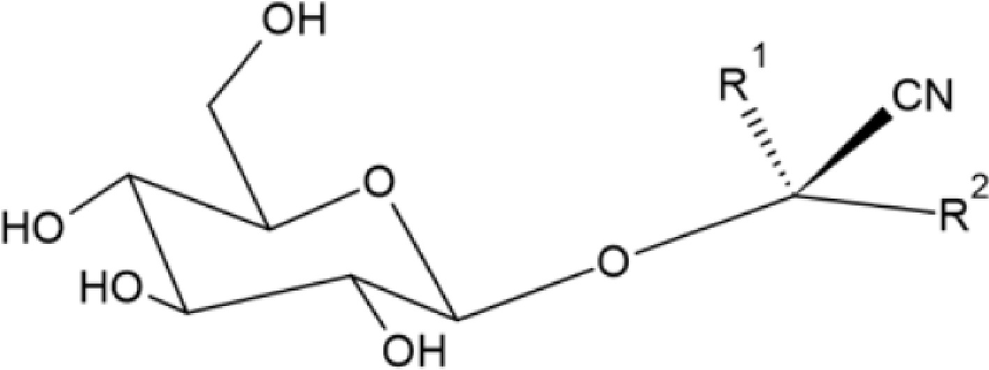
Figure 1.
General structure of cyanogenic glycosides (structure was made with ChemSketch software, version 2024.2.3).
They are found naturally in a wide variety of edible plants. For example, amygdalin and prunasin are cyanogenic glycosides present in almonds (Prunus dulcis Miller), where bitter varieties can contain between 2% and 4% amygdalin[46]. It is also found in peach pits, apricots, plums, black cherries, and apples. In medicinal plants such as Cnidoscolus aconitifolius, Piper carpunya, Parthenium hysterophorus, Taraxacum officinale, and Artemisia absinthium, CGs have been quantified, with Cnidoscolus aconitifolius having the highest concentration (5.02 ± 0.37 µg/g)[17]. Amygdalin, also known as vitamin B17 and, in synthetic form, as laetrile, has been investigated for its possible anticancer effects, but there is no scientific evidence to support its effectiveness in the treatment of cancer in humans. There is scientific evidence that being a CG, it releases cyanide during its hydrolysis.
The toxicity of HCN lies in its ability to inhibit cytochrome oxidase, thus blocking cellular respiration. The lethal dose of HCN in humans ranges from 0.5 to 3.5 mg/kg of body weight in a single dose. For example, ingesting large amounts of raw or poorly processed cassava can have fatal consequences[68]. Cassava (Manihot esculenta) is a staple food for much of the world's population, containing linamarin and lotaustralin in its roots and leaves, with a ratio of 93:7 and an average concentration of 1 mg/g of cyanogenic glycosides[17]. To mitigate its toxicity, proper processing is essential. In cassava, peeling, grinding, settling, and drying are processes that significantly reduce the CG and HCN content. A technologically advanced cassava starch production process can reduce cyanide concentrations by up to 89.12% and HCN by 88.9%[69].
Alkaloids
-
Alkaloids are among the most studied secondary metabolites of plants, especially due to their broad relationship with the development of modern pharmacology in the area of analgesia and pain treatment[70,71].
Glycoalkaloids (GAs)
-
Glycoalkaloids are secondary metabolites from plants, consisting of a nitrogen-containing steroid aglycone and a linked oligosaccharide. Figure 2 shows the structures of the most important glycoalkaloids.
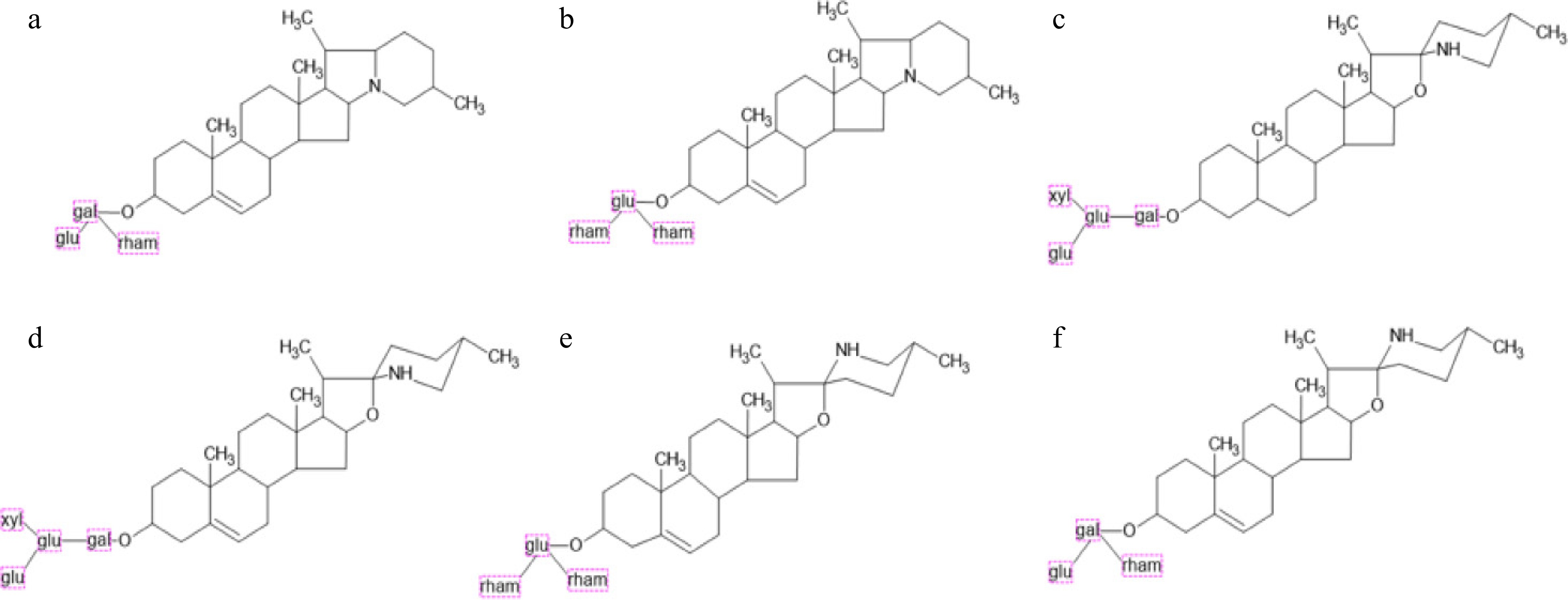
Figure 2.
Structures of the most important glycoalkaloids. (a) α-solanine, (b) α-chaconine, (c) α-tomatine, (d) dehydrotomatine, (e) solamargine, and (f) solasonine (structures were made with ChemSketch software, version 2024.2.3).
These types of alkaloids are associated with toxic steroidal glycosides that are found naturally in plants of the Solanaceae family[72,73]. Solanines are GAs that include α-solanine and α-chaconine[48] and are present in potatoes (Solanum tuberosum). These GAs can be harmful if consumed in large quantities; however, solanine poisoning is rare, as the amounts found in potatoes are usually low and further reduced by cooking; a concentration above 200 mg/kg in fresh potatoes is considered the upper safe limit[73]. GAs are also present in tomatoes (α-tomatine, α-dehydrotomatine) and eggplants (α-solamargine, α-solasonine); their function in the plant is to act as a defense mechanism against pests and pathogens[73]. Acute exposure to GAs can cause gastrointestinal symptoms such as nausea, vomiting, and diarrhea starting at 1 mg/kg of body weight, and doses of 3–6 mg/kg are considered potentially lethal. At the cellular level, they can reduce the integrity of gastrointestinal membranes, affecting nutrient transport and allowing the passage of high-molecular-weight compounds. They are also known for their ability to inhibit cholinesterase[17]. Detection is performed using techniques such as HPLC-UV, LC-ESI/MS, and ELISA[73].
Pyrrolizidine alkaloids (PAs)
-
The chemical structure of PAs consists of an acid fragment called necic acid and a bicyclic necic base with a hydroxymethyl substituent at position 1, and a hydroxyl group at position 7 (Fig. 3). PAs are secondary metabolites produced by more than 6,000 plant species, mainly from the Boraginaceae, Compositae, and Leguminosae families[74].
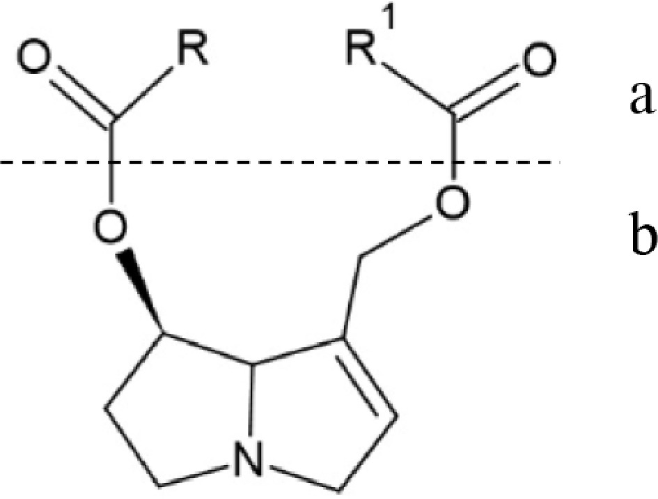
Figure 3.
General structure of Pyrrolizidine alkaloids. (a) Necic acid. (b) Necine base (structure was made with ChemSketch software, version 2024.2.3).
These compounds are a growing food safety issue as they can contaminate a wide variety of food products, including milk, meat, eggs, honey, tea, and herbal infusions, as a result of the consumption of plants containing PAs by animals or directly by humans. Pollen products are among the most contaminated, with averages of 555−576 μg/kg, and some exceeding 1,000 μg/kg[66]. PAs are pro-toxins that are metabolically activated in the body to exert their hepatotoxicity, pulmonary toxicity, genotoxicity, and carcinogenicity. Long-term intake can lead to cancer, and they also exhibit toxicity in human development. Cyclic diesters are considered the most toxic[75]. Current regulations establish maximum limits for PAs and their N-oxides (PA-N-oxides) in certain foods, ranging from 1.0 to 1,000 μg/kg[66]. The determination of these compounds is mainly performed by HPLC-MS/MS or UHPLC-MS/MS, although GC-MS is also used. However, GC-MS is not suitable for PA-N-oxides due to their thermal instability; however, the effect of thermal processing of food is limited and inconclusive[74]. In the preparation of teas, a transfer rate of PAs and PA-N-oxides of 16%−28% (monocrotaline 45%) from the raw material to the infusion has been observed[66].
Other alkaloids
-
Various alkaloids have been identified and quantified in medicinal plants such as Artemisia absinthium, Cnidoscolus aconitifolius, Parthenium hysterophorus, Piper carpunya, and Taraxacum officinale. It has been observed that high concentrations of alkaloids increase toxicity, which in turn is associated with the development of antitumor compounds, and their detection is carried out using spectrophotometric methods[75,76].
Lectins
-
Lectins are proteins present in vegetables, especially in legumes such as beans (Phaseolus vulgaris, Phaseolus acutifolius) and soybeans; they are also found in other foods such as quinoa, lentils, and rice[77]. They are considered antinutritional factors due to their ability to specifically bind to carbohydrates and agglutinate cells[78]. Phytohemagglutinin (PHA), a lectin found in common beans, can affect the weight of internal organs, such as the spleen, thymus, pancreas, heart, liver, kidneys, and small intestine[20,21]. Some lectins can interfere with nutrient absorption in the intestinal epithelium. However, some lectins also have anticancer properties by inhibiting the growth of cancer cells[79,80].
Lectins have been reported in tertiary structure, such as PHA, but also in quaternary structure, as is the case of leucoagglutinating phytohemagglutinin (PHA-L), which is a homotetramer. The three-dimensional structure of the PHA (Q8RVX6_PHAVU) and PHA-L (P05087·PHAL_PHAVU), both from Phaseolus vulgaris (Kidney bean), are shown in Fig. 4. The structure and amino acid sequence are available at the following links:
https://www.uniprot.org/uniprotkb/Q8RVX6/entry ;https://www.uniprot.org/uniprotkb/P05087/entry#sequences .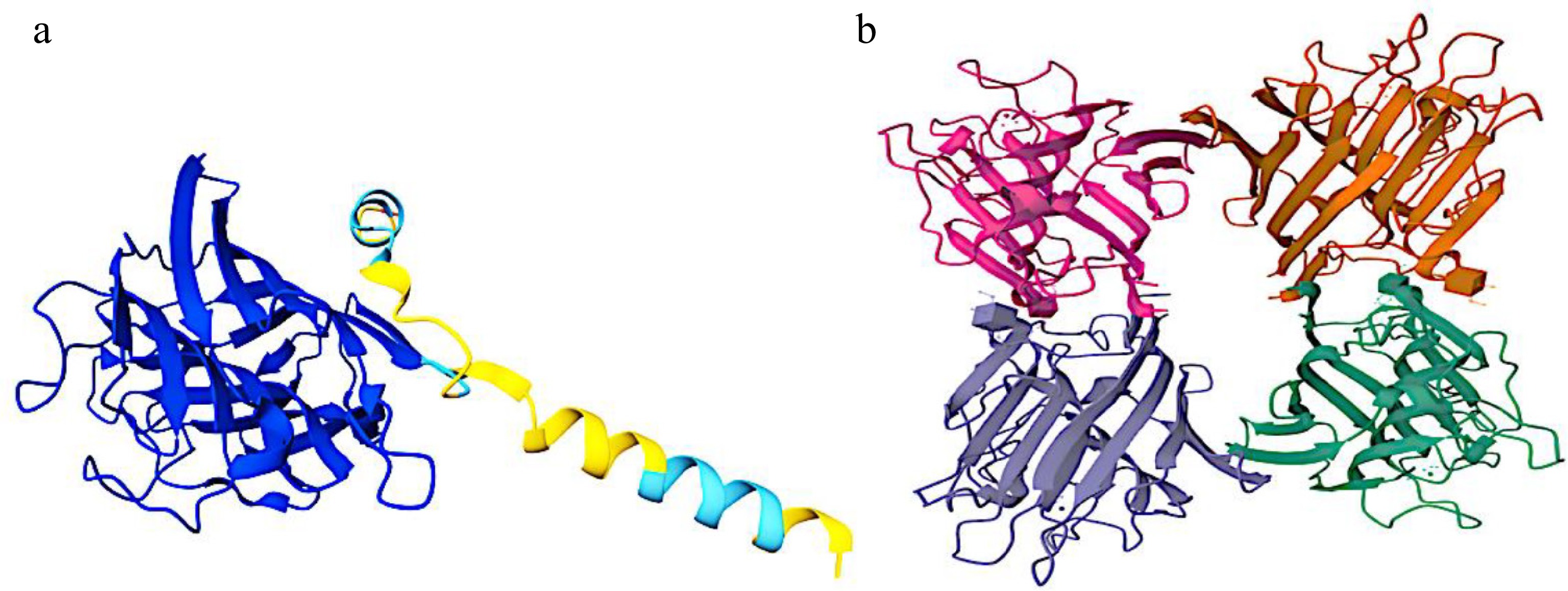
Figure 4.
Three-dimensional structures of the (a) PHA and (b) PHA-L of Phaseolus vulgaris.
Toxicity studies with semi-pure fractions of tepary bean lectin showed low oral toxicity in acute and subchronic studies in rats, although antinutritional effects such as decreased body weight and albumin levels were observed, which requires further investigation[79,80]. Lectin activity is measured with hemagglutination assays using erythrocytes, and chromatographic techniques (HPLC and LC-MS) are used for the analysis and purification of these biomolecules[77].
Heterocyclic aromatic amines (HAA)
-
HAA are hazardous chemical compounds formed primarily during the pyrolysis process, or high-temperature cooking of protein-rich foods such as meat. They are potentially carcinogenic compounds. Like the production of melanoidins, HAA are generated in a heat-dependent reaction between amino acids and sugars, producing pyridine, pyrimidine, or pyrazine, which then react with creatine. More than 25 HAA have been identified in well-cooked red meat[63]. The amount of HAA formed depends on the type of meat, temperature, and cooking time; these substances have been found in grilled beef, chicken, and fish, meat döner, chicken döner, and smoked meat products[63,64]. HAA are classified into several chemical subclasses, the most notable being aminoimidazoazarenes (AIA) and α/β-carbolines; Table 2 shows some chemical structures of these compounds.
Table 2. Structures and systematic names of some heterocyclic aromatic amines.
Subclass Common name Systematic name Chemical structure Aminoimidazoazarenes PhIP 2-amino-1-methyl-6-phenylimidazo[4,5-b]pyridine 
IQ 2-amino-3-methylimidazo[4,5-f]quinoline 
MeIQ 2-amino-3,4-dimethylimidazo[4,5-f]quinoline 
MeIQx 2-amino-3,8-dimethylmidazo[4,5-f]quinoxaline 
β-carbolines β-carboline-1 carboxylic acid 9H-pyrido[3,4-b]indole-1-carboxylic acid 
Harmane 1-methyl-9H-pyrido[3,4-b]indole 
Norharmane or β-carboline 9H-pyrido[3,4-b]indole 
(Structures were made with ChemSketch software, version 2024.2.3). AIA is formed by the heat-catalyzed reaction between amino acids and sugars, which react with creatine. An extensively studied AIA and the most abundant in well-cooked meat is 2-amino-1-methyl-6-phenylimidazo[4,5-b]pyridine (PhIP). Other examples include 2-amino-3-methylimidazo[4,5-f]quinoline (IQ), 2-amino-3,4-dimethylimidazo[4,5-f]quinoline (MeIQ) and 2-amino-3,8-dimethylmidazo[4,5-f]quinoxaline (MeIQx)[63]. α/β-Carbolines are formed from proteins or directly by pyrolysis of glutamic acid and tryptophan exposed to high temperatures. Notable examples are β-carboline-1 carboxylic acid (9H-pyrido[3,4-b]indole-1-carboxylic acid), harmane (1-methyl-9H-pyrido[3,4-b]indole), and norharmane (9H-pyrido[3,4-b]indole)[63].
Regarding their mechanisms of toxicity, HAA are of concern due to their toxic effects, which include neurotoxicity, genotoxicity, and carcinogenicity. These compounds have been identified as potentially affecting neurological function and are considered modifiable risk factors for neurodegenerative disorders such as Alzheimer's disease (AD) and Parkinson's disease (PD). Furthermore, they cross the blood-brain barrier, as AAHs such as PhIP and N-hydroxy-2-amino-1-methyl-6-phenylimidazo[4,5-b]pyridine (N-OH-PhIP), as well as harmane and its derivatives, have been observed to be present in the human or murine brain[63].
Exposure to HAA induces oxidative damage in neurons, a common feature in the progression of AD and PD. Mitochondria have also been shown to be a major target of HAA toxicity, which can lead to mitochondrial dysfunction and contribute to the onset of neurodegeneration. Harmane and its derivatives can inhibit the consumption of NAD+-bound O2 in rat liver mitochondria, suggesting a critical role of mitochondrial complex I[64]. For the analysis and quantification of HAAs, various chromatographic techniques are used, including HPLC coupled to tandem mass spectrometry (LC-MS/MS), GC-MS/MS, acetonitrile extraction coupled to UHPLC-MS/MS for the quantification of HAAs in meat products, solid phase extraction, and supercritical fluid chromatography coupled to triple quadrupole mass spectrometry[63].
Furan and furanocoumarins
-
Furan is an aromatic ring with four carbon atoms and one oxygen atom. It is a contaminant generated in food during high-temperature heating processes; examples include coffee, soups, sauces, canned foods, meat products, bakery products, and cereals, but it is of particular concern in baby food due to continuous exposure[47,81]. It has been shown to be genotoxic and carcinogenic, which can cause serious health problems in infants. It has also been linked to proliferative processes and epigenetic alterations in the liver and kidneys of rats[47]. Furan can cross the biological membranes of the intestine and lungs, causing it to be rapidly absorbed and integrated into the central metabolism. It can enter the human body through ingestion and inhalation during food preparation[47,81]. During its metabolism, the enzyme cytochrome P450 (CYP2E1) produces cis-but-2-ene-1,4-dialdehyde (BDA). BDA is a highly reactive compound and an active metabolite that forms several conjugated molecules with glutathione and cellular polyamines. Furan-induced toxicity has been demonstrated in in vitro and in vivo studies, due to this metabolic activation through CYP2E1[47].
On the other hand, furanocoumarins are compounds based on a furan ring fused to a coumarin (Fig. 5). The furan ring can be fused in various ways, resulting in different isomers. These chemical compounds are found in certain plants and also in citrus fruits, such as grapefruit, bitter oranges, lemons, and limes, which are known to be phototoxic. This phototoxicity manifests itself by altering and disrupting numerous biological processes in different types of cells and can cause skin reactions when exposed to sunlight or ultraviolet light. They react with biological macromolecules (DNA, RNA, proteins) and generate reactive oxygen species by transferring energy to molecular oxygen. Their involvement in cardiovascular effects has also been suggested. Chemically, furanocoumarins are coumarins to which a furan ring has been added, and they exhibit significant photosensitizing activity on cells, which is the basis of their toxic actions[48,82]. The detection of furan in food products is difficult due to its high volatility and low molecular weight[47]. The analytical techniques used for its quantification in foods include GC/MS with headspace solid-phase microextraction (HS-SPME), which has been verified as a rapid and simple method for determining furan concentration in fruit and vegetable-based baby foods[48] and automated headspace GC/MS (HS-GC-MS)[47].
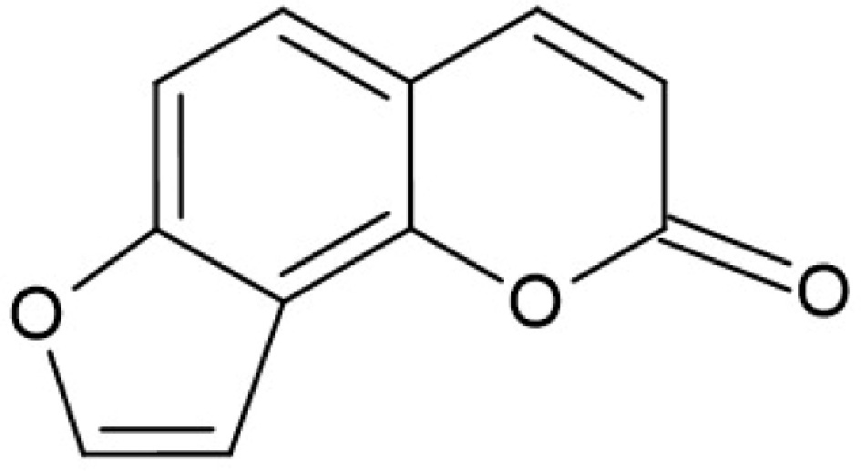
Figure 5.
Chemical structure of furanocoumarin (structure was made with ChemSketch software, version 2024.2.3).
Nitrates and nitrites
-
Nitrate is a natural compound that is part of the nitrogen cycle. Both nitrates and nitrites are substances that can be found in the environment and, by extension, in food. They are found naturally in a wide variety of vegetables and fruits such as leek, Swiss chard, celery, spinach, garden cress, green onion, turnip, radish, eggplant, squash, tomato, pepper, cucumber, mint, tarragon, and lettuce, among others[49]. The toxicity mechanisms of nitrates and nitrites are very similar, and in general, the main process of concern for human health focuses on the transformation of nitrate to nitrite and the subsequent formation of nitrosamines[49]. Nitrate, although a naturally occurring compound and considered non-toxic by itself, is transformed into nitrite in the stomach. This conversion occurs due to its reaction with amines. Once nitrites are formed in the stomach, they react with amines, leading to the formation of nitrosamines, which are considered hazardous substances to human health. They are formed by the reaction of secondary or tertiary amines with nitrites or nitrates, often under acidic conditions such as those found in the stomach. Nitrosamines can be present in various foods, tobacco products, and water, and can also be formed within the human body. They are organic compounds with a molecular structure that features a nitroso group (NO) attached to a secondary amine, which is crucial for their chemical reactivity and carcinogenic potential. The nitroso group and the N-N bond are what allow nitrosamines to be metabolized and form reactive intermediates that can damage DNA and cause cancer[49,83].
The determination of nitrite and nitrate content in plant extracts is carried out by spectrophotometric methods. The general analysis process involves the preparation of the macerated and homogenized sample, a nitrite and nitrate extraction, using the Griess diazotization reaction. For nitrate, a prior reduction to nitrite is performed, a quantity of homogenized sample is measured, and hot deionized water and disodium tetraborate are added. Subsequently, for the determination of nitrite content, a portion of the filtered sample is transferred to a volumetric flask, and specific solutions of sulfanilamide, hydrochloric acid, and N-(1-naphthyl) ethylenediamine dihydrochloride are added. Finally, the quantification is based on the standard method proposed by the International Organization for Standardization (ISO) 6,635:1,984[49,83].
Heavy metals
-
Heavy metals are a group of chemical elements that, above certain concentrations, can be toxic to living organisms. Their presence in the food chain is a public health concern due to their potential bioaccumulation and the various adverse effects they can cause. The study and monitoring of heavy metals in food is of vital importance to public health. Differentiating between chemical forms (as in the case of arsenic) is crucial to determining their actual toxicity. The development and validation of accurate analytical techniques to quantify these contaminants and establish standard parameters to minimize human exposure are essential; among the most used techniques are atomic absorption spectroscopy (AAS), atomic emission spectroscopy (AES), inductively coupled plasma-mass spectrometry (ICP-MS), and square wave anodic stripping voltammetry (SWASV)[32]. Environmental exposure to heavy metals can cause inhibition or overexpression of any component of palmitoylated glycoprotein signaling pathways, such as Wnt, transcription factors, receptors, ligands, or transducers, thereby preventing normal cellular function by negatively affecting, as well as contributing to diseases by deregulating Wnt signaling pathways[84].
Arsenic
-
Arsenic is a crystalline metalloid found naturally throughout the Earth's crust. Exposure to this element in the ecosystem and present in the human food chain comes from air, water, and soil. Dietary contamination depends on the presence of inorganic or organic forms, their oxidation state, water solubility, and the food matrix. Inorganic forms, such as arsenite (As[III]) and arsenate (As[V]), are considered highly toxic. Organic forms, such as arsenocholine and arsenobetaine, are commonly found in seafood and are considered to be of low toxicity[32,85]. Cereals and cereal products are the main source of arsenic exposure in different age groups, with rice and its derivatives being the main cause, since rice has a greater capacity to absorb arsenic compared to other cereals[86]. It has been reported that 75% of rice-based infant foods in Australia, as well as infant cereals in Spain and America, contain high concentrations of inorganic arsenic. Infant formulas may also play a significant role in inorganic arsenic exposure in infants and young children[34,35]. On the other hand, vegetables contribute to exposure to inorganic arsenic, with root vegetables accumulating the highest content, while edible parts have a lower amount[32,86]. Regarding its toxicity mechanism, it can be said that inorganic arsenic is almost completely absorbed (80%−90%) in the intestine and is metabolized by methylation to monomethylarsonic acid (MMAV), monomethylarsenious acid (MMAIII), dimethylarsinic acid (DMAV), and dimethylarsenious acid (DMAIII). Among these forms, MMAIII is considered the most cytotoxic, since it inhibits mitochondrial processes I and III, causing electron leakage through the electron transport chain, leading to the production of ROS and nitrogen species (RNS)[86]. Free radical production leads to DNA damage and impaired gene expression, inducing microRNA expression that impairs polymerase elongation function and the recruitment of splicing regulatory factors, contributing to carcinogenicity. Arsenic is also considered a promoter of inflammation, oxidative stress, and endothelial dysfunction[34,35].
Cadmium
-
Cadmium is a heavy metal that is toxic even at very low concentrations[85]. Aquatic organisms can bioaccumulate cadmium in their edible tissues (gills, kidneys, liver, and muscle) when living in a contaminated environment, so the consumption of fish internal organs, such as liver and kidney, can lead to increased cadmium accumulation in humans. Cadmium is also present in food at different concentrations depending on the origin or form of production of the food[85]. The toxicity of this element causes the induction of ROS, which leads to oxidative stress, which can lead to the development of cancer cells. On the other hand, cadmium and its compounds are classified by the International Agency for Research on Cancer (IARC) as Group 1 substances, known to cause cancer, and have been linked to the development of kidney, prostate, and pancreatic cancer[36]. Cadmium also causes multisystem failure, in addition, it affects musculoskeletal tissue since it interacts with bone cells and causes skeletal demineralization, inhibiting collagen production, which results in decreased bone density, fractures and osteoporosis, likewise, it affects the central and peripheral nervous system, cardiovascular, respiratory and reproductive, and since the bioaccumulation of cadmium in the body occurs in the kidney and liver, they turn out to be the most affected organs[36].
Mercury
-
Mercury is recognized as a heavy metal contaminant, and recent studies mainly mention methylmercury as the most relevant form of dietary exposure[61]. Fish consumption is the main source of exposure to mercury, especially in its methylmercury form[37,38]. During pregnancy, poisoning with this metal has been associated with changes in metabolic and inflammatory health biomarkers in children, and may also alter the expression of endotoxin-induced inflammatory cytokines in the liver[61]. Previous research has suggested that chronic mercury exposure, even at low doses, alters the plasticity of white adipose tissue. Furthermore, fish intake associated with mercury exposure has been evaluated in relation to the risk of cardiovascular disease, diabetes, and obesity. An association has been observed between blood mercury levels and overweight in adolescents[37,38].
-
Food is a source of nutrition and often provides health benefits; however, there are potential risks associated with the presence of certain chemical compounds that, depending on the frequency and dosage of consumption, can severely damage health. These chemical compounds can be naturally present in food, as part of its chemical composition, or they can be produced through food processing, such as the heat used during cooking. Therefore, it is crucial to continue scientific research on the presence of harmful compounds in food.
-
Toxic compounds and some antinutritional factors in food represent a multifactorial risk to human health; their presence may result from a plant's natural defense mechanisms, the formation of byproducts during thermal processing, or from environmental contaminants that impact the food chain. The chemical and biological diversity of these compounds determines a wide range of toxic effects, including enzyme inhibition, blockage of one or more metabolic pathways, and induction of oxidative stress, DNA damage, and carcinogenesis. Likewise, signaling pathways that are key regulators of cell homeostasis, that control many metabolic mechanisms, are affected. The risks of health problems from the consumption of foods containing cyanogenic glycosides, alkaloids, lectins, heterocyclic amines, furan, furanocoumarins, nitrates, nitrites, and heavy metals can be controlled through good agricultural practices, proper food processing, safe storage, and continuous monitoring of contaminants. On the other hand, the geolocation, genetics, and agricultural practices of each region vary the chemical composition of food; however, all must be governed by the strictest standards for the absence of harmful compounds, and every food safety program is required to guarantee food safety in order to reduce or eliminate all risks to the health of consumers.
To SECIHTI for the scholarship to study for a master's degree awarded to Jose Armando Narvaez Padilla (Grant No. 4036084).
-
Not applicable.
-
The authors confirm their contributions to the paper as follows: study conception and design: Díaz R; data collection, draft manuscript preparation: Díaz R, Narvaez-Padilla JA, Rodríguez-Requenes ME, Díaz-Godínez G; analysis and review: Díaz-Godínez G, Díaz R. All authors reviewed and approved the final version of the manuscript.
-
Data sharing not applicable to this review as no datasets were generated or analyzed during the current study.
-
The authors declare that they have no conflict of interest.
- Copyright: © 2026 by the author(s). Published by Maximum Academic Press on behalf of Nanjing Agricultural University. This article is an open access article distributed under Creative Commons Attribution License (CC BY 4.0), visit https://creativecommons.org/licenses/by/4.0/.
-
About this article
Cite this article
Narvaez-Padilla JA, Díaz-Godínez G, Rodríguez-Requenes ME, Díaz R. 2026. Toxic compounds and antinutritional factors in foods: a review. Food Materials Research 6: e003 doi: 10.48130/fmr-0026-0004 

Toxic compounds and antinutritional factors in foods: a review
- Received: 17 September 2025
- Revised: 19 December 2025
- Accepted: 19 January 2026
- Published online: 10 March 2026
Abstract: Toxic compounds and antinutritional factors are present in food. These compounds, which can be naturally occurring or arise due to technological, storage, or transportation processes, have the potential to bioaccumulate and cause acute or chronic toxicity, in addition to having adverse cellular, metabolic, and systemic effects. To mention a few, these are cyanogenic glycosides present in almonds and cassava, among others; alkaloids, which include glycoalkaloids from solanaceous plants and pyrrolizidine alkaloids, which are mainly found in angiosperm plant families: Asteraceae, Boraginaceae, Fabaceae, Orchidaceae, and Apocynaceae; and lectins found in legumes and other vegetables. Other compounds include amines, heterocyclic aromatic amines, furan and furanocoumarins, as well as some inorganic compounds such as nitrates and nitrites, and some heavy metals such as cadmium, mercury, and arsenic. Mechanisms of toxicity include blockage of critical enzymes, oxidative damage, interference with nutrient absorption, and genotoxic and carcinogenic processes. Techniques for detecting these compounds in food include the use of gas chromatography with mass spectrometry, spectrophotometry, and immunochemical methods, mainly. Quantifying these substances is essential for establishing regulatory thresholds and preventing acute and chronic poisoning. This review lists the main toxic and antinutritional compounds in food, chemical–analytical techniques for their detection and quantification, as well as some chemical structures of these compounds, seeking to provide an overview that will aid in the prevention and management of risks associated with the ingestion of these compounds.