










-
Fungi are key components of global ecosystems and represent a highly promising reservoir of biological resources, yet the exploration of their species diversity remains in its early stages[1]. They play indispensable roles in nutrient cycling, organic matter decomposition, symbiotic interactions, and ecosystem functioning, and also serve as important sources of food, medicine, and industrial products. It is estimated that approximately 2.5 million fungal species exist worldwide[2]; however, only about 160,000 have been formally described to date[3], indicating that more than 90% of fungal taxa remain undiscovered and hidden in natural environments. This vast gap underscores both the immense potential and the urgent need to advance fungal biodiversity research.
China, with its vast land area and remarkable environmental heterogeneity, encompasses a wide range of climate zones, from tropical rainforests to temperate coniferous forests. These diverse ecological conditions, shaped by complex topography and varied climatic gradients, have fostered exceptionally rich fungal resources. Consequently, China is recognized as a global hotspot of fungal diversity and a key region for advancing mycological research[4−8].
Through field investigations across multiple provinces, this study successfully identified 28 new fungal taxa based on morphology and multi-locus phylogenetic analyses. The discovery of these species not only further confirms the richness and uniqueness of China's fungal resources, providing empirical evidence to fill gaps in regional fungal flora research, but also supplements key branches of fungal research at the phylogenetic level, offering new materials for exploring species evolution and geographical distribution patterns. Meanwhile, the potential ecological functions and secondary metabolite values of these species lay the foundation for subsequent research on resource development and biological control[9].
This study aims to detail the morphological characteristics and phylogenetic positions of the 28 newly described fungal taxa, providing a scientific basis for the conservation and utilization of China's fungal resources. The abbreviations for the generic names referenced in this study are as follows: C. = Cortinarius, Cy. = Cystolepiota, Ec. = Echinoderma, En. = Entoloma, H. = Hygrocybe, Hy. = Hypholoma, Le. = Lepiota, Lc. = Leucocoprinus, Li. = Limacella, Ma. = Macrocystidia, Mar. = Marasmius, Me. = Melanoleuca, My. = Mycena, Ph. = Phloeomana, Pse. = Pseudobaeospora, Ps. = Pseudosperma, R. = Rhodophana, S. = Singerocybe, T. = Trogia, Tu. = Tubaria.
-
During macrofungal survey investigations in China, a total of 56 specimens representing 28 species were collected, each representing a distinct new species. These specimens were collected from seven provinces, namely Guangdong, Fujian, Heilongjiang, Jilin, Liaoning, Qinghai, and Shanxi, during the period from 6 August 2016 to 8 September 2025. Fresh basidiocarps were imaged in their natural habitats using a Canon 80D digital single-lens reflex (DSLR) camera, with simultaneous recording of habitat attributes. Color characteristics of the basidiocarps were documented and assigned color codes in accordance with the standardized procedure established by Kornerup & Wanscher[10]. All collected specimens were dried overnight at 40 °C in a forced-convection drying oven and were subsequently deposited in the Herbarium of Mycology, Jilin Agricultural Science and Technology University (HMJU).
Comprehensive macro-morphological observations and descriptions of the specimens were carried out based on fresh material. Microscopic features, encompassing basidiospores, basidia, cheilocystidia, pleurocystidia, and constituent elements of the pileipellis, were examined following the methodological guidelines described by Largent[11]. Prior to microscopic examination, sections of dried specimens were first treated with 3% potassium hydroxide (KOH), 1% Congo red, and Melzer's reagent for fixation.
The notations [n, p, m] are defined as follows: the data pertain to n basidiospores measured from p samples derived from m specimens. Basidiospore dimensions are presented in the format '(a–)b–av–c(–d)', where the interval 'b–c' encompasses no fewer than 90% of the measured values. Extreme values a and d are enclosed in parentheses. Q represents the length-width ratio of individual basidiospores, while Qm denotes the mean Q value computed from all measured basidiospores.
Scanning Electron Microscope (SEM) micrographs of basidiospores were obtained using the following protocol: free-hand sections of dried lamellae were prepared, directly mounted on double-sided adhesive tape attached to a metal specimen stub, and thereafter scanned at different magnifications under high-vacuum conditions. This experimental work was conducted using a Zeiss EVO 18 scanning electron microscope.
DNA extraction, PCR amplification, and sequencing
-
Genomic DNA extraction was performed using the EZup Column Fungi Genomic DNA Purification Kit (Sangon Biotech Co., Ltd., Shanghai, China), strictly following the operational protocols provided by the manufacturer. For polymerase chain reaction (PCR) amplification, the internal transcribed spacer (ITS) rDNA region was targeted with the primer pairs ITS1F/ITS1 and ITS4[12,13]. Nuclear large subunit (nrLSU) ribosomal RNA gene sequences were amplified using the primer set LR0R combined with LR5/LR7[14]. The second-largest subunit of RNA polymerase II gene (rpb2) was amplified specifically with primers rpb2-6F paired with rpb2-7cR/7.1R[15]. Additionally, the translation elongation factor 1-α (tef1-α) gene region was amplified utilizing the primer combinations tef1-983F and tef1-1567R/2218R[16].
For ITS regions, initial denaturation at 94 °C for 4 min, followed by 30 cycles of 94 °C for 1 min, 55 °C for 1 min, and 72 °C for 1 min, with a final extension of 72 °C for 5 min, and then held at 4 °C; nLSU region, initial denaturation at 94 °C for 4 min, followed by 30 cycles of 94 °C for 40 s, 52 °C for 45 s, and 72 °C for 40 s, with a final extension of 72 °C for 5 min, and then held at 4 °C; rpb2 region, initial denaturation at 94 °C for 3 min, followed by 35 cycles of 94 °C for 50 s, 59 °C for 1 min, and 72 °C for 1 min, with a final extension of 72 °C for 10 min, and then held at 4 °C; tef1-α region, initial denaturation at 94 °C for 3 min, followed by 30 cycles of 94 °C for 30 s, 49 °C for 35 s, and 72 °C for 30 s, with a final extension of 72 °C for 10 min, and then held at 4 °C. Following PCR product separation and visualization via 1% agarose gel electrophoresis on a JY 600 electrophoresis system (Beijing JUNYI Electrophoresis Co., Ltd, Beijing, China), all qualified amplicons were sent to BGI Co., Ltd (Beijing, China) for bidirectional Sanger sequencing.
Sequence alignment and phylogenetic analyses
-
To confirm the taxonomic positions of the new species, ITS and nrLSU, rpb2, and tef1-α sequences were combined and analyzed with Bayesian Inference (BI) and Maximum Likelihood (ML) methods. Sequence alignment was performed with MAFFT v7.0[17], and low-quality regions were trimmed with MEGA v7.0[18], then combined with Phylosuite v1.2.3[19,20]. The BI analyses was conducted using MrBayes v3.2.7a[21] in Phylosuite v1.2.3, which employs a Markov Chain Monte Carlo (MCMC) algorithm for a million generations until convergence was achieved, as indicated by an average standard deviation of split frequencies below 0.01. The initial 25% of sampled trees were discarded as burn-in. Nucleotide substitution models were determined by ModelFinder v 2.2.0. The ML analyses was performed using IQ-Tree v2.2.0[22] in Phylosuite v1.2.3, with the best model selected for each locus according to ModelFinder with 1,000 bootstrap replicates. The phylogenetic position of the new species were inferred using Maximum Likelihood (ML) and Bayesian Inference (BI) methods. The phylogenetic analyses were performed using IQ-TREE for Maximum Likelihood[23] and the Markov Chain Monte Carlo (MCMC) method, with MrBayes v3.2.2[21]. Bootstrap values ≥ 70% in Maximum Likelihood (ML) analyses and Bayesian Posterior Probabilities (PP) ≥ 0.70 are considered to provide significant support for phylogenetic nodes.
-
Cortinarius (Pers.) Gray, Nat. Arr. Brit. Pl. (London) 1: 627 (1821)
Initial taxonomic investigations of Cortinarius (Pers.) Gray sensu lato (s.l.) in Europe and the Americas relied primarily on the morphological features of basidiomata. Persoon originally classified these species as a section within the genus Agaricus L. Later, Gray[24] elevated the group to the rank of an independent genus. Fries subsequently performed a systematic analysis of 216 European Cortinarius species, categorizing them into six subgenera Dermocybe, Hydrocybe, Myxacium, Inoloma, Phlegmacium, and Telamonia based on the distinct attributes of the pileus, stipe, and veil. Following these foundational works, numerous scholars have expanded the taxonomy of Cortinarius s.l., with the classification schemes proposed by Moser & Horak[25], and Soop et al.[26,27] gaining widespread acceptance.
Moser & Horak[25] proposed a system comprising seven subgenera: Sericeocybe, Mycacium, Cystogenes, Telamonia, Leprocybe, Phlegmacium, Cortinarius, and Paramyxacium. In contrast, Soop[26] organized the genus into six subgenera (Dermocybe, Mycacium, Cortinarius, Telamonia, Orellani, and Phlegmacium). In this system, Sections Delibutus and Anomali were maintained as independent sections, while Sections Callistei, Leprocybe, and Limonei were merged into Subg. Orellani. Later, Soop et al.[27] utilized maximum likelihood methods (PhyML, RAxML) to analyze gene fragments (nrITS, nrLSU, rpb1, and rpb2) from 789 specimens. This phylogenetic study re-evaluated 37 existing sections, established 42 new combinations, and defined 20 clades, resulting in a granular sectional classification for the genus.
Despite these advances, earlier taxonomic efforts were largely confined to subgenus and section levels, leaving higher-level classifications unresolved. Although Soop's global sampling successfully identified monophyletic taxa at the sectional level, the boundaries between certain clades remained indistinct. Some species occupied transitional zones, making their placement difficult. Consequently, within current classification systems, subgeneric lineages often overlap or merge, resulting in taxonomic ambiguity.
To address this, Liimatainen et al.[28] conducted genomic and multi-gene concatenated analyses (using rpb1, rpb2, MCM7, GPD, and tef1) on a dataset comprising 19 species for genomic analysis and 245 species from both hemispheres. This study led to the splitting of Cortinarius s.l. into 10 distinct genera: Cortinarius sensu stricto (s.str.), Phlegmacium, Thaxterogaster, Calonarius, Aureonarius, Cystinarius, Volvanarius, Hygronarius, Mystinarius, and Austrocortinarius. Under this new framework, Cortinarius s.str. contains 11 subgenera: Cortinarius, Camphorati, Dermocybe, Illumini, Infracti, Iodolentes, Leprocybe, Myxacium, Orellani, Paramyxacium, and Telamonia. With the exception of Iodolentes and Infracti, these subgenera were redefined or reorganized from previous groups. Calonarius was established as a separate genus from the former Subg. Phlegmacium and includes three subgenera (Calonarius, Calochroi, and Fulvi). Similarly, Aureonarius (subgenera Aureonarius and Callistei), Cystinarius (subgenera Cystinarius and Crassi), Hygronarius (subgenera Hygronarius and Visincisi), and Thaxterogaster (subgenera Thaxterogaster, Cretaces, Multiformes, Riederorum, Scauri, and Variegati) were subdivided. The genus Phlegmacium was elevated from subgenus status and divided into four subgenera: Phlegmacium, Bulbopodium, Carbonella, and Cyanicium, while Mystinarius contains a single subgenus.
Molecular phylogenetic studies on Cortinarius s.l. abroad emerged in the late 20th century. Liu et al.[29] pioneered this field by using rDNA 5.8S and ITS sequences to explore relationships between Dermocybe and Cortinarius s.l., validating ITS as an effective tool for species identification. Garnica et al.[30] analyzed ITS and nuclear rDNA nLSU in 262 largely European species, integrating morphological data to demonstrate that combining macro- and micro-morphological traits aids in defining clades.
Liimatainen[28] also utilized 236 ITS type sequences to refine the classification of Subg. Phlegmacium, with a particular focus on exploring relationships among sections. In 2020, Liimatainen further expanded this approach to Subg. Telamonia, the largest subgenus, by examining 746 species using ITS sequence data to provide a more detail understanding of interspecific relationships. Meanwhile, Seidl[31] examined the relationship between Sect. Defibulati and Sect. Myxacium within Subg. Myxacium, suggesting that existing classification systems required further adjustment.
Although ITS is suitable for assessing intraspecific relationships, relying on a single genetic marker is inadequate for resolving genus and subgenus-level phylogeny. Many researchers have combined ITS and nLSU sequences with ITS, yet this approach often failed to clarify clade relationships. Frøslev et al.[32] were the first to apply a three-marker approach (ITS, rpb2, and rpb1) to Cortinarius s.l. Subsequently, Niskanen[33] used combined ITS and rpb2 sequences to describe five new Telamonia species from western North America. Similarly, Katri Kokkonen employed ITS and rpb2 markers alongside morphological analysis to identify four new Telamonia species from Finland.
In purely molecular studies, Stefani et al.[34] evaluated seven candidate barcodes (ITS, nLSU, gpd, mcm7, rpb1, rpb2, and tef1-α) on 68 Australian specimens, confirming the efficacy of ITS for species identification. Nouhra performed phylogenetic analyses using ITS and 28S sequences on 24 Thaxterogaster taxa from Patagonia, documenting them with photographs, SEM images of spores, and molecular data. Additionally, Ammirati defined 18 species within Subg. Leprocybe by synthesizing morphological data with DNA sequences (ITS and nLSU).
Cortinarius hyalocinnamomeus J.Z. Xu, sp. nov., Figs 1 and 2
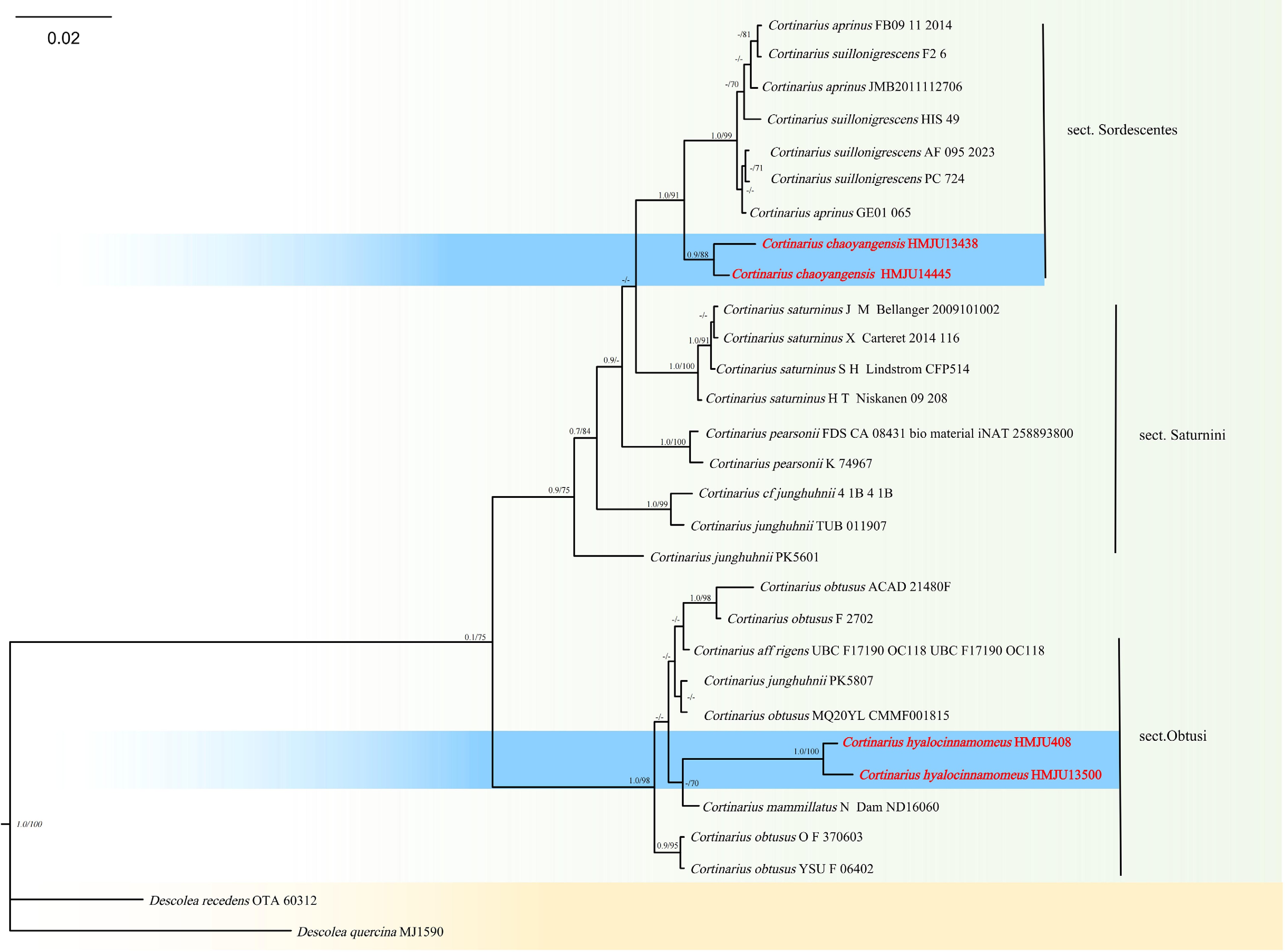
Figure 1.
Phylogram of Maximum Likelihood phylogenetic analysis based on combined ITS and nLSU sequences. The analysis includes 30 strains; total characters: 1,394 (ITS: 709, nLSU: 685). Descolea quercina (MJ1590) and Descolea recedens (OTA 60312) were used as the outgroup taxa. The best model used was TIM2 + F + R2. Estimated base frequencies were as follows: A = 0.248, C = 0.209, G = 0.212, T = 0.331. Bootstrap values for ML equal to or greater than 70% and BYPP values greater than 0.70 are labelled on the nodes. The type strains are in bold.
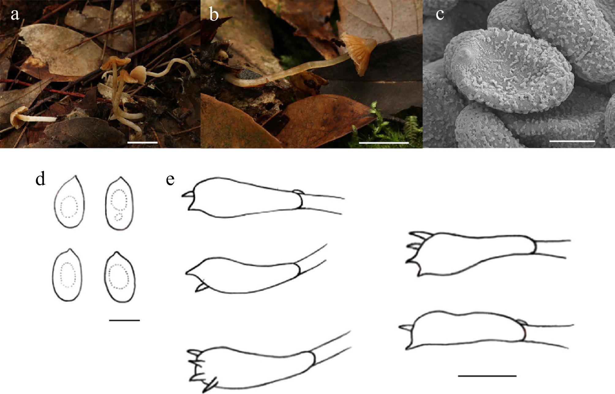
Figure 2.
Cortinarius hyalocinnamomeus (HMJU 408, holotype). (a), (b) Basidiocarps. (c) SEM images of basidiospores. (d) Basidiospores. (e) Basidia. Scale bar: (a), (b) 1 cm; (c) 3 µm; (d) 5 µm; and (e) 10 µm.
MycoBank Names: MB863386
Holotype: Guizhou Province, Tongren City, Fanjing Mountain, on decaying leaves, 27°52'58" N, 108°43'13" E, 14 July 2019, J.Z. Xu (HMJU 408, holotype).
Etymology: The specific epithet 'hyalocinnamomeus' refers to the transparent cinnamon-brown coloration at the center of the pileus.
Diagnosis: Cortinarius hyalocinnamomeus is characterized by small basidiomata, with the central part of the pileus translucent cinnamon-brown; the basidiospores are hyaline or yellowish-brown.
Description: Pileus 6–11 mm diam., conical, center transparent cinnamon brown (6D6), covered with chrome orange (6A8) striations on cinnamon brown (6D6) ground, margin covered with white (1A1) fibrils, context cinnamon brown (6D6), pileus context thin, subwaxy, pale chrome orange (6A8). Lamellae adnexed, distant, chrome orange (6A8), margin entire. Stipe 17–30 mm long, 1–2 mm thick, cylindrical, flexuous, transparent cinnamon brown (6D6), covered with white (1A1) fibrils, base tomentose, subwaxy, transparent chrome orange (6A8).
Basidiospores (8.0–)8.1–9.0(–9.1) × (4.7–)4.9–5.8(–6.4) μm, Q = 1.59–1.75, Qm = 1.58, ellipsoid to elongate, transparent or yellow-brown in KOH, without amyloid. Basidia (24–)25–30(–31) × (7–)8–9(–10) μm, clavate, both four and two sterigmata up to 2 μm, transparent or yellow-brown in KOH. Cheilocystidia absent. Pleurocystidia absent. Lamellar trama irregular, hyphae 3–8 μm wide, transparent in KOH. Clamp connections present in all hyphae.
Habitat: Scattered to gregarious on soil covered with leaf litter in forests.
Known distribution: Known only from Southwestern China.
Additional material examined: Guizhou Province, Tongren City, Fanjing Mountain, on decaying leaves, 27°52'58" N, 108°43'13" E, 17 July 2022, J.Z. Xu (HMJU 13500).
Notes: Cortinarius mammillatus Kałucka, Kytöv., Niskanen & Liimat. and Cortinarius obtusus (Fr.) Fr. forms a sister group (ML/PP = 98/1.00), but exhibits significant morphological differences. Cortinarius hyalocinnamomeus is morphologically similar to Cortinarius boreotrichus Kytov., Niskanen & Liimat., and C. mammillatus. C. boreotrichus is different in pileus brown, margin slightly grayish white, lamellae brown, margin irregular, stipe pale brown, and basidia hyaline[35]. C. obtusus features a larger pileus (20–50 mm), reddish brown to orange brown or yellowish brown, with yellow spores[36]. C. mammillatus is characterized by a larger pileus (15–30 mm), fulvous brown to ochraceous orange brown, lamellae rusty brown, a stipe with a universal veil, and lamella trama yellow[37].
Cortinarius chaoyangensis J. Z. Xu, sp. nov., Figs 1 and 3
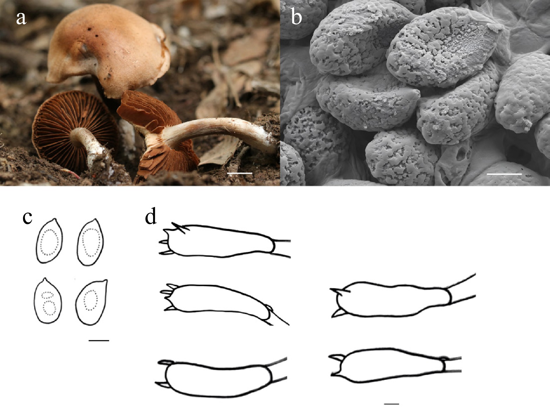
Figure 3.
Cortinarius chaoyangensis (HMJU 13438, holotype). (a) Basidiocarps. (b) SEM images of basidiospores. (c) Basidiospores. (d) Basidia. Scale bars: (a) 1 cm; (b)–(d) 5 µm.
MycoBank Names: MB863386
Holotype: Liaoning Province, Chaoyang City, Shuangta District, Fenghuang Mountain, on soil, 41°32'55" N, 120°30'55" E, 18 August 2025, W.L. Zhao (HMJU 13438, holotype).
Etymology: The specific epithet 'chaoyangensis' refers to the type locality, Chaoyang, Liaoning Province, China.
Diagnosis: Cortinarius chaoyangensis is characterized by large Basidiomata; the pileus is reddish golden with an undulate margin; the lamellae are reddish brown with an irregular margin.
Description: Pileus 41–61 mm diam., convex-lenticular, reddish golden (6C7), margin undulate, covered with minute velvety hairs concolorous with the pileus, with cracking, pileus context thick, cottony, pileus surface smoke brown (4F2), central part white (1A1). Lamellae free, subcrowded, reddish brown (6C7), margin irregular and entire. Stipe 39–74 mm long, 6–11 mm thick, cylindrical, base slightly swollen, cinnamon brown (6D6), fading to white (1A1) towards the base, covered with minute white (1A1) fibrils, stipe context cottony, white (1A1).
Basidiospores (8.4–)9.0–11.5(–11.6) × (6.0–)6.1–7.9(–8.0) μm, Q = 1.27–1.66, Qm = 1.51, broadly ellipsoid to ellipsoid, yellowish brown or transparent in KOH, without amyloid. Basidia (34–)35–43(–44) × (9–)10–14(–17) μm, transparent or yellowish brown, clavate, both four and two sterigmata to 2 μm. Cheilocystidia absent. Pleurocystidia absent. Lamella trama irregular, hyphae 2–8 μm wide, transparent in KOH. Clamp connections present in all hyphae.
Habitat: Scattered to gregarious on soil in deciduous forests.
Known distribution: Known only from Northeastern China.
Additional material examined: Liaoning Province, Chaoyang City, Shuangta District, Fenghuang Mountain, on soil, 41°32'55" N, 120°30'55" E, 7 September 2025, W.L. Zhao (HMJU 14445).
Notes: Phylogenetically, the taxon forms a well-supported sister clade with Cortinarius suillonigrescens Rob. Henry and Cortinarius aprinus Melot (MLBS/PP = 91/1.00). C. suillonigrescens is distinguished by the rust-brown maculae on the pileus, globose stipe base, and hyaline, smaller basidia in KOH (25–35 × 6.5–8.0 μm)[36].
Moreover, C. aprinus, Cortinarius pearsonii P.D. Orton, Cortinarius rigens (Pers.) Fr., and Cortinarius varius (Schaeff.) Fr. show considerable morphological similarity. However, C. aprinus is distinguished by its undulate lamellae margins that are brownish yellow with a purple tint, a stouter stipe, and a fugaceous pale-white cortina[38,39]. In contrast, C. pearsonii is characterized by an inrolled pileus margin with yellow cortinal remnants, a dark brown stipe covered with dense white fibrils towards the base, and smaller basidiospores (6.3–7.5 × 4.4–4.8 μm)[40]. Furthermore, C. rigens differs in having pale white lamellar margins, pale white stipe, and smaller basidiospores (7–9 × 4–5μm)[36]. C. varius is notable for a reddish-brown pileus center, lamellae with pale white margins, and a white stipe[36].
Cystolepiota Singer, in Singer & Digilio, Lilloa 25: 281 (1952) [1951].
The genus Cystolepiota was originally established by Digilio & Singer[41] to classify small lepiotoid fungi defined by epithelioid squamules and basidiospores that lack both amyloid and dextrinoid reactions. Later, Singer & Clémençon[42] proposed dividing the genus into two sections: Cystolepiota sect. Pseudoamyloideae Singer & Clémençon, containing species with basidiospores that become dextrinoid in Melzer's reagent (such as Cystolepiota icterina F. H. Møller ex Knudsen), and Cystolepiota sect. Cystolepiota Singer, comprising taxa with non-reactive spores. Independently, Bon[43] introduced the genus Pulverolepiota Bon for species featuring pileus scales made of elongated, inflated cells, an absence of clamp connections, and basidiospores that slowly develop a reddish-brown color in Melzer's reagent (exemplified by Cystolepiota petasiformis [Murrill] Vellinga ≡ Pulverolepiota petasiformis [Murrill] H. Qu, Damm & Z. W. Ge). Although Vellinga subsequently reduced this group to a section within Cystolepiota, recent phylogenetic evidence provided by Qu et al.[16] confirmed that Pulverolepiota represents a unique lineage separate from Cystolepiota, leading to its restoration as an independent genus.
In addition to established Cystolepiota species, certain taxa originally placed in Lepiota (Pers.) Gray are distinguished by pileal squamules consisting of sphaerocyst chains. Due to the presence of these spherocyte, Knudsen[44] initially moved Lepiota sect. Echinatae into Cystolepiota, though he later reversed this decision, maintaining the group as Lepiota sect. Echinatae. Bon[45] later reassigned these species to the genus Echinoderma (Locq. ex Bon) Bon. However, phylogenetic research by Hou & Ge[46] revealed that Echinoderma is polyphyletic: species with globose to ellipsoid basidiospores are members of Lepiota (e.g., Lepiota omninoflava Y. J. Hou & Z. W. Ge), whereas only those with subcylindrical spores are true members of Echinoderma (e.g., Echinoderma asperum [Pers.] Bon).
Cystolepiota rubellogrisea J.Z. Xu, sp. nov., Figs 4 and 5
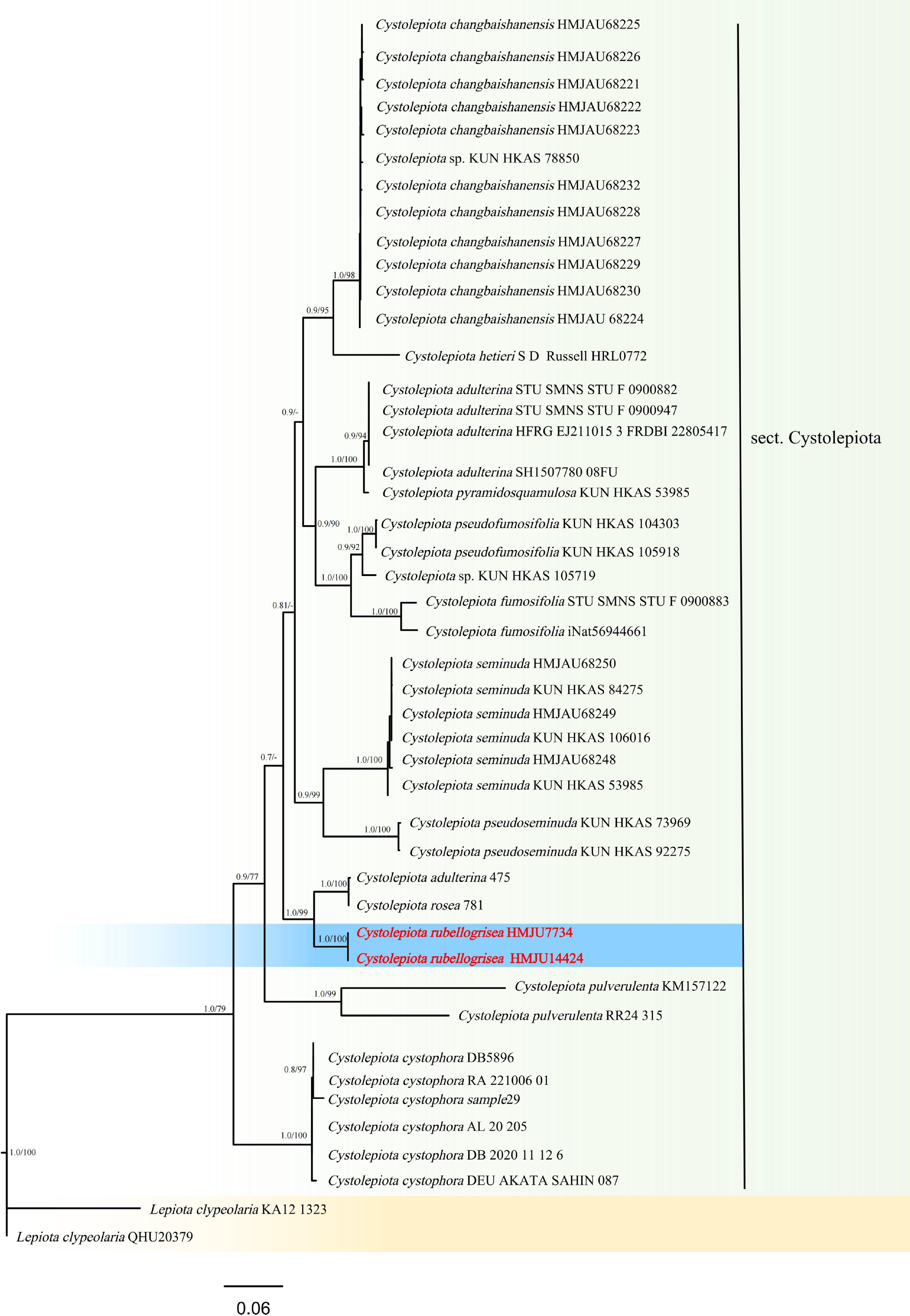
Figure 4.
Phylogram of Maximum Likelihood phylogenetic analysis based on combined ITS, nLSU, rpb2, and tef-1α sequences. The analysis includes 59 strains; total characters: 2,464 (ITS: 517, nLSU: 824, rpb2:616, tef-1α: 507). Lepiota clypeolaria (Bull.) P. Kumm. (KA12 1323) and Le. clypeolaria (QHU20379) were used as the outgroup taxa. The best model was TIM2e + I + G4. Estimated base frequencies were as follows: A = 0.251, C = 0.225, G = 0.253, T = 0.272. Bootstrap values for ML equal to or greater than 70% and BYPP values equal to or greater than 0.70 are labelled on the nodes. The type strains are in bold.
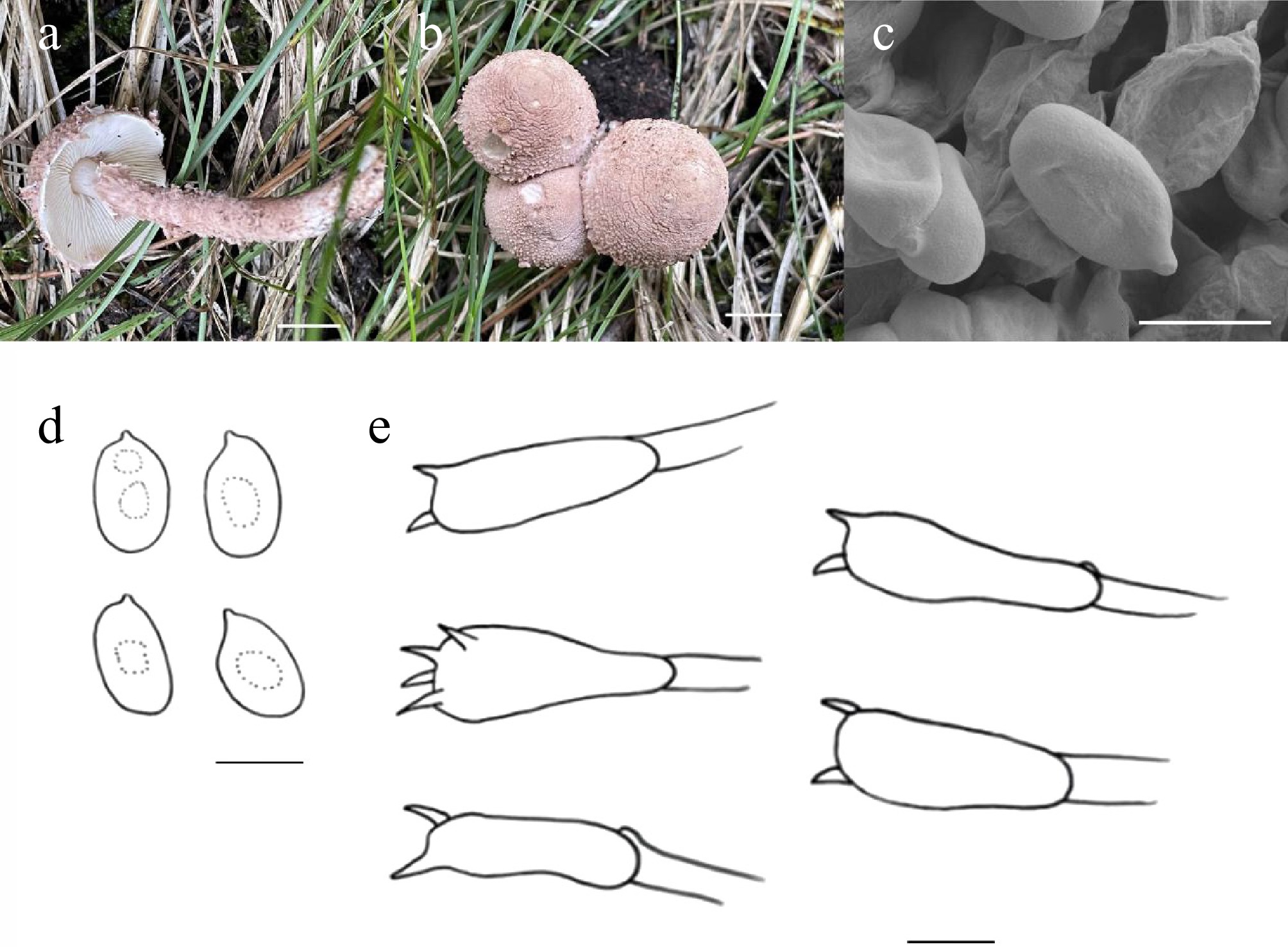
Figure 5.
Cystolepiota rubellogrisea (HMJU 7734, holotype). (a), (b) Basidiocarps. (c) SEM images of basidiospores. (d) Basidiospores. (e) Basidia. Scale bars: (a), (b) 1 cm; (c), (d) 3 µm; and (e) 5 µm.
MycoBank Names: MB863387
Holotype: Shanxi Province, Xinzhou City, Wuzhai County, Luya Mountain, on soil, 38°40'30" N, 111°52'45" E, 13 September 2022, L. Zhao (HMJU 7734, holotype).
Etymology: The specific epithet 'rubellogrisea' denotes that the entire basidiocarp of this fungus exhibits reddish-grey coloration.
Diagnosis: Cystolepiota rubellogrisea is characterized by a pileus that is dry and covered with dense scales; the stipe is white at the basal 1/10, with the remainder covered by reddish-grey fibrillose scales; cheilocystidia and pleurocystidia are absent, while clamp connections are present.
Description: Pileus 18–22 mm diam., convex when young, broadly convex at maturity, surface dry, covered with massive scales, margin gradually transitions to floccose rough flaky reddish-grey (10B2) scales, margin white (1A1) and exceeding the lamellae, pileus context thick, white (1A1). Lamellae free, crowded, white (1A1), margin entire. Stipe 32–48 mm long, 4–5 mm thick, cylindrical, 1/10 part white (1A1), remainder covered with reddish-grey (10B2) fibril scales, stipe context white (1A1). Annulus absent.
Basidiospores (3.9–)4.0–5.5(–5.7) × (1.8–)2.2–3.3(–3.6) μm, Q = 1.20–2.60, Qm = 1.77, oblong-ellipsoid, cylindric, transparent in KOH, inamyloid. Basidia (13–)14–22(–23) × (3–)4–5(–6) μm, clavate, mostly two sterigmata up to 2 μm long, transparent in KOH. Cheilocystidia absent. Pleurocystidia absent. Lamella trama regular, hyphae 2–9 μm wide, transparent in KOH. Clamp connections present in all hyphae.
Habitat: Scattered to gregarious on soil among grasses in meadows or open woodlands.
Known distribution: Known only from Northern China.
Additional material examined: Shanxi Province, Xinzhou City, Wuzhai County, Luya Mountain, on soil, 38°40'30" N, 111°52'45" E, 19 July 2023, L. Zhao (HMJU 14424).
Notes: Cystolepiota is the sister taxon to Cystolepiota rosea (Rea) Bon and Cystolepiota adulterina F.H. Møller ex Bon, with strong statistical support (MLBS/PP = 99/1.00). However, Cy. rosea is distinguished morphologically by its pink basidiomata and a stipe that lacks scales but bears conspicuous longitudinal striations[47]. Cy. adulterina is distinguished by its pileus covered with cream-ochre to greyish-yellow flocculose warts, larger spores (5.0–6.5 × 2.0–3.0 μm), and pleurocystidia that are parallel.
Morphologically, the new taxon most closely resembles Cystolepiota rhodella Sysouph. & Thongkl., Cystolepiota pulverulenta (Huijsman) Vellinga, Cystolepiota pseudofumosifolia M.L. Xu & R.L. Zhao, and Cystolepiota changbaishanensis T. Bau & X.Y. Zhou. Nevertheless, Cy. rhodella differs in having a brown-ruby pileus, an violet-brown lamellar edge, and cheilocystidia[48]. Moreover, Cy. pulverulenta is distinguished by its white to yellowish-brown pileus that turns red when touched and floccose pileipellis hyphae[49]. In addition, Cy. pseudofumosifolia is characterized by brown scales of the pileus and the presence of cheilocystidia[50]. The differences between Cy. changbaishanensis and Cy. rubellogrisea lies in the former having pale yellowish-grey verrucose scales on the surface, a pale reddish-brown stipe, and larger spores (4.6–6.0 × 2.1–3.0 μm)[51].
Echinoderma (Locq. ex Bon) Bon, Docums Mycol. 21(no. 82): 61 (1991)
Echinoderma (Locq. ex Bon) Bon[45] is classified among the lepiotaceous fungi, alongside allied genera such as Cystolepiota Singer, Lepiota (Pers.) Gray, Melanophyllum Velen., and Pulverolepiota Bon, due to their comparable macroscopic morphology. Phylogenetic analyses have revealed that Lepiota does not constitute a monophyletic genus on its own; however, a monophyletic clade is established when it is grouped together with Echinoderma, Cystolepiota, and Melanophyllum.
Morphologically, Echinoderma is distinguished by specific diagnostic attributes, including dextrinoid basidiospores, hyphae with clamp connections, and conical, sharply pointed squamules that detach easily and are composed of chains of subglobose to pyriform cells[52].
Bon[45] formally established Echinoderma as a genus, designating Ec. asperum as the type species. Taxa currently assigned to this genus were historically categorized under Lepiota section Echinatae Fayod. Members of this section share several morphological characteristics, such as free lamellae, cheilocystidia, a universal veil, and dextrinoid spores. The taxonomic status of this group remains a subject of debate: while some researchers continue to classify it as Lepiota section Echinatae[53], others accept it as the independent genus Echinoderma[54−57].
Phylogenetic reconstruction indicated that section Echinatae separates into two divergent lineages: a large-spored group, which contains species such as Lepiota aspera (Pers.) Quél., Lepiota hystrix F.H. Møller & J.E. Lange, and Lepiota perplexa Knudsen; and a small-spored group, comprising Lepiota jacobi Vellinga & Knudsen, Lepiota echinacea J.E. Lange, and two undescribed taxa.
Historically, Knudsen[44] initially treated Echinoderma as a subgenus of Cystolepiota, citing the prevalence of vesicular cells in the squamules as a primary distinguishing factor. However, he later reassigned these species to Lepiota. Furthermore, Johnson[58] utilized partial sequences from mitochondrial small subunit rDNA (mtSSU), internal transcribed spacer (ITS/5.8S), and ribosomal large subunit rDNA (nLSU) regions to reconstruct the phylogeny of lepiotaceous fungi.
Echinoderma petaloides J.Z. Xu, sp. nov., Figs 6 and 7
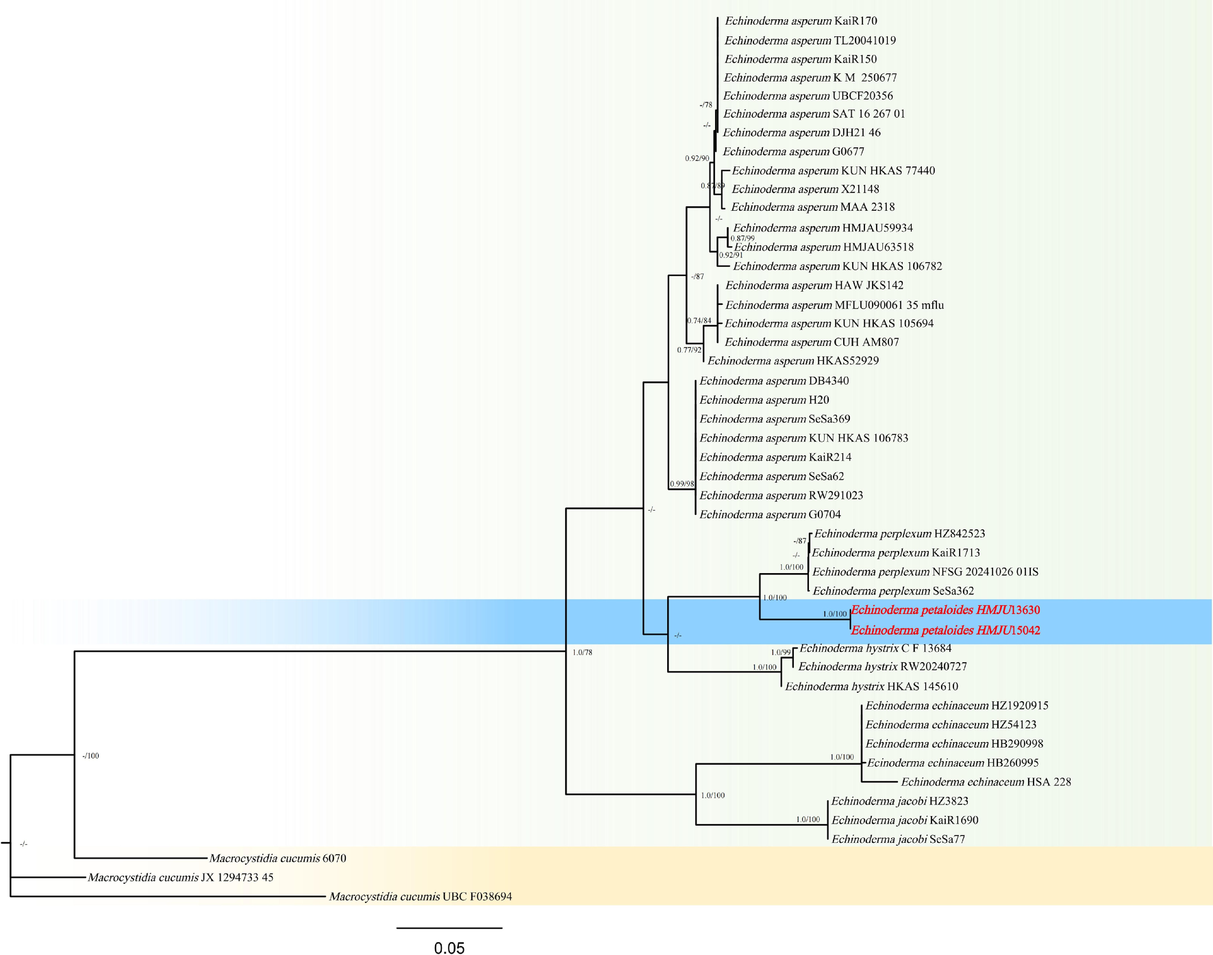
Figure 6.
Phylogram of Maximum Likelihood phylogenetic analysis based on combined ITS, nLSU, rpb2. The analysis includes 48 strains; total characters: 2,170 (ITS: 724, nLSU: 789, rpb2: 657). Macrocystidia cucumis (Pers.) Joss. (6070), and Ma. cucumis (JX 1294733 45), and Ma. cucumis (UBCF038694) were used as the outgroup taxa. The best model was TIM3e + G4. Estimated base frequencies were as follows: A = 0.250, C = 0.250, G = 0.250, T = 0.250. Bootstrap values for ML equal to or greater than 70% and BYPP values equal to or greater than 0.70 are labelled on the nodes. The type strains are in bold.
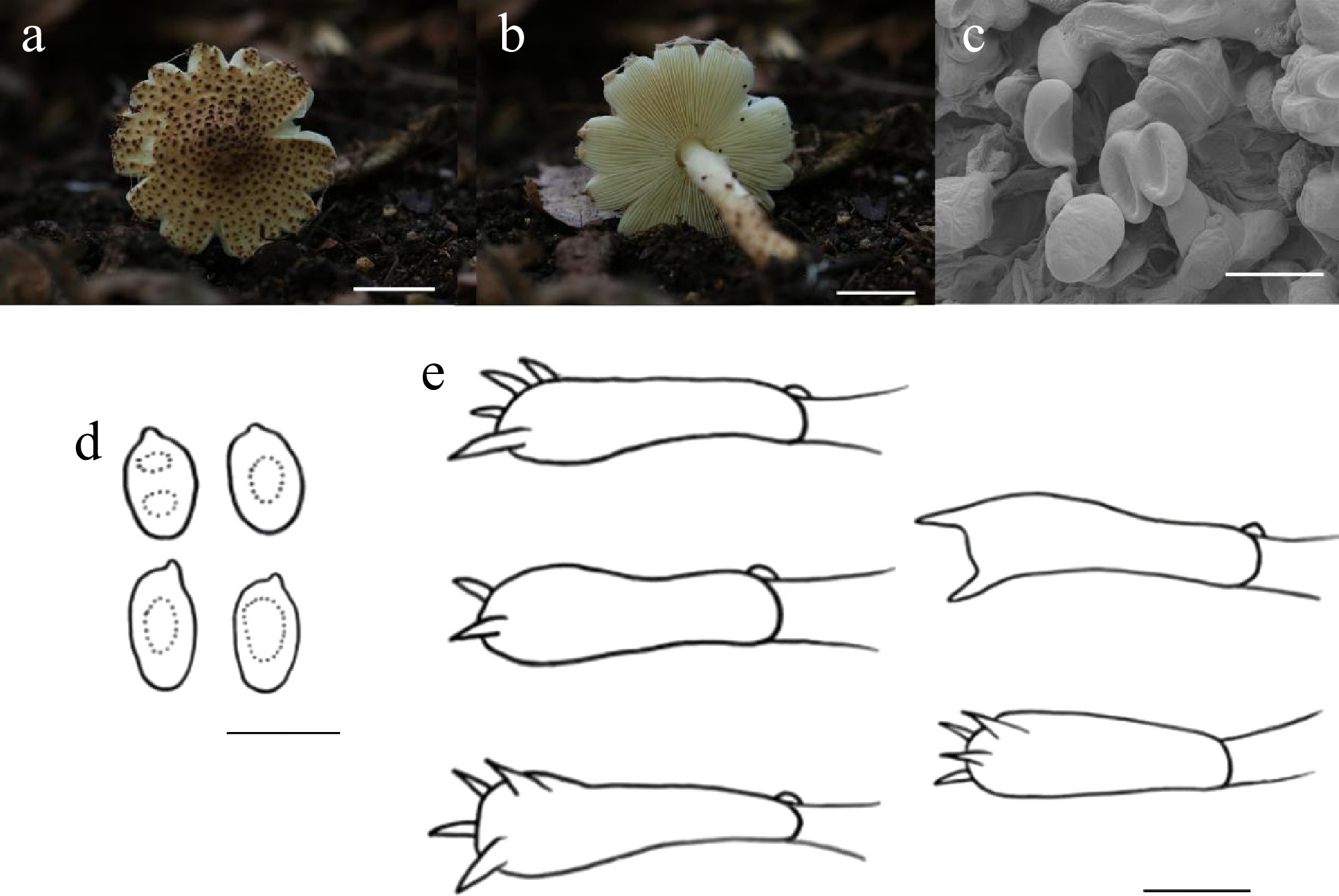
Figure 7.
Echinoderma petaloides (HMJU13630, holotype). (a), (b) Basidiocarps. (c) SEM images of basidiospores. (d) Basidiospores. (e) Basidia. Scale bars: (a), (b) 1 cm; (c)–(e) 5 µm.
MycoBank Names: MB863388
Holotype: Jilin Province, Meihekou City, Jiguan Mountain, on soil, 42°11′45″ N, 125°31′24″ E, 4 September 2025, J.Z. Xu (HMJU13630, holotype).
Etymology: The specific epithet 'petaloides' refers to the petal-like pileus margin of this species.
Diagnosis: Echinoderma petaloides is characterized by a petaloid-lobed pileus margin, with white fibrils between the lobes; the stipe is slightly curved, 1/3 of its surface is covered with white fibrils, and the remaining 2/3 is adorned with madder granular scales.
Description: Pileus 27–32 mm diam., plano-convex, pale orange (5A3) densely covered with madeia (8E5) granular scales, margin petaloid-lobed with white (1A1) fibrils between lobes, pileus context thin, white (1A1). Lamellae free, crowded, white (1A1), margin entire. Stipe 34–41 mm long, 3–6 mm thick, cylindrical, slightly curved, gradually thickened toward base, 1/3 part white (1A1), gradually darkening downward to pale orange (5A3), covered with white (1A1) fibrils, 2/3 part with madeia (8E5) granular scales, stipe context white (1A1). Annulus absent.
Basidiospores (4.0–)4.2–5.9(–6.0) × (1.6–)1.8–3.6(–3.8) μm, Q = 1.30–3.00, Qm = 1.93, ellipsoid to cylindric, transparent in KOH, dextrinoid. Basidia (10–)11–20(–21) × (3–) 4–6(–7) μm, both 4 and 2 sterigmata to 2 μm long, clavate, transparent in KOH. Cheilocystidia absent. Pleurocystidia absent. Lamella trama regular, hyphae 3–13 μm wide, transparent in KOH. Clamp connections present in all hyphae.
Habitat: Scattered on coniferous forest soil.
Known distribution: Known only from Northeastern China.
Additional material examined: Jilin Province, Meihekou City, Jiguan Mountain, on soil, 42°11'45" N, 125°31'24" E, 15 September 2025, J.Z. Xu (HMJU15042).
Notes: Phylogenetically, Ec. Petaloides forms a sister clade to Echinoderma perplexum (Knudsen) Bon (ML/PP = 100/1.00), but Ec. perplexum has significant morphological differences. Echinoderma petaloides is most similar morphologically to Echinoderma boertmannii (Knudsen) Bon, Echinoderma echinaceum (J.E. Lange) Bon, and Ec. perplexum. However, Ec. boertmannii has an intact pileus margin that is not petaloid, a pileus covered with reddish-brown scales, and larger basidia (25–40 × 8–11 µm)[45]. The pileus margin of Ec. echinaceum is non-petaloid, its pileus is dark brown, and the spores are yellow[45]. Ec. perplexum has a yellowish-brown pileus, a stipe with an annulus, larger spores (4.5–6.5 × 2.5–3 μm), and clavate marginal cystidia[45].
Entoloma (Fr.) P. Kumm., Führ. Pilzk. (Zerbst): 23 (1871)
Fries originally established Nolanea as a tribe under his broad concept of the genus Agaricus. Nomenclatural priority for this taxon dates back to 1829, when Loudon reclassified it as Agaricus subg. Nolanea. Later, Kummer[59] raised Nolanea to the level of an independent genus. Since then, the taxon has been assigned to two divergent taxonomic ranks: it is treated either as a distinct genus[60−65] or as a subgenus within Entoloma (syn. Rhodophyllus)[66−69].
Although originally defined by basidiocarps featuring a mycenoid habit, Nolanea has undergone significant re-evaluation and emendation in subsequent treatments. Largent & Benedict[70] emphasized the importance of a distinctive pileipellis structure—usually composed of repent hyphae and frequently characterized by a subpellis. Furthermore, it was reported that species within Nolanea and Claudopus possess high urea content, distinguishing them from taxa in Alboleptonia, Entoloma, and Leptonia; however, this biochemical trait was not adopted in later taxonomic systems.
Gilles & Romagnesi[66] added the hygrophanous quality of the pileus as a diagnostic feature for subg. Nolanea. Acting on a suggestion, Noordeloos further distinguished Nolanea from other subgenera by utilizing the morphology of tramal elements, specifically noting the presence of long, fusiform cells measuring 150–450 µm (or more) in length.
Entoloma liangshuiense J.Z. Xu, sp. nov., Figs 8 and 9
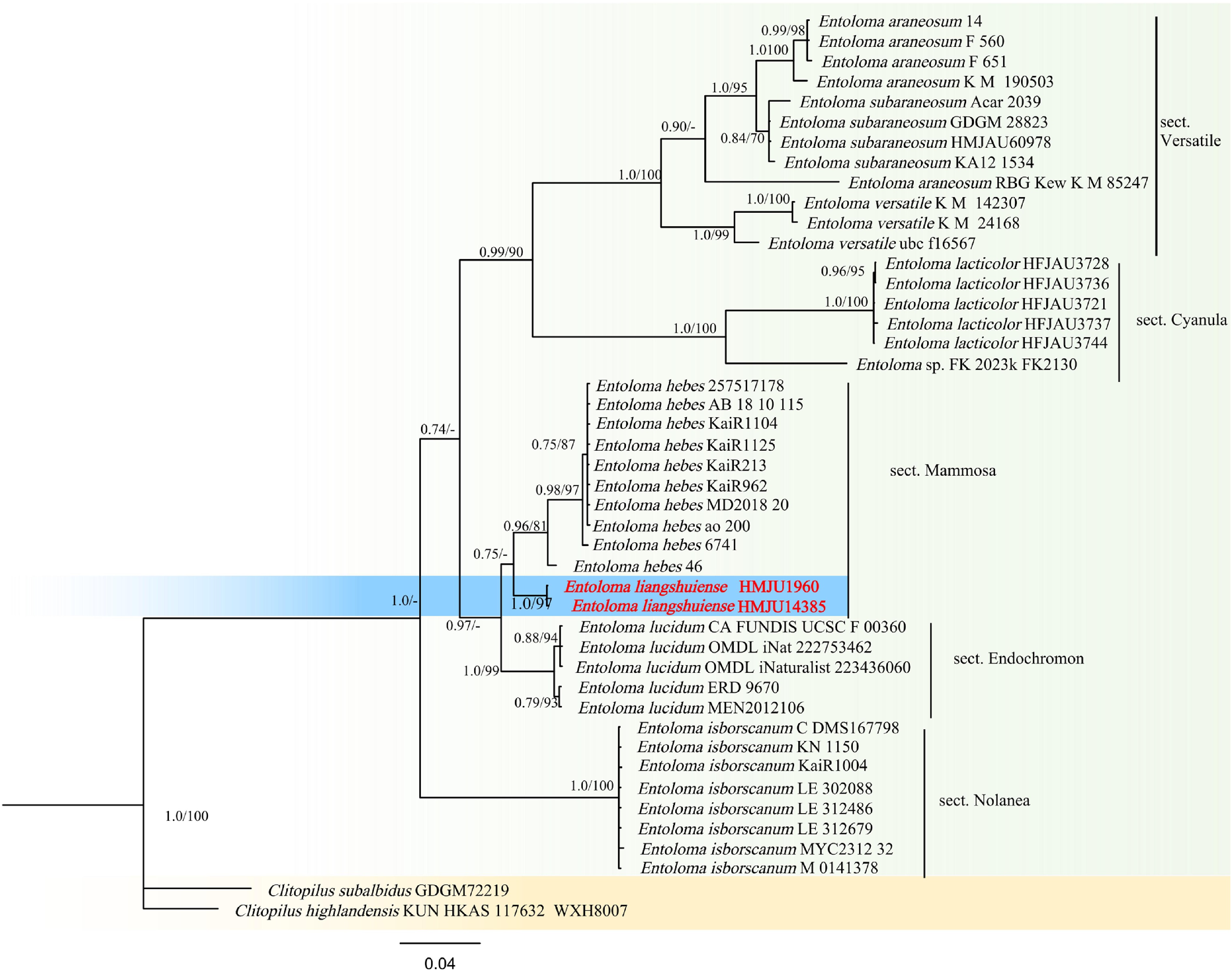
Figure 8.
Phylogram of Maximum Likelihood phylogenetic analysis based on combined ITS, nLSU, rpb2. The analysis includes 46 strains; total characters: 1,741 (ITS: 627, nLSU: 570, rpb2: 544). Clitopilus highlandensis (KUN HKAS 117632 WXH8007) and Clitopilus subalbidus (GDGM72219) were used as the outgroup taxa. The best model was TIM2 + G4 + F. Estimated base frequencies were as follows: A = 0.274, C = 0.193, G = 0.221, T = 0.312. Bootstrap values for ML equal to or greater than 70% and BYPP values greater than 0.70 are labelled on the nodes. Type strains are highlighted in bold
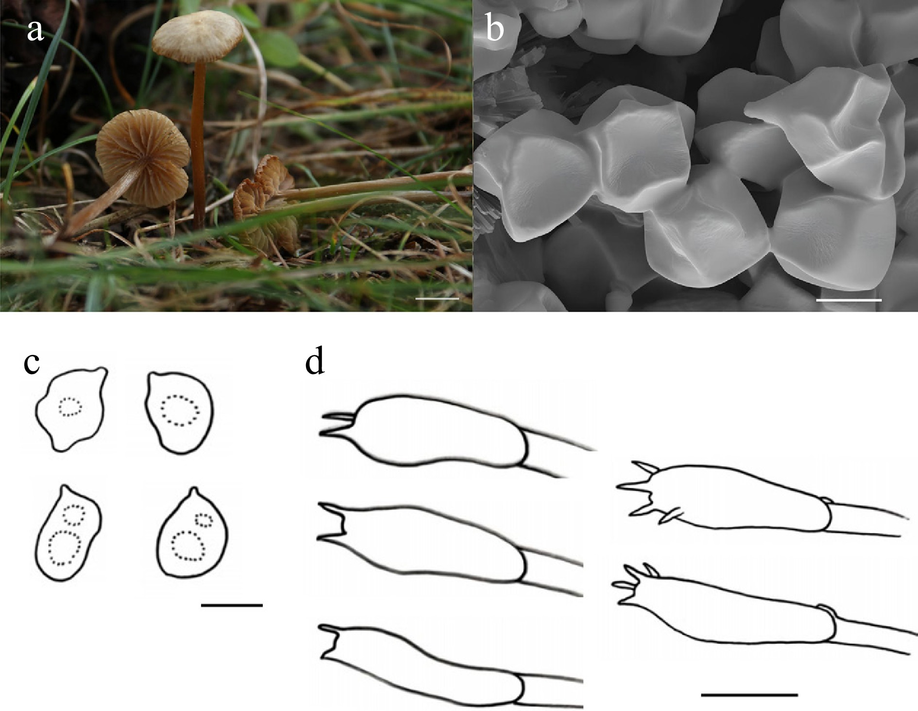
Figure 9.
Entoloma liangshuiense (HMJU 1960, holotype). (a) Basidiocarps. (b) SEM images of basidiospores. (c) Basidiospores. (d) Basidia. Scale bars: (a) 1cm; (b)–(d) 5 µm.
MycoBank Names: MB863390
Holotype: Heilongjiang Province, Yichun City, Dailing District, Liangshui National Nature Reserve, on soil, 47°10'50" N, 128°53'20" E, 10 September 2017, J.Z. Xu (HMJU 1960, holotype).
Etymology: The specific epithet 'liangshuiense' refers to the type locality, Liangshui Nature Reserve.
Diagnosis: Entoloma liangshuiense is characterized by a pileus with a champagne background, covered with henna vertical streaks and dense greyish-white filaments; the stipe is twisted and has a clamp connection present in all hyphae.
Description: Pileus 19–22 mm diam., applanate-convex, center slightly depressed, covered with pale henna (7E8) vertical streaks and dense greyish white (1B1) filaments in the champagne (4B4) ground, becoming sparse toward the pileus margin, pileus context thin, pale champagne (4B4). Lamellae adnexed, distant, champagne (4B4), margins darken to reddish golden (6C7) with age, pileus margin extends beyond the lamellae, with white (1A1) fibrillose material between the lamellae, margin entire. Stipe 48–78 mm long, 1–2 mm thick, cylindrical, twisted, cinnamon (6D6) covered with white (1A1) filaments, stipe context pale champagne (4B4).
Basidiospores (6.5–)6.8–8.8(–8.9) × (4.6–)4.9–6.8(–6.9) μm, Q = 1.08–1.56, Qm = 1.29, dimorphism with 4–7 angles, smooth, transparent in KOH, without amyloid. Basidia (19–)20–34(–35) × (6–)7–11(–12) μm, clavate, mostly 2 sterigmata up to 3 μm long, transparent in KOH. Cheilocystidia absent. Pleurocystidia absent. Lamella trama regular, parallel to subparallel, hyphae 5–10 μm wide; transparent in KOH. Clamp connections present in all hyphae.
Habitat: Scattered to gregarious on soil among grasses in meadows.
Known distribution: Known only from Northeastern China.
Additional material examined: Heilongjiang Province, Shuangyashan City, Baoqing County, Liangshui Nature Reserve, on soil, 47°10'50" N, 128°53'20" E, 5 August 2021, J.Z. Xu (HMJU 14385).
Notes: It forms a sister group with Entoloma hebes (Romagn.) Trimbach (ML/PP = 67/0.75); however, it has larger basidiomata (8.5–11.5 × 5.5–7.5 µm), a brown pileus, and pale orange-red lamellae[71]. Entoloma liangshuiense is morphologically most similar to Entoloma ameides (Berk. & Broome) Quél., Entoloma araneosum (Quél.) M.M. Moser, and Entoloma haastii G. Stev. However, En. ameides is characterized by a brownish pileus with a cracked margin when mature, grayish-beige lamellae, and larger spores (8.5–11 × 6.5–8.5 µm)[72,73]. En. araneosum has a silvery-gray pileus, gray lamellae that turn pink, larger spores (10–14 × 7–8.5 µm), clavate cystidia with long necks, and absent clamp connections[71]. En. haastii features a dark blue-black pileus with fine cracks and a reflexed margin, lamellae that are dark blue-gray initially and turn pale orange when mature[74].
Hygrocybe (Fr.) P. Kumm., Führ. Pilzk. (Zerbst): 26 (1871)
Hygrocybe conica (Schaeff.) P. Kumm. serves as the type species for the genus Hygrocybe and is classified within subgenus Hygrocybe (Fr.), section Hygrocybe, and subsection Hygrocybe (Fr.) P. Kumm. The members of subsection Hygrocybe, frequently referred to as the H. conica complex, are ubiquitous, visually distinctive, and widely encountered. Morphologically, this group is defined primarily by basidiomata that blacken with age or handling, a conical pileus that is usually fibrillose, and lamellae that range from free to narrowly adnate. Microscopic diagnostics include parallel hymenophoral trama hyphae (typically exceeding 200 μm in length) and a basidium to basidiospore length ratio generally below 5. Phylogenetic studies utilizing ITS and nLSU markers have confirmed that subsection Hygrocybe represents a monophyletic lineage[75,76]. Ecologically, these taxa act as significant environmental bioindicators, as they predominantly thrive in unpolluted grassland or woodland habitats[77,78].
Currently, 25 taxa worldwide conform to the morphological standards of subsection Hygrocybe. Europe is the type locality for 10 of these: Hygrocybe cinereifolia Courtec. & Priou and Hygrocybe nigrescens (Quél.) Kühner from France; H. conica from Germany; Hygrocybe conica var. aurantiolutea T. Borgen & Arnolds from Greenland; Hygrocybe conica var. conicopalustris (Bon) Arnolds and Hygrocybe conica var. minor Monthoux & Röllin from Switzerland; Hygrocybe conicoides (P.D. Orton) P.D. Orton & Watling and Hygrocybe olivaceonigra (P.D. Orton) M.M. Moser from the UK (England); Hygrocybe pseudoconica J.E. Lange from Denmark; and Hygrocybe veselskyi Singer & Kuthan from the Czech Republic[59,79−81].
Five taxa were originally identified in Africa: Hygrocybe astatogala (R. Heim) Heinem. from Madagascar; Hygrocybe astatogala var. laeticolor Heinem., Hygrocybe conica var. pallidipes Heinem., and Hygrocybe cortinata Heinem. from Zaire; and Hygrocybe chloroides (Malançon) Kovalenko from Hygrocybe conicoides[82]. Similarly, five taxa were described from North America: Hygrocybe albifolia (Hesler & A.H. Sm.) R. Valenz, Guzmán & J. Castillo, Hygrocybe foliirubens Murrill, Hygrocybe singeri (A.H. Sm. & Hesler) Singer, and Hygrocybe cuspidata (Peck) Murrill from the USA; and Hygrocybe conica var. atrosanguinea (Grund & K.A. Harrison) Malloch from Canada (Nova Scotia).
Asia is the origin of two taxa: Hygrocybe conica var. peradenyca (Sacc.) Pegler from Sri Lanka and Hygrocybe erinacea (Pat.) Singer from Vietnam. In the Caribbean region, two taxa have been recorded: Hygrocybe atrosquamosa Pegler from Martinique and Hygrocybe conica var. brevispora (Dennis) S.A. Cantrell & Lodge from Venezuela[83]. Finally, Hygrocybe conica var. tierneyi A.M. Young was originally described from Australia.
Hygrocybe santagouana J.Z. Xu, sp. nov., Figs 10 and 11
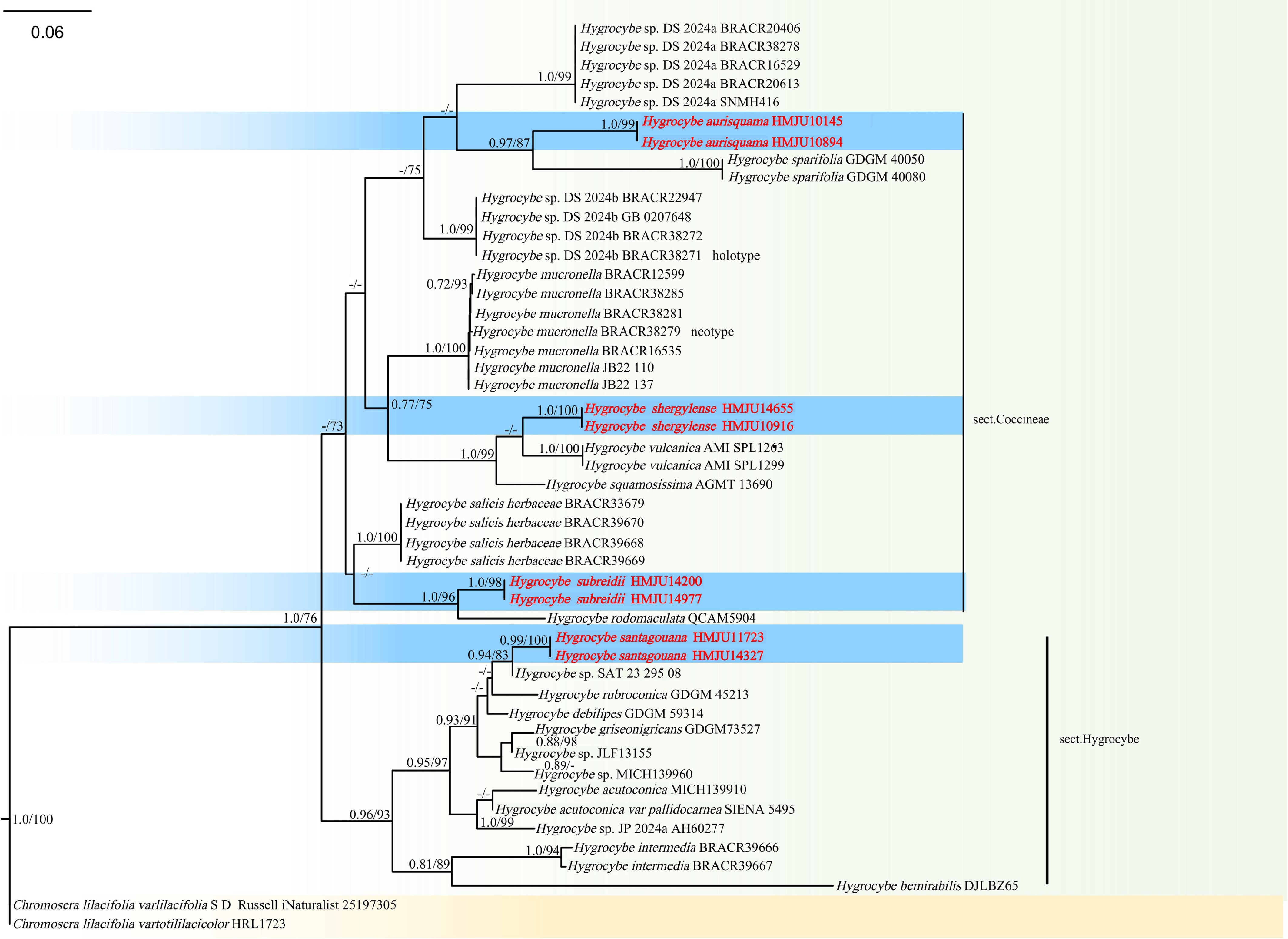
Figure 10.
Phylogram of Maximum Likelihood phylogenetic analysis based on combined ITS, nLSU, rpb2, and tef-1α sequences. The analysis includes 48 strains; total characters: 3,501 (ITS: 278, nLSU: 789, rpb2:1891, tef-1α: 543). Chromosera lilacifolia (varlilacifolia S D Russell iNaturalist 25197305) and Chromosera lilacifolia (vartotililacicolor HRL1723) were used as the outgroup taxa. The the best model being TIM2e + I + G4. Estimated base frequencies were as follows: A = 0.259, C = 0.221, G = 0.267, T = 0.253. Bootstrap values for ML equal to or greater than 70% and BYPP values equal to or greater than 0.70 are labelled on the nodes.The type strains are in bold.
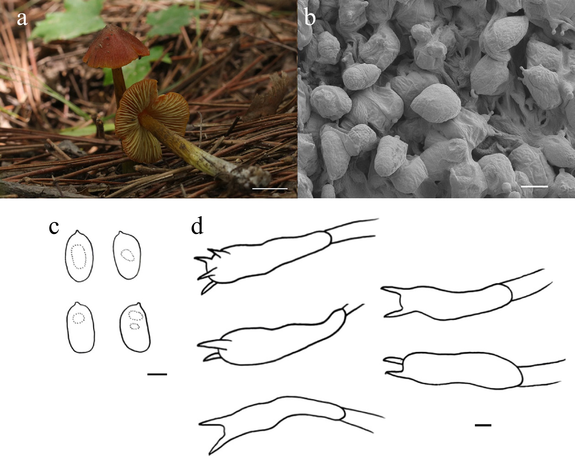
Figure 11.
Hygrocybe santagouana (HMJU 11723, holotype). (a) Basidiocarps. (b) SEM images of basidiospores. (c) Basidiospores. (d) Basidia. Scale bars: (a) 1 cm; (b)–(d) 5 µm.
MycoBank Names: MB863391
Holotype: Liaoning Province, Fuxin City, Fuxin Mongolian Autonomous County, Santagou Natural Scenic Area, on soil, 41°53'46" N, 121°48'06" E, 7 August 2024, J. Z. Xu (HMJU 11723, holotype)
Etymology: The specific epithet 'santagouana' refers to the type locality, Santagou Natural Scenic Area.
Diagnosis: Hygrocybe santagouana is characterized by a pileus conico-convex in shape; stipe turns black upon injury; basidiospores are ellipsoidal to citriform, and the sterigmata reach a maximum length of 4 μm.
Description: Pileus 19–26 mm diam., conico-convex, convex, henna (7E8), margin reddish orange (7A8) when young, slightly darker at centre, covered with henna (7E8) striations, pileus context henna (7E8). Lamellae adnexed, pale orange white (5A2), margins entire, medium to crowded. Stipe 45–48 mm long, 3–5 mm thick, cylindrical, gradually thickened toward base, 2/5 part red-haired (6C4), gradually transitioning downward to dyer broom (3A7), base white (1A1), stipe context pale red-haired (6C4), turns black when injured.
Basidiospores (7.5–)8.3–14.8(–15.3) × (4.6–)4.7–7.1(–7.2) μm, Q = 1.27–1.33, Qm = 1.92, broadly ellipsoid to ellipsoid, citriform to cylindrical, transparent in KOH, without amyloid. Basidia (17–)18–35(–38) × (4–)5–9(–10) μm, clavate, most two sterigmata to 4 μm, transparent in KOH. Cheilocystidia absent. Pleurocystidia absent. Lamella trama irregular, hyphae 3–18 μm wide, transparent in KOH. Clamp connections absent in all hyphae.
Habitat: Scattered on soil among pine needles in coniferous forests.
Known distribution: Known only from Northeastern China.
Additional material examined: Liaoning Province, Fuxin City, Fuxin Mongolian Autonomous County, Santagou Natural Scenic Area, on soil, 41°53'46" N, 121°48'06" E, 2 September 2025, J. Z. Xu (HMJU 14327).
Notes: Since relevant information cannot be determined for this sequence of Hygrocybe sp. SAT 2329508, and it is not possible to ascertain whether it has been published as a new species, it was therefore excluded as a taxon from the phylogenetic analysis conducted with the published species. Species morphologically similar to Hygrocybe calyptraeformis (Berk.) Fayod, Hygrocybe flavescens (Kauffman) Singer, and Hygrocybe acutoconica (Clem.) Singer, Hygrocybe conica (Schaeff.) P. Kumm. H. calyptraeformis is characterized by a pileus that is purple with an undulate margin, a stipe that is pale white tinged with pink, and clamp connections present[84]. H. flavescens features a pileus that is yellow with an entire margin, lamellae initially white, later turning yellow, and a stipe that is bright yellow[41]. H. acutoconica is different in pileus, yellow with papillate protrusion at the center, stipe pale yellow to orange, basidia larger (to about 55 μm long)[41]. It differs from H. conica in that the pileus turns black when mature, the stipe is yellow to orange-yellow, and the spores are pale brown in KOH[59].
Hygrocybe aurisquama J.Z. Xu, sp. nov., Figs 10 and 12
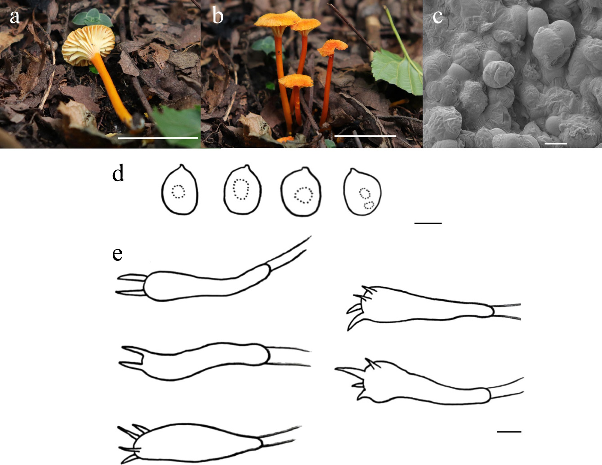
Figure 12.
Hygrocybe aurisquama (HMJU 10145, holotype). (a), (b) Basidiocarps. (c) SEM images of basidiospores. (d) Basidiospores. (e) Basidia. Scale bars: (a), (b) 2 cm; (c), (d) 5 µm; and (e) 10 µm.
MycoBank Names: MB863392
Holotype: Liaoning Province, Fushun City, Qingyuan Manchu Autonomous County, on soil, 42°06'13" N, 124°56'12" E, 20 August 2023, J. Z. Xu (HMJU 10145, holotype).
Etymology: The specific epithet 'aurisquama' refers to the pileus, which is densely covered with golden-yellow minute squamules.
Diagnosis: Hygrocybe aurisquama is characterized by pileus ornamented with minute golden-yellow granular scales, its margin presenting an undulate profile with a slight rimose; neither cheilocystidia nor pleurocystidia were observed, and clamp connections were also absent.
Description: Pileus 10–16 mm diam, centrally depressed, golden yellow (5B7), covered with golden yellow (5B7) tiny granular scales, margin wavy, slightly rimose, pileus context thick, golden yellow (5B7). Lamellae decurrent, slightly distant, medially disrupted; white (1A1); margins entire. Stipe 21–42 mm long, 4–7 mm thick, cylindrical, reddish orange (7A8), stipe context reddish orange (7A8).
Basidiospores (7.1–)7.3–10.4(–12.2) × (5.1–)5.3–8.5(–9.7) μm, Q = 1.00–1.80, Qm = 1.31, globose to rectangular, transparent in KOH, without amyloid. Basidia (29–)30–50(–51) × (5–)6–12(–13) μm, clavate, both four and two sterigmata up to 3 μm long, transparent in KOH. Cheilocystidia absent. Pleurocystidia absent. Lamella trama irregular, hyphae 3–11 μm wide, transparent in KOH. Clamp connections absent in all hyphae.
Habitat: Scattered to gregarious on soil covered with leaf litter in temperate forests.
Known distribution: Known only from Northeastern China.
Additional material examined: Liaoning Province, Fushun City, Qingyuan Manchu Autonomous County, on soil, 42°06'13" N, 124°56'12" E, 24 August 2024, J.Z. Xu (HMJU 10894).
Notes: Phylogenetically, the new species forms a well-supported sister clade to Hygrocybe sparifolia T.H. Li & C.Q. Wang (MLBS/PP = 87/0.97). However, it is characterized by a pileus with dark-brown fibrils, lamellae turning pink when injured, some basidia containing brown pigments, and brown pileipellis hyphae[85]. Moreover, the species shows morphological similarity with Hygrocybe cantharellus (Schwein.) Murrill, Hygrocybe reidii Kühner, and Hygrocybe turunda (Fr.) P. Karst. H. cantharellus is characterized by pale-yellow lamellae, a pale-yellow stipe base, and significantly larger basidiospores measuring 9–12 × 5–6 μm[86]. In contrast, H. reidii is different in pileus broadly convex or plano-convex, lamellae pale yellow, stipe base white[87]. H. turunda is characterized by yellow lamellae, an orange stipe that fades towards the base, and ovoid or pyriform spores[88].
Hygrocybe sejilensis J.Z. Xu, sp. nov., Figs 10 and 13
MycoBank Names: MB863393
Holotype: Xizang Autonomous Region, Linzhi City, Sejila Mountain, on soil, 29°36'41" N, 94°39'11" E, 1 August 2025, J.Z. Xu (HMJU 14655, holotype).
Etymology: The specific epithet 'sejilensis' refers to the type locality, Sejila Mountain.
Diagnosis: Hygrocybe sejilensis is characterized by lamellae pale orange-white in color; neither cheilocystidia nor pleurocystidia were observed; clamp connections were found to be present in all hyphae.
Description: Pileus 4–5 mm diam., hemispherical, reddish orange (7A8), smooth or covered with salmon (6B4) tomentose, pileus context thin, pale reddish orange (7A8). Lamellae free, distant, pale orange white (5A2), margins entire. Stipe 22–25 mm long, 1–3 mm thick, cylindrical, gradually thickened downward, base slightly swollen, gradually transitioning from reddish orange (7A8) to buttercup yellow (4A7).
Basidiospores (4.9–)6.5–11.0(–11.4) × (3.9–)4.3–7.8(–7.9), Q = 1.07–2.10, Qm = 1.55, subovoid to cylindrical, transparent in KOH, without amyloid. Basidia (19–)24–41(–42) × (3–)4–6(–7), clavate, most 2 sterigmata up to 4 μm, transparent in KOH. Cheilocystidia absent. Pleurocystidia absent. Lamella trama irregular, 3–8 μm wide, transparent in KOH. Clamp connections present in all hyphae.
Habitat: Scattered on soil.
Known distribution: Known only from Southwestern China.
Additional material examined: Xizang Autonomous Region, Linzhi City, Sejila mountain, on soil, 29°36'41" N, 94°39'11" E, 25 July 2022, J.Z. Xu (HMJU 10916).
Notes: Phylogenetically, the species forms a sister clade to H. sparifolia with moderate statistical support (MLBS/PP = 52/0.70). However, it is characterized by a pileus with dark-brown fibrils, lamellae turning pink when injured, some basidia containing brown pigments, and brown pileipellis hyphae. Moreover, the species shows morphological similarity to Hygrocybe miniata (Fr.) P. Kumm., Hygrocybe marchii (Bres.) F.H. Møller, and H. reidii. H. miniata differs in having a pileus densely covered with radial squamules, decurrent lamellae, a stipe tapering towards the base, and smaller basidiospores (6.5–9 × 4–5μm)[59]. In contrast, H. marchii is characterized by a stipe that is orange-yellow, lamellae that are orange, and spores that are smaller (7.5–8.6 × 4.3–6.1 μm)[89]. H. reidii is typified by a pileus with a crenate margin when young, and an orange stipe[89].
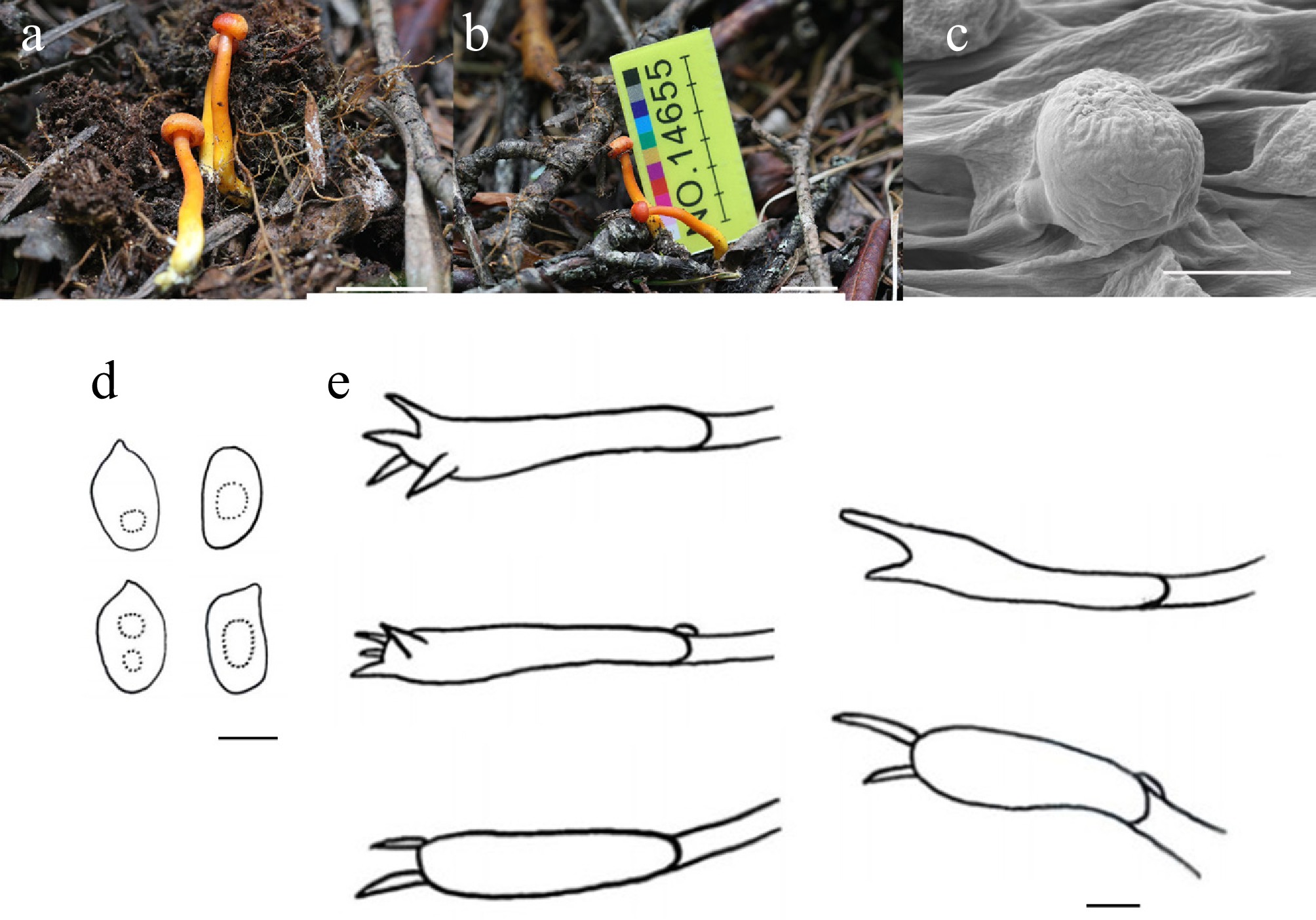
Figure 13.
Hygrocybe sejilensis (HMJU 14655, holotype). (a), (b) Basidiocarps. (c) SEM images of basidiospores. (d) Basidiospores. (e) Basidia. Scale bars: (a), (b) 1 cm; (c)–(e) 5 µm.
Hygrocybe subreidii J.Z. Xu, sp. nov., Figs 10 and 14
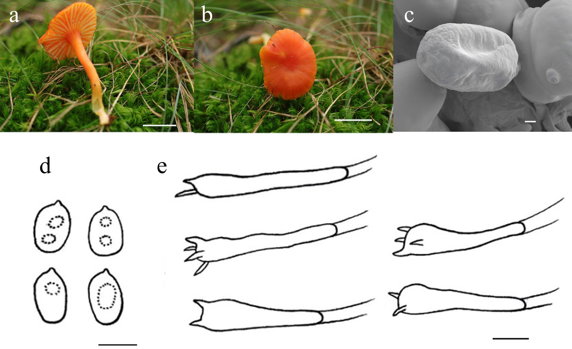
Figure 14.
Hygrocybe subreidii (HMJU 14200, holotype). (a), (b) Basidiocarps. (c) SEM images of basidiospores. (d) Basidiospores. (e) Basidia. Scale bars: (a), (b) 1 cm; (c)–(e) 5 µm.
MycoBank Names: MB863394
Holotype: Jilin Province, Baishan City, Fusong County, Songjiang River Baixi, on soil, 42°13'22" N, 127°42'18" E, 8 September 2025, Y. Qin (HMJU 14200, holotype).
Etymology: The specific epithet 'subreidii' indicates that this species is morphologically nearly identical to Hygrocybe reidii Kühner.
Diagnosis: Hygrocybe subreidii is characterized by lamellae decurrent, centrally partially split; stipe slightly flexuous; basidia predominantly 2–3-spored; sterigmata up to 2.81 μm.
Description: Pileus 13–27 mm diam., applanate, center slightly depressed, reddish orange (7A8), surface smooth, pileus context thin, hollow, reddish orange (7A8). Lamellae decurrent, distant, center partial split, light orange white (5A2), margins entire. Stipe 34–47 mm long, 3–5 mm thick, cylindrical, slightly flexuous, hollow, reddish orange (7A8), stipe context reddish orange (7A8).
Basidiospores (4.6–)4.9–7.7(–7.8) × (3.1–)3.2–5.3(–5.4) μm, Q = 1.30–2.20, Qm = 1.63, broadly ellipsoid to cylindrical, transparent in KOH, without amyloid. Basidia (24–)26–43(–48) × (3–)4–7(–8) μm, clavate, both four and two sterigmata up to 2 μm, transparent in KOH. Cheilocystidia absent. Pleurocystidia absent. Lamella trama irregular, hyphae 3–13 μm wide, transparent in KOH. Clamp connections absent in all hyphae.
Habitat: Scattered on moss-covered soil in grassy habitats.
Known distribution: Known only from Northeastern China.
Additional material examined: Jilin Province, Baishan City, Fusong County, Songjiang River Baixi, on soil, 42°13'22" N, 127°42'18" E, 15 August 2023, Y. Qin (HMJU 14977).
Notes: Phylogenetically, the species forms a well-supported sister clade to Hygrocybe rodomaculata A. Barili, C.W. Barnes & Ordoñez (MLBS/PP = 96/1.00). However, it is characterized by a white pileus, a white stipe, longer basidia (41–70 × 4–9 µm), and clamp connections[90]. Moreover, the newly described taxon shows morphological similarity with H. cuspidata, H. reidii, and H. acutoconica. H. cuspidata is different in that the pileus margin is faintly striate, the lamellae are orange, and the spores are constricted[91]. H. reidii is characterized by a pileus margin crenate, lamellae orange[87]. H. acutoconica features a pileus with papillate protrusions at the center and translucent striations on the margin, with yellow lamellae[92].
Hypholoma (Fr.) P. Kumm., Führ. Pilzk. (Zerbst): 21 (1871)
Taxonomically, the genus Hypholoma (Fr.) P. Kumm. is positioned within the subfamily Stropharioideae Singer, belonging to the family Strophariaceae Singer & A.H. Sm., order Agaricales, and phylum basidiomycota. The genus is defined by several diagnostic features: a pileus exhibiting intense pigmentation; a veil that, despite varying degrees of development, never forms a true membranous annulus on the stipe, and a spore print ranging from violaceous to purplish. Microscopic characteristics include basidiospores with prominent germ pores and thickened walls, as well as the presence of chrysocystidia[68].
Hypholoma is differentiated from its closest relative, Stropharia (Fr.) Quél., by the absence of acanthocytes in its rhizomorphs[93−95]. It is distinguished from another familial relative, Psilocybe (Fr.) P. Kumm., by the presence of chrysocystidia, which Psilocybe lacks[96−98]. According to Hawksworth, Hypholoma comprises roughly 30 species distributed globally across both tropical and temperate zones. These fungi typically colonize soil, mosses, living trees, or decaying wood[68].
The nomenclature of Hypholoma has been a subject of long-standing debate among taxonomists[99,100]. Historically, numerous species currently recognized as Hypholoma were classified under the genus Nematoloma P. Karst.[68], a name that was eventually rejected based on nomenclatural rules. Influenced by the work of Smith & Kühner, Noordeloos proposed reclassifying Hypholoma species as subgenera within a broad concept of Psilocybe (s.l.). However, this proposal has not achieved general acceptance in the mycological community[94,95] and is unsupported by recent molecular phylogenetic data[101]. Moncalvo recognizes Hypholoma as a distinct clade that includes most of the genus species, with the exception of Hypholoma subericaeum (Fr.) Kühner and Hypholoma aurantiacum (Cooke) Faus, whose phylogenetic placement within the 'stropharioid' clade remains unresolved.
Hypholoma salmopapillosum J.Z. Xu, sp. nov., Figs 15 and 16
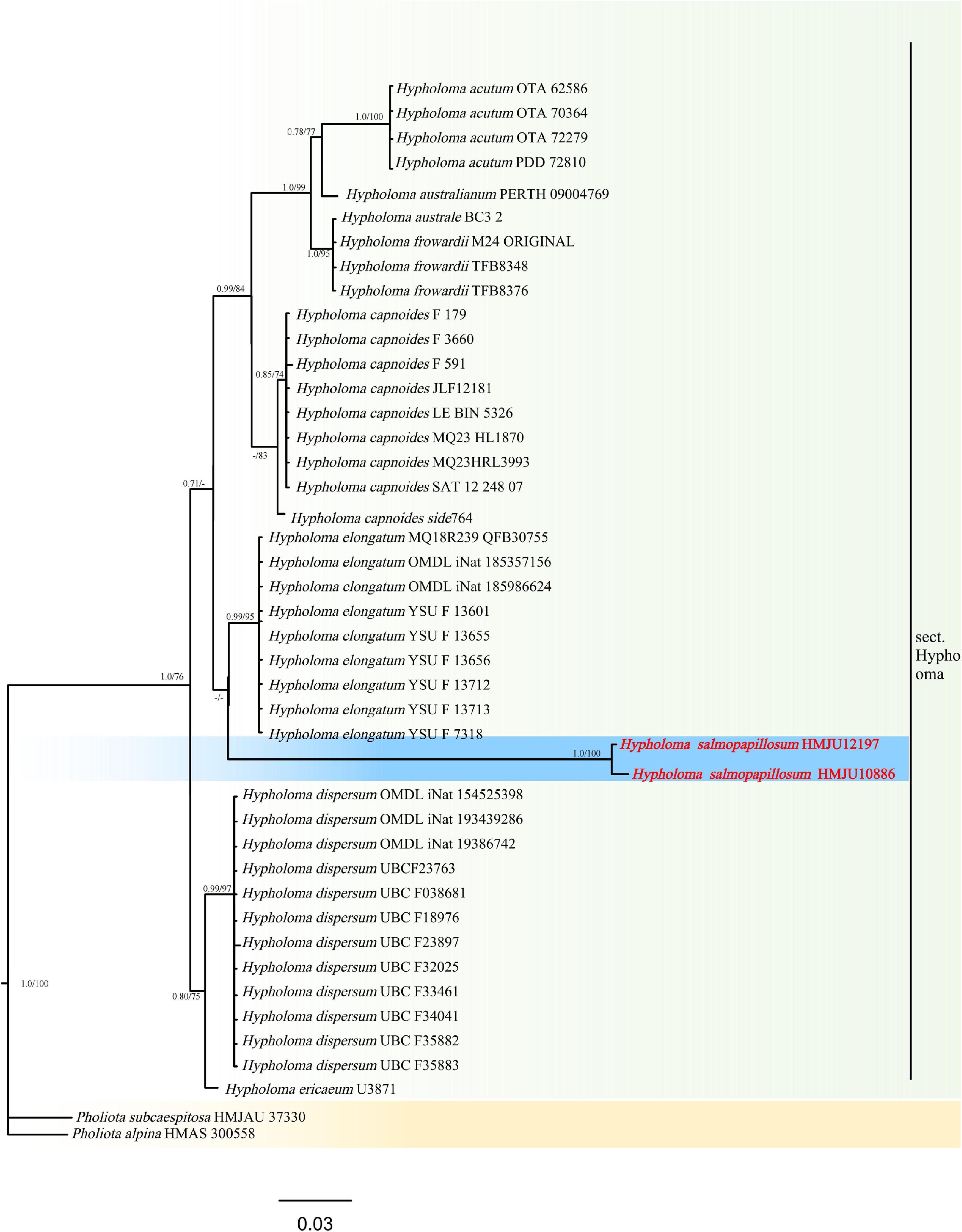
Figure 15.
Phylogram of Maximum Likelihood phylogenetic analysis based on combined ITS, nLSU, rpb2, and tef-1 sequences. The analysis includes 44 strains; total characters: 2,597 (ITS: 535, nLSU: 714, rpb2: 776, tef-1: 572). Pholiota subcaespitosa (HMJAU 37330) and Pholiota alpina (HMAS 300558) were used as the outgroup taxa. The best model was TPM2 + G4. Estimated base frequencies were as follows: A = 0.250, C = 0.250, G = 0.250, T = 0.250. Bootstrap values for ML equal to or greater than 70% and BYPP values equal to or greater than 0.70 are labelled on the nodes. The type strains are in bold.
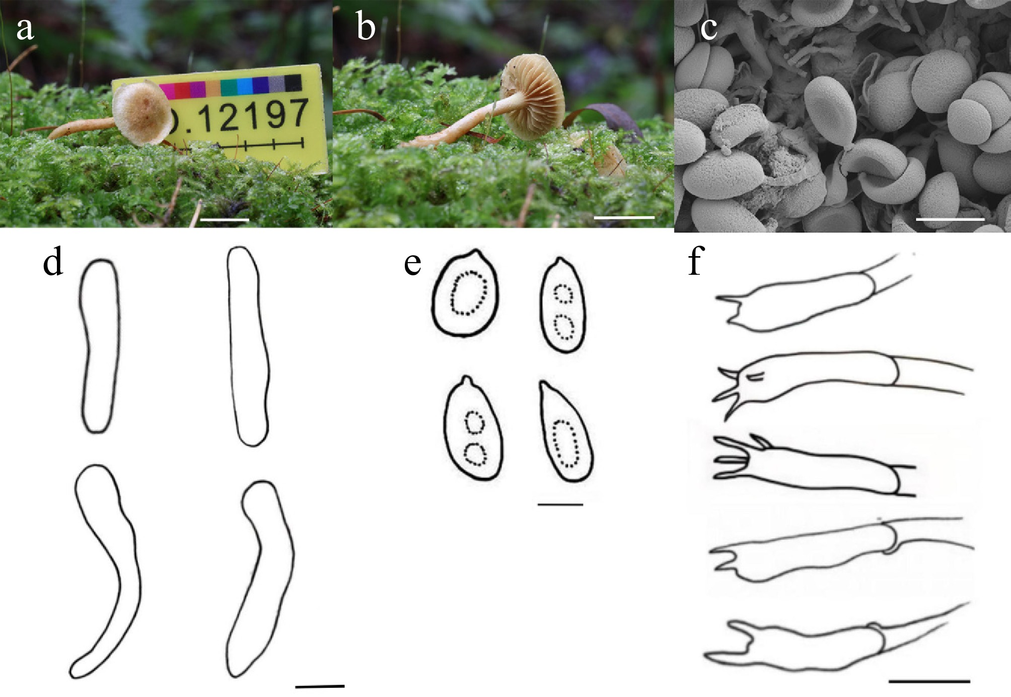
Figure 16.
Hypholoma salmopapillosum (HMJU 12197, holotype). (a), (b) Basidiocarps. (c) SEM images of basidiospores. (d) Basidiospores. (e) Basidia. Scale bars: (a), (b) 1 cm; (c) 5 µm; (d) 10 µm; (e) 3 µm; and (f) 10 µm.
MycoBank Names: MB863395
Holotype: Jilin Province, Baishan City, Changbai Mountain Primitive Forest, on soil, 42°12'58" N, 128°17'30" E, 11 August 2024, J.Z. Xu (HMJU 12197 holotype).
Etymology: The specific epithet 'salmopapillosum' indicates that the pileus is covered with salmon-colored (6A4) granular papillae.
Diagnosis: Hypholoma salmopapillosum is characterized by stipe beset with salmon verrucae; basidiospores brownish orange in KOH; cheilocystidia urticiform, hyaline or Chinese yellow in KOH; clamp connections present.
Description: Pileus 13–18 mm diam., applanate–convex, margin ascending, center to 3/4 of the pileus salmon (6A4), remainder cream (4A3), covered with salmon (6A4) granular papillae, pileus context thin, salmon (6A4). Lamellae free, distant, cream (4A3), margin entire. Stipe 19–23 mm long, 1–2 mm thick, cylindrical, gradually thickened toward base, flexuous, covered with salmon (6A4) verrucae, stipe context salmon (6A4). Annulus absent.
Basidiospores (4.6–)4.7–8.0(–8.1) × (2.2–)2.8–4.4(–4.5) μm, Q = 1.10–2.30, Qm = 1.71, ellipsoid to cylindrical, amygdaliform, brownish orange (6C8) in KOH, without amyloid. Basidia (14–)15–24(–25) × (4–)5–8(–9) μm, clavate, both 4 and 2 sterigmata up to 2 μm long, transparent in KOH. Cheilocystidia (37–)39–49(50) × (6–)7–8(–9) μm, urticiform, transparent or Chinese yellow (4B7) in KOH. Pleurocystidia absent. Lamella trama regular, hyphae 2–8 μm wide, transparent in KOH. Clamp connections present in all hyphae.
Habitat: Scattered on moss-covered soil in forest understory.
Known distribution: Known only from Northeastern China.
Additional material examined: Jilin Province, Baishan City, Changbai Mountain Primitive Forest, on soil, 42°12'58" N, 128°17'30" E, 27 August 2022, J.Z. Xu (HMJU 10886).
Notes: Phylogenetically, it forms a sister group with Hypholoma elongatum (Pers.) Ricken (ML/PP = 58/0.51); however, it differs significantly from Hy. salmopapillosum in this study in terms of macro-morphology.
It is morphologically similar to Hypholoma acutum (Sacc.) E. Horak, Hy. coronatum (Pers.) Ricken, Hy. elongatum, and Hypholoma ericaeoides P.D. Orton. Hy. acutum is characterized by a pileus bearing an orange-hued center, an undulate margin, and flattens out when mature; the stipe is thicker[102]. Hygrocybe coronatum is different in pileus with punctate maculae at the center, lamellae margins slightly serrulate, stipe apex covered with bran-like scales, and cheilocystidia capitate or fusiform-clavate[103]. Hy. elongatum has a milky-white, smooth pileus, milky-white lamellae, and lageniform and fusiform cheilocystidia[104]. Hy. ericaeoides features a campanulate pileus, a margin that splits into petal-like lobes when mature, and possesses larger spores (9.6–12.6 × 4.9–9.2 μm) and fusiform pleurocystidia[105].
Lepiota P. Browne, Civ. Nat. Hist. Jamaica: 77 (1756)
According to Singer[68], the genus Lepiota (Pers.) S.F. Gray is widely distributed throughout both temperate and tropical zones. Species delimitation within the genus relies on diverse pileipellis structures (such as trichoderms or cutises) and variations in spore morphology, which range from broadly ellipsoid to fusiform or spurred[106].
Taxonomists generally subdivide Lepiota into various sections based on the structural properties of the pileipellis and spore shape[68,106]. Lepiota sect. Stenosporae (J.E. Lange) Kühner was originally established by Lange to include all species possessing truncate or spurred basidiospores. Kühner later emended the section's definition, restricting it to taxa that combine spurred or truncate basidiospores with a pileus covering consisting of long, slender elements that are erect, ascending, or adnate. While this circumscription is accepted by the majority of taxonomists[68], some researchers have continued to classify species lacking these specific diagnostic traits within the section.
A primary example is Lepiota cristata P. Kumm., which features truncated basidiospores but possesses a hymeniderm pileus covering. Bon placed this species in sect. Stenosporae, then a classification subsequently supported by Candusso & Lanzoni[52]; however, Bon later removed it from the section. Conversely, Tofts[107] maintained the placement of L. cristata within sect. Stenosporae. Vellinga[108] determined that the structure of the pileus covering offers more taxonomically significant information than spore shape, concluding that L. cristata is distinct from sect. Stenosporae. Furthermore, phylogenetic analyses have identified four distinct clades within Lepiota, each containing species with heterogeneous morphological characteristics[108]; this indicates that traditional morphological traits are not fully consistent with current molecular phylogenetic evidence.
Lepiota adpileobrunnea J.Z. Xu, sp. nov., Figs 17 and 18
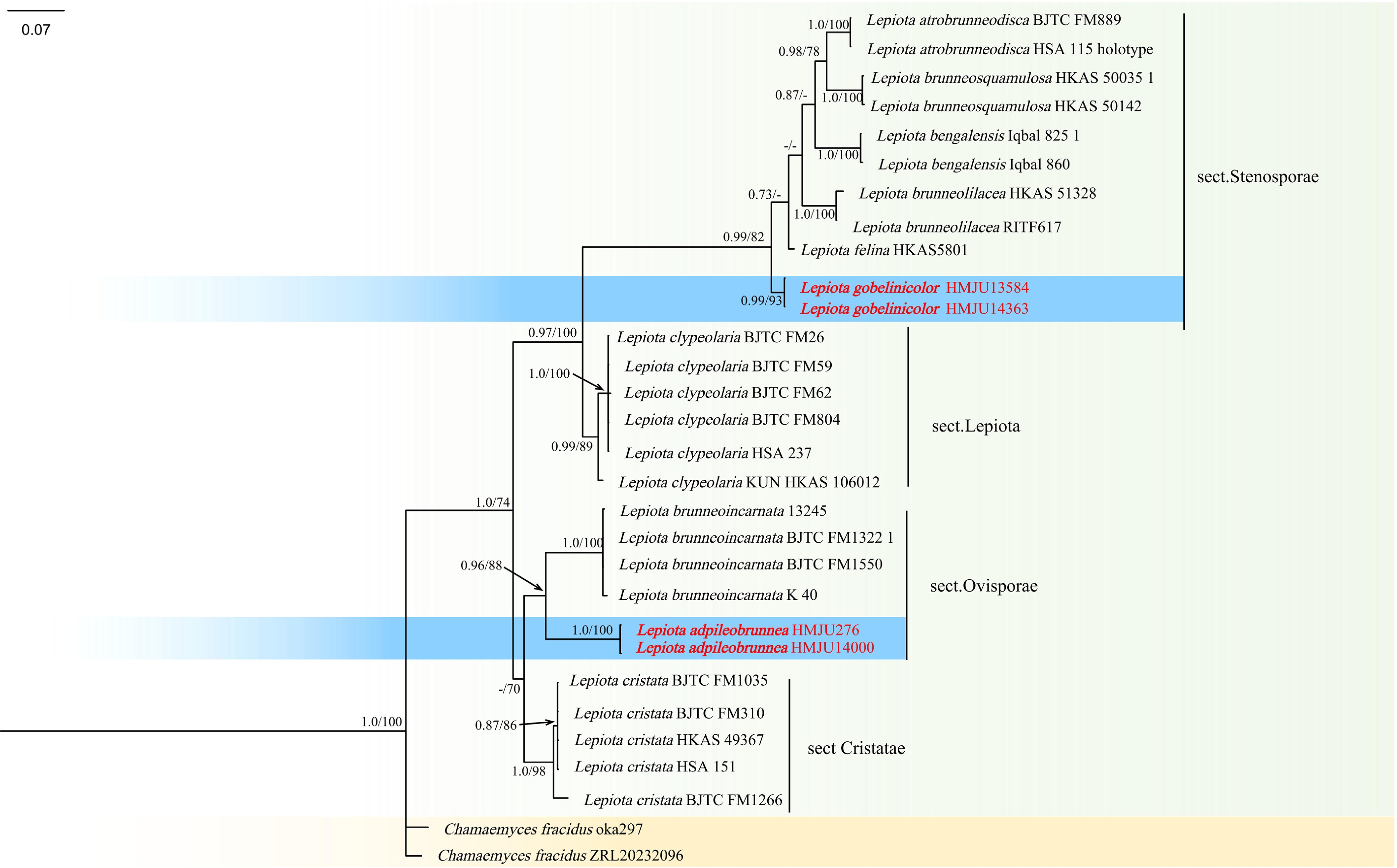
Figure 17.
Phylogram of maximum likelihood phylogenetic analysis based on combined ITS, LSU, rpb2, and tef-1α sequences. The analysis includes 37 strains; total characters: 1842 (ITS: 408, LSU: 390, rpb2:585, tef-1α: 459). Leucoagaricus naucinus (HMAS 8885) and Leucoagaricus naucinus (HMAS 88854) were used as the outgroup taxa. The best model was MIX{TPM2, TPM2} + I. Estimated base frequencies were as follows: A = 0.250, C = 0.250, G = 0.250, T = 0.250. The tree topology of the ML analysis is similar to the Bayesian analysis. Bootstrap values for ML equal to or greater than 70% and BYPP values equal to or greater than 0.70 are labelled on the nodes. The type strains are in bold.
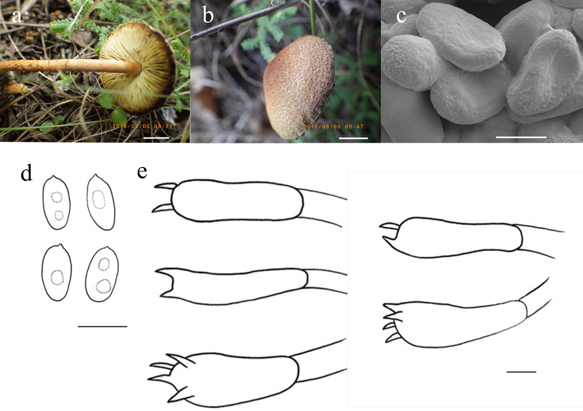
Figure 18.
Lepiota adpileobrunnea (HMJU276, holotype). (a), (b) Basidiocarps. (c) SEM images of basidiospores. (d) Basidiospores. (e) Basidia. Scale bars: (a), (b) 0.5 cm; (c)–(e) 3 µm.
MycoBank Names: MB863396
Holotype: Liaoning Province, Fuxin City, Haitang Mountain, on soil, 42°12'58" N, 128°17'30" E, 6 August 2016, J.Z. Xu (HMJU276, holotype).
Etymology: The specific epithet 'adpileobrunnea' indicates that the lamellae adjacent to the pileus transition to eye brown (7F6).
Diagnosis: Lepiota adpileobrunnea is characterized by a pileus ornamented with dense, burnt sienna fibrillose-granulose scales denser toward the center; lamellae are putty colored, transitioning to eye brown near the pileus; cheilocystidia, pleurocystidia, and clamp connections are absent.
Description: Pileus 10–27 mm diam., convex, the ground color white (1A1), covering dense burnt-sienna (7D8) fibrillose to granulose scales, gradually dense from the margin to the center in white (1A1) ground, pileus context thick, milk white(1A2). Lamellae free or adnexed, putty color (4B2), less crowded, and exhibit putty color (4B2), adjacent to the pileus transitions to eye brown (7F6), margin entire. Stipe 27–59 mm long, 1–2 mm thick, cylindrical, salmon (6A4), 1/3 part covering birch bark fibrils, 2/3 part covering scales, gradually increasing toward the base, stipe context milk white (1A2). Annulus absent.
Basidiospores (4.9–)5.0–7.8(–7.9) × (2.3–)2.4–4.1(4.2) μm, Q = 1.36–2.41, Qm = 1.90, ellipsoid to cylindrical, transparent in KOH, surface with gyrose ornamentation. Basidia (12–)16–27(–29) × (3–)5–7(–11) μm, both 4 and 2 sterigmata to 2 μm, clavate, transparent in KOH. Cheilocystidia absent. Pleurocystidia absent. Lamella trama irregular, hyphae 2–19 μm wide, transparent in KOH. Clamp connections absent in all hyphae.
Habitat: Solitary in coniferous-broadleaved mixed forests.
Known distribution: Known only from Northeastern China.
Additional material examined: Liaoning Province, Fuxin City, Haitang Mountain, on soil, 42°12'58" N, 128°17'30" E, 23 July 2020, J.Z. Xu (HMJU14400).
Notes: Phylogenetically, it forms a sister group with Lepiota brunneoincarnata Chodat & C. Martin (ML/PP = 88/0.96). However, Le. brunneoincarnata is characterized by a pileus with pinkish-brown scales arranged irregularly concentrically, lamellae that are creamy white, a stipe covered with dark-brown fibrous scales, with an indistinct woolly annulus, larger spores (8.9–10.2 × 4.8–5.5 μm), and cheilocystidia that are cylindrical or narrowly clavate[109]. It is morphologically similar to Lepiota decorata Zeller, Lepiota echinella Quél. & G.E. Bernard, Lepiota oreadiformis Velen., and Lepiota subgracilis Kühner. Le. decorata is characterized by an involute-margined pileus that ranges in color from rosé purple to pinkish purple, along with white lamellae, and relatively large spores with germ pores (6.5–9 × 5–6.5 µm)[110]. Le. echinella is characterized by a pileus with setose scales at the center, pinkish-white lamellae, and an orange-brown annulate stipe[111]. Le. oreadiformis features a pileus covered with fine light-brown scales, a white annulate stipe, clavate cheilocystidia, and the presence of clamp connections[112]. Le. subgracilis has an initially involuted pileus margin with fine brown or reddish-brown scales, white lamellae, and clavate or lageniform cheilocystidia[110].
Lepiota gobelinicolor J.Z. Xu, sp. nov., Fig. 18 and 19
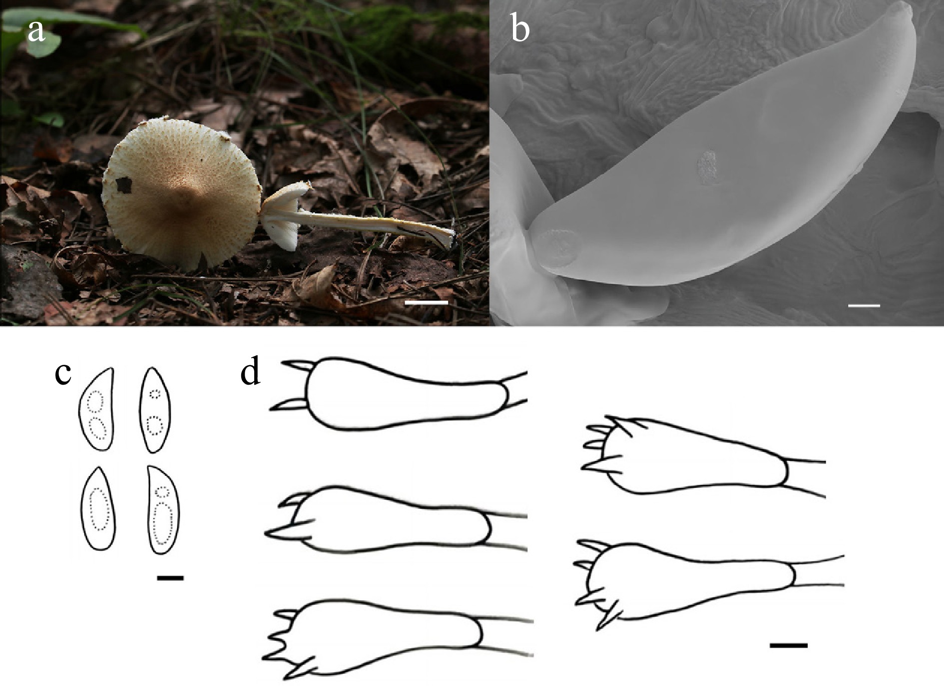
Figure 19.
Lepiota gobelinicolor (HMJU13584, holotype). (a) Basidiocarps. (b) SEM images of basidiospores. (c) Basidiospores. (d) Basidia. Scale bars: (a) 1 cm; (b)–(d) 5 µm.
MycoBank Names: MB863397
Holotype: Jilin Province, Meihekou City, Baoxing Village, on soil, 42°15'51" N, 125°19'03" E, 3 September 2025, J.Z. Xu (HMJU13584, holotype).
Etymology: The specific epithet 'gobelinicolor' indicates that the scales on the pileus are covered with a uniform gobelin pink color.
Diagnosis: Lepiota gobelinicolor is characterized by stipe-bearing reddish-golden granular scales on the lower two-thirds; cheilocystidia and pleurocystidia are lacking.
Description: Pileus 26–62 mm diam, convex, umbo at center and densely covered with gobelin pink (6A4) scales, becoming sparser toward the margin in the white (1A1) ground, margin has partial white (1A1) veil remnants, pileus context thin, white (1A1). Lamellae free, white (1A1), crowded, margin entire. Stipe 26–62 mm long, 4–8 mm thick, cylindrical, 1/3 part bears dense white (1A1) and gobelin pink (6A4) fibrils, white (1A1) fibrils, and reddish-golden (6C7) granular scales in the lower 2/3 part, hollow, stipe context white (1A1). Annulus absent.
Basidiospores (14.1–)14.8–16.9(–17.3) × (3.8–)3.9–6.0(6.2) μm, Q = 2.03–3.74, Qm = 2.75, narrowly cylindric to bacciliform, smooth, apex acuminate, transparent in KOH. Basidia (24–)25–32(–33) × (8–)9–12(–13) μm, both four and two sterigmata to 2 μm, clavate, transparent in KOH. Cheilocystidia absent. Pleurocystidia absent. Lamella trama inverse bilateral, hyphae 9–18 μm wide, transparent in KOH. Clamp connections absent in all hyphae.
Habitat: Scattered in broadleaved forest soil.
Known distribution: Known only from Northeastern China.
Additional material examined: Jilin Province, Meihekou City, Baoxing Village, on soil, 42°15'51" N, 125°19'03" E, 10 August 2024, J.Z. Xu (HMJU14363).
Notes: Phylogenetically, it forms a sister clade with Lepiota atrobrunneodisca L. Fan, L. Xia & N. Mao, Lepiota brunneosquamulosa J.F. Liang & Z.L. Yang, Lepiota bengalensis Hosen & T.H. Li, Lepiota brunneolilacea Bon & Boiffard, Lepiota felina (ML/PP = 82/0.99). However, Le. atrobrunneodisca is characterized by a central dark-brown pileus, with smaller basidiospores (5–7 × 3–4 μm), and possessing narrowly clavate cheilocystidia[113]. Le. brunneosquamulosa is different in having smaller basidiospores measuring (4.5–)5–6(–7) × 3–4 μm, and smaller basidia measuring 17–22 × 5–6 μm, with clamp connections present[114]. Le. bengalensis features a pastel-red pileus, the lamellae are pale yellow to yellowish white, and possesses clavate cheilocystidia[115]. Le. brunneolilacea is characterized by a pileus covered with brownish floccose squamules, an annulus rayish-green, and the stipe is brownish red. Le. felina is distinguished by a pileus that is dark brown, the lower part of the stipe is brown, and basidiospores that are smaller (6.5–8 × 3.5–4 μm)[116]. It is morphologically similar to Le. clypeolaria, Lepiota maculans Peck, and Lepiota recondita Tatti, Huijser & Vizzini. Le. clypeolaria differs in that the pileus is broadly campanulate or flattened when mature, the stipe has a yellowish to white annulus, and the spores are boletiform to fusiform, with clavate cheilocystidia[59]. Le. maculans features lamellae that turn orange pink when mature, a yellow stipe, clavate or subcylindrical cheilocystidia, and clamp connections[117]. Le. recondita is characterized by a stipe that turns pinkish brown when touched, a white annulus, yellowish lamellae, and cheilocystidia that are clavate, cylindro-clavate, sphaeropedunculate to submoniliform, and occasionally pyriform or cylindrical[118].
Leucocoprinus Pat., J. Bot., Paris 2: 16 (1888)
Patouillard originally distinguished Leucocoprinus Pat. as a separate entity from the genus Lepiota; however, he did not supply an initial circumscription for the new taxon. A detailed systematic characterization was later provided by Locquin[119], who organized the genus into four subgenera: subg. Leucocoprinus, subg. Leucoagaricus, subg. Hiatula (Fr.) Mont., and subg. Leucobolbitius J.E. Lange ex Locq. Geographically, Leucocoprinus is primarily found in subtropical and tropical zones[106,120]. It is a highly diverse genus, with the bulk of its species richness concentrated in the tropics[120]. Indeed, a significant number of novel species have recently been documented and described from these environments[121,122].
According to Vellinga[106], the genus is diagnostically defined by a specific set of traits: a plicate pileus margin, the presence of pseudoparaphyses surrounding the basidia, the absence of clamp connections, and metachromatic basidiospores. Since this definition was published, these morphological criteria have been widely accepted and utilized by researchers for delimiting species within Leucocoprinus[123,124].
Leucocoprinus margilongus J.Z. Xu, sp. nov., Figs 20 and 21
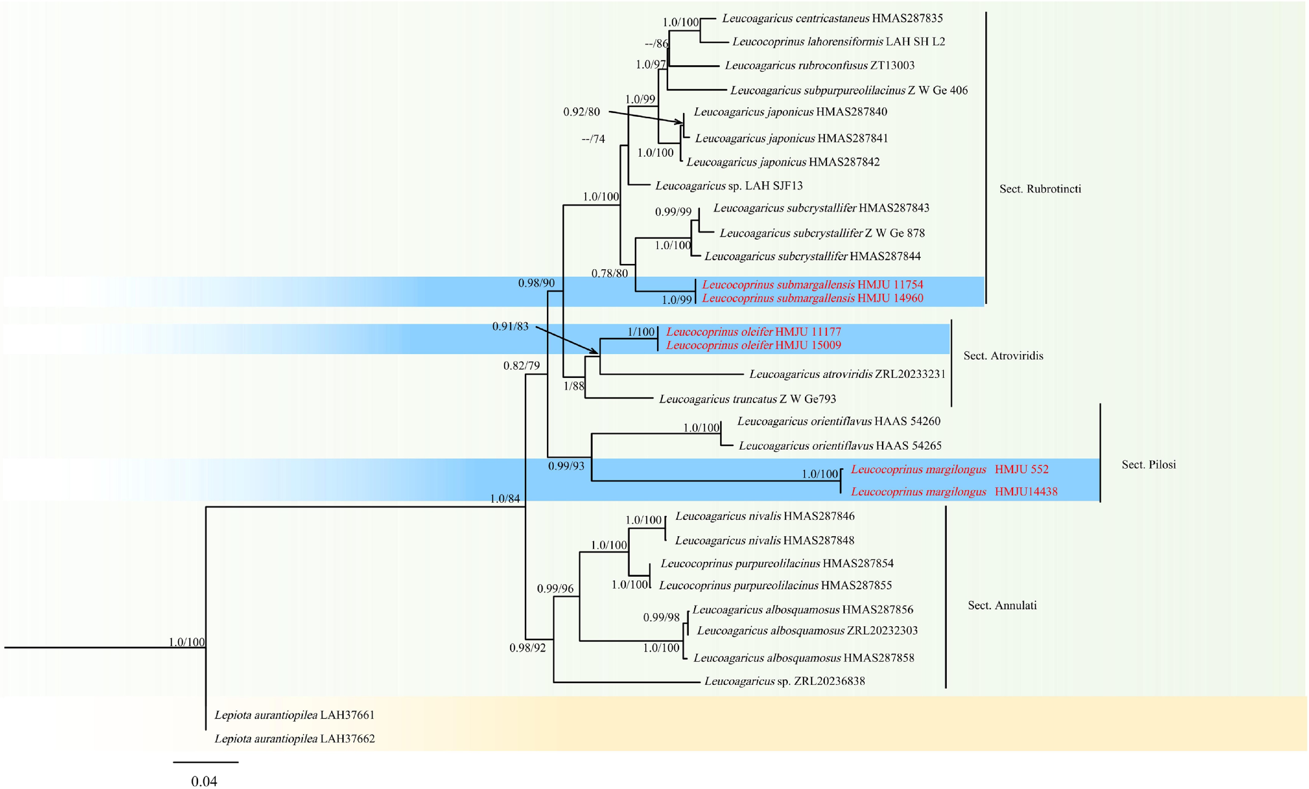
Figure 20.
Phylogram of Maximum Likelihood phylogenetic analysis based on combined ITS, nLSU, rpb2, and tef-1α sequences. The analysis includes 31 strains; total characters: 2,699 (ITS: 753, nLSU: 792, rpb2:659, tef-1α: 495). Lepiota aurantiopilea (LAH37661) and Lepiota aurantiopilea (LAH37662) were used as the outgroup taxa. The best model was SYM + I + G4. Estimated base frequencies were as follows: A = 0.250, C = 0.250, G = 0.250, T = 0.250. Bootstrap values for ML equal to or greater than 70% and BYPP values equal to or greater than 0.70 are labelled on the nodes. The type strains are in bold.
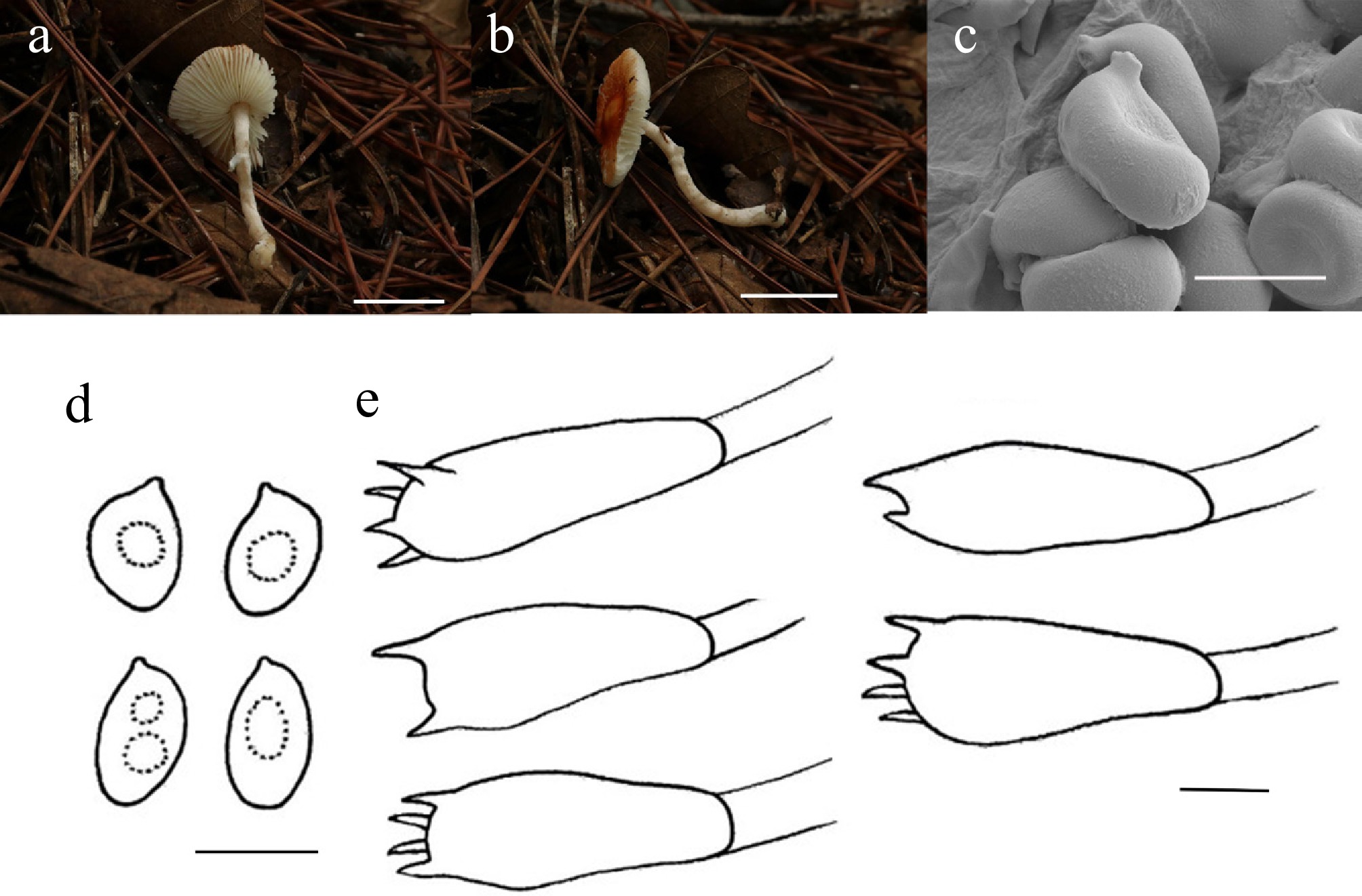
Figure 21.
Leucocoprinus margilongus (HMJU 552, holotype). (a), (b) Basidiocarps. (c) SEM images of basidiospores. (d) Basidiospores. (e) Basidia. Scale bars: (a), (b) 1 cm; (c)–(e) 5 µm.
MycoBank Names: MB863398
Holotype: Liaoning Province, Huludao City, Bailang Mountain, on soil, 40°47'42" N, 119°57'27" E, 10 August 2019, J.Z. Xu (HMJU 552, holotype).
Etymology: The specific epithet 'margilongus' refers to the fact that the lamellae margin is longer than the pileus.
Diagnosis: Leucocoprinus margilongusis characterized by lamellae exceeding the pileus in length; cheilocystidia, pleurocystidia, and clamp connections are lacking.
Description: Pileus 12–21 mm diam., convex to applanate, with a slight umbo present at the center, brownish orange (6C8) covered with brownish-orange (6C8) scales on an orange-white (5A2) background, gradually fading toward the margin, pileus context thin, white (1A1). Lamellae free, crowded, white (1A1); margin entire; longer than pileus. Stipe 21–32 mm long, 2–3 mm thick, cylindrical, base slightly swollen, 1/3 part has remnants of veil, covered with white (1A1) fibrils, stipe context white (1A1). Annulus white (1A1).
Basidiospores (4.6–)5.0–7.4(–7.5) × (3.7–)3.8–4.9(–5.2) μm, Q = 1.12–1.41, Qm = 1.46, broadly ellipsoid to ellipsoid, smooth, transparent in KOH, dextrinoid. Basidia (11–)12–23(–24) × (3–)5–8(–10) μm, clavate, both four and two sterigmata up to 2 μm long, transparent in KOH. Cheilocystidia absent. Pleurocystidia absent. Lamella trama regular, made up of parallel to subparallel, hyphae 3–18 μm wide; transparent in KOH. Clamp connections absent in all hyphae.
Habitat: Solitary in coniferous-deciduous mixed forest soil.
Known distribution: Known only from Northeastern China.
Additional material examined: Liaoning Province, Huludao City, Bailang Mountain, on soil, 40°47'42" N, 119°57'27" E, 16 August 2021, J.Z. Xu (HMJU 14438).
Notes: Phylogenetically, it forms a sister group with Leucocoprinus orientiflavus (Z.W. Ge) Asif, Saba & Vellinga (ML/PP = 93/0.99). However, Lc. orientiflavus is characterized by a pileus viscid when wet, a stipe pale yellow, and cheilocystidia clavate[125]. It is morphologically most similar to Leucocoprinus croceus S.M. Tang & K.D. Hyde, Leucocoprinus madagascarensis Ralaiv., Liimat. & Niskanen, and Lc. rosuliformis. However, Lc. croceus is characterized by pileus splits at maturity and a white stipe annulus[126]. Lc. madagascarensis has a black pileus center, a stipe with an annulus, spores larger (6.5–9.5 × 3.5–5 μm), and cheilocystidia cylindrical to clavate[127]. Lc. rosuliformis has a pileus covered with scales at the center, which become progressively sparser toward the margin; spores are larger (10.2–12 × 6.6–7.5 μm), and cheilocystidia are clavate[128].
Leucocoprinus submargallensis J.Z. Xu, sp. nov., Figs 20 and 22
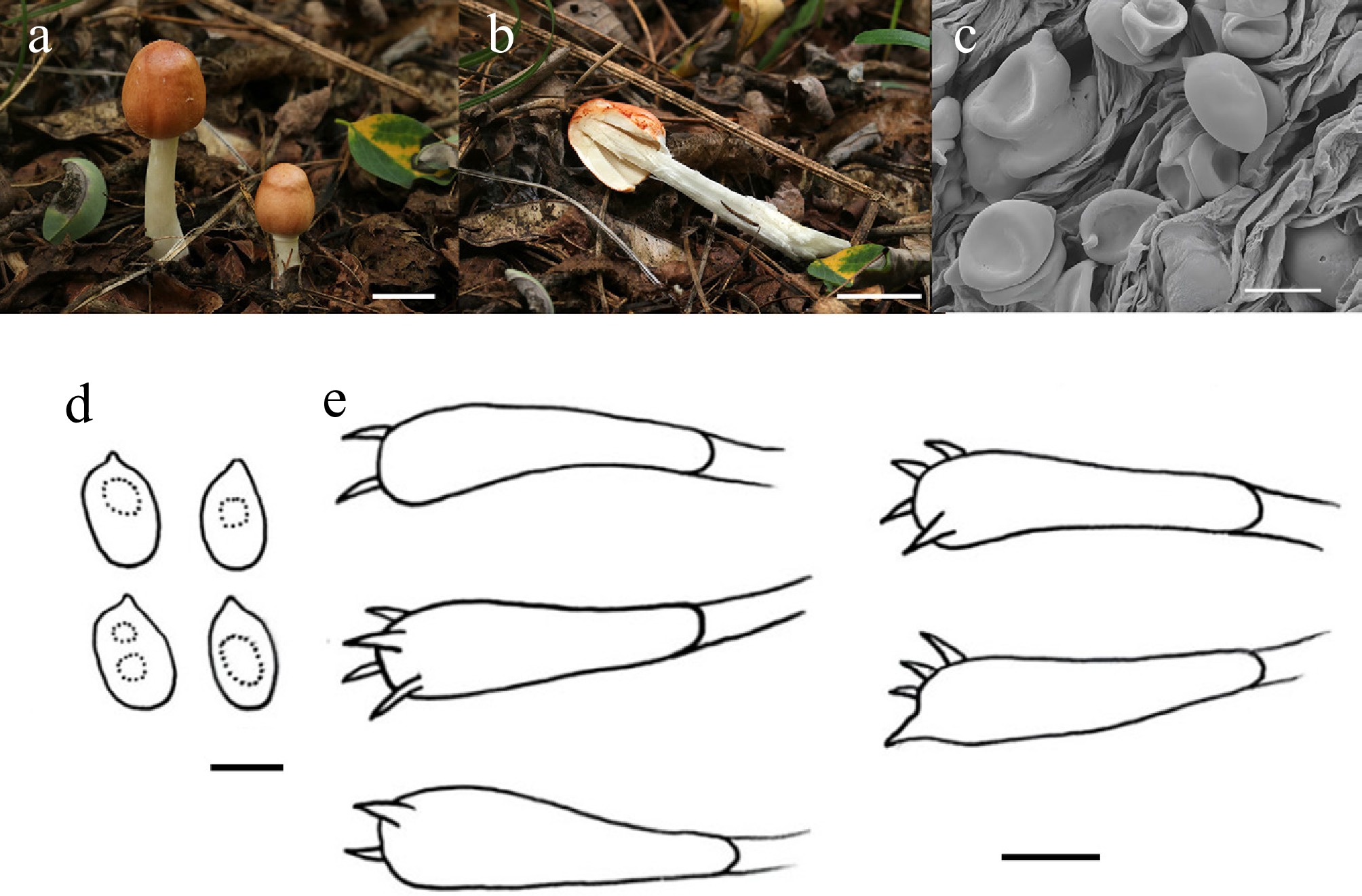
Figure 22.
Leucocoprinus submargallensis (HMJU 11754, holotype). (a), (b) Basidiocarps. (c) SEM images of basidiospores. (d) Basidiospores. (e) Basidia. Scale bars: (a) 1 cm; (b) 2 cm; and (c)–(e) 5 µm.
MycoBank Names: MB863399
Holotype: Liaoning Province, Huludao City, Jianchang County, Bailang Mountain, on soil, 40°49'19" N, 119°54'58" E, 8 August 2024, J.Z. Xu (HMJU 11754, holotype).
Etymology: The specific epithet 'submargallensis' refers to the species being morphologically similar to Leucocoprinus margallensis S. Ashraf, Naseer & Khalid.
Diagnosis: Leucocoprinus submargallensis is characterized by a stipe that is basally gradually bulbous; cheilocystidia, pleurocystidia, and clamp connections are lacking.
Description: Pileus 11–15 mm diam., parabolic, surface smooth, copper red (7C8), covered with fibrillose scales concolorous with the pileus and white (1A1) fibrils, pileus context thick, white (1A1). Lamellae free, crowded, white (1A1), margin entire. Stipe 23–42 mm long, 4–6 mm thick, cylindrical, gradually bulbous toward the base, white (1A1), covered with white (1A1) fibrils, stipe context white (1A1). Annulus absent.
Basidiospores (3.7–)4.0–7.4(–7.8) × (4.0–)4.5–7.8(–7.9) μm, Q = 1.21–1.53, Qm = 1.05, broadly ellipsoidal to ellipsoidal, smooth, transparent in KOH. Basidia (19–)20–27(–29) × (5–)6–8(–9) μm, clavate, most 2 sterigmata to 2 μm, transparent in KOH. Cheilocystidia absent. Pleurocystidia absent. Lamella trama hyphae 4–13 μm wide, transparent in KOH. Clamp connections absent in all hyphae.
Habitat: Solitary in coniferous-deciduous mixed forest soil.
Known distribution: Known only from Northeastern China.
Additional material examined: Liaoning Province, Huludao City, Jianchang County, Bailang Mountain, on soil, 40°49'19" N, 119°54'58" E, 7 August 2025 J.Z. Xu (HMJU 14960).
Notes: Forms a sister group with Leucocoprinus subcrystallifer (Z.W. Ge & Zhu L. Yang) Asif, Saba & Vellinga (ML/PP = 80/0.78). Lc. subcrystallifer is characterized by lamellae margin finely scalloped, spores larger (7)7.5–8.5(9) × 5–5.5(6) µm, and cheilocystidia clavate[129]. Morphologically most similar to Lc. margallensis and Leucocoprinus testaceumbonatus R.L. Zhao & J. Xin Li. Lc. margallensis features basidia smaller (13.08–23.5 × 7.7–8.72 μm), pale yellow in KOH, cheilocystidia clavate[126,129]. Lc. testaceumbonatus differs in spores, which are narrower (6.2–8.0 × 4.11–4.7 μm); cheilocystidia are clavate and subclavate[122].
Leucocoprinus oleifer J.Z. Xu, sp. nov., Figs 20 and 23
MycoBank Names: MB863400
Holotype: Liaoning Province, Fuxin City, Inner Mongolia Autonomous County, Haitang Mountain, on soil, 41°56'24" N, 121°49'19" E, 12 July 2024, J.Z. Xu (HMJU 11177, holotype)
Etymology: The specific epithet 'oleifer' refers to the presence of oily hyphae in the trama.
Diagnosis: Leucocoprinus oleifer is characterized by large basidiomata; pileus ornamented with minute grey scales, attenuating toward the margin; cheilocystidia and pleurocystidia lacking.
Description: Pileus 33–41 mm diam., convex, center with grey (1E1) umbo, margin fissured with age, covered with minute grey (1E1) scales on white (1A1) ground, becoming sparse toward the margin, pileus context thin, white (1A1). Lamellae adnexed, crowded, white (1A1), margin entire. Stipe 40–90 mm long, 4–7 mm thick, cylindrical, base slightly swollen, white (1A1), covered with white (1A1) fibrils, stipe context white (1A1). Annulus absent.
Basidiospores (5.0–)5.3–7.6 × (3.7–)3.8–5.1(–5.2) μm, Q = 1.18–1.80, Qm = 1.50, broadly ellipsoid to slightly oblong, elongated, smooth; transparent in KOH, dextrinoid. Basidia (14–)15–29(–32) × (6–)7–9(–10) μm, clavate, both 4 and 2 sterigmata to 2 μm, transparent in KOH. Cheilocystidia absent. Pleurocystidia absent. Lamella trama irregular, hyphae 7–22 μm wide, transparent in KOH. Clamp connections absent in all hyphae.
Habitat: Scattered in gravelly soil.
Known distribution: Known only from Northeastern China.
Additional material examined: Liaoning Province, Fuxin City, Inner Mongolia Autonomous County, Haitang Mountain, on soil, 41°56'24" N, 121°49'19" E, 24 July 2024, J.Z. Xu (HMJU 15009).
Notes: Phylogenetically, it forms a sister group with Leucocoprinus atroviridis Y.R. Ma, Z.W. Ge & X.D. Yu (ML/PP = 83/0.91). Lc. atroviridis features pileus with bottle-green scales, cheilocystidia, and broadly clavate[126,130]. Lc. griseosquamosus, Lc. brunneodiscus, and Lc. rosuliformis are mostly similar in morphology. Lc. griseosquamosus is characterized by lamellae that turn reddish white and erode when touched, spores larger (6.0–7.5 × 4.0–4.5 μm), and cheilocystidia clavate[48]. Lc. brunneodiscus has a plano-convex to flattened pileus with a dark-brown to reddish-brown center, a stipe with an annulus ranging from brownish orange to brown, and cheilocystidia clavate[131]. Lc. griseosquamosus is unique in that the pileus turns reddish white when touched, the lamellae are eroded, the basidia are smaller, and the cheilocystidia are clavate[48].
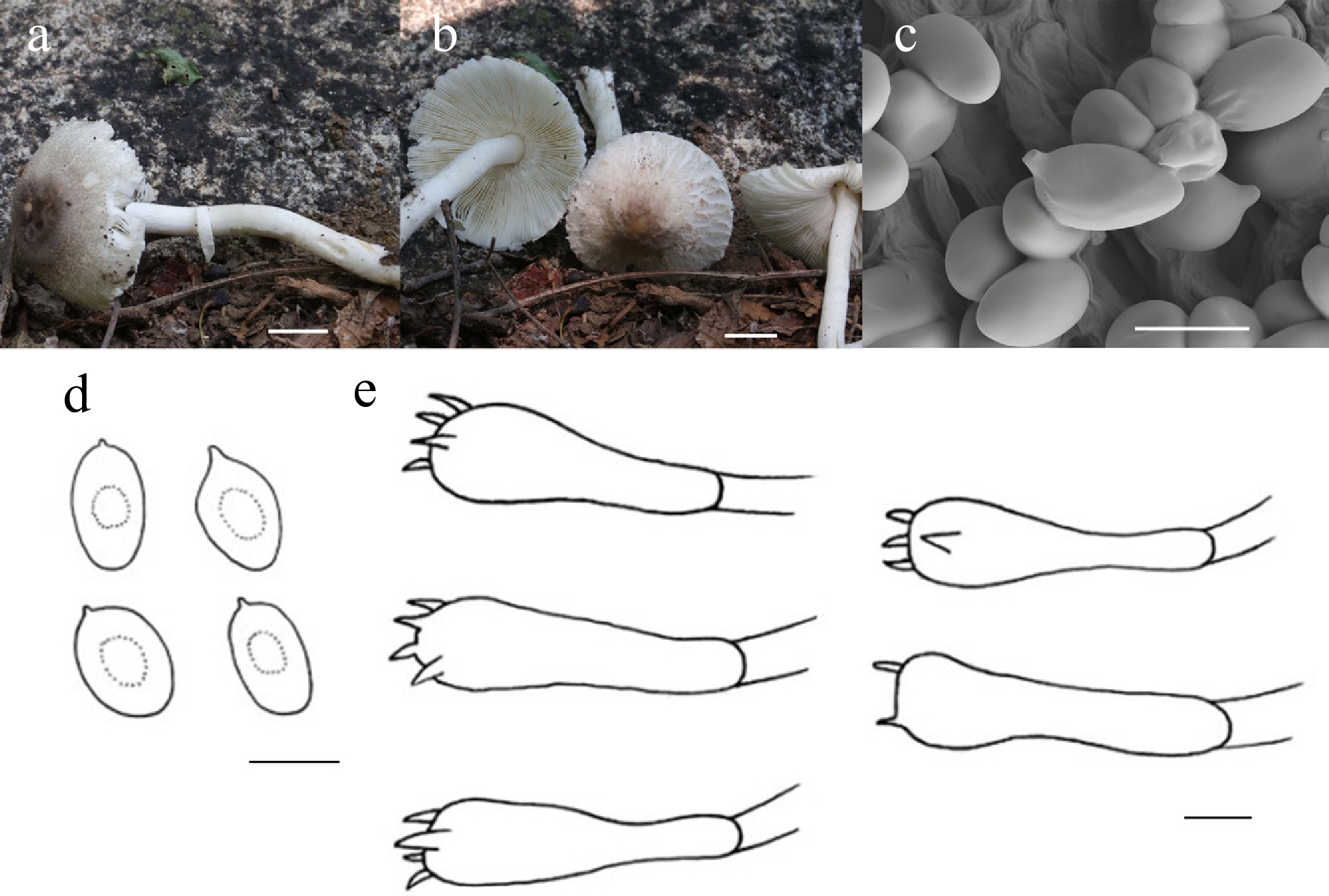
Figure 23.
Leucocoprinus oleifer (HMJU 11177, holotype). (a), (b) Basidiocarps; (c) SEM images of basidiospores; (d) Basidiospores; (e) Basidia. Scale bars: (a), (b) 1 cm; (c)–(e) 5 µm.
Limacella Earle, Bull. New York Bot. Gard. 5: 447 (1909)
Limacella Earle is an infrequently encountered genus within the order Agaricales, with a presumed cosmopolitan range[132]. Although approximately 50 taxa have been assigned to the genus, the current state of its taxonomy is chaotic, largely due to inconsistent and insufficient historical descriptions. Significant publications focusing on the morpho-taxonomy and molecular phylogeny of the group have aided in the macroscopic recognition of the genus and confirmed its classification within the family Amanitaceae[68,133−138].
Historically, the circumscription of Limacella relied on a suite of diagnostic morphological features: a slightly viscid to glutinous pileus lacking universal veil remnants; an epicutis consisting of ascending or erect terminal hyphal elements (modified or unmodified) embedded in a gelatinous matrix; lamellae that are free or nearly so with a fertile edge; and a bilateral hymenophoral trama with divergent lateral strata. Additionally, the genus is characterized by a white to cream spore print; globose to short-ellipsoid basidiospores that are inamyloid (rarely dextrinoid) and acyanophilous; a stipe that is either dry or viscid; a partial veil manifesting as either a membranous annulus or a glutinous layer; and a non-ectomycorrhizal nutritional mode[68,136]. However, several of these traditional characterizations are ambiguous, while others appear to be based on misconceptions regarding tissue ontogeny and the homology between tissues in Amanita and Limacella. Furthermore, some traits previously mentioned in taxonomic treatments have been subsequently neglected.
To address the confusion and vagueness persisting in earlier descriptions, Tulloss et al.[139] proposed a streamlined circumscription for Limacella. Under this revised definition, a species within the Amanitaceae belongs to Limacella if and only if it meets two criteria: (1) it possesses an agaricoid basidiome that does not originate via schizohymenial ontogeny; and (2) it exhibits a fertile lamellar margin combined with a gluten-bearing pileus, where the gluten is supported by anticlinally oriented structural elements.
Regarding infrageneric classification, Singer[68] separated Limacella into two sections based primarily on the viscosity of the stipe: sect. Lubricae and sect. Limacella [= Viscidae]. Section Lubricae contains species with a glutinous or viscid stipe, while sect. Limacella is reserved for those with a dry stipe. Later, based on the ecological and morphological study of European species, Gminder[140] established sect. Amanitellae to house Limacella guttata (Pers.) Konrad & Maubl and related taxa. This section is the most distinct of the three, defined by species possessing: (1) abundant conical or subconical terminal cells on the gluten-supporting hyphae, and (2) terminal cells that are frequently subtended by an inflated cell (or a short chain of such cells) which may be globose, subglobose, broadly ellipsoid, or otherwise inflated[140].
Limacella annulipendula J.Z. Xu, sp. nov., Fig 24 and 25
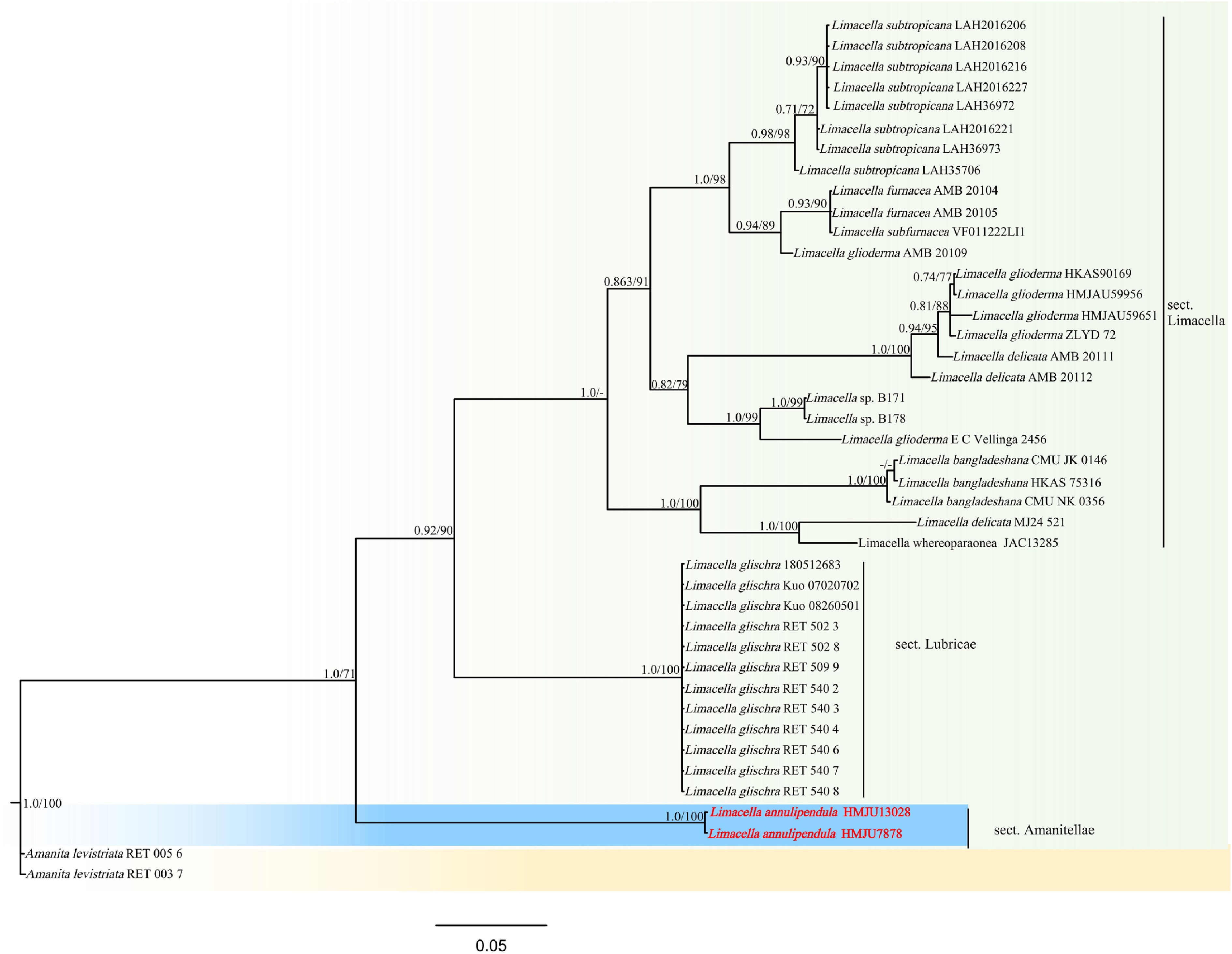
Figure 24.
Phylogram of Maximum Likelihood phylogenetic analysis based on combined ITS, nLSU, and sequences. The analysis includes 41 strains; total characters: 1,560 (ITS: 765, nLSU: 795). Amanita levistriata (RET 005 6) and Amanita levistriata (RET 003 7) were used as the outgroup taxa. The best model was TVM + F + I + G4. Estimated base frequencies were as follows: A = 0.299, C = 0.165, G = 0.227, T = 0.310. Bootstrap values for ML equal to or greater than 70% and BYPP values equal to or greater than 0.70 are labelled on the nodes. The type strains are in bold.
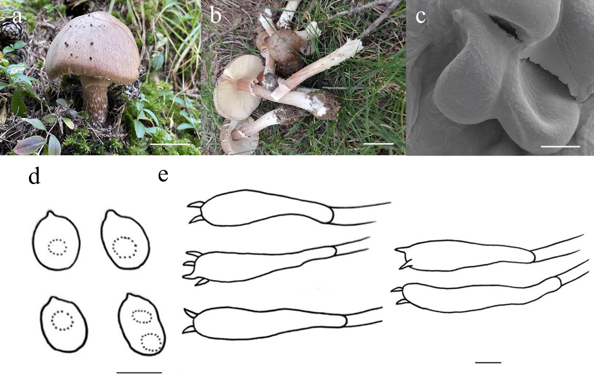
Figure 25.
Limacella annulipendula (HMJU 13028, holotype). (a), (b) Basidiocarps. (c) SEM images of basidiospores. (d) Basidiospores. (e) Basidia. Scale bars: (a), (b) 3 cm; (c)–(e) 5 µm.
MycoBank Names: MB863401
Holotype: Shanxi Province, Xinzhou City, Wuzhai County, Luya mountain, on soil, 38°40'02" N, 111°57'31" E, 17 September 2022, L. Zhao (HMJU 13028, holotype).
Etymology: The specific epithet 'annulipendula' refers to the annulus with a pendulous margin.
Diagnosis: Limacella annulipendula is characterized by a pileus that is hemispherical when young, with a margin adorned with red-haired striations; mature specimens have dense birch bark striations; the annulus is very pale, shell pink, densely fibrillose and undulate, bearing mandarin orange spots at the stipe-adjacent center; cheilocystidia and pleurocystidia are lacking.
Description: Pileus 42–65 mm diam., conical, finally becoming convex at maturity, covered with central pale orange (5A3), remainder oxblood red (9E7), margin covered with red-haired (6C4) striations when young, hemispherical, covering dense birch bark (6B2) striations on white (1A1) background when mature, pileus context thick, white (1A1). Lamellae free, crowded, wavy, whimargin entire. Stipe 76–88 mm long, 14–17 mm thick, cylindrical, base slightly bulbous-swollen, brick red (7D7), covered with white (1A1) fibrils or smooth, covering oxblood-red (9E7) striations on white (1A1) background, stipe context white (1A1). Annulus 1/5 part of the stipe; margin reflexed, overall, very pale shell pink (8A3), covered with dense fibrils, wavy; covering mandarin-orange (6B8) spots at center adjacent to the stipe.
Basidiospores (3.7–)4.0–7.2(–7.3) × (4.0–)4.2–6.7(–7.7) μm, Q = 1.02–1.21, Qm = 0.98, globose to broadly ellipsoidal, transparent in KOH, without amyloid. Basidia (17–)18–33(–34) × (4–)5–7(–8) μm, clavate, both 4 and 2 sterigmata up to 2 μm long, transparent in KOH. Cheilocystidia absent. Pleurocystidia absent. Lamella trama bilaterally, hyphae 5–16 μm wide, transparent in KOH. Clamp connections absent in all hyphae.
Habitat: Scattered to gregarious on soil among grasses in forests.
Known distribution: Known only from Northern China.
Additional material examined: Shanxi Province, Xinzhou City, Wuzhai County, Luya Mountain, 38°40'02" N, 111°57'31" E, on soil, 16 September 2022, L. Zhao (HMJU 7878).
Notes: It constitutes a separate section. The main differences distinguishing this genus from the currently known species are that the pilei of other species are glabrous and non-striate, with spores bearing reticulate ornamentation. Morphologically most similar to Limacella kauffmanii H.V. Sm., Limacella alachuana (Murrill) Pegler, and Limacella subfurnacea Contu. Li. kauffmanii differs from pileus glabrous, orange yellow at the center, stipe milky white, spores yellow in KOH[141]. Li. alachuana is unique in that its pileus is glabrous, applanate when mature, and yellowish brown; its stipe has a yellowish-brown annulus[142]. Li. subfurnacea is characterized by a pileus glabrous and viscid, a stipe with a yellowish-ochre annulus, and spores polygonal[143].
Macrocystidia Joss., Bull. Soc. Mycol. France 49(3–4): 373, 376 (1934) [1933]
Josserand[144] originally established the genus Macrocystidia, designating Macrocystidia cucumis as the type species. Although Kühner subsequently created the family Macrocystidiaceae to house this genus, its classification at the family level has historically been a matter of controversy[145]. Nevertheless, recent phylogenetic data point to a relationship between Macrocystidia and the Tricholomatineae lineage. Significantly, Vizzini et al.[146] presented definitive evidence placing Macrocystidia within the Macrocystidiaceae.
Conversely, Mycena (Pers.) Roussel represents a substantial genus within the order Agaricales, containing roughly 600 species assigned to the family Mycenaceae. The genus is typified by Macrocystidia galericulata (Scop.) Gray. Maas Geesteranus formulated the most widely accepted infrageneric system for Mycena, organizing the genus into various sections based on morphological attributes[147]. Within this diverse group, approximately 40 species spanning 18 distinct sections are known to be bioluminescent[148].
Macrocystidia tashanparensis J.Z. Xu, sp. nov., Figs 26 and 27
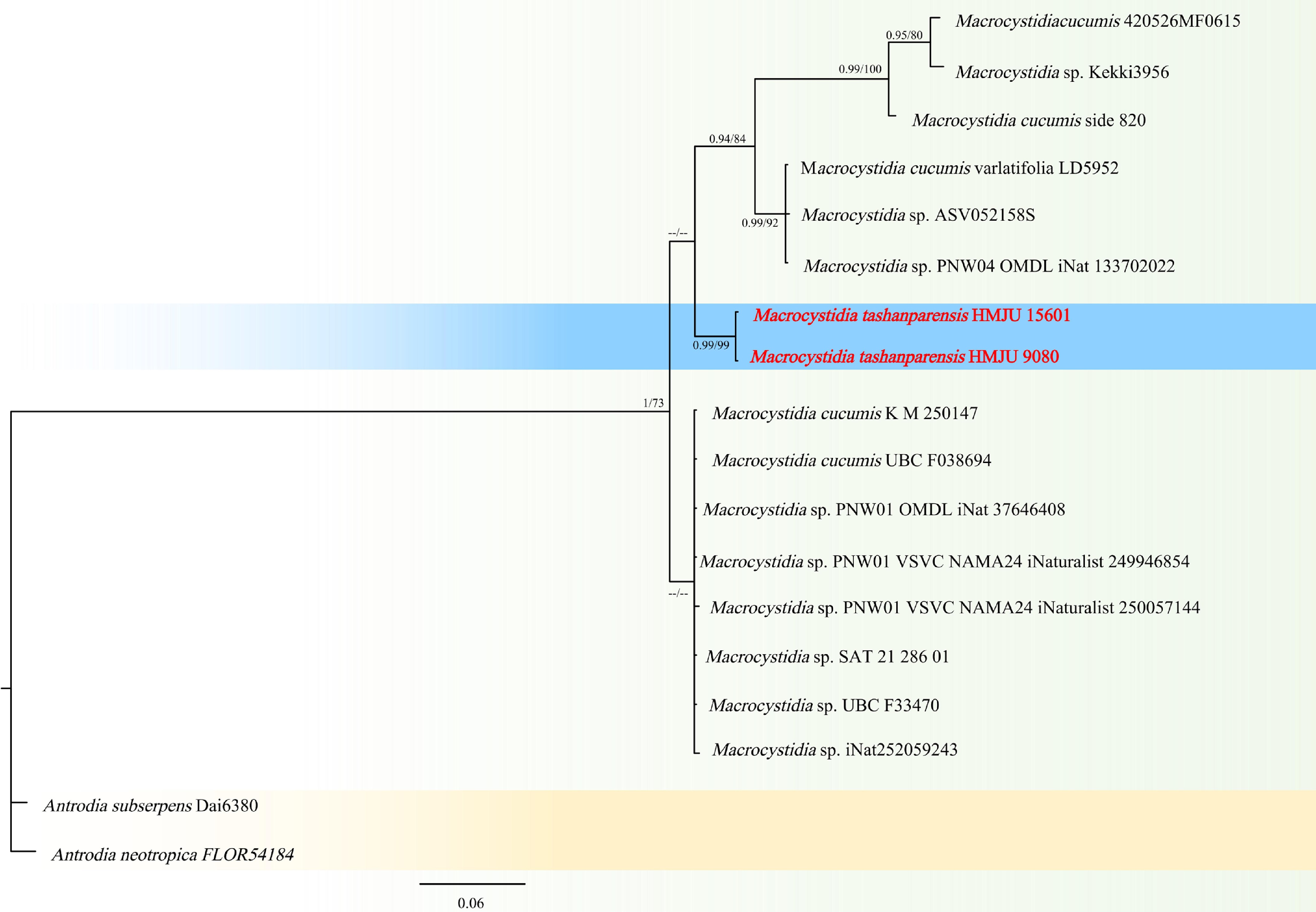
Figure 26.
Phylogram of Maximum Likelihood phylogenetic analysis based on combined ITS sequences. The analysis includes 18 strains; total characters: 586 (ITS: 586). Antrodia neotropica (FLOR 54184), and Antrodia subserpens (Dai 6380) were used as the outgroup taxa. The best model was HKY + F + I. Estimated base frequencies were as follows: A = 0.227, C = 0.232, G = 0.224, T = 0.317. Bootstrap values for ML equal to or greater than 70% and BYPP values equal to or greater than 0.70 are labelled on the nodes. The type strains are in bold.
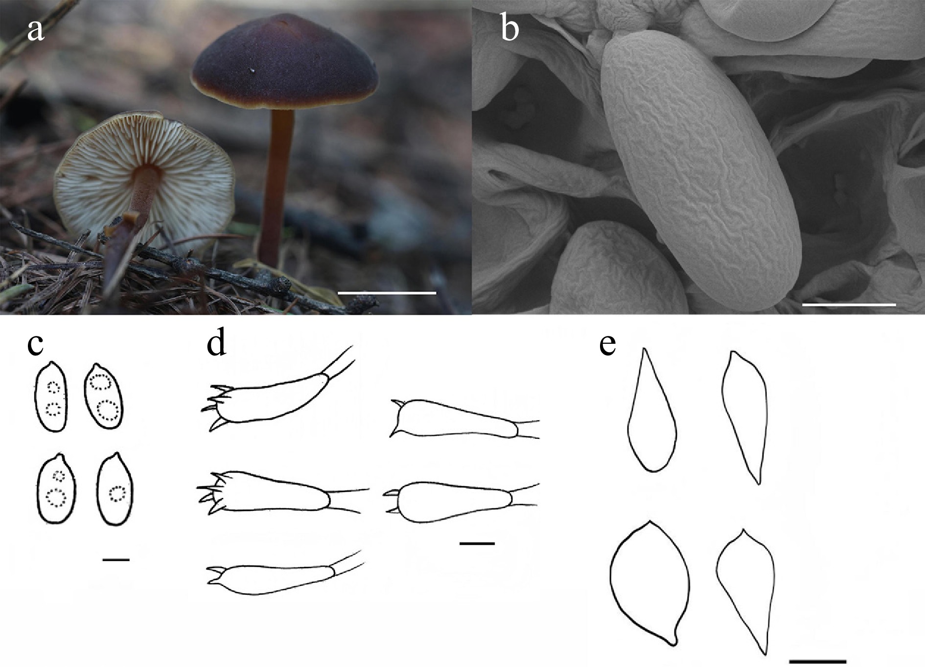
Figure 27.
Macrocystidia tashanparensis (HMJU 15601, holotype). (a) Basidiocarps. (b) SEM images of basidiospores. (c) Basidiospores. (d) Basidia. (e) Cheilocystidia. Scale bars: (a) 1 cm; (b), (c) 3 µm; and (d) 10 µm.
MycoBank Names: MB863402
Holotype: Liaoning Province, Huludao City, Lianshan District, on soil, 40°45'07" N, 120°52'20" E, 23 July 2025, J.Z. Xu (HMJU 15601, holotype).
Etymology: The specific epithet 'tashanparensis' refers to the type locality, Tashan Par.
Diagnosis: Macrocystidia tashanparensis is characterized by a pileus with an eye-brown disc and a birch bark margin; a stipe henna, darkening basally; basidia sparsely sterigmate; and cheilocystidia bulbous, apically acute.
Description: Pileus 19–21 mm diam., convex to broadly parabolic, tomentose, surface slightly cracked at the disc, disc eye brown (7F7), margin birch bark (6B2), pileus context thin, eye brown (7F7). Lamellae free, white (1A1), edge entire, concolorous. Stipe 17–33 mm long, 2–3 mm thick, cylindrical, henna (7E8), darkening towards the base, concolorous with the pileus, stipe context henna (7E8). Annulus absent.
Basidiospores (6.5–)6.8–8.2(–8.4) × (3.0–)3.5–4.3(–4.4) μm, Q = 1.56–1.84, Qm = 1.93, ellipsoid to rectangular, without germ pore, transparent in KOH, dextrinoid. Basidia (26–)29–39(–41) × (8–)9–11(–12) μm, clavate, both four and two sterigmata to 9 μm, with few sterigmata, transparent in KOH. Cheilocystidia (34–)37–53(–54) × (13–)14–22(–28) μm, bulbous with a pointed apex. Pleurocystidia absent. Lamella trama bilaterally arranged, hyphae 4–10 μm wide, transparent in KOH. Clamp connections absent in all hyphae.
Habitat: Scattered on coniferous forest soil.
Known distribution: Known only from Northeastern China.
Additional material examined: Liaoning Province, Huludao City, Lianshan District, on soil, 40°45'07" N, 120°52'20" E, 22 July 2019, J.Z. Xu (HMJU 9080).
Notes: Macrocystidia tashanparensis forms a sister group with Ma. Cucumis (ML/PP = 68/0.65), but Ma. Cucumis has a larger pileus (10–60 mm), a longer stipe (80 mm), and dark-olive spores. Macrocystidia africana Singer differs from Ma. tashanparensis by having broadly fusiform pleurocystidia, cheilocystidia with an obtuse apex, ochraceous lamellar trama, and a slightly striate pileus[42]. Macrocystidia incarnata Singer is distinguished from Ma. tashanparensis by its flesh-colored pileus, pruinose stipe, clavate and capitate cystidia, and the presence of dermatocystidia[42]. Macrocystidia reducta E. Horak & Capellano differs from Ma. tashanparensis in having a stouter fruiting body, broadly fusiform cystidia, and bigger spores (7.5–10 × 4.5–5 μm)[149].
Marasmius Fr., Fl. Scan.: 339 (1836) [1835]
Fries formally circumscribed the genus Marasmius Fr., selecting Marasmius rotula (Scop.) Fr. as the type species. Morphologically, the genus is defined by small to medium-sized basidiomata featuring campanulate to convex pilei; lamellae that are free, adnexed, or adnate (occasionally forming a collar); and stipes that may be either insititious or non-insititious. Diagnostic microscopic features include a hymeniform pileipellis, a white spore print, and basidiospores that are hyaline, smooth, thin-walled, and inamyloid. Ecologically, Marasmius functions primarily as a saprobe on leaf litter, dead branches, or soil within forest environments. While the genus is cosmopolitan, it exhibits exceptional species diversity in tropical and subtropical zones.
Historically, Singer[68,79,150] established a classification system comprising 12 sections based on spore traits and cortical anatomy. This framework remained the standard until the late 20th century; however, subsequent molecular investigations demonstrated that Marasmius sensu Singer is polyphyletic[151]. Phylogenetic reconstructions utilizing nLSU and nrITS rDNA sequences[151] have since delimited Marasmius sensu stricto (s. str.) as a monophyletic lineage. This restricted concept retains only five of Singer's original sections: Marasmius, Globulares, Sicci, Leveilleani, and Neosessiles, while excluding the others.
The first, subgenus Marasmius, is characterized by thin basidiomata, an insititious stipe, and trama that is either dextrinoid or inamyloid; it encompasses the sections Crinis-eques, Marasmius, Variabilispori, Sanguirotales, and Sicciformes. The second, subgenus Globulares, is defined by diverse basidiomatal forms, a non-insititious stipe with basal mycelium, and consistently dextrinoid trama; it includes the sections Globulares and Sicci.
China possesses remarkable macrofungal biodiversity, particularly in subtropical areas where complex topography and favorable climates create biodiversity hotspots. In recent years, at least 13 new species of Marasmius have been described from these regions[152−154].
Marasmius velutinus J.Z. Xu, sp. nov., Figs 28 and 29
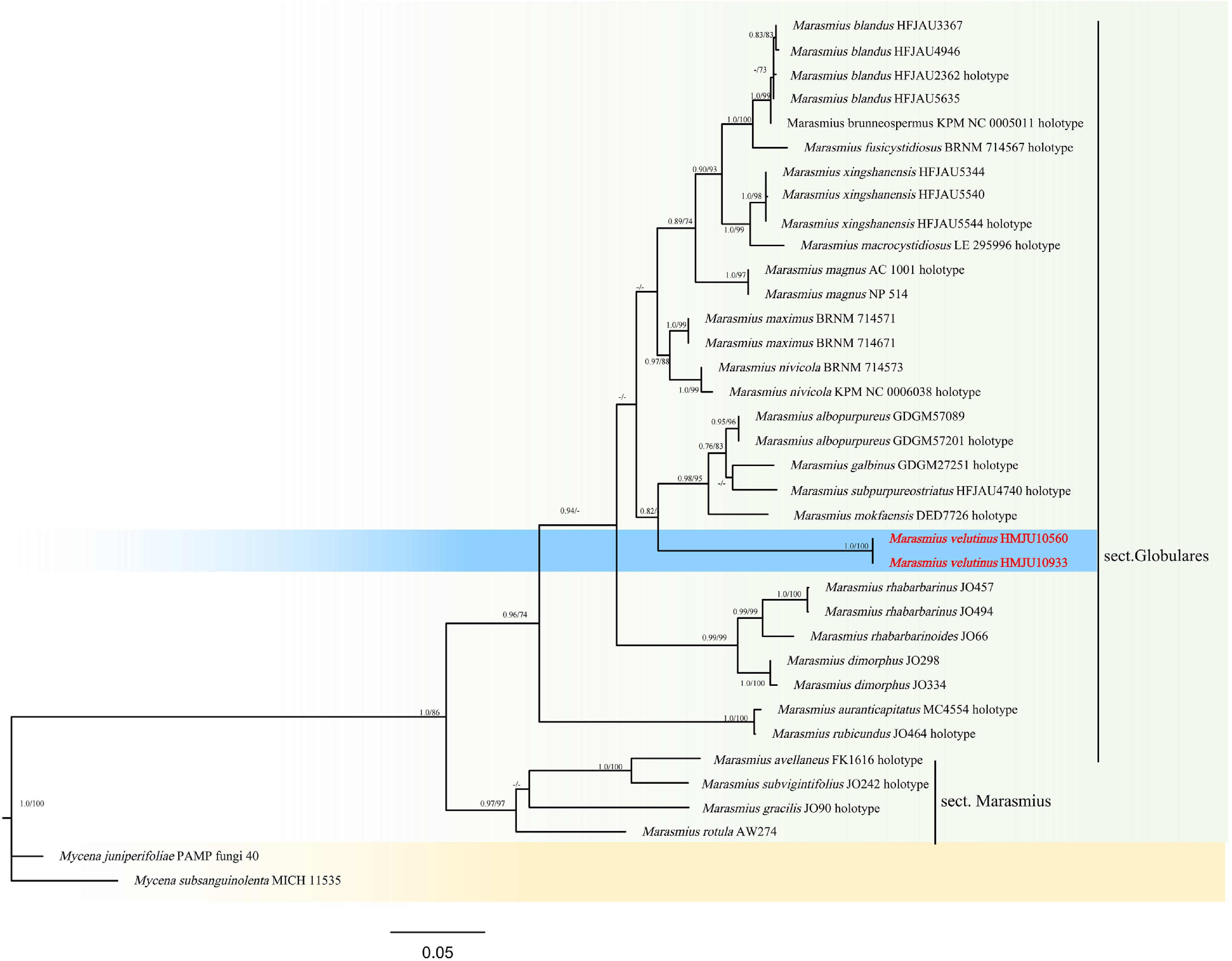
Figure 28.
Phylogram of Maximum Likelihood phylogenetic analysis based on combined ITS, nLSU, rpb2, and tef-1 sequences. The analysis includes 36 strains; total characters: 2,166 (ITS: 290, nLSU: 772, rpb2: 637, tef-1: 467). Mycena juniperifoliae (PAMP fungi 40) and Mycena subsanguinolenta (MICH 11535) were used as the outgroup taxa. The best model was TIM2 + F + I + G4. Estimated base frequencies were as follows: A = 0.258, C = 0.210, G = 0.273, T = 0.259. Bootstrap values for ML equal to or greater than 70% and BYPP values equal to or greater than 0.70 are labelled on the nodes. The type strains are in bold.
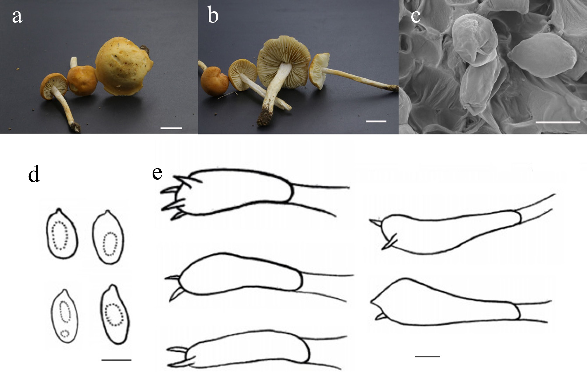
Figure 29.
Marasmius velutinus (HMJU 10560, holotype). (a), (b) Basidiocarps. (c) SEM images of basidiospores. (d) Basidiospores. (e) Basidia. Scale bars: (a), (b) 1 cm; (c)–(e) 5 µm.
MycoBank Names: MB863403
Holotype: Guangdong Province, Shenzhen City, Bao'an District, Hangcheng Street, Caowei Village, on soil, 22°37'18" N, 113°50'35" E, 23 April 2024, J.Z. Xu (HMJU 10560, holotype).
Etymology: The specific epithet 'velutinus' refers to the velutinate surface of the pileus, which is the most distinctive morphological character distinguishing it from closely related species.
Diagnosis: Marasmius velutinus is characterized by a pileus golden-wheat in color; stipe white on the upper third, shading to orange white; cheilocystidia and pleurocystidia lacking.
Description: Pileus 14–31 mm diam., convex, center-depressed, mandarin orange (6B8), covered with white (1A1) pruina, pileus context thin, white (1A1). Lamellae free, slightly crowded, margin entire. Stipe 51–72 mm long, 3–6 mm thick, cylindrical, 1/3-part white (1A1), transitioning to orange white (5A2), stipe context white (1A1).
Basidiospores (3.5–)4.1–10.6(–10.7) × (2.5–)3.0–5.7(–5.8) μm, Q = 1.30–2.74, Qm = 1.81, ellipsoidal to cylindrical, transparent in KOH, without amyloid. Basidia (16–)19–31(–35) × 5–13(–14) μm, clavate, both four and two sterigmata up to 2 μm, transparent in KOH. Cheilocystidia absent. Pleurocystidia absent. Lamella trama regular, hyphae 3–17 μm wide, transparent in KOH. Clamp connections absent in all hyphae.
Habitat: Scattered on the soil.
Known distribution: Known only from Southern China.
Additional material examined: Guangdong Province, Shenzhen City, Bao'an District, Hangcheng Street, Caowei Village, on soil, 22°37'18" N, 113°50'35" E, 5 May 2024, J.Z. Xu (HMJU 10933).
Notes: It forms a sister group with Marasmius albopurpureus T.H. Li & C.Q. Wang, Mar. galbinus T.H. Li & Chun Y. Deng, Mar. subpurpureostriatus J.Q. Yan & Hong Chen, and Mar. mokfaensis Wannathes, Desjardin & Lumyong (ML/PP = 68/0.82). However, Mar. albopurpureus is characterized by a pileus ranging from white to purple, a stipe pale purple in the upper part and brown in the lower part, and cheilocystidia clavate[155]. Mar. galbinus is distinguished by a pileus that is greenish white, lamellae that are yellowish white, and a stipe apex that is hyaline to white, with cheilocystidia that are irregularly clavate[152]. Mar. subpurpureostriatus is typified with a grayish-green pileus, with deep-purple striations, lamellae yellowish gray, and cheilocystidia clavate[8]. Mar. mokfaensis features a gray-toned pileus, a grayish-purple stipe apex, a context that turns yellow with age, and clavate, ventricose cheilocystidia[156]. Species morphologically similar to Mar. collinus (Scop.) Singer and Marasmius oreades (Bolton) Fr. Mar. collinus differs in having a pileus with a central umbo and translucent striations, and in having elliptical spores[157]. Mar. oreades is unique in that the pileus margin is undulate, ochraceous orange or brownish, with a glossy (oily) sheen, the lamellae are undulate, and the stipe is solid[36].
Melanoleuca Pat., Cat. Rais. Pl. Cellul. Tunisie (Paris): 22 (1897)
Melanoleuca Pat. is a genus traditionally assigned to the family Tricholomataceae. Although phylogenetic analyses conducted by Moncalvo et al.[145] and Matheny et al.[158] suggested that Melanoleuca forms a clade alongside Pluteus, the prevailing academic consensus maintains its taxonomic classification within the Tricholomataceae.
The genus was originally erected by the distinguished French mycologist Patouillard, who designated Melanoleuca vulgaris Pat. as the type species. Two years later, Agaricus adstringens Pers. was transferred to the genus as Melanoleuca adstringens (Pers.) Fayod, bringing the total number of recognized species to two. In 1897, Patouillard[159] formally emended the generic name from Melaleuca Pat. to Melanoleuca, thereby validating the combinations M. adstringens and the type species, M. vulgaris.
As new species were described, taxonomists proposed diverse frameworks for the infrageneric classification of Melanoleuca. Researchers such as Singer[68,160,161], Kühner[87], all advocated for subdividing the genus into distinct sections. For instance, Singer (1962) utilized macroscopic features, specifically stipe appendages and pileus coloration, to delineate four sections: Alboflavidae, Humiles, Oreinae, and Melanoleuca. Later, Bon[162] became the first to integrate microscopic characteristics into the classification system, defining seven sections based on a combination of micro- and morphological traits.
Focusing on micromorphology, Boekhout[163] prioritized the presence and shape of cystidia, establishing three subgenera: subg. Melanoleuca; subg. Urticocystis (defined by urticiform cheilocystidia and rare or absent pleurocystidia); and subg. Macrocystis (characterized by fusiform to lageniform cheilocystidia and pleurocystidia resembling the large cystidia). Boekhout also distinguished two urticiform sections based on cystidial shape: one featuring clavate cystidia (narrowing upwards) and the other featuring campanulate cystidia (tapering gradually toward the apex). Subsequently, Bon[45] proposed an alternative system based on spore Q-values and the distinction between lageniform and fusiform cystidia. Bon later reintroduced a simplified version of this scheme in 1999, which has achieved widespread acceptance among modern taxonomists. More recently, Aysenur et al. utilized combined nLSU and ITS gene sequences to classify 14 Turkish Melanoleuca species into two subgenera: subg. Melanoleuca and subg. Urticocystis[164,165].
Melanoleuca mozhugongkaensis J.Z. Xu, sp. nov., Figs 30 and 31
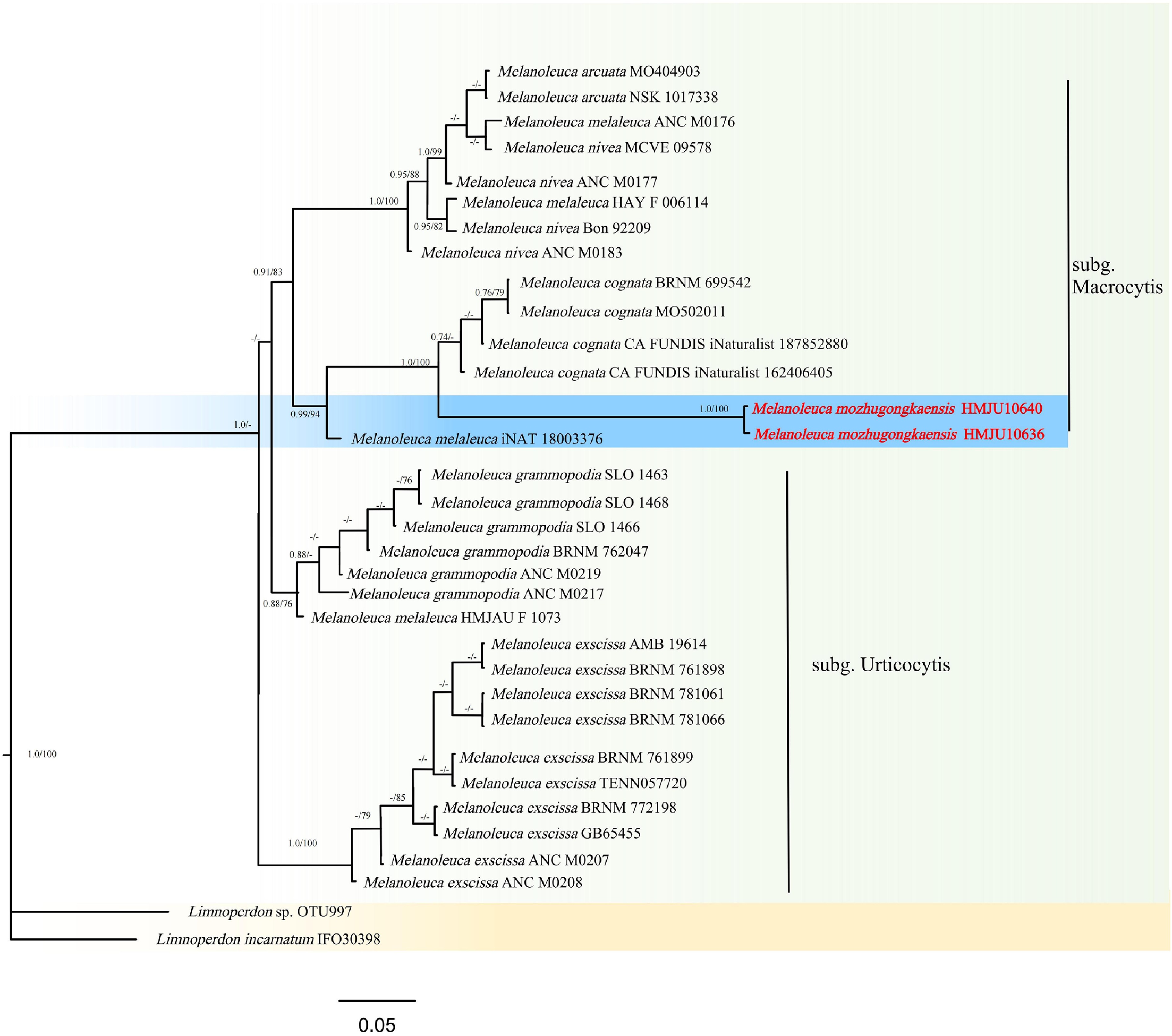
Figure 30.
Phylogram of Maximum Likelihood phylogenetic analysis based on combined ITS, nLSU, rpb2, and tef-1α sequences. The analysis includes 34 strains; total characters: 2,256 (ITS: 510, nLSU: 519, rpb2:720, tef-1α: 507). Limnoperdon sp. (OTU997) and Limnoperdon incarnatum (IFO30398) were used as the outgroup taxa. The best model was HKY + FO + G4. Estimated base frequencies were as follows: A = 0.257, C = 0.225, G = 0.232, T = 0.286. Bootstrap values for ML equal to or greater than 70% and BYPP values equal to or greater than 0.70 are labelled on the nodes. The type strains are in bold.
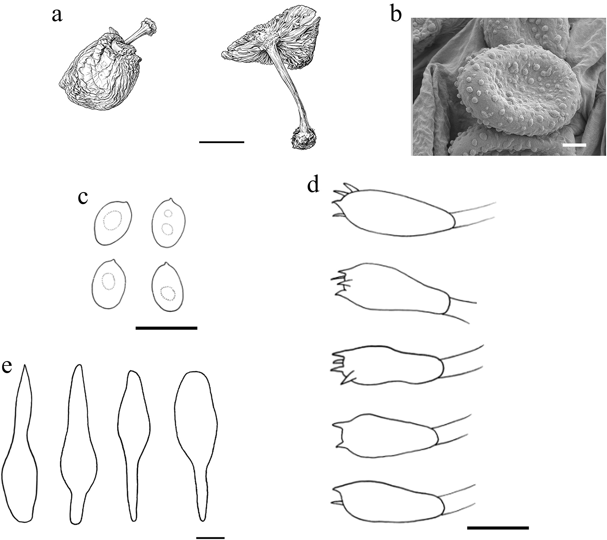
Figure 31.
Melanoleuca mozhugongkaensis (HMJU 10640, holotype). (a) Basidiocarps. (b) SEM images of basidiospores. (c) Basidiospores. (d) Basidia. (e) Pleurocystidia. Scale bars: (a) 5 cm; (b) 3µm; (c)–(e) 10 µm.
MycoBank Names: MB863404
Holotype: Xizang Autonomous Region, Lhasa City, Mozhugongka County, on soil, 29°47'32" N, 91°45'21" E, 13 July 2024, H. Zhang (HMJU 10640, holotype).
Etymology: The specific epithet 'mozhugongkaensis' refers to the type locality, Mozhugongka County, Xizang, China.
Diagnosis: Melanoleuca mozhugongkaensis is characterized by a pileus margin reflexed and elongate at maturity; colored snuff brown or silver grey, stipe base swollen; cheilocystidia lacking.
Description: Pileus 66–129 mm diam., applanate, smooth, umbo burnt umber (6F6), the remainder brownish beige (6E3), margin reflexed and elongated when mature, pileus context thin, pale grey (1B1). Lamellae free, crowded, marble white (5B2), margin entire. Stipe 55–93 mm long, 1–2 mm thick, cylindrical, center slightly depressed, base swollen, snuff brown (5F6) or silver grey (2B2), stipe context pale grey (1B1) to snuff brown (5F6). Annulus absent.
Basidiospores (6.2–)7.2–11.9(–12.5) × 4.5–9.0(–10.0) μm, Q = 1.21–1.72, Qm = 1.41, broadly ellipsoidal to rectangular, thick-walled, without amyloid. Basidia (12–)13–29(–30) × (6–)7–10(–12) μm, clavate, both four and two sterigmata to 3 μm, transparent in KOH. Cheilocystidia absent. Pleurocystidia (34–)36–66(–78) × (11–)12–17(–18) μm, urceolate, transparent in KOH. Lamella trama regular, hyphae 4–20 μm wide, transparent in KOH. Clamp connections absent in all hyphae.
Habitat: Scattered on the soil.
Known distribution: Known only from Southwestern China.
Additional material examined: Xizang, Linzhi City, Gongbujiangda County, on soil, 29°47'32" N, 91°45'21" E, 6 July 2024, H. Zhang (HMJU 10636).
Notes: Forms a sister group with Melanoleuca cognata (Fr.) Konrad & Maubl., but it exhibits striking morphological differences (ML/PP = 94/0.99). Morphologically similar to Melanoleuca schumacheri (Fr.) Singer, Me. cognata, and Melanoleuca rasilis (Fr.) Singer. However, Me. schumacheri has a different pileus with a pinkish luster, lamellae margins dentate, and smaller spores (5–6.5 × 7.5–9 μm)[160]. Me. cognata features a pileus with small protrusions, lamellae initially white, pleurocystidia, and cheilocystidia morphologically diverse[166]. Me. rasilis is typified with a pileus viscid when moist, lamellae grayish white, and spores smaller (7–8 × 6–7.5 µm)[160].
Mycena (Pers.) Roussel, Fl. Calvados, Edn 2: 64 ['46'] (1806)
With a global diversity of roughly 600 species, Mycena (Pers.) Roussel stands as one of the most speciose genera within the order Agaricales. Maas Geesteranus[167,168] formulated an infrageneric taxonomy for the genus that synthesizes microscopic and macroscopic features. In this system, species boundaries are defined macroscopically by the coloration of the basidiomata, specifically regarding the stipe, pileus, and the faces and edges of the lamellae.
Distinct pigmentation patterns characterize various sections. Bright hues such as yellow, white, red, or pink are typical of species formerly assigned to sect. Adonideae (Fr.) Quél. (now recognized as the independent genus Atheniella Redhead, Moncalvo, Vilgalys, Desjardin & B.A. Perry), as well as those in sect. Oregonenses Maas Geest. and sect. Aciculae Kühner ex Singer. Conversely, prominent violet tones are found in members of sect. Calodontes (Fr. ex Berk.) Quél., while dark pigmentation is a characteristic of sect. Rubromarginatae Singer ex Maas Geest.[169−172]. Microscopically, the morphology of the stipitipellis, pileipellis, cheilocystidia, and basidiospores is considered essential for subdividing the genus. Nevertheless, a comprehensive, unified classification for Mycena is currently lacking, and the morphology-based system proposed by Maas Geesteranus has yet to be fully validated. Recent phylogenetic evidence indicates that while certain sections—such as Sacchariferae Kühner ex Singer, Calodontes, and Amparoina T. Bau & Q. Na—are monophyletic, others are not[173]. Consequently, several taxa traditionally housed in Mycena, such as the Atheniella group, have been removed, and others may require transfer to genera including Cruentomycena R.H. Petersen, Kovalenko & O.V. Morozova, Favolaschia (Pat.) Pat., Hemimycena Singer, Panellus P. Karst., Resinomycena Redhead & Singer, and Roridomyces Rexer[174−178].
Currently, eight blue-tinted Mycena species spanning four sections have been documented. Five of these inhabit the Northern Hemisphere: Mycena subcaerulea Sacc. (North America); Mycena amicta (Fries) Quél. and Mycena cyanorhiza Quél. (Europe); and Mycena lazulina Har. Takah., Taneyama, Terashima & Oba and Mycena indigotica C.L. Wei & R. Kirschner (Asia)[179−181]. These five species typically exhibit a bluish sheen on the pileus or stipe. Four are assigned to sections Amictae Alexander H. Smith ex Maas Geesteranus, Sacchariferae, and Viscipelles Kühner, whereas My. indigotica remains unplaced due to its possession of tubular structures resembling those of Favolaschia[179−182].
In the Southern Hemisphere, three blue species are recognized: Mycena caesiocana Singer, Mycena cyanosyringea Singer, and Mycena interrupta (Berkeley) Sacc.[183,184]. These taxa are distributed across South America and Oceania—specifically Chile, Costa Rica, Australia, New Zealand, and New Caledonia—where they thrive in warm temperatures on decaying logs, dead wood, or tree stumps in forests dominated by trees such as Eucalyptus robusta Smith and Persea lingue (Ruiz & Pav.) Nees. These three allied species are readily distinguishable: My. caesiocana and My. cyanosyringea possess extremely small basidiomata (dimensions < 3 mm) and a storm-grey pileus, whereas Mycena interrupta (Berk.) Sacc. is defined by a blue stipe base. Additionally, the Chilean species Mycena cyanocephala Singer is regarded as a synonym of My. interrupta[184]. Although the northern species My. cyanorhiza also features a blue stipe base, it differs from My. interrupta by its pale-grey to pale-brown pileus and smaller cheilocystidia and basidiospores[172].
Mycena atrorubina J.Z. Xu, sp. nov. Figs 32 and 33
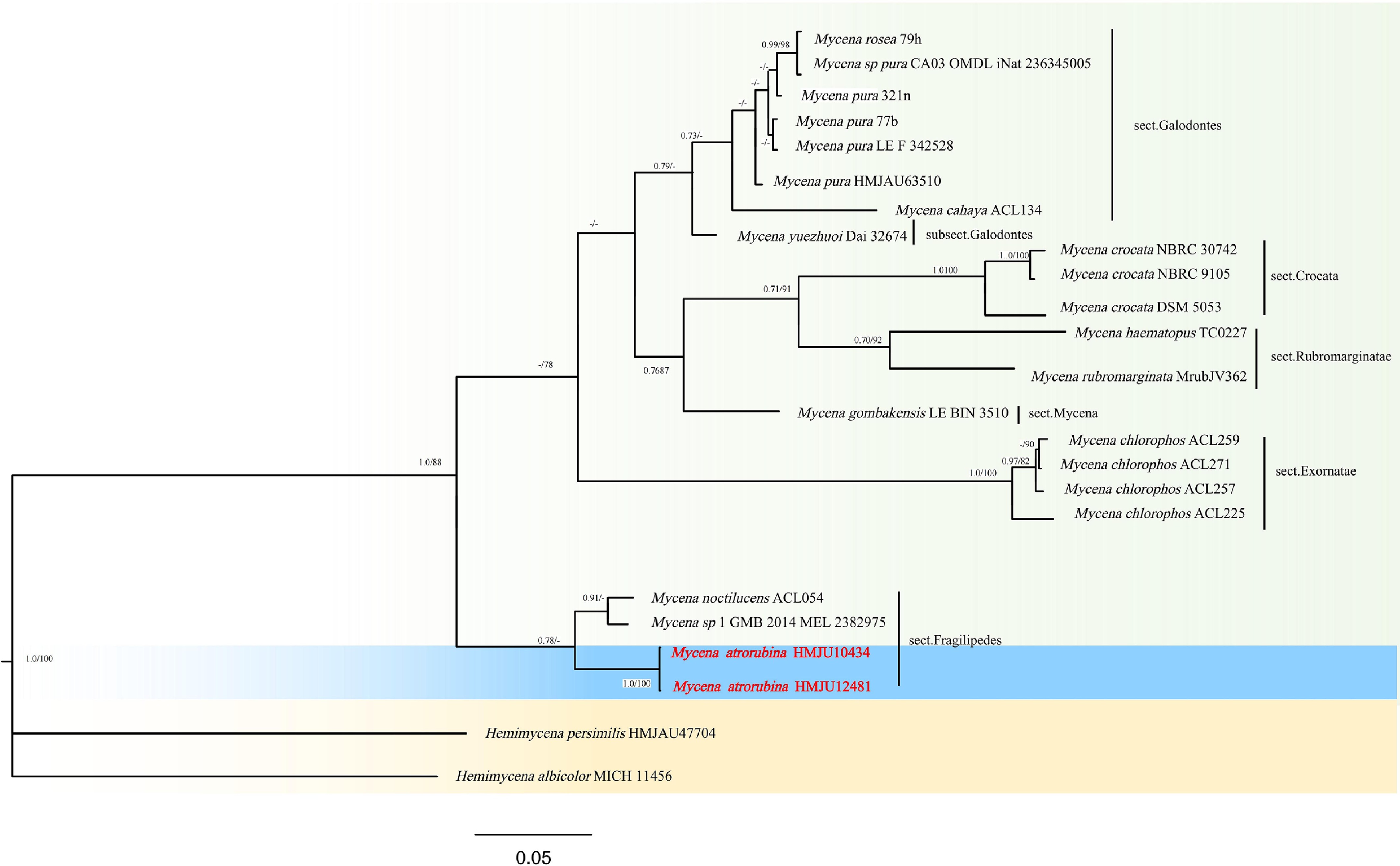
Figure 32.
Phylogram of Maximum Likelihood phylogenetic analysis based on combined ITS, nLSU, rpb2, and tef-1 sequences. The analysis includes 24 strains; total characters: 2,457 (ITS: 441, nLSU: 969, rpb2: 642, tef-1: 405). Hemimycena persimilis (HMJAU47704) and Hemimycena albicolor (MICH 11456) were used as the outgroup taxa. The best model was TIM2e + G4. Estimated base frequencies were as follows: A = 0.250, C = 0.250, G = 0.250, T = 0.250. Bootstrap values for ML equal to or greater than 70% and BYPP values equal to or greater than 0.70 are labelled on the nodes. The type strains are in bold.
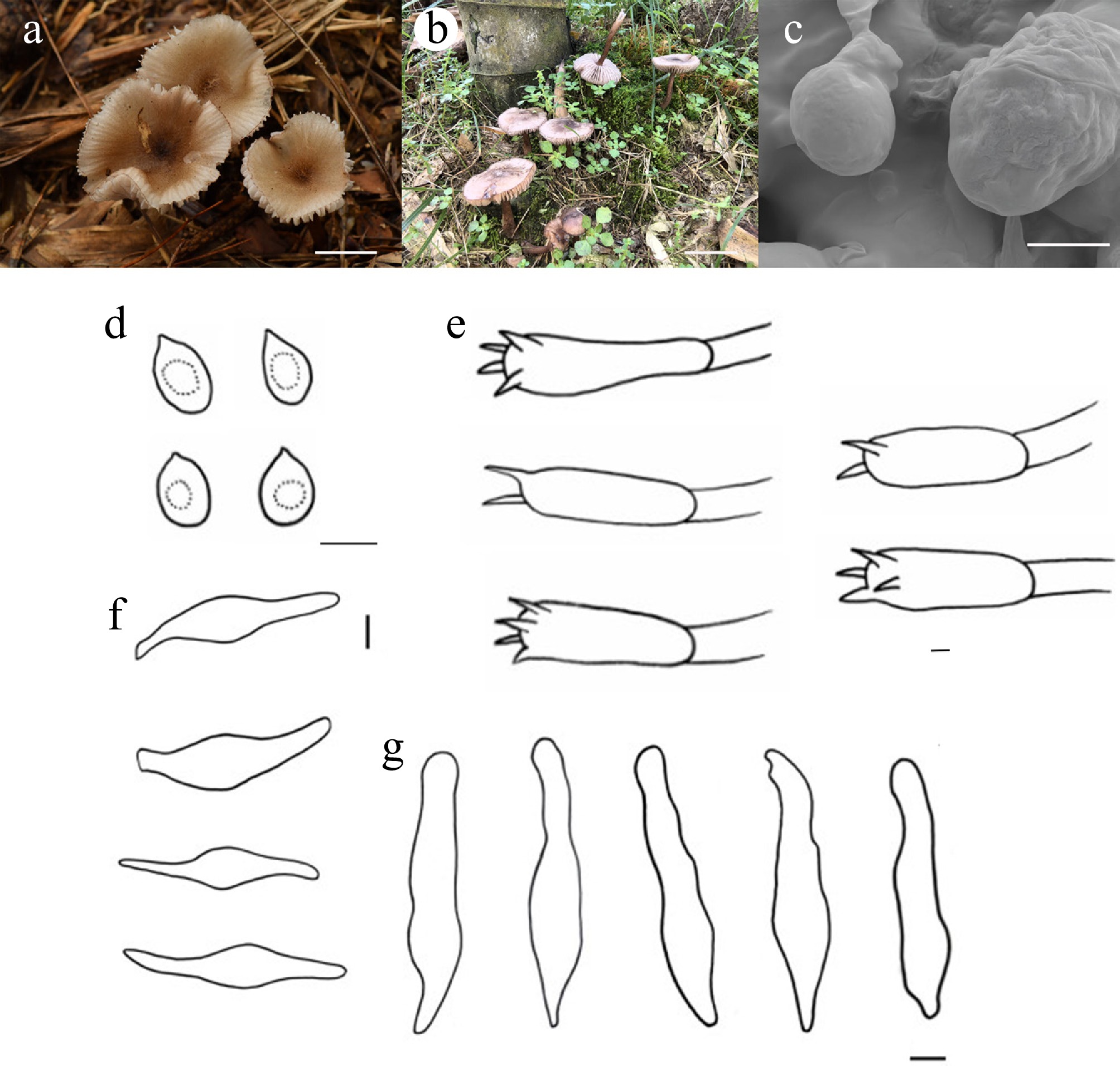
Figure 33.
Mycena atrorubina (HMJU 10434, holotype). (a), (b) Basidiocarps. (c) SEM images of basidiospores. (d) Basidiospores. (e) Basidia. (f) Pleurocystidia. (g) Cheilocystidia. Scale bars: (a) 3 cm; (b) 5 cm; (c)–(e), (g) 5 µm; and (f) 10 µm.
MycoBank Names: MB863405
Holotype: Fujian Province, Sanming City, Yong'an City, Keshan Village, on soil, 25°52'45" N, 117°20'32" E, 29 June 2024, J.P. Liao (HMJU 10434, holotype).
Etymology: The specific epithet 'atrorubina' refers to the pileus covered with dark-ruby (12F8) filaments, a diagnostic morphological character of the new species.
Diagnosis: Mycena atrorubina is characterized by a pileus adorned with dark-ruby striations and filaments; lamellae pale reddish white, margin caput mortuum, interlamellar transverse white striations present; cheilocystidia urceolate; pleurocystidia fusoid; clamp connections present.
Description: Pileus 41–100 mm diam., applanate, center slightly depressed, dingy hue, center dark ruby (12F8), covered with dark ruby (12F8) striations and filaments on reddish-grey (10B2) background, pileus context thick, milk white (1A2). Lamellae adnexed, distant, pale reddish white (10A2), margin caput mortuum (8F7), transverse white (1A1) striations between lamellae. Stipe 48–101 mm long, 3–6 mm thick, cylindrical, twisted, center depressed, oxied red (8E8), covered with eye brown (7E6) scales, stipe context milk white(1A2). Annulus absent.
Basidiospores (3.5–)4.1–10.2(–10.8) × (2.5–)3.0–8.0(–8.1) μm, Q = 1.01–1.89, Qm = 1.38, globose to broadly rectangular, transparent in KOH, without amyloid. Basidia (14–)15–29(–32) × (5–)6–9(–10) μm, clavate, both 4 and 2 sterigmata to 4 μm, transparent in KOH. Cheilocystidia (24–)25–61(–64) × (5–)6–12(–13) μm, urceolate, transparent in KOH. Pleurocystidia (50–)55–78(–87) × (12–)13–17(–18) μm, fusoid, transparent in KOH. Lamella trama regular, hyphae 13–22 μm wide, transparent in KOH. Clamp connections present in all hyphae.
Habitat: Gregarious in deciduous broad-leaved forests.
Known distribution: Known only from Southern China.
Additional material examined: Fujian Province, Sanming City, Yong'an City, Shangping Village, on soil, 25°52'45" N, 117°20'32" E, 14 October 2023, J.P. Liao (HMJU 12481).
Notes: Forms a sister group with Mycena noctilucens Corner, but exhibits significant macroscopic differences (ML/PP = 50/0.78). Morphologically similar to My. noctilucens, Mycena pura (Pers.) P. Kumm., Mycena rosea Gramberg, and Mycena pearsoniana Dennis ex Singer. But, My. noctilucens is different with pileus pale brown to gray, lamellae white to pale gray, clamp connections absent[185]. My. pura is distinguished by a pileus that is white or yellow, a white stipe, and spores that are amyloid[59]. My. rosea is characterized by a pileus without yellow color at the center, lamellae pink, a stipe base densely covered with white or pale-yellow fibrils, amyloid spores, and pleurocystidia and cheilocystidia morphologically diverse. My. pearsoniana differs in concave lamellae margins, and pleurocystidia absent[150].
Phloeomana Redhead, Index Fungorum 15: 2 (2013)
Phloeomana speirea, frequently referred to by the common name 'bark bonnet', is a member of the family Porotheleaceae. Typically found in temperate forest ecosystems, this agaric inhabits tree bark and produces fruit bodies that range in color from whitish to fuscous. Morphologically, these basidiomes are notable for displaying a blend of both omphalinoid and mycenoid characteristics.
The species was first formally described by Elias Fries, who originally named it Agaricus speireus. It was later reclassified the taxon to the newly erected genus Phloeomana, designating it as the type species.
Phloeomana flavomaculata J.Z. Xu, sp. nov. Figs 34 and 35
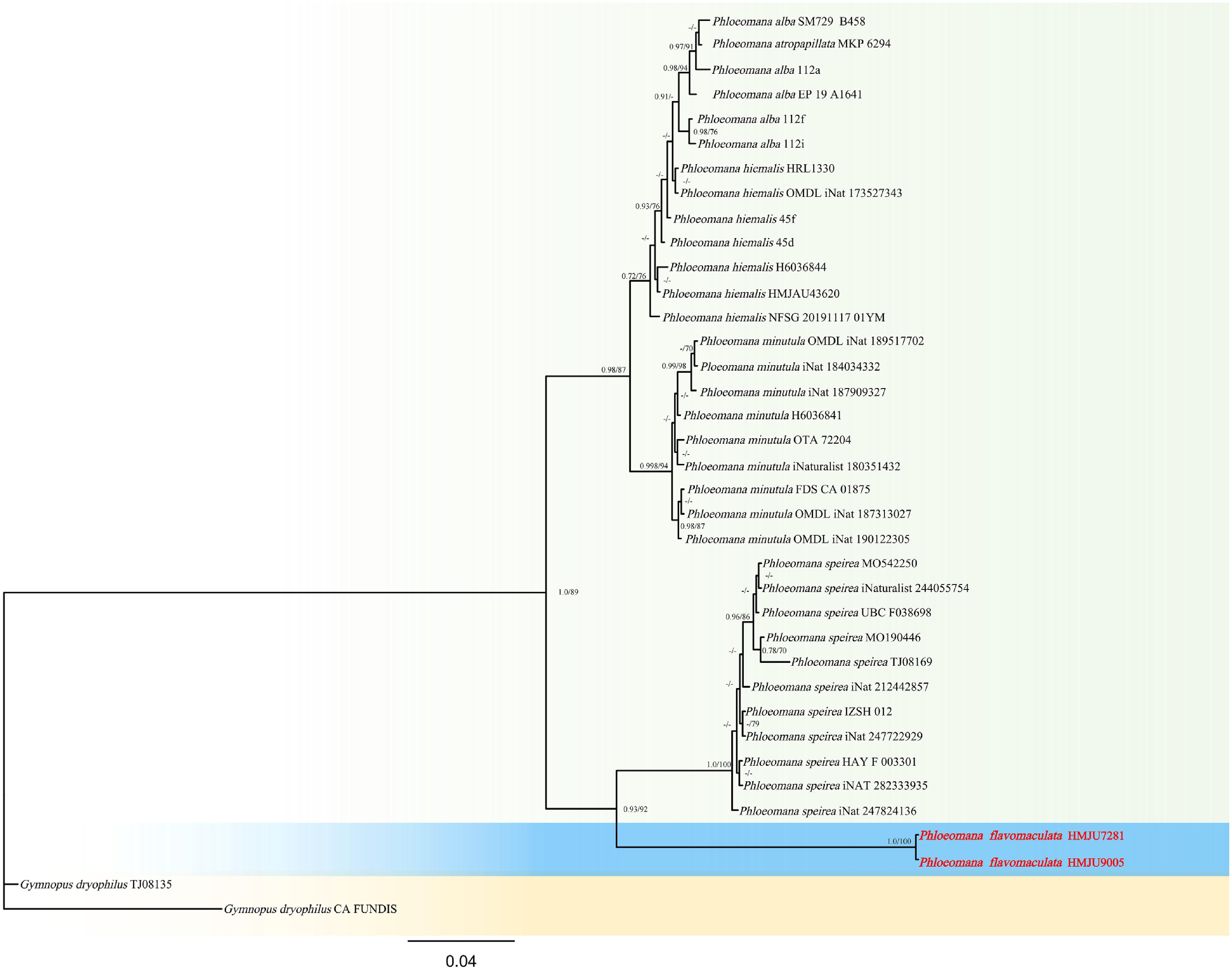
Figure 34.
Phylogram of Maximum Likelihood phylogenetic analysis based on combined ITS, nLSU. The analysis includes 37 strains; total characters: 1,748 (ITS: 920, nLSU: 828). Gymnopus dryophilus (TJ08135) and Gymnopus dryophilus (CA FUNDIS) were used as the outgroup taxa. The best model was TPM2u + F + G4. Estimated base frequencies were as follows: A = 0.258, C = 0.209, G = 0.206, T = 0.327. Bootstrap values for ML equal to or greater than 70% and BYPP values equal to or greater than 0.70 are labelled on the nodes. The type strains are in bold.
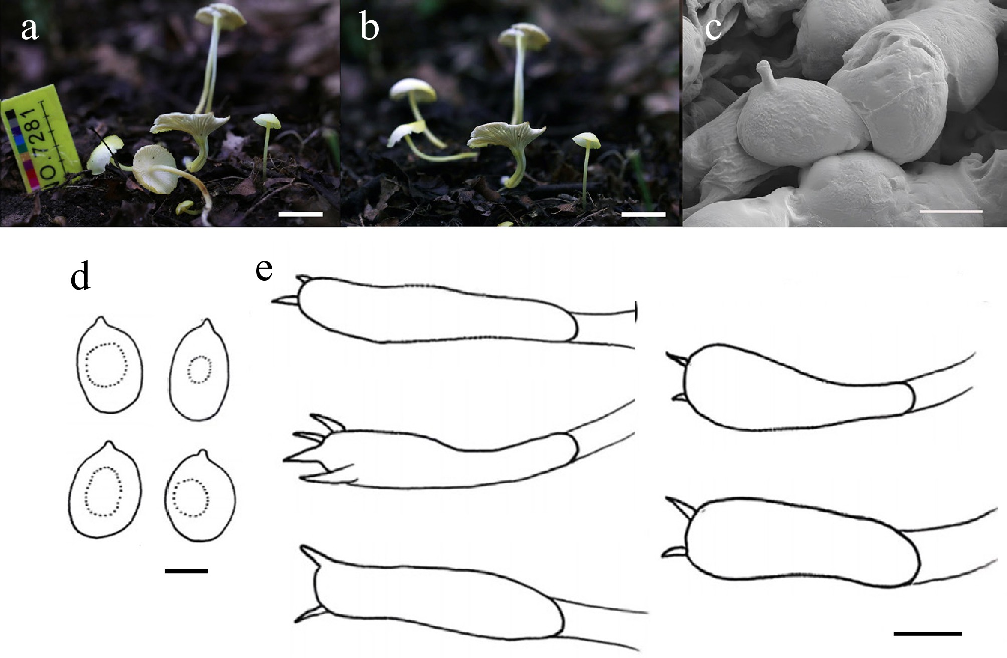
Figure 35.
Phloeomana flavomaculata (HMJU 7281, holotype). (a), (b) Basidiocarps. (c) SEM images of basidiospores. (d) Basidiospores. (e) Basidia. Scale bars: (a), (b) 1 cm; (c) 3 µm; and (d), (e) 5 µm.
MycoBank Names: MB863406
Holotype: Jilin Province, Baishan City, Hunjiang District, Hangou Village, on soil, 42°01'40" N, 126°25'37" E, 26 July 2023, J.Z. Xu (HMJU 7281, holotype).
Etymology: The specific epithet 'flavomaculata' refers to the irregular yellow maculae on the pileus, a diagnostic characteristic that distinguishes this species from other species in Phloeomana.
Diagnosis: Phloeomana flavomaculata is characterized by a pileus that is pastel yellow, otherwise white, with a thin, translucent margin extending to the lamellae; the lamellae margin is lead grey and serrate; the stipe is erect or flexuous, pastel yellow, and darkens gradually to somali basally; cheilocystidia and pleurocystidia are lacking.
Description: Pileus 9–26 mm diam., convex when young, applanate in age, smooth, pastel yellow (2A4), remainder white (1A1), margin thin and translucent, extending to the lamellae, pileus context thin, milk white(1A2). Lamellae free or decurrent, crowded, white (1A1), margin lead grey (2D2); margin serrate. Stipe 22–42 mm long, 2–3 mm thick, cylindrical, erect or flexuous, pastel yellow (2A4), gradually darkening somali (7E5) toward the stipe base, stipe context milk white(1A2).
Basidiospores (3.7–)4.3–7.2(–7.6) × (2.5–)3.0–5.0(–5.3) μm, Q = 1.10–2.20, Qm = 1.56, subglobose to broadly cylindrical, surface tuberculate, transparent in KOH. Basidia (14–)15–26(–29) × (2–)3–9(–10) μm, clavate, both four and two sterigmata up to 1 μm, apex bearing refractive material, transparent in KOH. Cheilocystidia absent. Pleurocystidia absent. Lamella trama regular, hyphae 2–8 μm wide, transparent in KOH. Clamp connections absent in all hyphae.
Habitat: Solitary in broad-leaved forests.
Known distribution: Known only from Northeastern China.
Additional material examined: Jilin Province, Baishan City, Hunjiang District, Hangou Village, on soil, 42°01'40" N, 126°25'37" E, 5 July 2024, J.Z. Xu (HMJU9005).
Notes: This species forms a sister group with Phloeomana speirea but exhibits significant morphological differences (ML/PP = 92/0.93). Morphologically similar to Ph. speirea, Phloeomana minutula (Sacc.) Redhead, and Phloeomana alba (Bres.) Redhead, but Ph. speirea is distinguished by a pileus centrally depressed, pale grayish brown or pale yellowish brown, a stipe pale yellow or brown, and cheilocystidia morphologically diverse[186]. Ph. minutula is characterized by veined lamellae, basidia larger (22–27 × 6–7 μm), pleurocystidia, and cheilocystidia diverse[187]. Ph. alba has a pileus with a faint pink tinge, a pale gray stipe, and larger spores (6.5–8 μm)[187].
Pseudobaeospora Singer, Lloydia 5: 129 (1942)
The genus Pseudobaeospora Singer became a significant subject of scientific interest following the work of Bas[188], who demonstrated that Dutch herbarium specimens actually represented two undescribed species, rather than the two European taxa previously recognized. This discovery precipitated a surge of taxonomic activity across Europe[189−192], which collectively expanded the genus by adding 15 new species, two varieties, and two provisionally described taxa. Furthermore, Bas[190] amended Singer's original definition of the genus; the revised circumscription now includes taxa displaying a hymeniform pileipellis—in addition to those with a cutis-type structure, as well as species whose basidiocarps undergo color changes when treated with KOH.
More recently, a second phase of taxonomic research has emerged, focusing on the critical re-examination of established species[193]. Geographically, the genus extends well beyond Europe; Desjardin[194] identified five species in North America. Taxonomists rely on diagnostic traits to delimit species within Pseudobaeospora, including basidiocarp, spores, and the presence or absence of both cheilocystidia and clamp connections.
Pseudobaeospora vulpecula J.Z. Xu, sp. nov. Figs 36 and 37
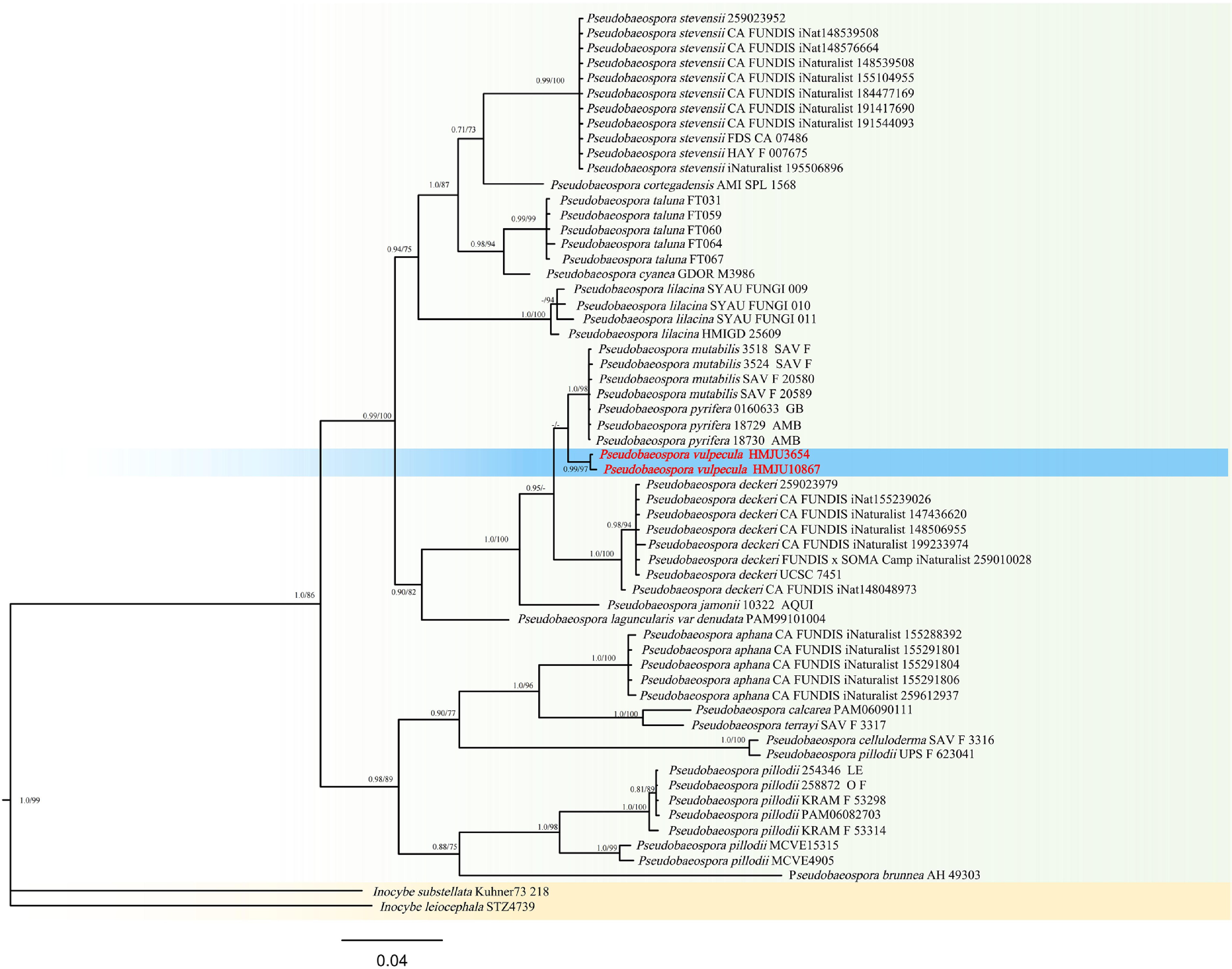
Figure 36.
Phylogram of Maximum Likelihood phylogenetic analysis based on combined ITS, nLSU, rpb2, and tef-1α sequences. The analysis includes 60 strains; total characters: 2,248 (ITS: 570, nLSU: 667, rpb2: 507, tef-1α: 504). Inocybe substellata (Kuhner73 218) and Inocybe leiocephala (STZ4739) were used as the outgroup taxa. The best model was GTR + F + G4. Estimated base frequencies were as follows: A = 0.278, C = 0.191, G = 0.232, T = 0.299. Bootstrap values for ML equal to or greater than 70% and BYPP values equal to or greater than 0.70 are labelled on the nodes. The type strains are in bold.
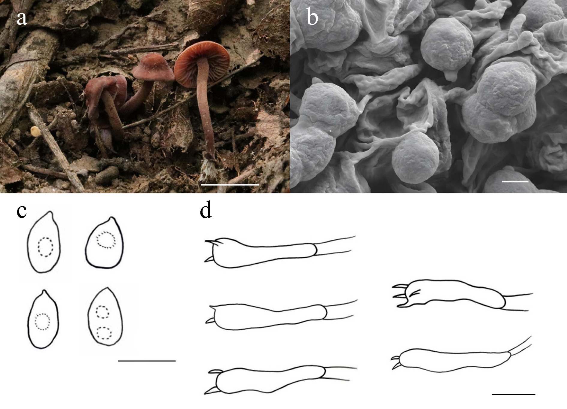
Figure 37.
Pseudobaeospora vulpecula (HMJU 3654, holotype). (a) Basidiocarps. (b) SEM images of basidiospores. (c) Basidiospores. (d) Basidia. Scale bars: (a) 5 cm; (b) 3 µm; and (c), (d) 5 µm.
MycoBank Names: MB863407
Holotype: Liaoning Province, Fuxin City, Haitang Mountain, on soil, 41°55'15" N, 121°49'21" E, 25 July 2021, J.Z. Xu (HMJU 3654, holotype).
Etymology: The specific epithet 'vulpecula' refers to the diagnostic morphological characteristic of the new basidiome, which is fox colored.
Diagnosis: Pseudobaeospora vulpecula is characterized by a campanulate pileus, umbo fox and slightly obtuse; umbo margin extending to 2/3 of the pileus, madeira, remainder cube, entirely white tomentose; stipe adorned with tiny white fibrils.
Description: Pileus 19–31 mm diam., campanulate, fox (8D7) umbo slightly obtuse, umbo margin towards pileus 2/3 madeira (8E5), the remainder cube (9E8), entirely covered with white (1A1) tomentose, pileus context thick, madeira (8E5). Lamellae free, slightly distant, reddish grey (10B2); margin entire. Stipe 37–59 mm long, 4–5 mm thick, cylindrical, fox (8D7), covered with tiny white (1A1) fibrils, stipe context fox (8D7).
Basidiospores (2.8–)3.1–5.7(–8.3) × (1.8–)2.2–4.0(–4.1) μm, Q = 1.01–2.90, Qm = 1.39, globose to broadly cylindrical, transparent in KOH, surface with gyrose ornamentation. Basidia (11–)12–23(–24) × (1–)2–5(–6) μm, clavate, both four and two sterigmata to 2 μm, transparent in KOH. Cheilocystidia absent. Pleurocystidia absent. Lamella trama regular, hyphae 3–8 μm wide, transparent in KOH. Clamp connections absent in all hyphae.
Habitat: Scattered to gregarious on soil among leaf litter and decaying wood in forests.
Known distribution: Known only from Northeastern China.
Additional material examined: Liaoning Province, Fuxin City, Haitang Mountain, on soil, 41°55'15" N, 121°49'21" E, 13 July 2025, J.Z. Xu (HMJU10867).
Notes: Forms a sister group with Pseudobaeospora pyrifera Bas & L.G. Krieglst. and Pseudobaeospora mutabilis Bas & Adamcík (ML/PP = 56/0.72). Pse. pyrifera is different in lamellae margins slightly irregular; cheilocystidia morphologically diverse; clamp connections present[193]. Pse. mutabilis has a low umbo, smaller spores (3.0–3.7 × 2.7–3.5 μm), clavate cheilocystidia, and clamp connections[195]. Morphologically most similar to Pseudobaeospora aciculifera Voto & Soop, Pseudobaeospora deckeri Schwarz, and Pseudobaeospora aphana Vellinga. Pse. aciculifera is characterized by a pileus that is curved at maturity, basidia larger (19.5–28 × 4.5–5.7 μm), and cheilocystidia that are morphologically diverse[196]. Pse. deckeri is typified with lamellae with anastomoses, lamellae slightly irregular, stipe squamulose, spores smaller (3.6–3.8 × 3.3–3.5 μm), and clamp connections present[197]. Pse. aphana is unique in that its pileus is brownish, its lamellae are cream yellow, and clamp connections are present[198].
Pseudosperma Matheny & Esteve-Rav., in Matheny, Hobbs & Esteve-Raventós, Mycologia 112(1): 93 (2019)
When defined in the broad sense (sensu lato), Inocybe (Fr.) Fr. (order Agaricales, family Inocybaceae) constitutes a highly diverse genus of ectomycorrhizal fungi, containing approximately 735 globally distributed species[199]. While the genus exhibits a cosmopolitan range, it is frequently encountered in temperate zones but appears less often in tropical environments[200,201]. The monophyletic status of the Inocybaceae has been substantiated through multi-locus phylogenetic studies performed by Matheny et al.[200]. Within the family, Matheny delineated seven primary lineages, named each clade, and proposed they be informally recognized as distinct genera.
One of these seven major lineages is the Pseudosperma clade[200], which corresponds to Inocybe section Rimosae in the strict sense[201,202]. Historically, this group was categorized within subgenus Inosperma[203,204]. Morphologically, taxa belonging to this clade are distinguished by a rimose pileus surface, a stipe surface ranging from furfuraceous to furfuraceous-fibrillose, and the absence of both pleurocystidia and metuloids. Additionally, they possess cylindrical to clavate cheilocystidia and smooth basidiospores that are elliptical or vaguely phaseoliform. Although species in the Mallocybe and Inosperma clades[200], along with the genera Auritella Matheny & Bougher and Tubariomyces Esteve-Rav. & Matheny, similarly lack pleurocystidia, the Pseudosperma clade is distinct because its basidia are hyaline and do not show necropigmentation. The Nothocybe clade is monotypic; its single species also lacks pleurocystidia but is differentiable via molecular phylogenetic analysis. Within the Pseudosperma clade, certain lineages are composed of numerous cryptic species. These fungi establish ectomycorrhizal relationships with a diverse range of plant hosts, encompassing both angiosperms and gymnosperms[203].
Based on a six-locus phylogenetic study of the Inocybaceae, Matheny et al.[205] formally assigned generic status to these clades. The proposed genera include Inocybe sensu stricto, Nothocybe Matheny & K.P.D. Latha, and Pseudosperma Matheny & Esteve-Rav., as well as Inosperma (Kühner) Matheny & Esteve-Rav. and Mallocybe (Kuyper) Matheny, Vizzini & Esteve-Rav., both of which were elevated from subgeneric rank. These newly recognized genera complement the previously described Auritella and Tubariomyces. The authors opted to formalize this generic classification to facilitate clearer scientific communication and to offer the taxonomic resolution necessary for identifying biodiversity hotspots and advancing conservation initiatives.
Pseudosperma lacteoconicum J.Z. Xu, sp. nov. Figs 38 and 39
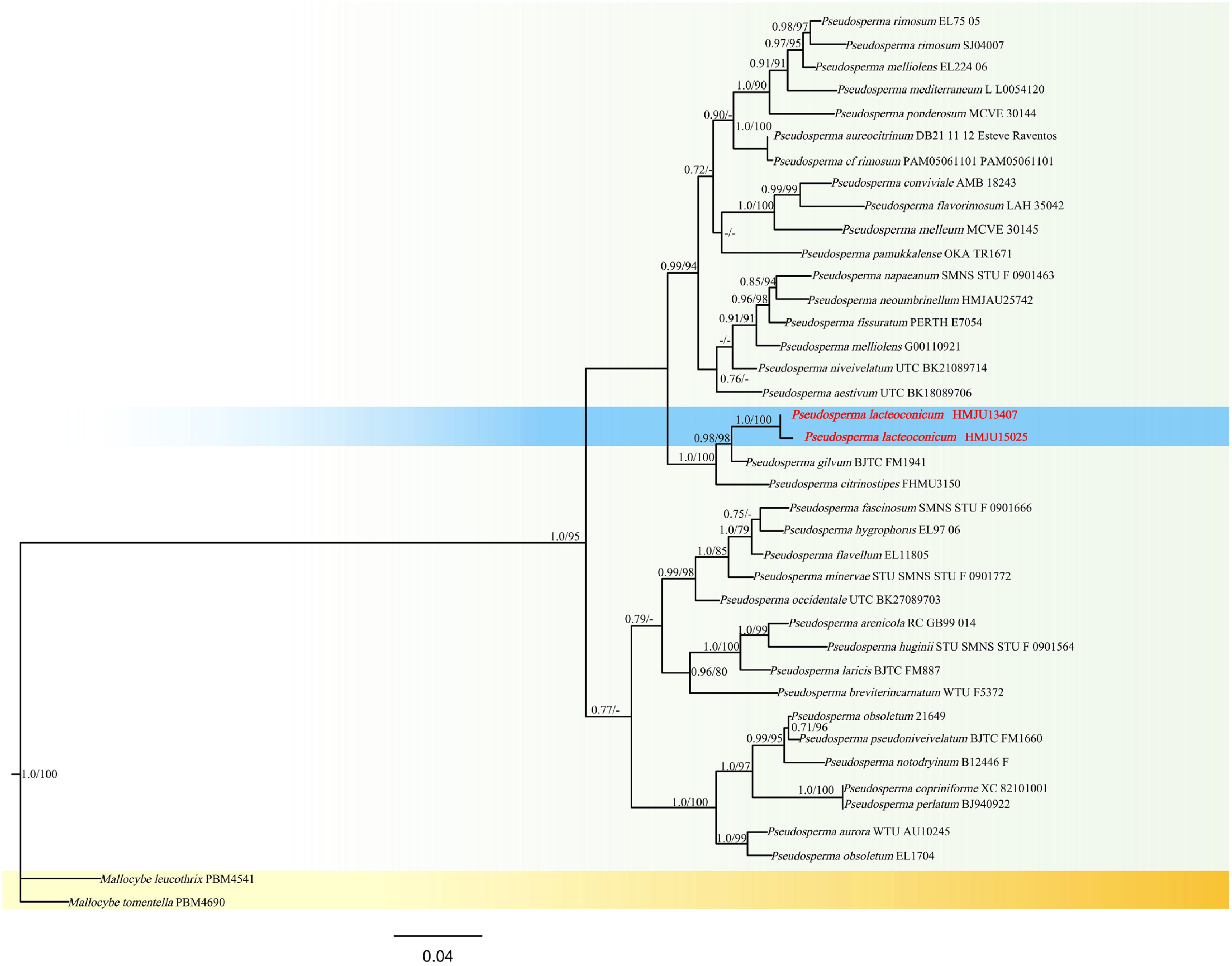
Figure 38.
Phylogram of Maximum Likelihood phylogenetic analysis based on combined ITS, nLSU, rpb2. The analysis includes 39 strains; total characters: 2,115 (ITS: 701, nLSU: 847, rpb2: 567). Mallocybe leucothrix (PBM4541) and Mallocybe tomentella (PBM4690) were used as the outgroup taxa. The best model was TIM2 + F + G4. Estimated base frequencies were as follows: A = 0.258, C = 0.205, G = 0.205, T = 0.333. Bootstrap values for ML equal to or greater than 70% and BYPP values equal to or greater than 0.70 are labelled on the nodes. The isolate of the current study is in purple, while the type strains are in bold.
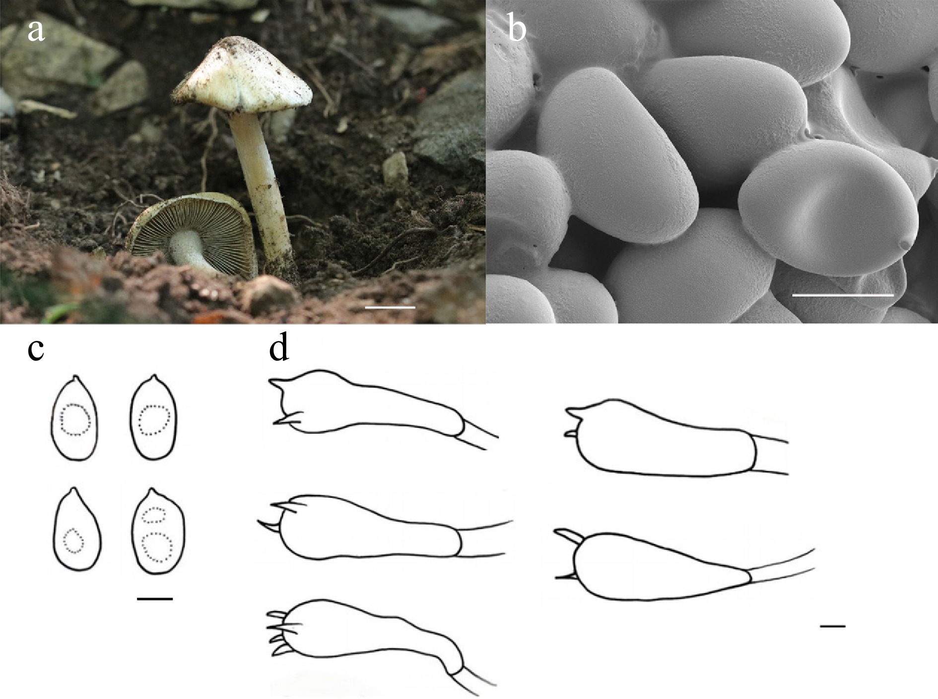
Figure 39.
Pseudosperma lacteoconicum (HMJU 13407, holotype). (a) Basidiocarps. (b) SEM images of basidiospores. (c) Basidiospores. (d) Basidia. Scale bars: (a) 1 cm; (b)–(d) 5 µm.
MycoBank Names: MB863408
Holotype: Liaoning Province, Chaoyang City, Shuangta District, Fenghuang Mountain, on soil, 41°32'56" N, 120°30'55" E, 18 August 2025, J.Z. Xu (HMJU 13407, holotype).
Etymology: The specific epithet 'lacteoconicum' refers to the milky-white color of the entire basidcarp and the conical pileus, which distinguishes it from other congeners in the genus Pseudobaeospora.
Diagnosis: Pseudosperma lacteoconicum is characterized by a milk-white pileus, a margin slightly rimose; stipe concolorous with pileus, adorned with tiny white fibrils; cheilocystidia clavate; pleurocystidia absent.
Description: Pileus 27–31 mm diam., conical, milk white (1A2), smooth, margin slightly rimose, pileus context thick, milk white (1A2). Lamellae free, crowded, putty (4B2), margin entire. Stipe 23–52 mm long, 5–7 mm thick, cylindrical, milk white (1A2), covered with tiny white (1A1) fibrils, stipe context milk white (1A2). Annulus absent.
Basidiospores (7.8–)8.4–12.8(–13.0) × (4.9–)5.4–7.8(–8.2) μm, Q = 1.30–1.94, Qm = 1.62, ellipsoidal to rectangular, transparent in KOH, without amyloid. Basidia (18–)28–46(–47) × (8–)9–14(–15) μm, clavate, both 4 and 2 sterigmata up to 2 μm long, transparent in KOH. Cheilocystidia (38–)40–55(–60) × (9–)10–13(–15) μm, clavate, hyaline in KOH. Pleurocystidia absent. Lamella trama regular, hyphae 4–7 μm wide, transparent in KOH. Clamp connections absent in all hyphae.
Habitat: Solitary in sandy soil.
Known distribution: Known only from Northeastern China.
Additional material examined: Liaoning Province, Chaoyang City, Shuangta District, Fenghuang Mountain, on soil, 41°32'56" N, 120°30'55" E, 8 September 2025, J.Z. Xu (HMJU 15025).
Notes: Phylogenetically forms a sister group with Pseudosperma gilvum L. Fan & N. Mao (ML/PP = 98/0.98). Ps. gilvum is unique in that its lamellae turn yellowish brown when mature, and its cheilocystidia are morphologically diverse[206]. Pseudosperma citrinostipes Y.G. Fan & W.J. Yu differs in lamellae fimbriate to serrate, stipe covered with yellowish fibrils, and basidia with pale yellow contents[207]. Species is morphologically similar to Pseudosperma arenarium Y.G. Fan, Fei Xu, Hai J. Li & Vauras. Ps. arenarium is characterized by a pileus that is yellowish, a stipe that is tinged with pink, spores that are larger (14–20 × 7–9.2 μm), yellowish brown, and cheilocystidia that are fusiform, with clamp connections present[208].
Rhodophana Kühner, Bull. Soc. Mycol. France 62(3-4): 193 (1947) [1946]
Two primary lineages are recognized within the family Entolomataceae Kotl. & Pouzar: the Entoloma clade and the Rhodocybe-Clitopilus clade. In terms of global diversity, the Rhodocybe-Clitopilus clade is significantly smaller, containing roughly 300 species, whereas the Entoloma clade comprises approximately 1,200 species.
Based on a phylogenetic analysis using a limited selection of representative taxa, Co-David et al.[209] proposed that the Entolomataceae should be classified into just two genera: Entoloma Fr. ex P. Kumm. sensu lato and Clitopilus (Fr. ex Rabenh.) P. Kumm. This taxonomic concept was based on the observation that the morphological traits traditionally utilized to separate segregate genera within the Entoloma clade were not supported phylogenetically; a pattern that also applied to the Rhodocybe-Clitopilus clade.
Expanding on this foundation, Baroni et al.[210] performed a phylogenetic study of the Entolomataceae with a larger sample size, using the same three gene markers as Co-David[209]. This research identified four distinct, well-supported monophyletic groups within the Rhodocybe-Clitopilus clade. Subsequently, Kluting et al.[211,212] greatly expanded the sampling of the Rhodocybe-Clitopilus clade and examined three specific partial protein-coding genes (atp6, tef1, and rpb2). Their results established five statistically robust clades, each morphologically delimitable as a distinct genus: Clitopilus (Fr. ex Rabenh.) P. Kumm., Clitopilopsis Maire, Rhodophana Kühner, a redefined Rhodocybe Maire, and the newly established genus Clitocella Kluting, T.J. Baroni & Bergemann. Phylogenetically, Clitocella was resolved as the sister taxon to Clitopilus and Rhodocybe.
Rhodophana rimosula J.Z. Xu, sp. nov. Figs 40 and 41
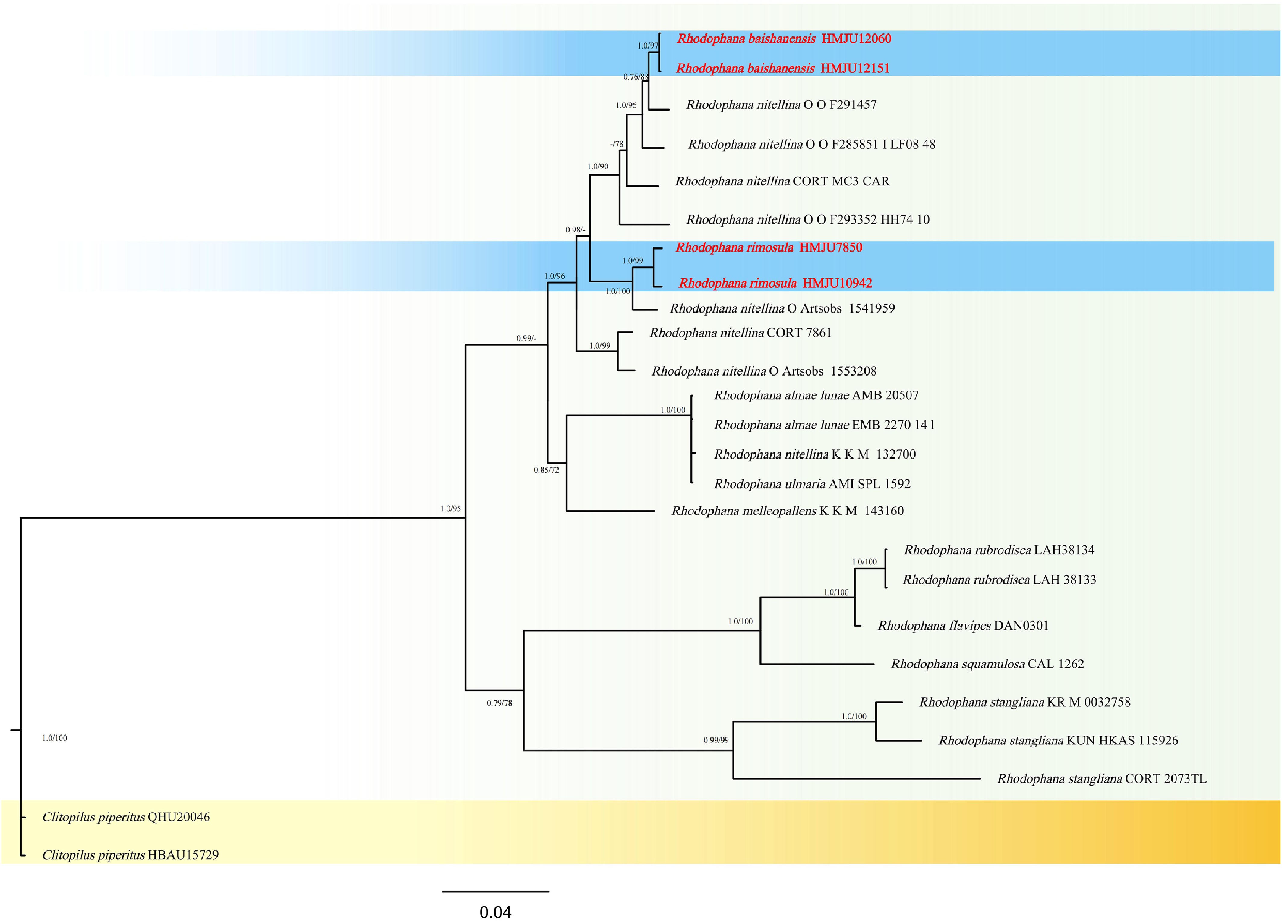
Figure 40.
Phylogram of Maximum Likelihood phylogenetic analysis based on combined ITS, nLSU, rpb2, and tef-1α sequences. The analysis includes 25 strains; total characters: 2,819 (ITS: 672, nLSU: 846, rpb2:519, tef-1α: 782). Clitopilus piperitus (QHU20046) and Clitopilus piperitus (HBAU15729) were used as the outgroup taxa. The best model was TIM2 + F + G4. Estimated base frequencies were as follows: A = 0.272, C = 0.205, G = 0.248, T = 0.276. Bootstrap values for ML equal to or greater than 70% and BYPP values equal to or greater than 0.70 are labelled on the nodes. The isolate of the current study is in purple, while the type strains are in bold.
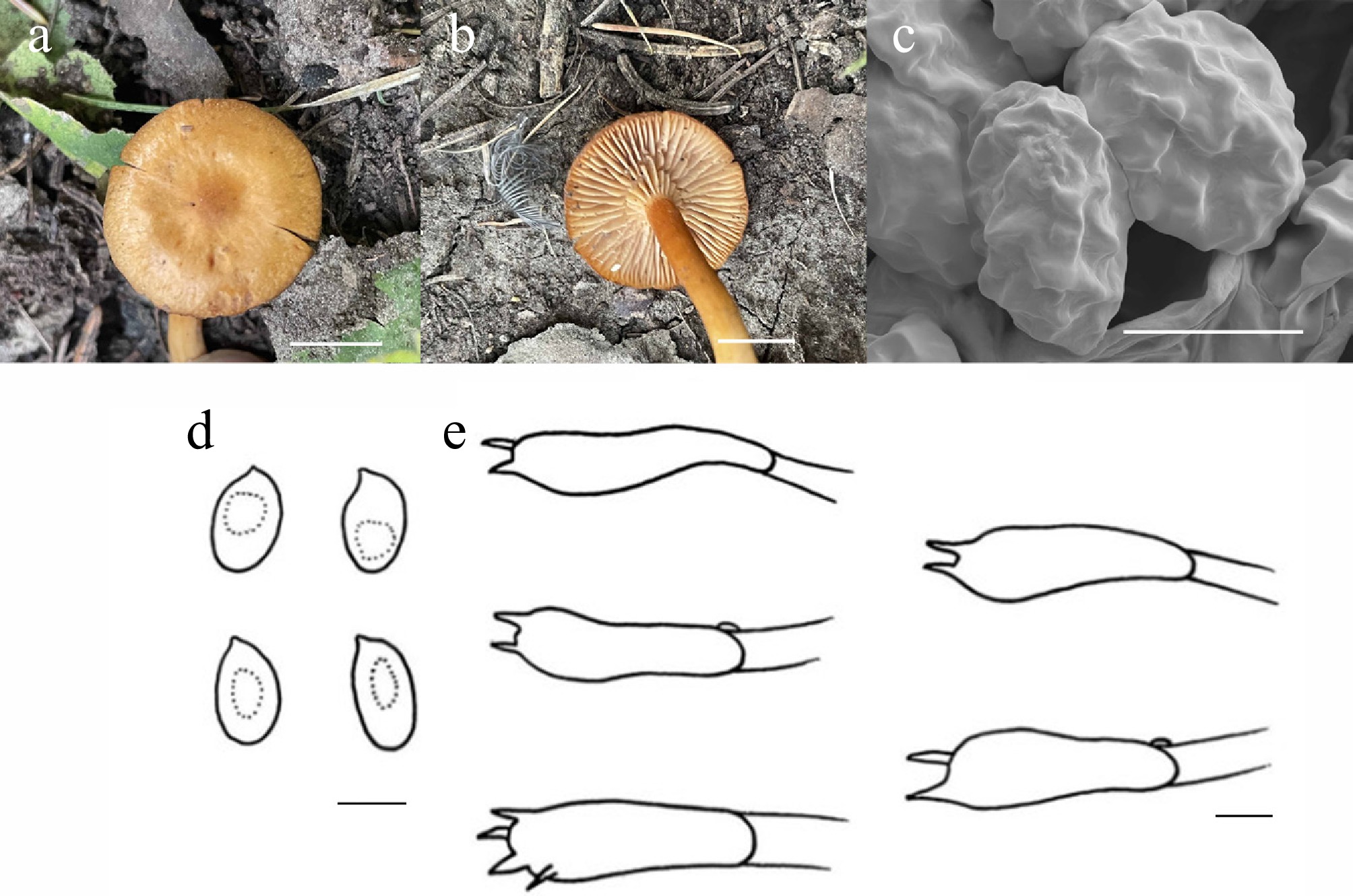
Figure 41.
Rhodophana rimosula (HMJU 7850, holotype). (a), (b) Basidiocarps. (c) SEM images of basidiospores. (d) Basidiospores. (e) Basidia. Scale bars: (a), (b) 1 cm; (c)–(e) 5 µm.
MycoBank Names: MB863409
Holotype: Shanxi Province, Xinzhou City, Wuzhai County, Luya mountain, on soil, 38°40'02" N, 111°57'31" E, 13 September 2022, L. Zhao (HMJU 7850, holotype).
Etymology: The species epithet 'rimosula' refers to the diagnostic morphological characteristic of the pileus cracking when dry.
Diagnosis: Rhodophana rimosula is characterized by a concolorous pileus and stipe, mandarin orange; pileus surface with irregular depressions or protrusions; cheilocystidia and pleurocystidia absent; clamp connections present.
Description: Pileus 20–29 mm diam., plano-convex, center depressed, brownish orange (6C8), dry, the remainder mandarin orange (6E8), margin madeira (8E5), rimose, irregular depressions or protrusions, pileus context thin, pale brownish orange (6C8). Lamellae free, slightly crowded, orange white (5A2), covered with white (1A1) fibrils on sides, margin entire. Stipe 33 –42 mm long, 1–4 mm thick, cylindrical, mandarin orange (6E8), stipe context pale brownish orange (6C8) to white.
Basidiospores (5.4–)6.1–9.4(–9.7) × (3.2–)3.6–6.3(–6.9) μm, Q = 1.29–2.04, Qm = 1.65, broadly ellipsoidal to broadly cylindrical, surface irregular, transparent in KOH, without amyloid. Basidia (17–)18–25(–26) × (6–)7–9(–10) μm, clavate, both four and two sterigmata up to 3 μm, transparent in KOH. Cheilocystidia absent. Pleurocystidia absent. Lamella trama regular, hyphae 5–12 μm wide, transparent in KOH. Clamp connections present in all hyphae.
Habitat: Solitary to scattered, saprobic on humus-rich soil in forests.
Known distribution: Known only from Northern China.
Additional material examined: Shanxi Province, Xinzhou City, Wuzhai County, Luya Mountain, on soil, 38°40'02" N, 111°57'31" E, 27 August 2023, L. Zhao (HMJU 10942).
Notes: Forms a sister group with Rhodophana nitellina (Fr.) Kühner(ML/PP = 97/1.0), but differs in having a reddish-brown pileus with a striate margin, lamellae pale cream to orange and undulate, and cystidia clavate. Species morphologically similar to Rhodophana fuscofarinacea (Noordel. & Kosonen) Cons., Noordel., Dima & Eyssart. and Rhodophana melleopallens (P.D. Orton) Kluting, T.J. Baroni & Bergemann. R. fuscofarinacea is characterized by a pileus that is brownish with papillate protrusion, a stipe that is dark brown, and basidia that are larger (21–32 × 6–9 μm)[213]. R. melleopallens differs in having a pileus with a central protrusion and a striate margin, and spores are smaller (4.5–7 × 3–4)[212].
Rhodophana baishanensis J. Z. Xu, sp. nov. Figs 40 and 42
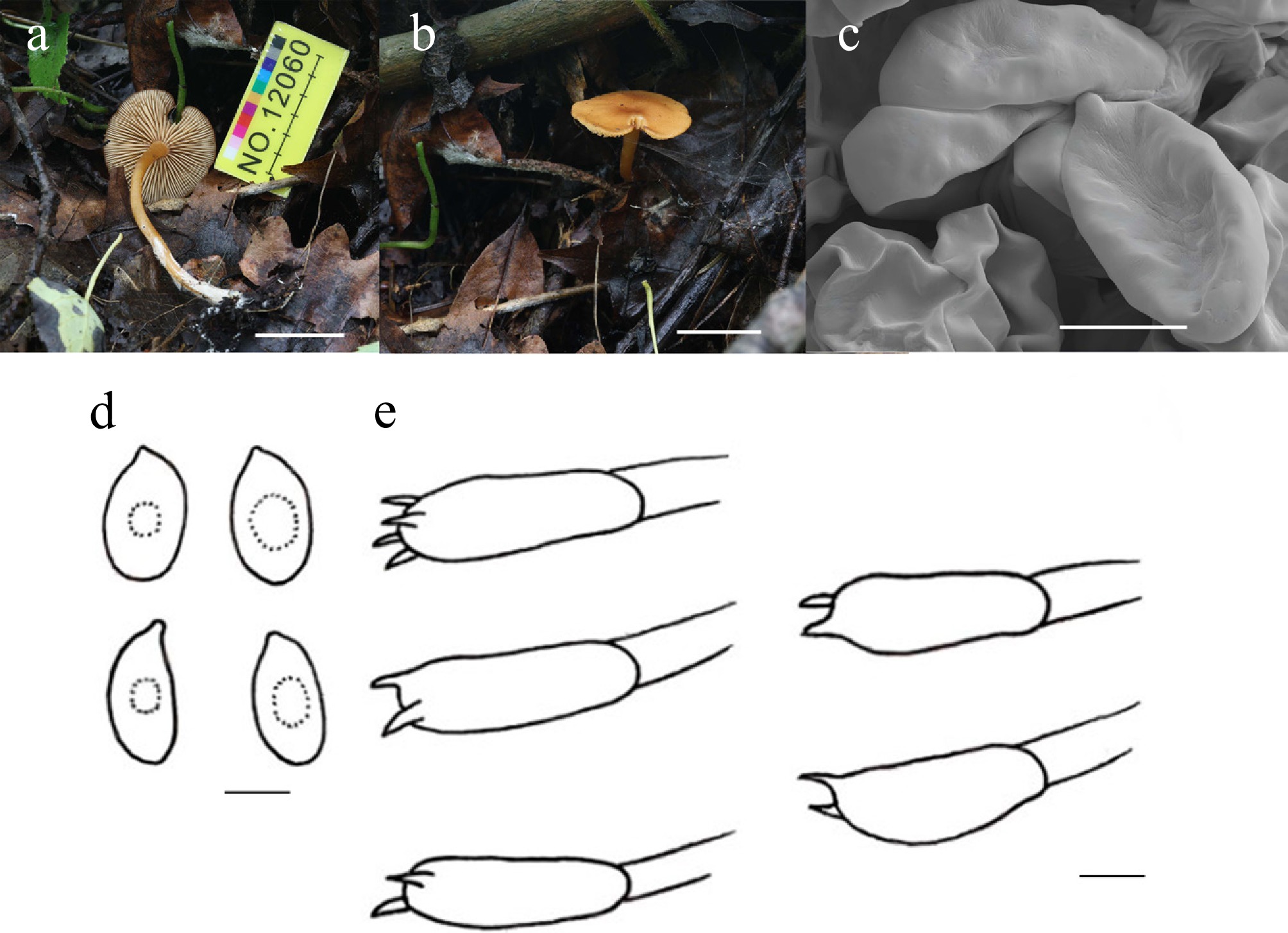
Figure 42.
Rhodophana baishanensis (HMJU 12060, holotype). (a), (b) Basidiocarps; (c) SEM images of basidiospores; (d) Basidiospores; (e) Basidia. Scale bar: (a), (b) 2 cm; (c)–(e) 5 µm.
MycoBank Names: MB863410
Holotype: Jilin Province, Baishan City, Jiangyuan District, Xincun Village, on soil, 42°05'43" N, 126°40'18" E, 12 August 2024, J.Z. Xu (HMJU 12060, holotype).
Etymology: The specific epithet 'baishanensis' refers to the type locality, Baishan City.
Diagnosis: Rhodophana baishanensis is characterized by a centrally depressed pileus, brownish orange at the disc, mandarin orange elsewhere, surface moist; stipe lower portion adorned with dense white fibrils.
Description: Pileus 27–30 mm diam., applanate, center depressed, brownish orange (6C8), the remainder mandarin orange (6E8), surface moist, pileus context thin, mandarin orange (6E8). Lamellae free, white (1A1), margin entire. Stipe 42–52 mm long, 2–5 mm thick, cylindrical, salmon (6A4), lower part covered with dense white (1A1) fibrils, stipe context pale salmon (6A4).
Basidiospores (4.9–)5.1–8.0(–8.3) × (3.2–)3.4–5.8(–5.9) μm, Q = 1.12–2.20, Qm = 1.56, subglobose to broadly cylindrical, transparent in KOH, without amyloid. Basidia (13–)15–24(–26) × (3–)4–9(–10) μm, clavate, both 4 and 2 sterigmata up to 2 μm, transparent in KOH. Cheilocystidia absent. Pleurocystidia absent. Lamella trama regular, hyphae 2–6 μm wide, transparent in KOH. Clamp connections absent in all hyphae.
Habitat: Solitary on soil covered with dead leaves.
Known distribution: Known only from Northeastern China.
Additional material examined: Jilin Province, Baishan City, Changbai Mountain, Primitive forest spring water line, on soil, 42°05'43" N, 126°40'18" E, 11 August 2024, J. Z. Xu (HMJU 12151).
Notes: Forms a sister group with Rhodophana nitellina, but exhibits significant morphological differences (ML/PP = 99/1.0). Morphologically similar to R. nitellina and Rhodophana stangliana (Bresinsky & Pfaff) Vizzini. But R. nitellina is distinguished by a complete, hemispherical pileus with an orange-yellow margin, and cheilocystidia clavate[36]. R. stangliana is characterized by a pileus with silky longitudinal striations, a stipe rose ochre, and basidia larger (28.0–35.0 × 7.5–9.5 µm)[214].
Singerocybe Harmaja, Karstenia 27(2): 71 (1988) [1987]
Clitocybe sect. Bulluliferae Singer, defined by the presence of swollen elements in the pileus epicutis, was originally classified by Singer[160] under Clitocybe subgen. Eu-Clitocybe, with Clitocybe kuehneri Singer designated as the type species. Singer subsequently reassigned this section to either subgen. Pseudobophyllum Singer or subgen. Cystoditus Singer within the genus Clitocybe (Fr.) Staude.
Because the name C. kuehneri was invalid due to the lack of a Latin description or reference, Harmaja emended the group as Clitocybe section Bulluliferae (Singer) Harmaja, establishing Clitocybe hydrogramma (Bull.) P. Kumm. as the new type. Later, Harmaja elevated the group to the rank of an independent genus named Singerella to house C. hydrogramma, which was then the only validly recognized species in the section. A second species, Singerella clitocytoides (basionym: Agaricus clitocytoides), was incorporated.
However, because Singerella Harmaja was considered a later homonym of Singeria (Perr.), Harmaja introduced the replacement name Singerocybe Harmaja. This genus included three species: Singerocybe phaeophthalma (Pers.) Harmaja, Singerocybe hydrogramma (Bull.) Harmaja (syn. C. hydrogramma), and the newly designated type species, Singerocybe viscida Harmaja. S. clitocytoides was omitted from this taxonomic revision. Notwithstanding these revisions, the taxonomic validity of Singerocybe and its precursor Singerella has not been broadly endorsed by the mycological community[68].
Singerocybe was initially circumscribed based solely on European taxa. In North America, species such as Clitocybe adirondackensis (Peck) Sacc. and Clitocybe jalapensis (Murr.) Singer—both placed in C. sect. Bulluliferae by Singer[68]—as well as Clitocybe caespitosa Peck, C. kuehneri, Clitocybe fritilliformis (Lasch) Gillet, and Clitocybe anastomosica Velen., have been treated as synonyms of either Clitocybe phaeophthalma (Pers.) Kuyper or C. hydrogramma. Similarly, species from Australia and New Zealand, including Leucopaxillus otagoensis Stevenson and Clitocybe clitocyboides (Cooke & Massee) Pegler, are morphologically very similar to C. phaeophthalma or C. hydrogramma[102].
Various Asian taxa possessing cuticular vesicles have also been recorded. Examples include Cantharellus humilis Berk. & Broome from tropical Asia, Clitocybe trogioides Corner from Sri Lanka, Clitocybe trogioides var. odorifera Har. Takah. from Japan, Clitocybe alboinfundibuliforme Seok et al. from South Korea, and various unnamed Chinese collections. Because phylogenetic research on these groups is scarce, their evolutionary relationships remain largely unresolved.
Singerocybe rugospora J.Z. Xu, sp. nov. Figs 43 and 44
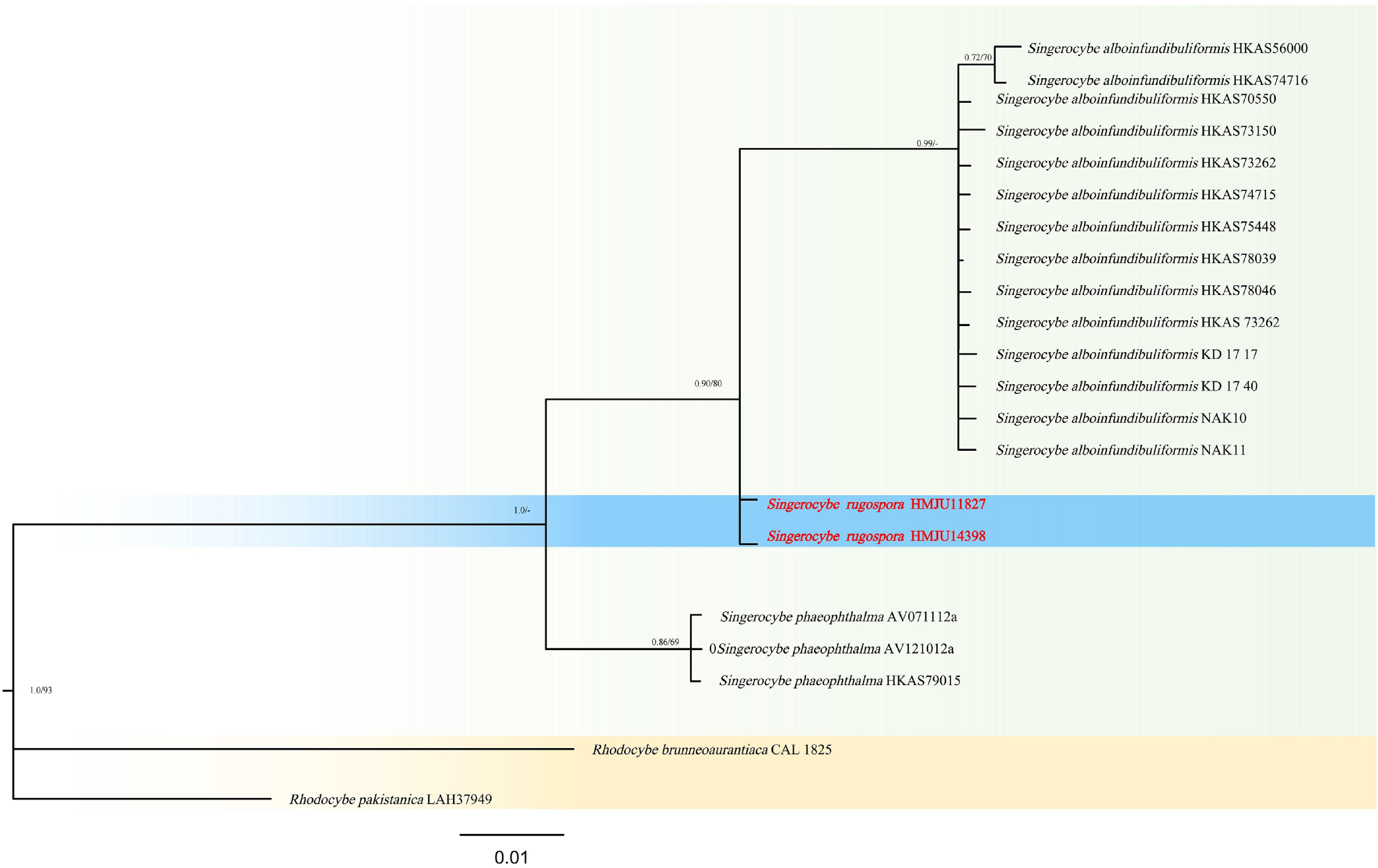
Figure 43.
Phylogram of Maximum Likelihood phylogenetic analysis based on combined ITS, nLSU, rpb2, and tef-1 sequences. The analysis includes 21 strains; total characters: 2,213 (ITS: 261, nLSU: 810, rpb2: 570, tef-1: 572). Rhodocybe brunneoaurantiaca (CAL 1825) and Rhodocybe pakistanica (Dai 6380) were used as the outgroup taxa. The best model was TIM + F + G4. Estimated base frequencies were as follows: A = 0.264, C = 0.208, G = 0.257, T = 0.271. Bootstrap values for ML equal to or greater than 70% and BYPP values equal to or greater than 0.70 are labelled on the nodes. The isolate of the current study is in purple, while the type strains are in bold.
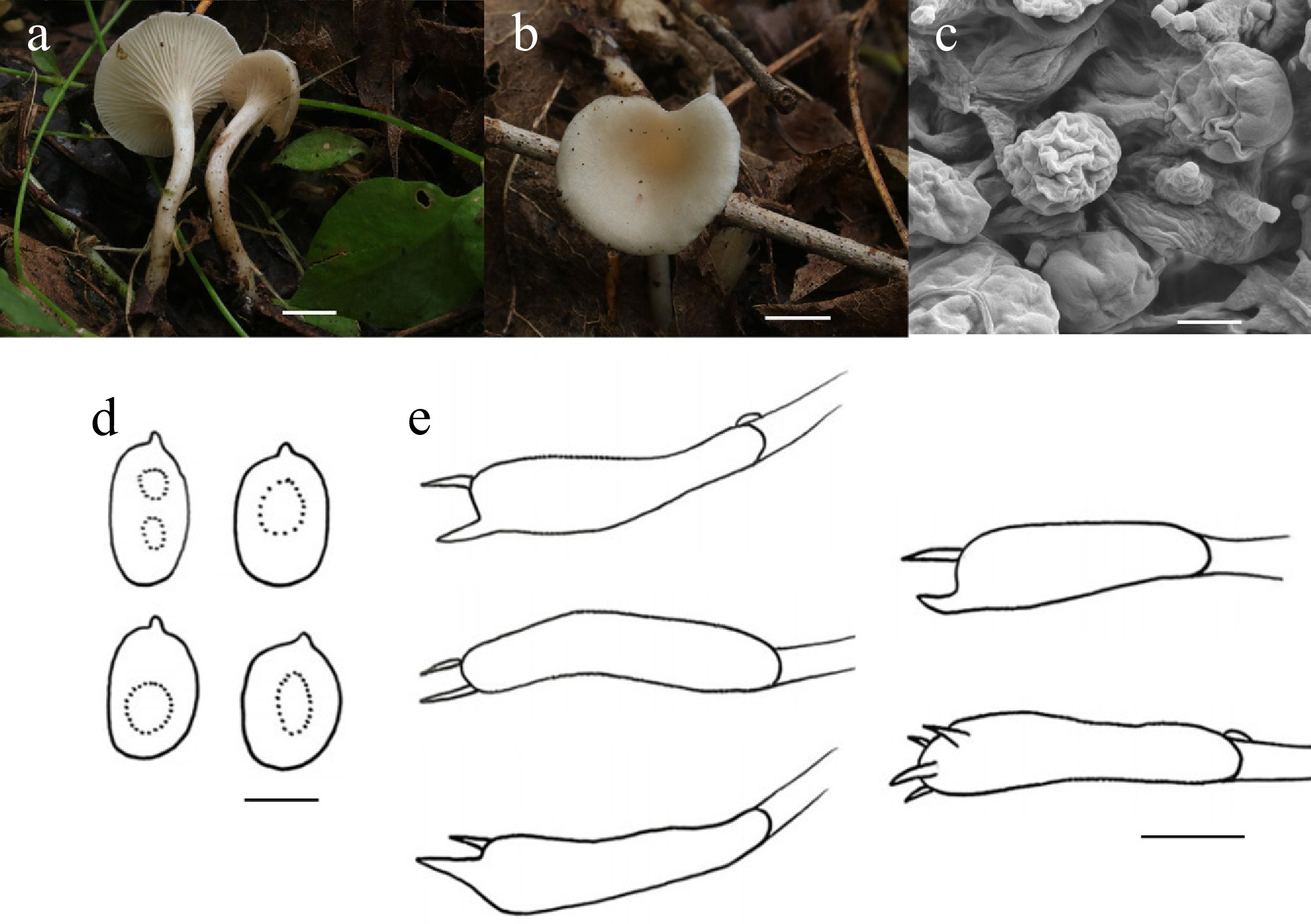
Figure 44.
Singerocybe rugospora (HMJU 11827, holotype). (a), (b) Basidiocarps. (c) SEM images of basidiospores. (d) Basidiospores. (e) Basidia. Scale bars: (a), (b) 0.5 cm; (c), (d) 3 µm; and (e) 5 µm.
MycoBank Names: MB863411
Holotype: Liaoning Province, Huludao City, Jianchang County, Bailang Mountain, on soil, 40°47'42" N, 119°57'27" E, 8 August 2024, J.Z. Xu (HMJU 11827, holotype).
Etymology: The specific epithet 'rugospora' refers to the wrinkled surface of the spores, which is the diagnostic micromorphological character of the new species.
Diagnosis: Singerocybe rugospora is characterized by a stipe surface adorned with irregular, birch bark-like patches; cheilocystidia and pleurocystidia absent; clamp connections present.
Description: Pileus 8–16 mm diam., infundibuliform, white, surface covered with white tomentum, center marble white (5B2), gradually shifting to white (5A1) towards the margin, pileus context thin, white (1A1). Lamellae decurrent, white (1A1). Stipe 21–23 mm long, 1–2 mm thick, white (1A1), cylindrical, curved, slightly broadening towards the base, surface adorned with irregular, birch bark-like (6B2) patches, stipe context white (1A1).
Basidiospores (3.7–)4.5–6.9(–8.0) × (3.1–)3.3–5.2(–5.3) μm, Q = 1.01–1.72, Qm = 1.28, wrinkled, globose to broadly rectangular, transparent in KOH, inamyloid. Basidia (13–)14–20(–22) × (3–)4–6(–7) μm, clavate, both 4 and 2 sterigmata to 2 μm, transparent in KOH. Lamella trama bilaterally, hyphae 2–6 μm wide, transparent in KOH. Clamp connections present in all hyphae.
Habitat: Scattered on soil among leaf litter in forests.
Known distribution: Known only from Northeastern China.
Additional material examined: Liaoning Province, Huludao City, Jianchang County, Bailang Mountain, on soil, 40°47'42" N, 119°57'27" E, 15 August 2025, J.Z. Xu (HMJU 14398).
Notes: Forms a sister group with Singerocybe alboinfundibuliformis (S.J. Seok et al.) Zhu L. Yang, J. Qin & Har. (ML/PP = 80/0.90), but exhibits significant morphological differences. Singerocybe umbilicata Zhu L. Yang & J. Qin differs from S. rugospora in that it has a dark violet-blue pileus, a yellowish-brown stipe, inamyloid tissues, and bigger spores (11–13 × 7.5–10 μm)[215]. Singerocybe adirondackensis (Peck) Zhu L. Yang & J. Qin differs in its pileus being larger (10–60 mm), tan, with striate[215]. In Singerocybe phaeophthalma, the pileus margin is striate, and spores have a prominent apical projection[216]. S. alboinfundibuliformis is distinguished by its pileus sulcate, anastomosing veins on the stipe, and subbulbous stipe base[215]. Singerocybe clitocyboides (Cooke & Massee) Zhu L. Yang, J. Qin & G.M. Gates is characterized by a larger and pale-yellow pileus, sometimes with reddish flush, longer stipe, and smaller spores (5 × 2 μm)[215].
Trogia Fr., Fl. Scan.: 339 (1836) [1835]
The genus Trogia, formally established by Elias Magnus Fries in 1835 and assigned to the Marasmiaceae (Agaricales, Basidiomycota), designates Trogia montagnei Fr. as its type species. This genus is morphologically defined by a growth habit ranging from clitocyboid to omphalinoid, a tough basidiomatal texture, and the ability for in situ revival[217]. Ecologically, most Trogia species are saprophytic, typically found on dead wood or other plant debris within forest ecosystems[218].
Corner[219] published the first global monograph on Trogia, employing a broad generic circumscription that encompassed 56 species, with the presence of a sarcodimitic tramal structure as the principal defining characteristic. In contrast, Singer's generic concepts were applied, the species recognized by Corner would be segregated into at least four distinct genera, including but not limited to Gerronema Singer, Hydropus Kühner ex Singer, Hemimycena Singer, and Mycena (Pers.) Roussel. Indeed, in his 1986 work, The Agaricales in Modern Taxonomy, Singer firmly rejected Corner's[219] broad definition of Trogia, acknowledging only three species within the genus. Singer distinguished Trogia from closely allied genera such as Gerronema, Clitocybe (Fr.) Staude, and Neoclitocybe Singer by a combination of traits: basidiomata that readily revive, narrow and often furcated lamellae composed of interwoven hyphae, and a distinctive pigmented trichodermial epicutis. Redhead further challenged Corner's methodology, arguing that relying exclusively on sarcodimitic tissues for generic delimitation was taxonomically invalid, as this feature is shared across numerous agaric genera. Consequently, Redhead transferred taxa exhibiting sarcodimitic tissues to the family Xerulaceae Jülich. Corner[218] subsequently defended his original concept of Trogia and documented 90 species from the Neotropics, Asia, and Australasia.
The type species, T. montagnei, originally described from Southern India, remains inadequately characterized, and its type specimen is currently untraceable. This absence contributes to the ongoing uncertainty regarding the precise taxonomic circumscription of Trogia[220,221]. To elucidate the relationships among marasmioid and gymnopoid fungi, Desjardin & Wilson[151] for the first time incorporated Trogia infundibuliformis Berk. & Broome into phylogenetic analyses; this taxon formed a sister group to the marasmioid clade, though with modest statistical support. Approximately 7 years later, Yang et al.[220] in their description of Trogia venenata Zhu L. Yang, Y.C. Li & L.P. Tang, employed a combined dataset of nrLSU and ITS rDNA to demonstrate that Trogia forms a strongly supported clade with other genera, including Megacollybia Kotl. & Pouzar, Gerronema, Clitocybula (Singer) Singer ex Métrod, Porotheleum Fr., and Calyptella Quél.
Trogia albotuberculata J.Z. Xu, sp. nov. Figs 45 and 46
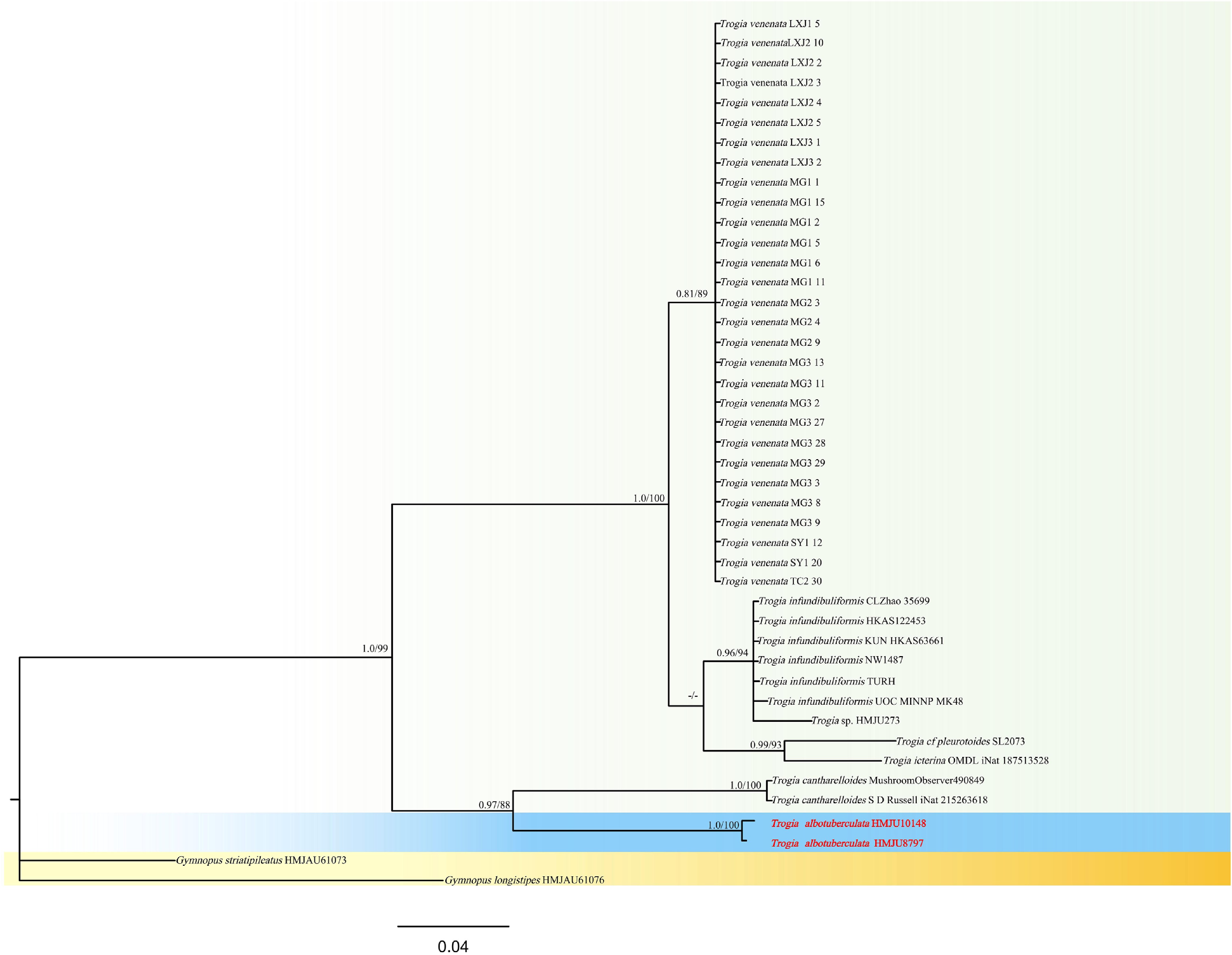
Figure 45.
Phylogram of Maximum Likelihood phylogenetic analysis based on combined ITS sequences. The analysis includes 44 strains; total characters: 617 (ITS: 617). Gymnopus striatipileatus (HMJAU61073) and Gymnopus longistipes (HMJAU61076) were used as the outgroup taxa. The best model was HKY + F + G4. Estimated base frequencies were as follows: A = 0.236, C = 0.209, G = 0.211, T = 0.344. Bootstrap values for ML equal to or greater than 70% and BYPP values equal to or greater than 0.70 are labelled on the nodes. The isolate of the current study is in purple, while the type strains are in bold.
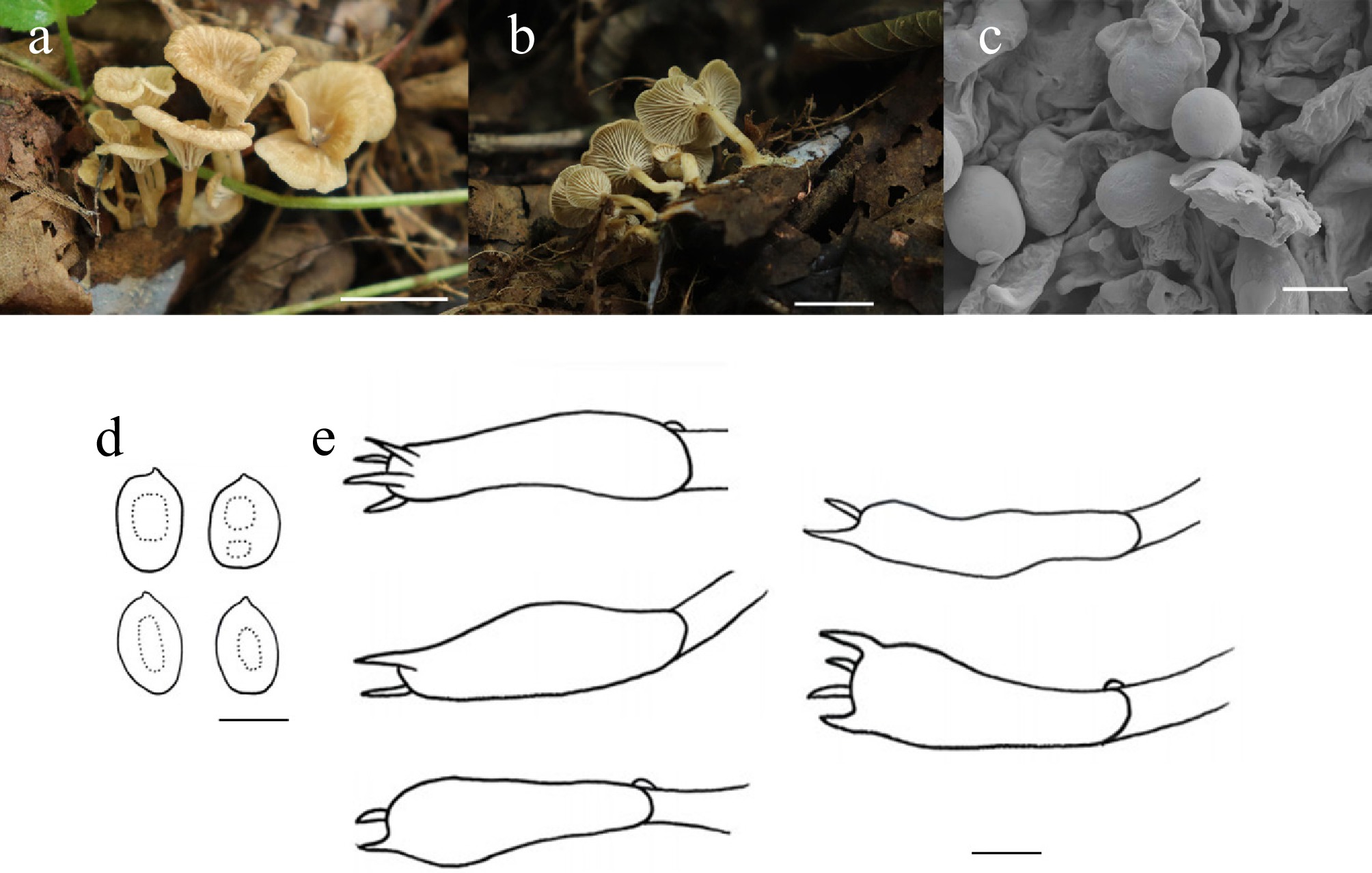
Figure 46.
Trogia albotuberculata (HMJU 10148, holotype). (a), (b) Basidiocarps; (c) SEM images of basidiospores; (d) Basidiospores; (e) Basidia. Scale bars: (a), (b) 1 cm; (c)–(e) 5 µm.
MycoBank Names: MB863412
Holotype: Liaoning Province, Fushun City, Qingyuan Manchu Autonomous County, on soil, 42°05'29" N, 124°54'60" E, 20 August 2023, J.Z. Xu (HMJU 10148, holotype).
Etymology: The specific epithet 'albotuberculata' refers to the white, tuberculate-covered structures of the species.
Diagnosis: Trogia albotuberculata is characterized by a slightly infundibuliform pileus, applanate at maturity, centrally depressed, margin reflexed and exceeding the lamellae, fading gradually to henna toward the center, entirely covered with white tubercles and fibrils; clamp connections present.
Description: Pileus 4–13 mm diam., slightly infundibuliform, becoming applanate after maturity, center depressed, margin reflexed, exceeding the lamellae, ground color natural (4B3) gradually fades to henna (7E8) toward the center, covered with white (1A1) tuberculate and fibrils; pileus context thin, white (1A1) to pale grey (1B1). Lamellae decurrent, crowded, white (1A1), margin entire. Stipe 8–23 mm long, 1–2 mm thick; cylindrical, flexuous, orange white (5A2), 2/3 part covered with white (1A1) fibrils, stipe context near white (1A1), waxy. Annulus absent.
Basidiospores (4.5–)4.7–9.2(–14.3) × (4.20–)4.2–9.2(–9.5) μm, Q = 0.58–1.44, Qm = 0.96, subglobose to ellipsoidal, transparent in KOH, without amyloid. Basidia (17–)19–37(–44) × (3–)4–9(–13) μm, clavate, both 4 and 2 sterigmata to 3 μm, transparent in KOH. Cheilocystidia absent. Pleurocystidia absent. Lamella trama irregular, hyphae 2–11 μm wide, transparent in KOH. Clamp connections present in all hyphae.
Habitat: Gregarious on soil covered with decaying leaves.
Known distribution: Known only from Northeastern China.
Additional material examined: Liaoning Province, Fushun City, Qingyuan Manchu Autonomous County, on soil, 42°05'29" N, 124°54'60" E, 24 July 2019, J.Z. Xu (HMJU 8797).
Notes: Forms a sister group with Trogia cantharelloides (Berk. ex Sacc.) Singer (ML/PP = 88/0.97), but T. cantharelloides differs in having a purple pileus with a reflexed margin, a stipe base that is inflated, and lamellae margins that are serrated[222]. Species morphologically similar to T. infundibuliformis, T. venenata, and Trogia benghalensis K. Acharya & A.K. Dutta. T. infundibuliformis is characterized by a purplish pileus, longer sterigmata (5 µm), and cheilocystidia morphologically diverse[223]. T. venenata features a pink pileus, margin sometimes radially split, basidia larger with longer sterigmata (38–50 × 6.5–8 μm), hyphae pale yellow or yellowish brown[220]. T. benghalensis is different in that the pileus is sometimes eroded, the lamellae margin is eroded, and the cheilocystidia are clavate[221].
Tubaria (W.G. Sm.) Gillet, Hyménomycètes (Alençon): 538 (1876) [1878]
Members of the genus Tubaria are generally identified by a suite of diagnostic morphological and ecological traits. These include small basidiomes (pileus diameter usually under 5 cm) that are brownish in color, a stipe that is centrally attached, and lamellae that are adnate to subdecurrent. Microscopically, they produce brown or ochraceous spore prints, possess cheilocystidia, and typically bear basidiospores that are smooth and lack a germ pore[223]. Anatomically, the pileipellis is filamentous, and ecologically, these fungi are saprotrophs.
While smooth spores are typical, at least three species described from Asia or subtropical zones are distinct for possessing verrucose spores: Tubaria thermophila Singer, Tubaria verruculospora Pegler, and Tubaria lithocarpicola M. Zang[61]. Furthermore, spores ranging from punctate to rugose are observed in two species from North America or Europe: Tubaria dispersa (L.: Fr.) Singer and Tubaria decurrens (Peck) Murrill. According to Singer[68], these five unique taxa, along with various other species described from regions outside Europe, have not yet been incorporated into a unified systematic framework for the genus. It is worth noting, however, that Tu. thermophila was recently reassigned to the genus Crepidotus following combined morphological and molecular analyses[224].
Regarding infrageneric taxonomy, Bon[225] developed a classification scheme specifically for European Tubaria species; however, this system did not accommodate certain taxa, such as Tubaria confragosa (Fr.) Harmaja, which remained unassigned to any infrageneric group.
Tubaria qingyuanensis J.Z. Xu, sp. nov. Figs 47 and 48
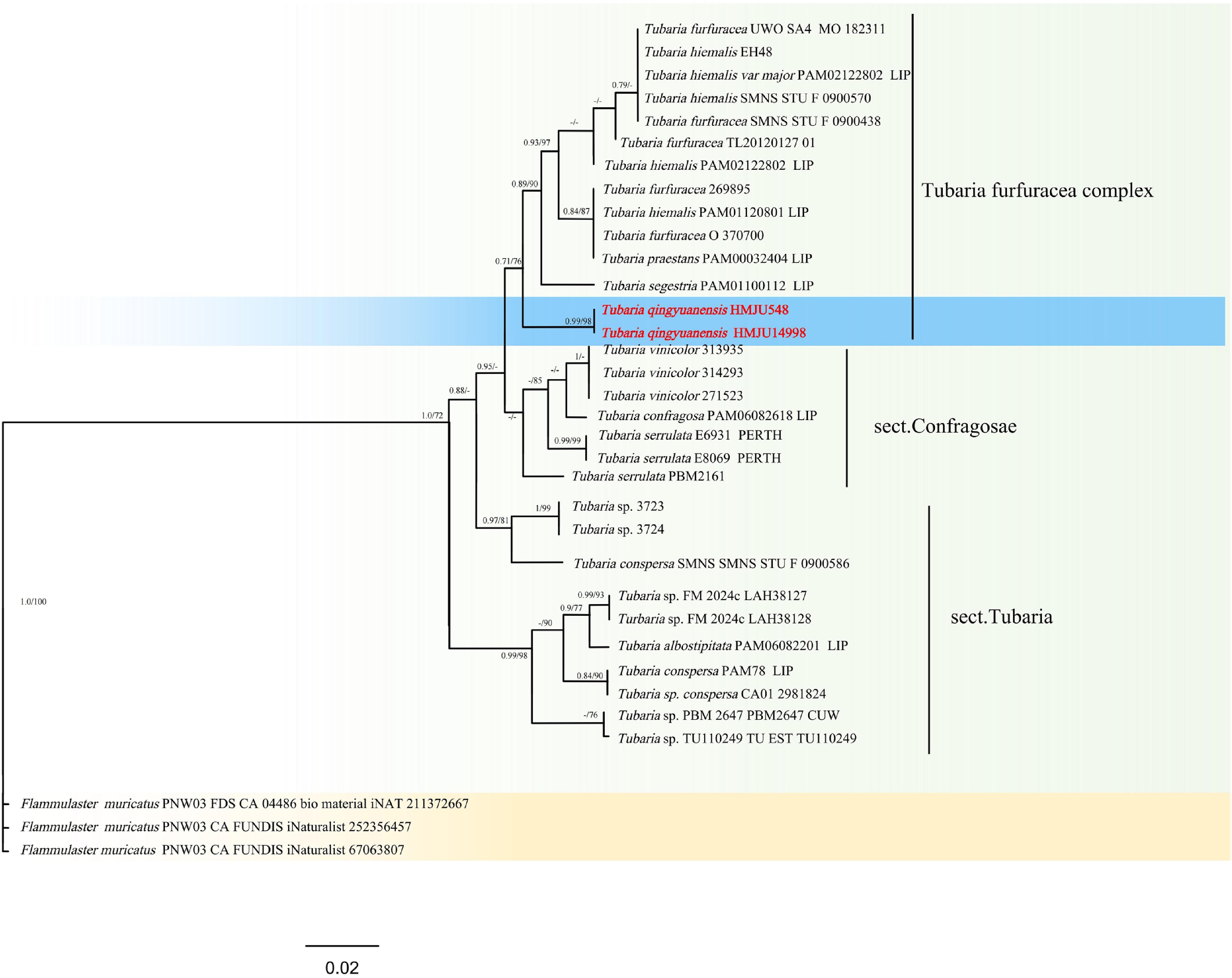
Figure 47.
Phylogram of Maximum Likelihood phylogenetic analysis based on combined ITS and nLSUsequences. The analysis includes 34 strains; total characters: 1,433 (ITS: 604, LSU: 829). Flammulaster muricatus (PNW03 FDS CA 04486 bio material iNAT 211372667), Flammulaster muricatus (PNW03 CA FUNDIS iNaturalist 252356457), and Flammulaster muricatus (PNW03 CA FUNDIS iNaturalist 67063807) were used as the outgroup taxa. The best model was TIM2 + F + G4. Estimated base frequencies were as follows: A = 0.245, C = 0.207, G = 0.246, T = 0.302. Bootstrap values for ML equal to or greater than 70% and BYPP values equal to or greater than 0.70 are labelled on the nodes. The isolate of the current study is in purple, while the type strains are in bold.
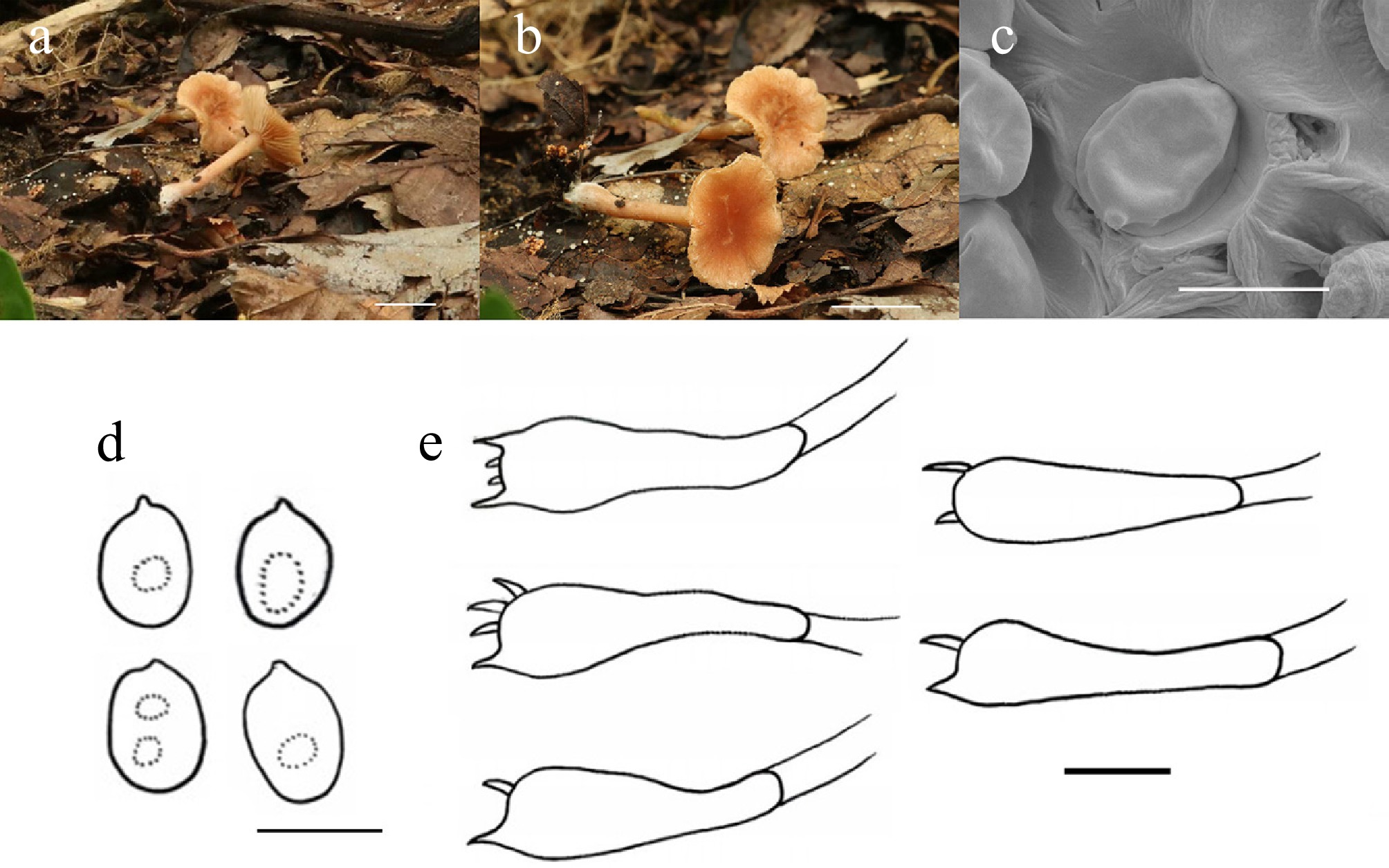
Figure 48.
Tubaria qingyuanensis (HMJU 548, holotype). (a), (b) Basidiocarps. (c) SEM images of basidiospores. (d) Basidiospores. (e) Basidia. Scale bars: (a), (b) 1 cm; (c)–(e) 5 µm.
MycoBank Names: MB863413
Holotype: Liaoning Province, Fushun City, Qingyuan Manchu Autonomous County, Huangdaigou, on soil, 42°15'09" N, 124°43'54" E, 20 August 2023, J.Z. Xu (HMJU 548, holotype).
Etymology: The specific epithet 'qingyuanensis' refers to the type locality, Qingyuan Manchu Autonomous County in Liaoning Province, China.
Diagnosis: Tubaria qingyuanensis is characterized by a centrally depressed pileus, copper, covered with minute white setulae, margin adorned with whitestriations, irregular flesh-colored patches present at the center, overlaid with minute white fibrils; basidia predominantly 2-spored; cheilocystidia and pleurocystidia absent.
Description: Pileus 13–16 mm diam., applanate, center depressed, copper (7C8), covering minute white (1A1) setulae, margin covered with white (1A1) strations, center has irregular flesh (6B3) patches, covered with minute white (1A1) fibrillose, pileus context thin, pale copper (7C8). Lamellae free, subcrowded, orange white (5A2), margin entire. Stipe 10–11 mm long, 1–2 mm thick, cylindrical, orange white (5A2), covered with sparsely white (1A1) fibrils, base slightly swollen, covering white (1A1) dense fibrils, stipe context orange white (5A2). Annulus absent.
Basidiospores (3.7–)4.3–6.2(–6.3) × (3.4–)3.6–4.7(–4.9) μm, Q = 1.00–1.50, Qm = 1.25, subglobose to ellipsoidal, surface wrinkled, without amyloid, transparent in KOH. Basidia (12–)13–19(–24) × (3–)4–7(–8) μm, clavate, most two sterigmata up to 2 μm; transparent in KOH. Cheilocystidia absent. Pleurocystidia absent. Lamella trama irregular, hyphae 2–8 μm wide, transparent in KOH. Clamp connections absent in all hyphae.
Habitat: Solitary on soil covered with dead leaves.
Known distribution: Known only from Northeastern China.
Additional material examined: Liaoning Province, Fushun City, Qingyuan Manchu Autonomous County, Huangdaigou, on soil, 42°15'09" N, 124°43'54" E, 29 August 2024, J.Z. Xu (HMJU 14998).
Notes: Forms a sister group with Tubaria furfuracea (Pers.) Gillet, Tubaria hiemalis Romagn. ex Bon, Tubaria praestans (Romagn.) Romagn and Tubaria segestria (Fr.) Boud. Tu. furfuracea is unique with cream-ochraceous pileus, margin finely striate with fimbriate universal veil remnants, stipe with indistinct annulus, cheilocystidia clavate[226]. Tu. hiemalis is characterized by a reddish-brown pileus, spores larger (7.1–8.6 × 4.6–5.4 µm), cheilocystidia capitate, and clamp connections present[227]. Tu. praestans features dark-brown lamellae; the stipe is longer, the spores are pale brown to dark yellow, and clamp connections are present (Kühner). Tu. segestria is characteried by a lenticular pileus, reddish brown; stipe with white annulus; spores yellowish brown[228]. Species morphologically similar to Tu. dispersa, Tubaria elongatispora Musumeci, and Tubaria serrulata (Cleland) Bougher & Matheny. Tu. dispersa is distinguished by its lamellae, which are orange yellow; its spores are larger (5.8–7.3 × 3.7–4.7 µm), and its cheilocystidia are cylindrical, urceolate, and clavate; clamp connections are present[229]. Tu. serrulata is characterized by a pileus that is brownish, brown lamellae, a stipe with a pale-white annulus, spores that are brownish ferruginous and larger[230], and cheilocystidia present. Tu. elongatispora is unique in having a pileus subglobose to hemispherical, surface fibrillose, lamellae subdecurrent, and spores larger (6–9 µm)[231].
-
Cortinarius can form symbiotic relationships with some species of both woody and herbaceous plants. The associated woody plant families include Dipterocarpaceae Blume, Fagaceae Dumort., Myrtaceae Juss., Nothofagaceae Kuprian., Pinaceae Spreng. ex F. Rudolphi, Rhamnaceae Juss., Salicaceae Mirb., and Rosaceae Juss., while the associated herbaceous plant families comprise Cyperaceae Juss., Orchidaceae Juss., and Polygonaceae Juss. These symbiotic associations endow Cortinarius with significant ecological and socioeconomic values. Additionally, some species within the genus hold considerable edible and medicinal importance, such as Cortinarius caperatus (Pers.) Fr.[232].
The hyphae of Cystolepiota and Echinoderma can efficiently degrade organic residues such as leaf litter and dead branches, transform complex organic matter, and directly enhance soil fertility, endowing the genera with significant ecological value.
Entoloma species can thrive in diverse habitats, including tundra, swamps, grasslands, shrublands, and forests, and have been documented in temperate, subtropical, and tropical rainforest ecosystems. Most species are saprotrophic fungi, capable of decomposing plant litter or wood. Some Entoloma species are widely consumed by humans, such as Entoloma abortivum (Berk. & M.A. Curtis) Donk., Entoloma clypeatum (L.) P. Kumm., Entoloma sarcopum Nagas., Entoloma saundersii (Fr.) Sacc., and Entoloma turbidum (Fr.) Quél. However, ingestion of certain species can induce varying degrees of toxic reactions, including neuropsychiatric, gastroenteritic, hepatotoxic, and respiratory and circulatory failure syndromes. Examples of such toxic species include Entoloma album Hiroë, Entoloma chalybeum (Pers.) Noordel., Entoloma incanum (Fr.) Hesler, Entoloma opacum Noordel., Entoloma rhodopolium (Fr.) P. Kumm., and Entoloma sinuatum (Bull.) P. Kumm.[233−235].
The hyphae of Hygrocybe can secrete extracellular enzymes such as cellulases and hemicellulases, which efficiently degrade complex polysaccharides in herbaceous plant residues. This process not only increases soil organic matter content but also improves soil structure and enhances aeration, thereby playing a crucial role in nutrient cycling within nutrient-poor grassland ecosystems[77].
Hypholoma species are predominantly distributed in the North Temperate Zone, growing on dead wood, peat, soil, or plant debris during autumn and winter. Most species of the genus have a bitter taste and low edibility; several, including Hypholoma fasciculare (Huds.) P. Kumm., Hypholoma lateritium (Schaeff.) P. Kumm., and Hypholoma capnoides (Fr.) P. Kumm., are toxic. Ingestion of these toxic species can induce adverse reactions such as nausea, diarrhea, and muscle spasms, and such poisoning incidents have been frequently reported. Additionally, Hypholoma species contain a variety of bioactive compounds, including sesquiterpenoids, triterpenoids, and ergosterol, which confer significant medicinal value[236].
Species of the genus Lepiota exhibit a cosmopolitan distribution, occurring across all regions of the globe. They play a pivotal role in sustaining ecological equilibrium in forest ecosystems, as they can break down both lignin and cellulose in their environments. Furthermore, these fungi can serve as bioindicators for monitoring heavy metal contamination in environmental systems. In contrast, some members of this genus are edible and prized for their delicate flavor, such as Le. clypeolaria and Le. alba, others contain potent toxins that can be life-threatening if ingested. Nonetheless, the toxic peptides and amatoxins produced by these hazardous species have considerable research significance across disciplines such as life sciences and medical biology[237−239].
The genus Leucoagaricus is species-rich, with most taxa aggregating in low-latitude regions. Some species of Leucoagaricus are edible and provide nutrients for humans. This genus can secrete yellow laccase, which degrades anthracene and other polycyclic aromatic hydrocarbons, thereby promoting environmental remediation. However, certain species are toxic; ingestion can cause gastrointestinal poisoning, accompanied by symptoms such as nausea and diarrhea[240,241].
Limacella functions as either a saprotrophic or ectomycorrhizal fungus[242]. It has significant ecological value and plays an important role in regulating forest ecosystems and maintaining ecological environments.
Some species of the Macrocystidia emit a cucumber-like odor; Ma. cucumis is a typical example. These fungi can decompose dead leaves and humus, thereby facilitating the breakdown of organic matter in the environment.
Marasmius is an important genus of edible and medicinal fungi, with common species such as Mar. oreades and Marasmius albogriseus (Peck) Singer. Beyond their edible value, substances derived from Marasmius conigenus (Pers.) P. Karst of this genus exhibits potent antibacterial activity after industrial processing. Additionally, enzymes secreted by some Marasmius species can facilitate the decomposition of environmental pollutants[243].
Most species of the genus Melanoleuca possess considerable nutritional and economic value, being rich in proteins, minerals, and crude fiber. For instance, the basidiocarps of M. arcuata contain high levels of proteins and essential amino acids, as well as 10 types of mineral elements. Meanwhile, other species within the genus exhibit antioxidant and antibacterial activities, which are of significant importance to life sciences[244−246].
Species of the genus Mycena are not only key saprotrophic decomposers in forest ecosystems but also exhibit multiple values in biological interactions and resource utilization. They are widely distributed on leaf litter and decaying wood, providing the driving force for material cycling in forest ecosystems[247]. Some species can efficiently promote the germination of Gastrodia elata seeds and the growth and development of medicinal Orchidaceae plants, possessing unique ecological application value and development potential. In addition, fungi of this genus are rich in various bioactive components, which serve as a valuable resource reserve for the research and development of new drugs and biological agents.
Pseudobaeospora are rarely recorded in Northern China. They can participate in the nitrification process, promote nitrogen cycling, and thus maintain ecosystem stability.
Pseudosperma species predominantly occur in forests dominated by genera such as Betula, Cedrus, and Populus, as they can establish ectomycorrhizal symbioses with a range of plant hosts. It is noteworthy that some members of this genus are highly toxic, containing various alkaloids, muscarine, amatoxins, and other toxic compounds, rendering them inedible.
Rhodophana plays a crucial role in decomposing leaf litter and humus in forest ecosystems. To date, no edible or medicinal value has been documented for any species within this genus[248].
Singerocybe is predominantly saprotrophic, occurring on dead wood or in soil containing decaying leaves. It has a widespread distribution across multiple provinces in China, including Yunnan, Shanxi, Shandong, Guizhou, Hubei, and Jilin. Singerocybe alboinfundibuliformis is an edible species and has been the subject of extensive research.
Most Trogia species grow on the surface of decaying wood or on felled tree stumps, where they contribute to the decomposition of organic matter. Notably, species within this genus are highly toxic, and accidental ingestion can be life-threatening.
Most Tubaria species inhabit decaying wood and have a cosmopolitan distribution. The mycelia produced during their growth can enhance soil aeration and facilitate interactions within microbial communities.
As one of the most species-rich groups on Earth, fungi are not only an important component of biodiversity but also play a crucial role in material cycling, energy flow, and biological interactions. Although diversity of macrofungi has been extensively studied in China recently, and many new taxa have been established or described[249−256], because of the variable climate and vegetation in the country, many unknown species still exist. The discovery of the new species in this study has filled the gap in fungal species diversity[257−265]. Their taxonomic status was clarified from the perspectives of morphology and molecular phylogeny, supplementing the species information of related taxa. The discovery of the new species also has important ecological and application values; their unique metabolic pathways may be involved in the decomposition of specific substances, providing new research resources for the fields of medicine, pesticides, and industry[266−269].
It should be noted that there are certain limitations in the related research on fungal diversity in this article. The discussion on the new species only clarifies their morphological characteristics and taxonomic status, while the analysis of their physiological and metabolic mechanisms and ecological functions is insufficient. Further exploration of fungal species resources in subsequent studies is needed to provide theoretical support for more comprehensive fungal research.
-
The authors confirm their contributions to the paper as follows: conceptualization, experiment, software, morphological analysis: Zhang AQ; formal analysis, constructive discussions: Xu JZ; writing − review and editing, writing − original draft preparation: Zhang CL. All authors reviewed the results and approved the final version of the manuscript.
-
These data were derived from the following resources available in the public domain: NCBI GenBank (www.ncbi.nlm.nih.gov/genbank).
-
This study was supported by the Sanjiang Laboratory Project (No. SJ2025006-3), and the Science and Technology Department of Jilin Province (20240602031RC). We would like to thank the Agricultural College, Jilin Agricultural Science and Technology College, Yue-Xin Liu, Tong Li, Jing-Yi Jie, Shao-Yuan Zhang, Yu-Hao Huang, Ming-Yang Liu, and Jun-Jie Peng; Jilin Agricultural University, Sheng Ming, Beijing Forestry University, Yu-cheng Dai, Heng Zhao, as well as Qujing Normal University Samantha C. Karunarathana for their contributions to this paper.
-
The authors declare that they have no conflict of interest.
-
#Authors contributed equally: Ji-Ze Xu, An-Qi Zhang
- Copyright: © 2026 by the author(s). Published by Maximum Academic Press, Fayetteville, GA. This article is an open access article distributed under Creative Commons Attribution License (CC BY 4.0), visit https://creativecommons.org/licenses/by/4.0/.
-
About this article
Cite this article
Xu JZ, Zhang AQ, Zhang CL. 2026. Morphological and molecular analyses reveal 28 new macrofungal species in China. Mycosphere 17: e007 doi: 10.48130/mycosphere-0026-0007 

Morphological and molecular analyses reveal 28 new macrofungal species in China
- Received: 08 February 2026
- Revised: 15 April 2026
- Accepted: 27 April 2026
- Published online: 27 June 2026
Abstract: Fungi are vital resources on Earth, playing crucial roles in promoting the decomposition of organic matter and sustaining ecosystem stability. Many fungal taxa also represent important edible and medicinal resources, holding significant values for human well-being. However, our understanding of fungal species diversity remains incomplete, and exploration in this field is still ongoing. This study focuses on macroscopic fungi from different regions of China. Based on detailed morphological comparisons and multi-gene phylogenetic analyses, 28 new species are described herein: Cortinarius hyalocinnamomeus, Cortinarius chaoyangensis, Cystolepiota rubellogrisea, Echinoderma petaloides, Entoloma liangshuiens, Hygrocybe santagouana, Hygrocybe aurisquama, Hygrocybe sejilensis, Hygrocybe subreidii, Hypholoma salmopapillosum, Lepiota adpileobrunnea, Lepiota gobelinicolor, Leucocoprinus margilongus, Leucocoprinus submargallensis, Leucocoprinus oleifer, Limacella annulipendula, Macrocystidia tashanparensis, Marasmius velutinus, Melanoleuca mozhugongkaensis, Mycena atrorubina, Phloeomana flavomaculata, Pseudobaeospora vulpecula, Pseudosperma lacteoconicum, Rhodophana rimosula, Rhodophana baishanensis, Singerocybe rugospora, Trogia albotuberculata, and Tubaria qingyuanensis. Descriptions, illustrations, multi-locus phylogenetic analyses, and detailed comparisons with closely related taxa are provided for each newly described species. The discovery of these 28 novel macrofungal species provides an essential taxonomic framework for future studies on fungal systematics and conservation of fungi in China.
-
Key words:
- Agaricales /
- Entolomataceae /
- Amanitaceae /
- Hygrocybaceae /
- Taxonomy